Structural study of Cu2-xSe alloys produced by mechanical alloying
Abstract
The crystalline structures of superionic high temperature copper selenides Cu2-xSe () produced by Mechanical Alloying were investigated using X-ray diffraction (XRD) technique. The measured XRD patterns showed the presence of the peaks corresponding to the crystalline superionic high temperature -Cu2Se phase in the as-milled sample, and its structural data were determined by means of a Rietveld refinement procedure. After a heat treatment in argon at 200∘C for 90 h, this phase transforms to the superionic high temperature -Cu1.8Se phase, whose structural data where also determined through the Rietveld refinement. In this phase, a very low occupation of the trigonal 32(f) sites (%) by Cu ions is found. In order to explain the evolution of the phases in the samples, two possible mechanisms are suggested: the high mobility of Cu ions in superionic phases and the intense diffusive processes in the interfacial component of samples produced by Mechanical Alloying.
keywords:
Mechanical alloying , x-ray diffraction , semiconductors.PACS:
61.10.Nz , 81.05.Cy , 81.07.Bc , 81.20.Ev, , , , , ,
1 Introduction
Due to their physical and chemical characteristics, selenium-based alloys are very important from technological and scientific points of view, mainly those alloys containing germanium, zinc or copper because of their very interesting optical properties. Recently, cation-deficient copper selenide Cu2-xSe, which is a mixed ionic-electronic superionic conductor with a homogeneity range of , has been extensively studied because of its interesting properties and potential application in solar cells [1], window material [2], optical filter [3], superionic conductor [4], electro-optical devices [5], thermoelectric converter [6] and also as a precursor for preparation of copper-indium-diselenide CuInSe2 (CIDS) [7]. The superionic transition from phase (low temperature) to phase (high temperature) occurs at 414 K for Cu2Se and decreases with increasing deviation from stoichiometry. At room temperature, the superionic phase is stable for [8]. Cu2-xSe films are typically p-type, highly conducting, semitransparent, with band gap between 1.1 and 1.4 eV, very suitable for solar energy conversion. A solar cell (Schottky type) employing a semi-transparent layer of Cu2-xSe as window material on n-type semiconductor is estimated to have excellent photovoltaic properties and conversion efficiency around 9 % [9]. Many preparation methods of Cu2-xSe alloys have been reported including sonochemical syntesis [10, 11], chemical bath deposition [12, 13, 14], photochemical route [15], -irradiation [16], solid state reaction [17], microwave assisted heating [18], hydrothermal method [19], electrodeposition [20, 21] and Mechanical Alloying (MA) [22].
The crystalline structure of Cu2-xSe has been studied several times [17, 23, 24, 25, 26, 27, 28, 29, 30] but the precise distribution of Cu ions and the structure itself are still under discussion and diverging crystallographic data can be found in the literature. Rahlfs [23] and Borchert [24] proposed a structural model for -Cu2-xSe consisting of a cage of F3m symmetry, built by Se atoms in 4(a) sites and Cu ions in 4(c) sites, and a mobile cation subsystem formed by the remaining Cu ions distributed over the interstitial sites (tetrahedral 4(d), octahedral 4(b), and trigonal 16(e) sites). Heyding and Murray [25] considered an fcc cage formed only by Se atoms and a mobile subsystem formed by Cu ions in tetrahedral 8(c) and trigonal 32(f) sites, with an overall symmetry Fm3m. Neutron diffraction results [26], on the other hand, suggest that Cu ions are distributed over the trigonal 32(f) sites leaving the octahedral sites empty. The -Cu2-xSe phase are either described as monoclinic or tetragonal [29, 30]. The ordering of Cu ions in the -phase results in a complicated superstructure very sensitive to the composition and preparation technique.
In this paper we report results obtained for Cu2-xSe alloys produced by the MA technique [31]. MA has been used for almost two decades to produce many unique materials. These include, for instance, nanostructured alloys, amorphous compounds and unstable and metastable phases [32, 33, 34, 35, 36, 37, 38]. This method has several intrinsic advantages, like low temperature processing, easy control of composition, relatively inexpensive equipment, and the possibility of scaling up. Although the MA technique is relatively simple, the physical mechanisms involved are not yet fully understood. In order to make use of this technique in industrial applications, a better understanding of these physical mechanisms is desirable.
2 Experimental procedures
A binary mixture of high-purity elemental powders of copper (Vetec 99.5%, particle size m) and selenium (Alfa Aesar 99.999% purity, particle size m) with nominal composition Cu2Se was sealed together with several steel balls into a cylindrical steel vial under an argon atmosphere. The ball-to-powder weight ratio was 5:1. A Spex Mixer/Mill model 8000 was used to perform MA at room temperature. The mixture was continuously milled for 72 h. A ventilation system was used to keep the vial temperature close to room temperature. For this sample, an x-ray diffraction (XRD) measurement was performed. After that, it was heat treated at 200∘C for 90 h in quartz capsule containing argon, and a new XRD measurement was collected. All XRD measurements were recorded on a Siemens diffractometer with a graphite monochromator in the diffracted beam, using the Cu Kα line ( Å).
3 Results and discussion
Figure 1 shows the x-ray diffraction (XRD) pattern taken for the as-milled sample (AM-Cu2-xSe). A comparison of this pattern with JCPDS cards indicated the presence of Cu2Se phase in the F23 structure given by JCPDS card 76-0136. The main peaks of this phase are seen at about 2, , and , in very good agreement with that card. This is a high temperature phase of the Cu2-xSe system, indicating that MA performed at room temperature can produce these phases. The pattern shown in Fig. 1 was simulated using the Rietveld procedure [39], and the simulation is also seen in this figure. In Ref. [12], using a chemical bath deposition method, Garcia et al. obtained the Cu1.85Se phase corresponding to the mineral berzelianite (JCPDS card 06-0680). Danilkin et al. [17] produced the low temperature monoclinic -Cu2Se phase using solid state reactions. We also investigated these possibilities but Rietveld refinements using these structures did not furnish good results. The lattice parameters obtained for -Cu2Se phase was Å. The occupation of the sites was also refined, and these data are given in Table 1.
| Atom | Site | Position | Occupation (%) |
|---|---|---|---|
| Cu | 4(c) | 100 | |
| Cu | 4(a) | 42 | |
| Cu | 16(e) | 8 | |
| Cu | 16(e) | 8 | |
| Se | 4(a) | 100 |
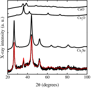
In addition to the peaks of the -Cu2Se phase, the XRD pattern of AM-Cu2-xSe has some other peaks, which were identified as belonging to the oxides Cu2O (JCPDS card 78-2076) and CuO (JCPDS card 80-1917). Although the sample was kept under argon atmosphere during the milling, it was probably contaminated by oxygen during its preparation and also during the XRD measurement. About 73% of the crystalline phases present in this sample is given by the -Cu2Se phase, whereas the contaminant phases are responsible for 18% (Cu2O) and 9% (CuO), respectively. The contribution of the three phases to the XRD pattern of AM-Cu2-xSe is also shown in Fig. 1. It should be noted that the strong diffuse scattering seen in Fig. 1 is a well-established characteristic of the -Cu2Se phase, due to the large number of vacancies in the sublattice of Cu atoms [40]. Thus, we did not consider it as an amorphous part in the Rietveld refinement. The refined lattice parameters of the oxides are Å, for Cu2O (space group Pnm), and Å, Å, Å and (space group Cc). The average crystallite sizes found using the Scherrer formula [41] for -Cu2Se, Cu2O and CuO are 60 Å, 78 Å and 50 Å, respectively, showing that all of them are in nanometric form.
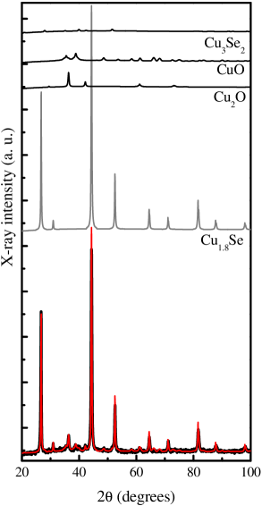
To study the stability of the -Cu2Se phase, a thermal treatment was made at 200∘C for 90 h. The XRD pattern of the heat treated Cu2-xSe sample (HT-Cu2-xSe) is shown in Fig. 2. A comparison with Fig. 1 indicates the presence of a phase different from those found in AM-Cu2-xSe. The peaks at about , , , and are the most intense of the superionic high temperature phase -Cu1.8Se given in JCPDS card 71-0044, that corresponds to one of the forms of the mineral berzelianite. This phase belongs to the space group Fmm, and its refined lattice parameters are Å. The Rietveld refinement indicated that the Cu ions are found mainly in the tetrahedral 8(c) sites. The trigonal 32(f) sites are almost unoccupied. In addition, the refined position of the trigonal 32(f) sites agrees with those given in the ICSD card 238, as can be seen in table 2, which gives the refined data concerning this phase. Danilkin et al. [17] have also studied this phase, but they found the trigonal 32(f) sites in a different position and with a different occupation. Our results agree with the neutron diffraction study of Oliveira et al. [26], which did not give any indication of octahedral occupation.
| Atom | Site | Position | (Å2) | Occupation (%) |
|---|---|---|---|---|
| Cu | 8(c) | 5.2 | 80 | |
| Cu | 32(f) | 1.7 | 3 | |
| Se | 4(a) | 3.6 | 100 |
In addition to the -Cu1.8Se phase, the HT-Cu2-xSe sample contains the two oxides already seen in AM-Cu2-xSe, and a small quantity of tetragonal Cu3Se2 phase known as umangite, given in JCPDS card 41-1745 (space group Pm). About 75% of the crystalline phases found in HT-Cu2-xSe are given by the -Cu1.8Se phase, and the contribution of the other phases is 6% of Cu2O, 16% of CuO and 3% of Cu3Se2. The refined lattice parameters obtained for the contaminant phases are Å for Cu2O, Å, Å, Å and for CuO and Å and Å for Cu3Se2.
With the heat treatment at 200∘C, the -Cu2Se phase changes to the -Cu1.8Se phase and the Cu2O phase decreases while CuO increases. These processes could be explained by a combination of two factors. First, in the superionic phase Cu ions have a very high mobility, which could be explained by a very small activation energy for jumps of Cu between sites. Second, it is well known that MA produces materials with two components, the nanocrystalline component, which preserves the crystalline structure of the crystals, and the interfacial region, formed by defect centers. The diffusive processes in the interfacial component can be much more intense than they are in the nanocrystalline one, and Cu, Se and O atoms belonging to this region could have reacted during the heat treatment and produced the structural changes seen in the XRD pattern of the heat treated sample. An indication that these processes really take place in the alloy is the large increase in the average crystallite size of the phases. The values obtained are 390 Å, 225 Å, 113 Å and 144 Å, respectively, for -Cu1.8Se, Cu2O, CuO and Cu3Se2. The crystallinity of all phases improves during heat treatment, in particular the crystallinity of the -Cu1.8Se phase, whose average crystallite size increases more than six times.
4 Conclusion
From the results shown above, we conclude that:
-
1.
The superionic high temperature -Cu2Se phase can be produced by Mechanical Alloying at room temperature. It is the majority phase (about 73%) in the as-milled Cu2-xSe sample. During the preparation of the sample a contamination by oxygen occurred, and two oxides, Cu2O and CuO, were formed.
-
2.
The -Cu2Se phase present in the as-milled Cu2-xSe sample is found in the space group F23, and its refined structural parameters, shown in table 1, are a little different from those seen in the literature. The oxides are also found in nanometric form.
-
3.
After a heat treatment in argon at 200∘C for 90 h, the structure of -Cu2Se changes, and the superionic high temperature -Cu1.8Se phase is formed. It is found in the space group Fmm, and its refined structural parameters are found in table 2. Almost all Cu ions are found in tetrahedral 8(c) sites, and the trigonal 32(f) sites have a very small occupation (%). There is no indication of octahedral occupation, in agreement with results from Refs. [26, 27]. The crystallinity of all phases are much improved during the heat treatment, in particular the crystallinity of the Cu1.8Se phase, whose average crystallite size increases by more than six times, from 60 Å to 390 Å. There are at least two possible reasons for the structural changes occurred in the sample with the heat treatment. First, Cu ions in the superionic phases have a high mobility since their activation energy for jumps is expected to be very small, and the heat treatment could furnish this energy. Second, atoms in the interfacial component of the sample produced by Mechanical Alloying have also a very high mobility, and the diffusive processes in this component can be very intense. The combination of the two processes could explain the structural changes and crystallinity improvement occurred during the heat treatment, and also the formation of the superionic phase at room temperature.
References
- [1] S. T. Lakshmikumar, Mater. Solut. Cells 32 (1994) 7.
- [2] S. K. Haram, K. S. V. Santhanam, M. N. Spallart, C. L. Clement, Mater. Res. Bull. 27 (1992) 1185.
- [3] H. Toyoji, Y. Hiroshi, Jpn. Kokai Tokkyo Kaho JP 02 (1990) 175.622.
- [4] W. S. Chen, J. M. Stewart, R. A. Mickelsen, J. Appl. Phys. Lett. 46 (1985) 1095.
- [5] R. C. Kainthla, D. K. Pandya, K. L. Chopra, J. Electrochem. Soc. 2 (1980) 127.
- [6] H. Rau, J. Phys. Chem. Solids 28 (1967) 903.
- [7] Y. Ueno, H. Kawai, T. Sugiura, H. Minorra, Thin Solid Films 157 (1989) 159.
- [8] H. H. Abrikosov, V. F. Bankina, M. A. Korzhuev, G. K. Demski, O. A. Teplov, Sov. Phys. Solid State 25 (1983) 1678.
- [9] H. Okimura, T. Matsumae, R. Makabe, Thin Solid Films 72 (1980) 53.
- [10] S. Xu, H. Wang, J. J. Zhu, H. Y. Chen, J. Cryst. Growth 234 (2002) 263.
- [11] Y. Xie, X. W. Zheng, X. C. Jiang, J. Lu, L. Y. Zhu, Inorg. Chem. 41 (2002) 387.
- [12] V. M. Garcia, P. K. Nair, M. T. S. Nair, J. Cryst. Growth 203 (1999) 113.
- [13] C. L. Clement, M. N. Spaliart, S. K. Haram, K. V. S. Santhanam, Thin Solid Films 302 (1997) 12.
- [14] H. M. Pathan, C. D. Lokhande, D. P. Amalnerkar, T. Seth, Appl. Surface Sci 211 (2003) 48.
- [15] Y. Yan, X. Qian, H. Xu, J. Yin, Z. Zhu, Inor. Chem. Commun. 6 (2003) 34.
- [16] Z. P. Qian, Y. Xie, J. G. Xu, X. M. Liu, Y. J. Zhu, Y. T. Qian, Can. J. Chem. 78 (2000) 1143.
- [17] S. A. Danilkin, A. N. Skomorokhov, A. Hoser, H. Fuess, V. Rajevac, N. N. Bickulova, J. Alloys Comp. 361 (2003) 62.
- [18] J. J. Zhu, O. Palchik, S. G. Chen, A. Gendanken, J. Phys. Chem. B 104 (2000) 7344.
- [19] W. Z. Wang, Y. Geng, P. Yan, F. Liu, Y. Xie, Y. T. Qian, J. Am. Chem. Soc. 121 (1999) 4062.
- [20] D. Lippkow, H.-H. Strehblow, Electrochim. Acta 43 (1998) 2131.
- [21] R. N. Battacharya, A. M. Fernandez, M. A. Contreras, J. Keane, A. L. Tennant, K. Ramanathan, J. R. Tuttle, R. N. Noufi, A. M. Hermann, J. Electrochem. Soc. 143 (1996) 854.
- [22] T. Ohtani, M. Motoki, K. Koh, K. Ohshima, Mater. Res. Bull. 30 (1995) 1495.
- [23] P. Rahlfs, Z. Physik. Chem. B 31 (1936) 157.
- [24] W. Borchert, Z. Kristallogr. 106 (1945) 5.
- [25] R. D. Heyding, R. M. Murray, Can. J. Chem. 54 (1976) 841.
- [26] M. Oliveira, R. K. McMullan, B. J. Wuensch, Solid State Ionics 28–30 (1988) 1332.
- [27] J. B. Boyce, T. M. Hayes, J. C. Mikkelsen, Solid State Ionics 5 (1981) 497.
- [28] K. Yamamoto, S. Kashida, J. Solid State Chem. 93 (1991) 202.
- [29] L. A. de Montereuil, Econom. Geol. 70 (1975) 384.
- [30] O. Milat, Z. Vučić, B. Ruščić, Solid State Ionics 23 (1987) 37.
- [31] C. Suryanarayana, Prog. Mater. Sci. 46 (2001) 1.
- [32] J. C. de Lima, K. D. Machado, V. Drago, T. A. Grandi, C. E. M. Campos, D. M. Trichês, J. Non-Cryst Solids 318 (2003) 121.
- [33] A. W. Weeber, H. Bakker, Physica B 153 (1988) 93.
- [34] D. K. Mukhopadhyay, C. Suryanarayana, F. H. Froes, Scripta Metall. Mater. 30 (1994) 133.
- [35] A. R. Yavari, P. J. Desré, T. Benameur, Phys. Rev. Lett. 68 (1992) 2235.
- [36] C. E. M. Campos, J. C. de Lima, T. A. Grandi, K. D. Machado, P. S. Pizani, Sol. State. Commun. 123 (2002) 179.
- [37] K. D. Machado, J. C. de Lima, C. E. M. de Campos, T. A. Grandi, D. M. Trichês, Phys. Rev. B 66 (2002) 094205.
- [38] K. D. Machado, J. C. de Lima, C. E. M. Campos, T. Grandi, A. A. M. Gasperini, Sol. State. Commun. 127 (2003) 477.
- [39] R. A. Young, J. Appl. Cryst. 28 (1995) 366.
- [40] T. Sakuma, A. Ayoama, H. Takahashi, Y. Shimoto, Y. Morii, Physica B 213–214 (1995) 399.
- [41] H. P. Klug, L. E. Alexander, X-Ray Diffraction Procedures for Polycrystalline and Amorphous Materials, 2nd Edition, John Wiley and Sons, New York, 1974.