Electron channels in biomolecular nanowires
Abstract
We report a first-principle study of the electronic and conduction properties of a quadruple-helix guanine wire (G4-wire), a DNA-derivative, with inner potassium ions. The analysis of the electronic structure highlights the presence of energy manifolds that are equivalent to the bands of (semi)conducting materials, and reveals the formation of extended electron channels available for charge transport along the wire. The specific metal-nucleobase interactions affect the electronic properties at the Fermi level, leading the wire to behave as an intrinsically p-doped system.
*email: calzolari.arrigo@unimore.it
I Introduction
Fueled by the ever increasing drive for miniaturization and improved performance in electronic devices and by potential applications in the nanotechnologies, the research effort to investigate the properties of novel nanowire materials is undergoing an impressive growth. It is foreseen that the conventional solid-state technology could be replaced by new generations of devices based on molecular components, which take advantage of the quantum mechanical effects that rule the nanometer scale. Molecular electronics 1 is currently explored as a long-term alternative for increasing the device density in integrated circuits. However, to keep up with these expectations, the new trend must provide flexible, reproducible and well structured architectures, easy to wire in a programmable way.
By virtue of their recognition and self-assembling properties, DNA molecules seem particularly suitable to fulfill these requirements. Both the intrinsic combinatory principles of nucleic acids and their chemistry can be exploited to build precise, miniaturized and locally modulated patterns, where the drawing of functional arrays is obtained through a series of programmed chemical reactions and not by the physical handling of the samples. The realization of DNA-based wired architectures via self-assembly is a viable route to scale down the size of devices to the molecular level. 2 ; 3 ; 4 However, whereas it was demonstrated that the self-assembling capabilities of DNA make it suitable as a template to wire metallic materials, 5 its ability as an intrinsic conductor is questioned by experimental results. 6 Depending on base sequence, molecule length, environmental conditions, substrates, and electrode materials, the direct measurements of the dc conductivity of DNA-based structures in solid-state devices 7 report insulating character, 8 semiconductor-like transport characteristics, 9 ohmic behavior, 10 and proximity-induced superconductivity. 11 Indeed, even in the cases in which charge transport has been observed, the current is very low, with resistances of the order of 0.1-1 G cross the length of the DNA molecules (variable between 10 m and 10 nm). Thus, due to its apparent poor intrinsic conductivity, DNA might be reasonably considered as a bad insulator rather than a viable electrical molecular wire.
Besides the standard DNA, other nucleotide-based helical molecules, such as guanine quadruple helices (G4-wires) or ”metal-manipulated” duplexes, may offer the desired mechanical, recognition, and self-assembly properties, that make DNA so attractive. With respect to native DNA, these derivatives have metal cations in the inner core of the base stack. Whereas the interactions between external ions and the double helix have been largely studied both experimentally and theoretically, the effects of their inclusion inside the helix are largely unknown. The presence of internal metal ions may drastically affect the bonding pattern with and among the bases, introducing novel features in the structural and electronic properties of the system. 12 A promising pathway for the exploitation of DNA as a conductor in molecular devices is indicated by the evidence that metal ions incorporated in the helix core may modify the conductivity of DNA-based wires. 13 ; 14
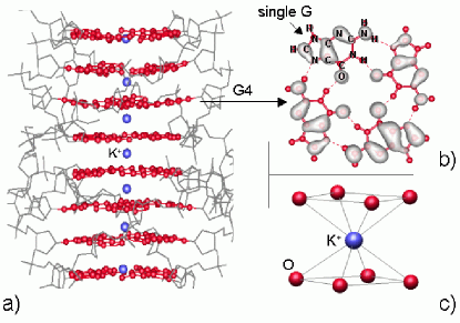
In this paper, we focus on nanowires known as G4-wires 15 (or quadruplexes), consisting of stacked guanine (G) tetrads (G4): the structure of these systems is illustrated and described in Figure 1. These one-dimensional polymers are becoming appealing as prospective candidates for bio-molecular electronics because, due to the low ionization potential of guanine (the lowest among nucleic-acid bases), they might be suitable to mediate charge transport by hole conduction along the helix, and have even been suggested as nano-mechanical extension-contraction machines. 16 In the presence of appropriate metal cations (especially K+ and Na+), solutions of homoguanylic strands in water, 17 ; 18 as well as lipophilic guanosine monomers in organic solvents, 19 self-assemble in right-handed quadruple helices. The G4-motif has been identified in both cases; however, while the quadruplexes obtained from guanylate strands have an outer mantle of sugars and phosphates (as in DNA) that connects the adjacent G4 planes, those obtained from lipophilic guanosine derivatives have no interplanar connection: a continuous linker between consecutive tetramers is not necessary to form the wires. The guanine quadruplexes have recently attracted interest because of their possible role in biological systems: 20 their biological relevance has propelled a large number of investigations (e.g. X-ray 21 and NMR 22 ) aimed at characterizing such unusual supramolecular structures. Quadruple helices have been obtained in the presence of different monovalent (K+, Na+, NH4+) 19 a, 23 and divalent (Ca2+, Ba2+, Sr2+, Pb2+) 19 b, 24 cations. Despite the different chemical nature of the constituents, all these G4-wires are characterized by an inner core of stacked G4’s intercalated by metal cations: one cation in every tetrad and one cation in every other tetrad, in the case of mono- and di-valent ions, respectively. The self-assembly capability of the G4-helices allows for the formation of quite long (100-1000 nm) and stiff wires. AFM images 25 of four-stranded helices obtained from G-rich oligonucleotides on substrates revealed the G4-wires to be uniform and relatively rigid polymers, with few bends, kinks, or branches.
Despite the large amount of structural investigations, the conduction properties of these nanowires are basically unknown and a direct measurement of electrical properties of G4-wires is still missing. From the theoretical point of view, quantum chemistry and molecular dynamics studies focused on the energetics and on the geometry of isolated G-quartets or finite clusters of stacked G4’s, 26 whereas the electronic properties of these materials were so far investigated to a much lesser extent. 27
In the following, we present a first-principle investigation of the electronic and conduction properties of periodically repeated G4-wires. Recently, 27 we described the structure and the energetics, as well as some basic features of the electronic structure, of an infinite G4-wire with and without the presence of K+ ions in the inner cavity. In this paper, we focus entirely on the electronic properties of the same system and present a complete and thorough analysis of the guanine-guanine and metal-guanine interactions, discussing the effects on the conduction properties of the tubular system.
II Computational approach
We performed ab-initio calculations of the electronic and conduction properties of infinite G4-wires in the presence of K+ ions, within the Density Functional Theory (DFT) approach, 28 using the PW91 29 gradient-corrected exchange-correlation functional. DFT is lately gaining a large credit in the scientific community as a reliable and accurate method to describe large-scale biomolecular aggregates, 30 including guanine-based stacks. 31
Our total-energy-and-force calculations 32 allowed us to attain a simultaneous description of the optimized atomic configuration and of the corresponding electronic structure for the selected systems (see Figure 1): the finite planar guanine tetrad (G-quartet) and the infinite helical G4-wire filled with K+ ions in the inner cavity (label 3G4/K+). The single-particle electron wave-functions were expanded in a plane-wave basis set with a kinetic-energy cutoff of 25 Ry. Two special k-points in the irreducible wedge were employed for Brillouin Zone (BZ) sums in the case of the G4-wire. The infinite helix was simulated by repeated supercell containing three stacked G4 tetrads, 27 employing periodic boundary conditions in the three spatial direction a thick vacuum layer ( Å) in the directions perpendicular to the helical axis prevented spurious interactions between adjacent replicas of the wire. For the isolated G-quartet, the same vacuum thickness was employed also in the third direction perperdicular to the plane of the tetrad, and only the point was used in the BZ sampling.
The electron-ion interaction was described by non-norm-conserving pseudopotentials 33 for all the species (C, N, O, H) except K, for which a norm-conserving pseudopotential 34 was used. For the latter species, both the valence and the semi-core shells contributed to the system with valence electrons. This treatment represented a significant refinement towards a complete description of the electronic structure, with respect to the simplified results 27 obtained by fixing the shell in the frozen core and by applying Non Linear Core Corrections 35 (NLCC) to account for partial core relaxation.
The starting atomic configuration was obtained from the results of the X-ray analysis 21 of the d(TG4T) quadruple helix. Motivated by the observation that G4-wires form with 17 ; 18 and without 19 the covalent skeleton and by our specific interest in the base stack as a channel for charge mobility, we neglected the external backbone in our simulations and focused on the central core of the helix, constituted of guanines and metal cations (see Fig. 1a). This choice is supported by theoretical reports 36 for approximated DNA structural models, which assert that if any current flows in such systems, it does through the base-stacking, without involving the external mantle in the transport phenomena. The same evidence was more recently confirmed by DFT calculations of the electronic properties of real DNA sequences (A-DNA 8 a and Z-DNA 37 ): these simulations showed that the orbitals related to the sugar-phosphate backbone are a few eV below the Highest Occupied Molecular Orbital (HOMO) and above the Lowest Unoccupied Molecular Orbital (LUMO) of the system. In principle, one should expect that the environment surrounding the base stack plays an important role in the overall conductivity properties of DNA molecules. 37 ; 38 ; 39 Recent studies 37 ; 39 b pointed out that a random distribution of counter-ions in the unit cell modifies the electrical properties of DNA both reducing the bulk quantum conductance and introducing localized empty states in the energy gap. However, this result is coherent with a picture where the external ions quantitatively affect the global transport properties of the system, but do not constitute an alternative pathway for the electron/hole transport through the helix. 7 Therefore, we believe that the bare guanine core will be well representative of the essential electronic properties of the quadruplexes, allowing for the inspection of the charge-migration mechanisms and of the key features of metal-molecule interaction. An explicit account for the effects of the backbone and of the surrounding counterions 37 ; 39 would instead be demanded for a quantitative evaluation of the quantum conductance to be compared with measured transport characteristics, which is way beyond the purpose of the present work.
III Results and discussions
By means of the first-principle approach outlined above, we optimized both the isolated G-quartet and the periodic G4-stack.
The analysis of the electronic structure of the planar G-quartet shows interesting features that will help understanding the electrical properties of G4-wires. As also found for the other planar G-aggregates (e.g. dimers, ribbons), 31 the H-bonds among the guanines do not favor the formation of supramolecular orbitals extended on the whole G-quartet and the existence of dispersive bands. On the other hand, the intermolecular interactions split each guanine energy level into a multiplet structure: each multiplet is composed of four (the number of G’s in the tetrad) energy levels and has a total width of about 200 meV. The orbitals that contribute to a manifold have identical character and are localized on the individual G molecules. Figure 1b shows an isosurface plot of the convolution of four electron states (the HOMO’s of the four guanines in the tetrad) which form the -like HOMO manifold.
By exploiting the square-symmetry of the planar tetrad and the 30∘ twist angle between consecutive tetrads, we simulated a quadruplex of infinite length with a periodically repeated unit supercell containing three stacked G-quartets and three intercalated K+ ions in the unit cell. 27 Each potassium ion was symmetrically located between two consecutive tetrads, and bipyramidally coordinated with the eight (four above and four below) nearest-neighboring oxygen atoms (Fig. 1c). The atomic positions were relaxed until the forces vanished, within an accuracy of 0.03 eV/Å. The structure, the energetics, and the metal-induced stability of the tube were described elsewhere. 27 We find now that the explicit inclusion of the semi-core electrons of K in the valence shell for the pseudopotential calculations does not alter the results obtained previously within the NLCC approximation, 27 and does not change the understanding of the system from the structural point of view. The refined treatment of the semi-core electrons of K allows us to gain a deeper insight into the electronic structure.
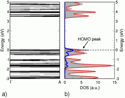
The band-structure of the G4-wire 3G4/K+ is shown in Figure 2a: the unoccupied states (above 3.5 eV) are separated from the occupied states (below 0 eV) by a large energy gap (affected by the typical DFT underestimation 28 ), and some of the states around zero energy are partially filled. We previously demonstrated 31 that - coupling among guanines may give rise to delocalized Bloch-type orbitals, whose band dispersion along the stacking direction depends on the relative rotation angle between nucleobases in adjacent planes. Taking into account the detected rotation angle 21 of 30∘ for the guanine quadruple helices, we now report that the electronic band-structure of the 3G4/K+ column is represented by dispersionless energy bands, indicating that no complete supramolecular orbital delocalization is to be expected for such a degree of helicity. This behavior leads to the conclusion that in G4-wires the - superposition is not sufficient to induce a sizable coupling between the molecular states of guanines and/or the atomic orbitals of potassium, which would lead to purely dispersive bands similar to those of (semi)conducting materials. However, as for the isolated G-quartet, the band-structure of the 3G4/K+ column identifies the presence of manifolds that are equivalent to effective bands: each manifold contains a number (or multiplet) of levels that can be explained in terms of the number of nucleobase molecules and of potassium ions in the periodic supercell, as we discuss later. The orbitals in a multiplet have identical symmetries and some degree of hybridization between neighboring bases. The levels within a manifold are so close (dense) in energy (E 20meV) that an external weak interaction (e.g. the thermal fluctuations or an exterinal electric field) may let the orbitals mix, giving an effective dispersive band. This interpretation suggests a description of the electronic structure where the spreading of the energy levels of the manifold leads to the formation of effective band-like peaks in the Density of States (DOS) (Fig. 2b, shaded gray area). The DOS of the 3G4/K+ wire thus appears similar to those of materials with a dispersive band-structure.
It is important to note that there is a one-to-one correspondence between the band structure and the ground state conduction properties (e.g., the quantum conductance spectrum) of a periodic system in the coherent transport regime: 40 at any given energy, the quantum conductance is a constant value proportional to the number of transmitting channels available for charge mobility, which are equal (in the absence of external leads) to the number of bands at the same energy. Therefore, the formation of effective dispersive bands in the investigated G4-wire is a signal of the intrinsic capability of the material of hosting electron ”energy channels” available for charge migration, whithin continuous energy ranges. On the contrary, the energy spikes that would characterize a discrete spectrum would not be efficient for transport. The extended orbitals (see below) related to the effective bands turn out to be the corresponding viable ”space channels” for carrier mobility, e.g. the pathways through which the carriers may migrate in the wire.
A comparison between the manifolds of the isolated G-quartet and of the G4-wire highlights an increased density of energy levels in the manifolds of the quadruple helix with respect to the tetrad. This effect is due to two factors: (i) the number of energy levels in each multiplet; (ii) the in-plane and out-of-plane guanine-guanine and guanine-metal interactions, that couple the electron states stemming from the various structural elements (either from G or from K) in the supercell. The number of electron states in each manifold depends upon the number of molecules and ions in the unit cell: therefore, whereas each manifold of the G-quartet contains four levels, the manifolds of the 3G4/K+ wire contain twelve levels from the twelve guanines, and additional twelve levels from the K+ ions in the energy range where metal-base hybridization occurs. We come back to this point later when we discuss the DOS. The number of levels in a manifold is not the only element that determines the details of the bandstructure: another key feature is the band-width, which is instead controlled by the specific interactions. Whereas in the case of the G4 tetrad only H-bonding plays a role, in the case of the 3G4/K+ wire there are different contributions from H-bonding and stacking between the bases, and from the coupling between the bases and the metal ions. It is worth noting that the formation of dense manifolds and the consequent establishment of effective band-like potential conduction channels is a common characteristic of stacked H-bonded nucleobase aggregates. In fact, not only we identified the same features in guanine quadruplexes both in the presence and absence of inner metal cations, 27 but similar energy manifolds were also detected in the electronic band-structure of two different DNA duplexes. 7 ; 8 a, 37 For instance, in their simulation of an eleven base-pair poly(dG)-poly(dC) 8 a sequence, de Pablo and coworkers found a HOMO-manifold derived from the eleven states of guanines. In that case, the topmost valence band had a bandwidth of only 40 meV, smaller than that calculated in our G4-wire ( 700 meV). This confirms that, while the splitting of the energy levels into multiplets is a fingerprint of stacked rotated H-bonded nucleobases, the peculiar characteristic of the manifolds depend on the intrinsic stacking properties (e.g. the - coupling) of each type of helix.
To investigate more closely the effects of the metal cations and of the metal-molecule interaction, we projected 41 the total density of states of 3G4/K+ (shaded gray area in Fig. 2b) on G (thin red curve) and K (thick blue curve) atomic orbitals. The G- and K-Projected DOS’s (PDOS’s) show that in a large portion of the spectrum (below -2 eV and above 3.5 eV) only the guanine electron states contribute to the DOS. For instance, the LUMO peak at 3.5 eV has a pure guanine character, being the convolution of the only twelve LUMO orbitals of twelve G’s. The contribution of the K+ ions to the total DOS is mainly localized at the top of the valence band (between -2 eV and 0 eV). The presence of both guanine and potassium contributions to the DOS (as seen from the superposition of the blue and red peaks in Fig. 2b) at the top of the occupied bands is the consequence of a metal-molecule interaction. The HOMO peak of the 3G4/K+ wire (see the double-peak indicated by an arrow in Figure 2b) is due to a convolution of 24 electron states deriving from the coupling between the twelve G HOMO’s and the twelve potassium orbitals. Indeed, one would expect that the K+ ions would contribute to the DOS only with the filled orbitals, after the electrons are removed upon ionization of the system. However, if this were the case, by counting the levels in the manifolds we would find only nine completely occupied levels due to the metal (three orbitals from each K ion), in addition to the guanine-based manifolds containing twelve levels each. Instead, the number of computed occupied levels (twelve more than those coming from the guanine counting, and all concentrated in the HOMO double-peak) and the occurrence of partial occupation for some of them (those around the computed Fermi level, see Figure 2b) indicates hybridization in potassium when inserted in the helical guanine complex. Therefore, potassium does not contribute separately with the and the filled shells, but with a partially occupied shell: 7 equivalent electrons in 4 orbitals, which become 6 electrons in 4 orbitals when the system is ionized as the 3G4/K+ wire. Consequently, the highest-energy component of the HOMO double-peak has an occupation factor of 3/4 the Fermi level is pinned at the top of the effective valence band (Fig. 2b) and the G4-wire in the presence of K+ ions behaves as an intrinsically p-type doped system. The observation of delocalized holes at the topmost valence band is in agreement with the description of the electronic structure of potassium discussed above, where each ionized K+ atom contributes to the whole electronic structure not with three fully occupied -levels, but with four partially occupied -states.
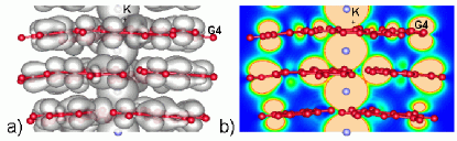
The spread -orbitals of potassium easily interact with the surrounding molecular orbitals of the tetrads, giving hybrid metal-molecule states. We visualized this effect by drawing the convolution of the effective HOMO of the wire. Figure 3a shows an isosurface of the HOMO charge density in the unit cell, obtained from the convolution of the 24 highest occupied orbitals that contribute to the HOMO double-peak. By comparison with Figure 1b, it is possible to recognize a uniform distribution of the orbitals deriving from the tetrads, but also the inner delocalized density stemming from the orbitals of the K+ ions. By cutting the HOMO charge density with a plane perpendicular to the G-quartets, we further inspect the features of the metal-molecule interaction (Fig. 3b). Due to the high degree of charge delocalization along the wire axis, the G4-wires may be described as good electron/hole channels for mobile charges. Two types of pathways for such mobile charges can be identified in Figure 3b and contribute to the conductivity channels. The first one stems from the low-energy component of the HOMO double-peak, is extended through the guanine core of the helix, and is due to the base-base interaction. Similar channels are also observed for the other manifold-derived peaks (e.g. LUMO) and in the empty (K free) quadruple helix. 27 The second type of pathway is due to the high-energy component of the HOMO peak and results from the metal-base interaction. This hybrid pathway is centered around the potassium ions in the central cavity of the wire, and it clearly shows the coupling with the coordinated oxygen atoms of the G molecules. Hence, the inclusion of the metal cations inside the helix drastically influences the electronic properties of the system. The details of the metal-nucleobase interactions depend upon the nature of the cation included in the helix. In the present case of potassium, its coupling with the G bases modifies the topmost valence band, favoring the formation of extended orbitals along the stacking direction and enhancing the conduction properties of the empty guanine structure. 27
To get further insights into the metal-base coupling, we compared the 3G4/K+ wire with other two similar neutral systems: the empty quadruple helix (labeled 3G4) and the G4-wire with neutral K atoms (labeled 3G4/K). The structures 3G4 and 3G4/K do not describe real systems, but are useful models for better understanding the 3G4/K+ quadruplexes. The changes of the inner core do not basically modify the geometry (bond lengths and angles) of the quadruple helix, but affect the electronic structure. Figure 4 shows the comparison among the DOS (shaded gray areas) and the PDOS (thin red and thick blue curves) of the systems. To compare the three structures we aligned the bottom energy levels, and fixed the origin of the energy scale at the top of the valence band of the empty 3G4 helix (taken as reference), with the purpose to outline the effects of the inclusion of metals into the empty guanine supramolecular structure.
As already mentioned, in the DOS of all the studied G4-wires we observe the occurrence of broad G-derived peaks, reflecting the same guanine aggregation state. The empty tube has the valence band completely occupied and the Fermi level lays in the middle of the gap; in the presence of potassium (3G4/K and 3G4/K+), the Fermi level is pinned at the top of a partially occupied valence band. In the 3G4 case, the HOMO peak is the convolution of the HOMO’s of guanines ( in Fig. 4), which are completely occupied; in the other two cases, instead, the HOMO derives from the interactions between the HOMO’s of guanine () and the hybrid states of potassium (labeled in Fig. 4), which are partially filled. One may consider the 3G4/K structure as obtained by adding three electrons to the 3G4/K+. The inclusion of such electrons increases (from 3/4 to 7/8 filling factor) but does not complete the occupation of the topmost band; thus, the helix with the atomic K would also be an intrinsic p-type doped wire.
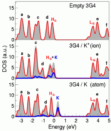
By analyzing the position and the width of the band-like peaks for the three systems, we can underline further details about the K-G interactions. As the K-derived states are found at the top of the valence band, far away from that range the spectra are identical, e.g. peaks have the same width, shape and positions in the three plots. Also the G-like LUMO peak remains basically unchanged. On the contrary, in the 3G4/K+ wire (central panel in Figure 4) the occurrence of a non-vanishing K-PDOS in the [-2;-1] eV range strongly modifies the peaks and with respect to the empty wire: the electrostatic coupling between the -like peak and the orbitals of the K+ ions shifts the position of this peak to lower energies and reduces its height. All the other peaks in Fig. 4 have a -like character and are less influenced by the G-K+ coupling. The displacement of peak also modifies peak , which is sharper and higher in 3G4/K+ than in the free-standing tube. In the neutral 3G4/K helix, the electrostatic coupling between K and G is more effectively screened because of the larger number of electrons in the system: as a consequence, the shift of peak is reduced with respect to the 3G4/K+ case, and peak remains similar to that of the empty wire. Another difference that we remark is the composition of the HOMO peaks: whereas for the 3G4 empty tube the HOMO is purely G-like (), it is a mixed G-K-like double-peak () for the charged 3G4/K+ wire. For the neutral 3G4/K wire, the HOMO (Fig. 4, middle panel) is split into two sharper peaks (Fig. 4, bottom panel): a lower energy component similar to the peak and a higher component with a prevalent character. The decrease in the degree of K-G orbital mixing in 3G4/K with respect to 3G4/K+ is most likely attributable to the increased occupation of the shell of potassium, which becomes more inert because more similar to a closed shell.
These changes in the electronic structure due to different charge states of the inner metals underline the delicate equilibrium that rules the stability and the mutual interactions in this hybrid organic/inorganic molecule/metal complex. Differently from M-DNA,13 where the metal cation substitutes for an immino hydrogen atom and is covalently bonded to a nucleobase, in G4-wires there is no direct charge sharing between the metal and the guanines. The stability and the electronic properties of the system are ruled by the coordination ratio among the K+ and the eight nearest-neighboring oxygen atoms, symmetrically located above and below the ion (Fig. 1c). The metal cations stabilize the structure via electrostatic interactions, but also constitute an effective bridge between the electronic charge density around the oxygen atoms: the HOMO contour plot shown in Figure 3b is the result of the - coupling between the oxygens of consecutive tetrads, mediated by the electronic structure of the K+ ion.
The possibility of changing the electronic properties of the G4-wire, by using different metal ions to stabilize the stack, is expected to be a powerful tool for tuning the conduction properties of the nanowires. We are currently exploring the inclusion of other cations with different outer-shell electronic configurations (e.g. the transition metals), that may be exploited to further change the conductivity of G4-wires.
IV Conclusions
The first principle study of the electronic and conduction properties of infinite G4-wires, stabilized by K+ inner cations, shows that the - coupling among stacked planar tetrads is insufficient to induce the formation of dispersive bands in the wires. However, the presence of closely spaced energy levels leads to the formation of manifolds, whose density of states suggests a band-like behavior. The coupling among guanine-localized molecular orbitals, which may be easily induced by a weak external interaction, gives rise to extended electron channels, suitable to host mobile charge carriers along the wire.
While the formation of split manifolds seems to be a general feature of base-base interactions in H-bonded stacked supramolecular nucleobase aggregates, the effects due to the presence of the metals depend on the identity of the cations. In the case of potassium, the inclusion of the cations enhances the conduction properties of the system, generating additional extended electronic channels stemming from the metal-guanine interaction. The mixed -orbitals of K+ hybridize with the HOMO’s of guanine, and this coupling gives origin to a partially filled HOMO band, which makes the system equivalent to a p-doped wide-bandgap semiconductor.
The above results, along with their attractive mechanical and self-assembly properties, suggest that G4-wires may be explored as viable DNA-based conductors for nanoscale molecular electronics. The computed properties are the equivalent of those of a bulk material: of course, the actual behavior as a wire in a device setting depend on the specific device implementation, is affected from conditions such as electrode-wire and substrate-wire coupling, and leaves several open issues for further investigation and exploitation.
V Acknowledgments
We gratefully thank Joshua Jortner for illuminating discussions. This work was partially supported by the EC through project ”DNA-based nanowires” IST-2001-38951, by INFM through ”Progetto calcolo parallelo” which provided computer time at CINECA (Bologna, Italy), and by MIUR (Italy) through grant ”FIRB-NOMADE”.
References
- (1) (a) Joachim, C.; Gimzewski, J. K.; Aviram, A. Nature 2000, 408, 541-548. (b) Heath, J. R.; Ratner, M. A. Physics Today 2003, 56, 43-49.
- (2) Seeman, N.C. Nature 2003, 421, 427-431.
- (3) Niemeyer, C. M. Angew. Chem Int. Ed. 2001, 40, 4128-4158.
- (4) Yan, H.; Zhang, X.; Shen, Z.; Seeman, N. C. Nature 2002, 415, 62-65.
- (5) (a) Braun, E.; Eichen Y.; Sivan, U.; Ben-Yoseph, G. Nature 1998, 391, 775-778. (b) Keren, K.; Krueger, M.; Gilad, R.; Ben-Yoseph, G.; Sivan, U.; Braun, E. Science 2002, 297, 72-75. (c) Warner, M. G.; Hutchison, J. E. Nature Materials 2003, 2, 272-277.
- (6) Dekker, C.; Ratner, M. A. Phys. World 2001, 8, 29-34.
- (7) Porath, D.; Cuniberti, G.; Di Felice, R. to be published in Long-range charge transfer in DNA, Schuster, G., Ed.; Springer-Verlag, Berlin 2003.
- (8) (a) de Pablo, P. J.; Moreno-Herrero, F.; Colchero, J.; Gómez Herrero, J.; Herrero, P.; Baró A. M.; Ordejon, P.; Soler, J. M.; Artacho, E. Phys. Rev. Lett. 2000, 85, 4992-4995. (b) Storm, A.J.; van Noort, J.; de Vries, S.; Dekker, C. Appl. Phys. Lett. 2001, 79, 3881-3883.
- (9) (a) Porath, D.; Bezryadin, A.; de Vries, S.; Dekker, C. Nature 2000, 403, 635-638 . (b) Cai, L.; Tabata, H.; Kawai, T. Appl. Phys. Lett. 2000, 77, 3105-3107.
- (10) Fink, H.-W.; Schönenberger, C. Nature 1999, 398, 407-410.
- (11) Kasumov, A. Yu.; Kociak, M. Guéron, S.; Reulet, B.; Volkov, V. T.; Klinov, D. V.; Bouchiat, H. Science 2001, 291, 280-282.
- (12) Richter, J. Physica E 2003, 16, 157-173.
- (13) Rakitin, A.; Aich, P.; Papadopoulos, C.; Kobzar, Yu.; Vedeneev, A. S.; Lee, J. S.; Xu, J. M. Phys. Rev. Lett. 2001, 86, 3670-3673.
- (14) Tanaka, K.; Tengeiji, A.; Kato, T.; Toyama, N.; Shionoya, M. Science 2003, 299, 1212-1213.
- (15) The term G-wire has previously appeared in literature to label quadruple helices deriving both from guanine rich sequences (e.g. G4T2G4) and from lipophilic guanosine monomers. In this paper we will use G4-wirei to describe the four stranded homoguanylic rods.
- (16) (a) Alberti, P.; Mergny, J. L. Proc. Natl. Acad. Sci. USA 2003, 100, 1569-1573. (b) Li, J. J.; Tan, W. Nano Lett. 2002, 2, 315-318.
- (17) Saenger, W. Principles of Nucleic Acids, Springer-Verlag, NY 1984.
- (18) Gottarelli, G.; Spada, G. P.; Garbesi, A. in Comprehensive Supramolecular Chemistry vol. 9, Atwood, J. L.; Davies, J. E. D.; MacNicol, D. D.; Vögtle, F., Eds.; Pergamon 1996.
- (19) (a) Marlow, A. L.; Mezzina, E.; Spada, G. P.; Masiero, S.; Davis, J. T.; Gottarelli, G. J. Org. Chem. 1999, 64, 5116-5123. (b) Kotch, F. W.; Fettinger, J. C.; Davis, J. T.; Org. Lett. 2000, 2, 3277-3280.
- (20) (a) Williamson, J. R.; Raghuraman, M. K.; Cech, T. R. Cell 1989, 59, 871-880. (b) Parkinson, G. N.; Lee, M. P. N.; Neidle, S. Nature 2002, 417, 876-880. (c) Dapic, V.; Abdomerovic, V.; Marrington, R.; Peberdy, J.; Rodger, A.; Trent, J. O.; Bates, P. J. Nucleic Acids Res. 2003, 31, 2097-2107.
- (21) (a) Laughlan, G.; Murchie, A. I. H.; Norman, D. G.; Moore, M. H.; Moody, P. C. E.; Lilley, D. M. J.; Luisi, B. Science 1994, 265, 520-524. (b) Phillips, K.; Dauter, Z.; Murchie, A. I. H.; Lilley, D. M. J.; Luisi, B. J. Mol. Biol. 1997, 273, 171-182. X-ray structure ID UDF062.
- (22) (a) Aboul-ela, F.; Murchie, A. I. H.; Norman, D. G.; Lilley, D. M. J. J. Mol. Biol. 1994, 243, 458-471. (b) Rovnyak, D.; Baldus, M.; Wu, G.; Hud, N. V.; Feigon, J.; Griffin, R. G. J. Am. Chem. Soc. 2000, 122, 11423-11429.
- (23) (a) Schultze, P.; Hud, N. V.; Smith, F. W.; Feigon, J. Nucleic Acids Res. 1999, 27, 3018-3028. (b) Marathias, V. M.; Bolton, P. H. Biochemistry 1999, 38, 4355-4364. (c) Basu, S.; Szewczak, A. A.; Cocco, M.; Strobel, S. A. J. Am. Chem. Soc. 2000, 122, 3240-3241. (d) Wong, A.; Fettinger, J. C.; Forman, S. L.; Davis, J. T.; Wu, G. J. Am. Chem. Soc. 2002, 124, 742-743. (e) Črnugelj, M.; Hud, N. V.; Plavec, J. J. Mol. Biol. 2002, 320, 911-924.
- (24) (a) Deng, J.; Xiong Y.; Sundaralingam, M. Proc. Natl. Acad. Sci. USA 2001, 98, 13665-13670. (b) Shi, X.; Fettinger, J. C.; Davis, J. T.; J. Am. Chem. Soc. 2001, 123, 6738-6739. (c) Miyoshi, D.; Nakao, A.; Sugimoto, N. Nucleic Acids Res. 2003, 31, 1156-1163.
- (25) (a) Marsh, T. C.; Vesenka, J.; Henderson, E. Nucleic Acids Res. 1995, 23, 696-700. (b) Porath, D. private communication.
- (26) (a) Töhl, J.; Eimer, W. Biophys. Chem. 1997, 67, 177-186. (b) Spackova, N.; Berger, I.; Sponer, J. J. Am. Chem. Soc. 1999, 121, 5519-5534. (c) Gu, J.; Leszczynski, J. J. Phys. Chem. A 2000, 104, 6308-6313. (d) Gu, J.; Leszczynski, J. J. Phys. Chem. A 2002, 106, 529-532. (e) Chowdhury, S.; Bansal, M. J. J. Phys. Chem. B 2001, 105, 7572-7578. (f) Meyer, M.; Steinke, T.; Brandl, Sühnel, J. J. Comp. Chem. 2001, 22, 109-124. (g) Louit, G.; Hocquet, A.; Ghomi, M.; Meyer, M.; Sühnel, J. PhysChemComm 2003, 6, 1-5.
- (27) Calzolari, A.; Di Felice, R.; Molinari, E.; Garbesi, A. Appl. Phys. Lett. 2002, 80, 3331-3333.
- (28) Wimmer, E. Density Functional Approaches for Molecular and Materials Design; Laird, B. B.; Ross, R. B.; Ziegler, T., Eds.; American Chemical Society, Washington DC, 1996; p. 423.
- (29) Perdew, J. P.; Chevary, J. A.; Vosko, S. H.; Jackson, K. A.; Pederson, M. R.; Singh, D. J.; Fiolhais, C. Phys. Rev. B 1992, 46, 6671-6687.
- (30) Friesner, R. A.; Dunietz, B. D. Acc. Chem. Res. 2001, 34, 351-358.
- (31) (a) Di Felice, R.; Calzolari, A.; Molinari, E.; Garbesi, Phys. Rev. B 2002, 65, 045104; (b) Calzolari, A.; Di Felice, R.; Molinari, E.; Garbesi, A. Physica E 2002, 13, 1236-1240.
- (32) We used the code PWSCF by Baroni, S; Dal Corso, A.; de Gironcoli, S.; Giannozzi, P., available at http://www.pwscf.org.
- (33) Vanderbilt, D. Phys. Rev. B 1990, 41, 7892-7895.
- (34) Troullier, N.; Martins, J. L. Phys. Rev. B 1992, 46, 1754-1765.
- (35) Louie, S. G.; Froyen, S. ; Cohen, M. L. Phys. Rev. B 1982, 26, 1738-1742.
- (36) (a) Ladik J. J.; Ye, Y.-J. Phys. Stat. Sol. (b) 1998, 205, 3-10; (b) Ye, Y.-J.; Jiang, Y. Int. J. Quant. Chem. 2000, 78, 112-130.
- (37) Gervasio, F. L.; Carloni, P.; Parrinello, M. Phys. Rev. Lett. 2002, 89, 108102.
- (38) (a) Lee, H.-Y.; Tanaka, H.; Otsuka, Y.; Yoo, K.-H.; Lee, J.-O.; Kawai, T. Appl. Phys. Lett. 2002, 80, 1670-1672, (b) Das, R.; Mills, T. T.; Kwok, L. W.; Maskel, G. S.; Millett, I. S.; Doniach, S.; Finkelstein, K. D.; Herschlag, D.; Pollack, L. Phys. Rev. Lett. 2003, 90, 188103.
- (39) (a) Adessi, Ch.; Walch, S.; Anantram, M. P. Phys. Rev. B 2003, 67, 081405(R); (b) Adessi, Ch.; Anantram, M. P. Appl. Phys. Lett. 2003, 82, 2353-2355.
- (40) Datta, S.; Electronic transport in mesoscopic systems, Cambridge University Press 1995.
- (41) The G projection was obtained by summing over the projections onto the atomic orbitals of all the species contained in the guanine molecules.