Characterization of low-energy magnetic excitations in chromium
Abstract
The low-energy excitations of Cr, i.e. the Fincher-Burke (FB) modes, have been investigated in the transversely polarized spin-density-wave phase by inelastic neutron scattering using a single- crystal with a propagation vector parallel to . The constant-momentum-transfer scans show that the energy spectra consist of two components, namely dispersive FB modes and an almost energy-independent cross section. Most remarkably, we find that the spectrum of the FB modes exhibits one peak at 140 K near and two peaks near , respectively. This is surprising because Cr crystallizes in a centro-symmetric bcc structure. The asymmetry of those energy spectra decreases with increasing temperature. In addition, the observed magnetic peak intensity is independent of suggesting a transfer of spectral-weight between the upper and lower FB modes. The energy-independent cross section is localized only between the incommensurate peaks and develops rapidly with increasing temperature.
pacs:
PACS numbers: 75.30Fv, 75.50.Ee, 75.40.Gb, 75.30.DsI Introduction
Although chromium exhibits a simple bcc structure and consists only of a single element the magnetism is very complicated and one of the most intriguing subjects in condensed matter physics. fawcett88 . Below the Néel temperature K, the magnetic structure exhibits an incommensurate antiferromagnetic transverse spin-density-wave (TSDW) with the moments oriented perpendicular to the ordering wavevectors () Werner67 . Below the spin-flop temperature K the moments arrange along in a longitudinal spin-density-wave (LSDW). The pitch of the modulation is approximately at low temperatures.
The inelastic magnetic cross section shows also a surprisingly rich behavior (Fig. 1). High-velocity excitations (often called spin waves) emerge from the incommensurate positions fincher81 ; boeni98 . The excitations exhibit longitudinal and transverse polarization lorenzo94 ; fukuda96 , the former one being assigned to a phason mode fishman96 . In particular, while the longitudinal mode dominates below an energy transfer meV, the incommensurate excitations become isotropic above 8 meV. These modes are present in the LSDW as well as in the TSDW phase.
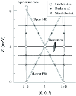
In addition to the high-energy excitations at , Fincher et al., fincher79 ; fincher81 Burke et al. burke83 and Sternlieb et al. sternlieb93 found another magnetic mode in the TSDW phase at low energies that is located between the incommensurate peaks. The dispersion relation of this so-called Fincher-Burke (FB) mode was reported to emanate from the positions and to merge into the high-energy excitations near meV (Fig. 1). The two FB-branches cross each other at meV and . (Hereafter, we redefine the two branches as upper and lower FB mode, respectively, with the boundary energy of .) By means of neutron scattering with polarized neutrons it was shown that the FB modes are longitudinally polarized pynn92 ; pynn97 ; boeni98 .
Very recently, Hiraka et al. re-investigated the FB mode using unpolarized neutrons and found by means of constant-momentum-transfer scans (constant-Q scans) a new excitation mode hiraka03 that emerges from the point towards a direction transverse to within the plane. Interestingly, the gap-type mode extends with a low velocity up to at least and shows a decreasing intensity with increasing . Using a cold neutron triple-axis spectrometer with high-resolution Böni et al. investigated the FB mode in detail boeni02 using constant- and constant-energy-transfer scans (constant- scans). They pointed out that the FB mode neither shows a simple linear dispersion nor obeys a simple spin-wave picture with respect to intensity. In addition, the intensity contour for indicated that the FB mode is asymmetric with respect to . However, it was not clear if this was due to spurious scattering.
In order to obtain a coherent picture of the low-energy excitations in Cr we have performed a detailed investigation of the FB modes in the TSDW phase by means of constant- scans using thermal- and cold-neutron spectrometers. Previous measurements were mostly performed by constant-E scans (Fig. 1). However, the intense high-energy excitations made the quantitative analysis of the weak response of the FB modes difficult.
The major result of this work is a confirmation of the asymmetry of the energy spectra with respect to near using a different sample and different spectrometers. We also observed that the energy spectra become more symmetric at high temperatures. In addition, we show that the FB modes conserve intensity along (or ), in contrast to the transverse mode that shows a drastic depression of intensity along and hiraka03 . The nearly energy-independent component shows a strong -dependence of the cross section that may be closely related to the commensurate scattering fincher81 ; fukuda96 or the critical scattering discussed by Sternlieb et al. sternlieb93 .
II Experimental procedure
For the present experiments we have used the identical single crystal of cm3 that was previously investigated hiraka03 . The single- structure pointing along was induced by cooling the sample through in a strong magnetic field yielding a domain population %. The crystal was mounted inside a closed cycle refrigerator with the scattering plane horizontal. The measurements with thermal and cold neutrons were conducted on the triple-axis spectrometer TOPAN installed at the JRR-3M research reactor at the Japan Atomic Energy Research Institute and the triple-axis spectrometer SPINS at the National Institute for Standard and Technology, respectively. The reflection of pyrolytic graphite was used to monochromate and analyze the neutron energy. The final neutron energy of TOPAN (SPINS) was fixed at 14.7 meV (3.7 meV), and the horizontal-collimation sequence was set to 30’-60’-30’-60’ (guide-40’-40’-open) from before the monochromator to after the analyzer. The energy resolution of the spectrometers was estimated to be less than 1.2 meV and 0.4 meV, respectively. Higher-order neutrons were removed by means of a pyrolytic graphite filter and a BeO filter for TOPAN and SPINS, respectively.
III Experimental results
In Fig. 2 we show the salient results of the present work. It is clearly seen that spectra evolve from a single peak at towards a double-peak structure at . The significance of this result was confirmed by reproducing the scans under different experimental conditions.
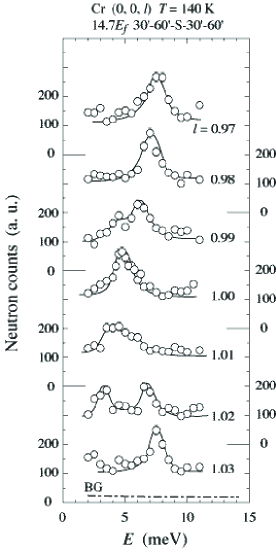
Figure 3 shows detailed measurements at the commensurate position and at the intermediate position for temperatures below and above . The so-called Fincher peak at fincher79 is broader than the -resolution. A broadening of the peaks is also noticed in the scan at . Below , the peak structure disappears completely. At the same time the -independent magnetic scattering decreases too, but it is significantly larger than the background of counts/5 min that was measured at various points in the Brillouin zone. The behavior of the -independent component is consistent with “commensurate scattering” which has already been observed before by means of constant- scans using unpolarized fukuda96 and polarized neutrons boeni98 .
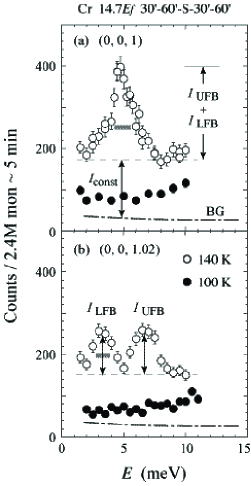
In order to parametrize the data we have fitted the peaks in Figs. 2 and 3 after subtraction of the background to one or two Lorentzians plus a constant, . We express peak heights of the former part as and for the upper and lower FB mode, respectively (Fig. 3). The latter is considered to be independent of but depends on . Figure 4(a) summarizes the -dependence of the energy position of the FB mode. The vertical thick bars indicate the width of the peaks which are, roughly speaking, constant against . It is clearly seen that the dispersion is extremely asymmetric with respect to because no peaks are observed at the low- side of the lower FB branch.
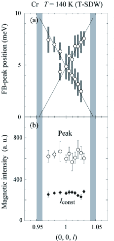
Figure 4(b) shows the peak intensity defined as a sum of versus , being almost constant between the high-energy incommensurate scattering. At this stage, no corrections have been made except taking into account a squared magnetic form factor for Cr2+ free ions. The -independent scattering contributes about half of the above mentioned peak intensity near and it is insensitive to also. Therefore, the intensity of the FB modes () is independent of . At the high- side a transfer of intensity from the upper to the lower FB branch takes place while keeping the total intensity fixed.
Böni et al. have already found an asymmetry of the FB mode for the first time boeni02 based on an intensity contour map constructed from constant- and constant- scans. They found a blob of scattering at near 3 meV. However, the quality of the data did not allow to clearly establish the pronounced (and unexpected) asymmetry in scattering unambiguously boeni-private . Our new measurements clearly reproduce the asymmetry between the scattering at and as shown in Fig. 5 using a different single- crystal on a different spectrometer with different resolution.
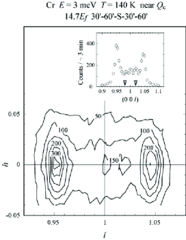
We notice here once more, that it will be difficult to obtain new insight from constant- scans as shown in the inset of Fig. 5, unless the incommensurate scattering at can be correctly evaluated and convoluted with the instrumental resolution function. We emphasize that it is the constant- technique that allows the unambiguous determination of dispersion relations and not the constant- technique that leads often to wrong conclusions as proven in the past.
Figures 6 and 7 show the evolution of the inelastic intensity at and with , respectively. The magnetic intensity increases much faster than expected on the basis of the thermal population factor, , indicating that the scattering is not due to spin waves clementyev04 . In particular, a remarkable growth is seen in as shown with shades. The increase is also not due to conventional critical scattering near because the measurements were performed far away from K, i.e. at reduced temperatures of 0.58 and 0.71 for 180 K and 220 K, respectively. We point out that with increasing the “missing” peak develops at near 3 meV (Fig. 7), i.e. the spectrum becomes more symmetric with increasing and approaches the cross section at (Fig. 6).
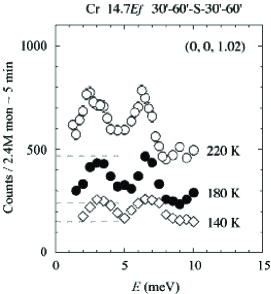
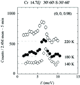
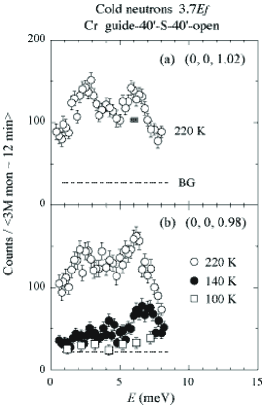
Figure 8 shows (i) once more the asymmetry of the scattering with respect to and (ii) the reduction of unbalance for two peaks at with increasing . These high-resolution measurements ( meV in FWHM) confirm the results from Fig. 7 ( meV) that were performed with thermal neutrons. Due to the very different resolution conditions the asymmetry of the scattering is less pronounced with cold neutrons.
IV Discussions
We have shown that the FB modes can be measured more precisely by means of constant- scans than by means of constant- scans. As a result, it was possible to clearly identify an asymmetry of the magnetic cross sections with respect to the commensurate position . The asymmetry decreases with increasing . We have proven by using very different experimental conditions and different spectrometers that the asymmetry is not due to a resolution effect. The asymmetry is also not an artifact of the sample because similar results were obtained using another Cr single crystal and another spectrometer boeni02 . It might be interesting to investigate the asymmetry of the scattering in other Brillouin zones.
Our results also indicate that the FB modes are not spin-wave modes because the peak intensity is independent of (Fig. 4(a)). Moreover, we observe a transfer of intensity between the lower and upper branches of the excitations. Finally, the intensity of the modes increases significantly faster with increasing than given by , which is in agreement with previous works fincher81 ; clementyev04 . One may speculate that we have to deal with a thermally activated process. Another possible explanation may be reached by considering mode-mode coupling between the upper and lower FB branches harada71 .
The present results strongly suggest the existence of magnetic scatterting that develops around the commensurate position. Its intensity increases also significantly faster than given by and finally dominates the inelastic scattering between in agreement with previous constant- works fincher81 ; fukuda96 . should contribute to the significant critical scattering near fincher81 . In contrast to the FB modes this scattering is polarization independent boeni98 and exists already in the LSDW phase below the spin-flop transition. The energy-independent feature of is also present transverse to but drastically decreases with increasing and hiraka03 . We do not know the origin yet. One may speculate that the FB modes as well as the -independent scattering may be explained by the complicated shape of the Fermi surface that gives rise to many different modes. We mention in particular that already a three-band model gives rise to at least 25 magnetic modes fishman96 .
V Conclusion
We have reinvestigated the low-energy FB excitations of Cr in the TSDW phase by making extensive use of constant- scans. We have observed an asymmetry in the inelastic scattering near that is at variance with the simple centro-symmetric bcc structure of Cr. The asymmetry of energy spectra decreases with raising . The observed peak intensity of the FB modes satisfies a sum rule with regard to Q. The intensity of the dispersive excitations and the -independent scattering increase with increasing much faster than expected according to the temperature factor . Therefore, the FB modes are not spin-wave modes. We expect that the present results challenge the development of a theory to explain these results and resolve the puzzle of the magnetic excitations in Cr.
Acknowledgements.
We thank Y. Endoh, R. S. Fishman, S. A. Werner and B. J. Sternlieb for helpful discussions. Present research was supported by the U.S.-Japan Cooperative Neutron-Scattering Program. Work at Tohoku University was supported by the Ministry of Monbu-Kagaku-shou of Japan. Work at Brookhaven was supported by the Division of Material Sciences, U.S. Department of Energy under contract DE-AC02-76CH00016. Work at SPINS was based upon activities supported by the NSF under DMR-9986442.References
- (1) E. Fawcett, Rev. Mod. Phys. 60, 209 (1988).
- (2) S. A. Werner, A. Arrott, and H. Kendrick, Phys. Rev. 155, 528 (1967).
- (3) C. R. Fincher, G. Shirane, and S. A. Werner, Phys. Rev. B 24, 1312 (1981).
- (4) P. Böni, B. J. Sternlieb, G. Shirane, B. Roessli, J. E. Lorenzo, and S. A. Werner, Phys. Rev. B 57, 1057 (1998).
- (5) J. E. Lorenzo, B. J. Sternlieb, G. Shirane, and S. A. Werner, Phys. Rev. Lett. 72, 1762 (1994).
- (6) T. Fukuda, Y. Endoh, K. Yamada, M. Takeda, S. Itoh, M. Arai, and T. Otomo, J. Phys. Soc. Jpn. 65, 1418 (1996).
- (7) R. S. Fishman and S. H. Liu, Phys. Rev. Lett. 76, 2398 (1986); Phys. Rev. B 54, 7252 (1986).
- (8) C. R. Fincher, G. Shirane, and S. A. Werner; Phys. Rev. Lett. 43, 1441 (1979).
- (9) S. K. Burke, W. G. Stirling, K. R. A. Ziebeck, and J. G. Booth, Phys. Rev. Lett. 51, 494 (1983).
- (10) B. Sternlieb, G. Shirane, S. A. Werner, and E. Fawcett, Phys. Rev. B 48, 10217 (1993).
- (11) R. Pynn, W. G. Stirling, and A. Severing, Physica B 180 & 181, 203 (1992).
- (12) R. Pynn, R. T. Azuah, W. G. Stirling, and J. Kulda, Proceedings of a NATO Advanced Study Institute on The Dynamics of Unconventional Magnetic Systems held in Geilo, Norway April 2-12, 1997.
- (13) H. Hiraka, P. Böni, M. Fujita, Y. Endoh, K. Yamada, and G. Shirane, Phys. Rev. B 67, 064423 (2003).
- (14) P. Böni, E. Clementyev, Ch. Stadler, B. Roessli, G. Shirane, and S. A. Werner, Appl. Phys. A 75, 1 (2002).
- (15) P. Böni, private communication.
- (16) E. S. Clementyev, P. Böni, F. Demmel, and G. Shirane, accepted for publication by Elsevier Science.
- (17) J. Harada, J. D. Axe and G. Shirane, Phys. Rev. B 4, 155 (1971).