Supersonic strain front driven by a dense electron-hole plasma
Abstract
We study coherent strain in (001) Ge generated by an ultrafast laser-initiated high density electron-hole plasma. The resultant coherent pulse is probed by time-resolved x-ray diffraction through changes in the anomalous transmission. The acoustic pulse front is driven by ambipolar diffusion of the electron-hole plasma and propagates into the crystal at supersonic speeds. Simulations of the strain including electron-phonon coupling, modified by carrier diffusion and Auger recombination, are in good agreement with the observed dynamics.
pacs:
61.10.Nz, 63.20.-e, 42.65.RESubpicosecond laser-induced electron-hole plasmas in semiconductors can produce large amplitude lattice strain and rapid loss of translational order. These effects have been studied extensively in ultrafast linear and nonlinear reflectivity experiments Shank and Auston (1975); Shank et al. (1982); Tom et al. (1988); Sokolowski-Tinten et al. (1995); Sokolowski-Tinten et al. (1998); Thomsen et al. (1984, 1986); Chigarev et al. (2000); Wright et al. (2001) and, more recently, in time-resolved x-ray Bragg scattering experimentsRose-Petruck et al. (1999); Lindenberg et al. (2000); Siders et al. (1999); Cavalleri et al. (2000, 2001); Solokolowski-Tinten et al. (2001). X-ray diffraction has the advantage that it can provide quantitative structural information.Many of the x-ray experimentsRose-Petruck et al. (1999); Lindenberg et al. (2000); Reis et al. (2001) have been analyzed using the thermoelastic model put forward by Thomsen et al.Thomsen et al. (1986) in which the strain is caused by differential thermal expansion. Deviations from this model are discussed in the work of Thomsen et al. and have been seen in x-ray diffraction Lindenberg et al. (2000); Reis et al. (2001); DeCamp et al. (2001); Cavalleri et al. (2000, 2001). Cavalleri et al.Cavalleri et al. (2000, 2001) studied coherent strain near the thermal melting threshold in Ge and concluded that the strain is produced over a region which is thick compared to the optical penetration depth due to ambipolar diffusion. However, their experiment was only sensitive to structural changes in the near surface region.
In this letter we report on measurements of coherent strain generation in Ge following ultrafast laser-excitation using a bulk sensitive structural probe. We use time-resolved ultrafast x-ray transmission to measure strain propagation deep within the crystal, providing information about the generation process. Initially, the strain front advances at speeds greater than the sound speed. In our experiments, the laser intensity is sufficient to impulsively generate a dense electron-hole plasma at the crystal surface, the dynamics of which are governed by ambipolar diffusion Young and van Driel (1982) and Auger recombination. The plasma couples to the lattice through the deformation potential. In order to probe the resulting coherent acoustic pulse as it travels deep within the bulk, we utilize the Laue geometry whereby the x-rays traverse the full thickness of the crystal, emerging on the other side as two mutually coherent beamsBatterman and Cole (1964). We have recently shown that a short acoustic pulse can coherently transfer energy between these two beams on a time scale inconsistent with the thermo-elastic model. Following the initial transient, the beam intensities oscillate as a function of the pump-probe delayDeCamp et al. (2001). In the experiments reported here, the strain generation is studied as a function of the incident laser fluence. The relative phase of the oscillations and the amplitude of the transient provide information about the strain generation process at times shorter than the x-ray probe duration.
In the Laue geometry, two linearly independent wave solutions propagate through the crystal. Transverse to the propagation, these two solutions are standing waves whose wavelengths are twice the spacing of the diffracting planes. The solutions are usually labelled and with the convention that has its nodes, and its antinodes on the diffracting planes. In the case that all atoms lie on these planes, is maximally transmitted and is maximally absorbed. Because the two solutions interact with different electron densities, they propagate with different velocities.
Outside the crystal, two diffracted beams are produced: one in the direction of the input beam (forward-diffracted or “0” beam), and the other in the direction determined by the vector sum (deflected-diffracted or “H” beam). Here () corresponds to the wavevector of the forward-diffracted (deflected-diffracted) beam and is the reciprocal lattice vector corresponding to the diffracting planes. These beams are linear combinations of the two internal solutions, and . The external intensities are given by:
| (1) | |||||
| (2) |
where () is the diffracted intensity of the forward (deflected) beam, is the complex wave field inside the crystal, is the complex wavevector of the solutions (including absorption), and are determined by the crystal orientation. The two internal modes oscillate in and out of phase as they propagate through the crystal. The wavelength of the interference, , is known as the Pendellösung length which is typically a few to tens of microns and is often shorter than the absorption length.
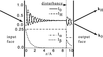
For a crystal that is thick compared to the -absorption length, only the solution survives and there are no interference effects. This is the anomalous transmission of x-rays, known as the Borrmann Effect Batterman and Cole (1964). A distortion of the lattice can cause a redistribution of the interior wave solutions Authier et al. (1996). Figure 1 shows the effect of a thin region of distortion regenerating the solution after it has decayed away in a thick crystal for the case of zero absorption. When this occurs close enough to the crystal exit, the regenerated wave does not decay away and interference occurs at the exit face, despite the fact that the crystal is thick.
In our experiments, a short acoustic pulse is generated at the surface of a thick crystal. This pulse can be considered as a moving lattice disturbance. The diffracted intensities will oscillate in time as the pulse travels into the crystal bulk with a period that is given by the Pendellösung length divided by the speed of sound. Deviations from the impulsive strain generation will be evident in the phase and/or amplitude of the x-ray modulation as a function of pump-probe delay.
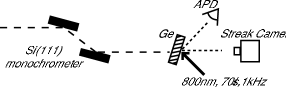
The experiments were performed at the 7-ID undulator beamline at the Advanced Photon Source. The x-ray energy was set to 10keV using a cryogenically cooled Si 111 double crystal monochromator leading to a 1.410-4 fractional energy spread. The x-ray beam is masked by tantalum slits to ensure that the x-ray spot is smaller than the laser spot on the sample and to provide x-ray collimation. The sample is a 280m thick, (001) Ge single crystal. The crystal was oriented such that the x-rays diffracted in the asymmetric Laue geometry. In this geometry, and at 10 keV, the Pendellösung length is 6.2 m and the absorption length is 19m, normal to the surface. Therefore, in the unperturbed crystal, only survives at the exit. The only difference between the two diffracted beams is in their direction and a mismatch in their amplitudes due to details of the boundary conditions on the exit face Batterman and Cole (1964).
Coherent strain pulses are produced on the x-ray output face of the crystal by sub-100fs, 800 nm laser pulses at a 1kHz repetition rate. The excitation is fully reversible between subsequent laser pulses. The laser is phase-locked to the x-ray beam to better than the x-ray pulse duration. The laser is timed to the x-rays using a combination of a digital delay generator and an electronic phase shifter in the phase-locked loop. In this manner the pump-probe delay may be set across a range of 1 ms with 19 ps precision. A fast silicon avalanche photodiode (APD) and a picosecond x-ray streak camera Chang et al. (1996) were used as the time-resolved detectors. The APD sampled the deflected-diffracted beam intensity and the streak camera sampled the forward-diffracted beam (see Fig. 2). The x-ray bunch separation was 152 ns, large enough to allow electronic gating and measurement of a single x-ray pulse.
Following laser-excitation, high contrast oscillations are observed in the pump-probe data over a large span of excitation densities. Figure LABEL:f:oscillations shows these oscillations in the deflected-diffracted beam. The period of oscillation agrees with the Pendellösung length divided by the longitudinal speed of sound. At an incident fluence of 35, the behavior near shows a large transient that is unresolved with the 100 ps x-ray probe beam. After the transient, the oscillations show a significant phase-shift with respect to oscillations that occur following an excitation of 2. The amplitude and frequency of the oscillations are relatively insensitive to the fluence. However, as shown in Fig. LABEL:f:phaseshift, the phase is strongly dependent on the fluence and is correlated with the amplitude of the initial transient. The relative phase of the oscillation was defined with respect to the 2 excitation and was retrieved from a least squares fit Barkhudsen et al. (1985); Wise et al. (1987). The amplitude of the transient is defined as the diffracted intensity at a delay of 200 ps. Most of the energy transfer occurs in 40ps, measured with the forward diffracted beam using a streak camera (see the inset in fig.LABEL:f:oscillations). At relatively high fluences ( 10), the intensity of the deflected diffracted beam approximately doubles while the forward diffracted beam is cut by more than 75%. At relatively low fluences ( 2), there is no transient.
Inspection of (1) and (2) shows that the maximum energy transfer between the forward and deflected beams near the exit of a thick crystal occurs if the and solutions are coupled at a depth of . This implies that the transient behavior is due to a perturbation to the lattice that reaches a depth of more than 1.5 m into the bulk. In the simplified picture that a moving interface couples the and solutions, the excitation must propagate into the bulk at greater than 37,000 m/s, more than seven times the longitudinal speed of soundfoo .
The strain pulse has a finite spatial extent and is comprised of a spectrum of phonons with different wavevectors. We expect that the phonon component with wavelength equal to the Pendellösung length will resonantly couple the two interior wave solutions Entin et al. (1978). To model this phenomenon, we solve the equations of dynamical diffraction within the crystal, taking into account the laser-induced time-dependent strain profiles, using the Takagi-Taupin formalism adapted for Laue geometry Takagi (1962); Burgeat and Taupin (1968). In this method, the differential equations coupling the and branches are solved numerically. The depth-dependent strain profile for a given time is taken into account by noting that local strain is equivalent to a change in the local Bragg angle. Details of this approach (for Bragg geometry) can be found in the original work of TakagiTakagi (1962) and Taupin Burgeat and Taupin (1968). The means by which the method can be adapted for Laue geometry are implicit in the work of Zachariasen Zachariasen (1945) and Batterman and Cole Batterman and Cole (1964).
Pure thermoelastic models of strain propagation do not predict the observed fluence dependence of the phase and amplitude of the Pendellösung oscillations. A proper model must include the effects of the coupling of the photoexcited plasma to the crystal lattice. The strain is comprised of both diffusive and elastic components: the diffusive strain is determined by the instantaneous temperature and carrier density profiles, modified by Auger recombination; the elastic strain is driven by changes in the temperature and carrier density, and propagates into the crystal at the speed of sound. In the absence of diffusion, a bipolar pulse develops in the time given by the optical penetration depth divided by the speed of soundThomsen et al. (1986). For LA phonons with wavectors along [100] and a 0.2 m penetration depth, this corresponds to 40ps. Including diffusion, the electron-hole plasma extends 1 m in the same time, leading to a strain front that has propagated into the bulk faster than the speed of sound.
Figure LABEL:f:ampsim shows the calculated diffraction intensity as a function of laser delay at an absorbed laser fluence of (corresponding to a carrier density of cm-3). Good qualitative agreement with the experiment is seen (Fig. LABEL:f:oscillations). The sharp initial rise in diffraction intensity is reproduced, as well as the frequency and phase of the time-resolved Pendellösung oscillations. Figure LABEL:f:phasesim shows the calculated phase shift and the diffracted intensity as a function of absorbed optical fluence. After taking into account the surface reflectivity of the sample, good agreement with the experiment is obtained (Fig. LABEL:f:phaseshift).
In conclusion we have demonstrated a bulk sensitive probe of lattice dynamics using time-resolved x-ray anomalous transmission. We have observed that electron-phonon coupling modified by carrier diffusion is a dominant mechanism for energy transport in laser-excited Ge. This work could be extended to study how the elastic response of the material can modify the electronic transport properties of semiconductors.
Acknowledgements.
We thank Bernhard Adams, Marcus Hertlein, Don Walko, and Jared Wahlstrand for technical assistance and stimulating discussions. We also thank Jin Wang for use of the intensified CCD camera. This work was conducted at the MHATT-CAT insertion device beamline at the Advanced Photon Source and was supported in part by the U.S. Department of Energy, Grants No. DE-FG02-99ER45743 and No. DE-FG02-00ER15031, by the AFOSR under contract F49620-00-1-0328 through the MURI program and from the NSF FOCUS physics frontier center. One of us (SF) acknowledges the financial support of Science Foundation Ireland. Use of the Advanced Photon Source was supported by the US Department of Energy Basic Energy Sciences, Office of Energy Research under Contract No. W-31-109-Eng-38.References
- Shank and Auston (1975) C. Shank and D. Auston, Phys. Rev. Lett. 34, 479 (1975).
- Shank et al. (1982) C. V. Shank, R. Yen, and C. Hirlimann, Phys. Rev. Lett 51, 900 (1982).
- Tom et al. (1988) H. W. K. Tom, G. D. Aumiller, and C. H. Brito-Cruz, Phys. Rev. Lett. 60, 1438 (1988).
- Sokolowski-Tinten et al. (1995) K. Sokolowski-Tinten, J. Bialkowski, and D. von der Linde, Phys. Rev. B 51, 14186 (1995).
- Sokolowski-Tinten et al. (1998) K. Sokolowski-Tinten et al., Phys. Rev. B 58, R11805 (1998).
- Thomsen et al. (1984) C. Thomsen et al., Phys. Rev. Lett. 53, 989 (1984).
- Thomsen et al. (1986) C. Thomsen et al., Phys. Rev. B 34, 4129 (1986).
- Chigarev et al. (2000) N. V. Chigarev et al., Phys. Rev. B 61, 15837 (2000).
- Wright et al. (2001) O. B. Wright et al., Phys. Rev. B 64, 081202 (2001).
- Rose-Petruck et al. (1999) C. Rose-Petruck et al., Nature 398, 310 (1999).
- Lindenberg et al. (2000) A. M. Lindenberg et al., Phys. Rev. Lett. 84, 111 (2000).
- Siders et al. (1999) C. Siders et al., Science 286, 1340 (1999).
- Cavalleri et al. (2000) A. Cavalleri et al., Phys. Rev. Lett. 85, 586 (2000).
- Cavalleri et al. (2001) A. Cavalleri et al., Phys. Rev. B 63, 193306 (2001).
- Solokolowski-Tinten et al. (2001) K. Solokolowski-Tinten et al., Phys. Rev. Lett 87, 225701 (2001).
- Reis et al. (2001) D. A. Reis et al., Phys. Rev. Lett. 86, 3072 (2001).
- DeCamp et al. (2001) M. F. DeCamp et al., Nature 413, 825 (2001).
- Young and van Driel (1982) J. Young and H. van Driel, Phys. Rev. B 26, 2147 (1982).
- Batterman and Cole (1964) B. Batterman and H. Cole, Rev. Mod. Phys. 36, 681 (1964).
- Authier et al. (1996) A. Authier, S. Lagomarsino, and B. Tanner, eds., X-ray and Neutron Dynamical Diffraction: Theory and Applications (NATO ASI Series, 1996).
- Chang et al. (1996) Z. Chang et al., Appl. Phys. Lett. 69, 133 (1996).
- Barkhudsen et al. (1985) H. Barkhudsen et al., J. Mag. Res. 61, 465 (1985).
- Wise et al. (1987) F. W. Wise, M. J. Rosker, G. L. Millhauser, and C. L. Tang, J. Quant. Elec. 23, 1116 (1987).
- (24) Modest strains can reduce the Pendellösung length by more than one order of magnitude due to the change in the diffraction condition or crystal structure factor. However, the associated fringe visability due to this effect will be less than 5 percent.
- Entin et al. (1978) I. R. Entin et al., Sov. Phys. Solid State 20, 754 (1978).
- Takagi (1962) S. Takagi, Acta Cryst. 15, 1311 (1962).
- Burgeat and Taupin (1968) J. Burgeat and D. Taupin, Acta Crystallogr. A 24, 99 (1968).
- Zachariasen (1945) W. H. Zachariasen, Theory of X-ray Diffraction in Crystals (John Wiley and Sons, Inc., 1945).