Ultrafast Optical Manipulation of Ferromagnetic Order in InMnAs/GaSb
Abstract
We have performed a two-color time-resolved magneto-optical Kerr effect (MOKE) study of a ferromagnetic InMnAs/GaSb heterostructure. We observed ultrafast photo-induced changes in the MOKE signal induced by a large density of spin-polarized transient carriers created only within the InMnAs layer using intense 140 fs mid-infrared pulses. Our data clearly demonstrates that magnetic properties, e.g., remanence and coercivity, can be strongly modified. The dependence of these changes on the time delay, pump polarization, pump intensity, and sample temperature is discussed.
1 Introduction
Recently, there has been significant interest in ultrafast spin dynamics in ferromagnets, both from scientific and technological viewpoints.zhang Itinerant ferromagnets such as nickel, cobalt, iron, and CoPt3 have been studied extensively using ultrafast optical and magneto-optical spectroscopies, exhibiting an array of new phenomena.beaurepaire ; hohlfeld ; scholl ; aeschlimann ; ju ; beaurepaire1 ; gudde ; koopmans ; guidoni In particular, the discovery of ultrafast demagnetizationbeaurepaire suggested a novel ultrafast scheme for writing data in magneto-optical recording applications. At the same time, exactly how a laser pulse can effectively change the magnetic spin moment in an ultrafast manner is an open question, motivating intensive experimental and theoretical investigations.gudde ; koopmans ; guidoni ; zhang1 In extreme cases, intense laser pulses were shown to increase the electron temperature to even above the Curie temperature, driving a ferromagnetic to paramagnetic phase transition in the femtosecond time scale.beaurepaire1
III-V ferromagnetic semiconductors such as InMnAsohno ; munekata and GaMnAsohno1 can add new dimensions to this problem. The carrier-induced nature of ferromagnetism in these semiconductors, whose microscopic origin is a matter of controversy,theory has paved a natural path to electricalohno2 and opticalkoshihara control of magnetic order. Since ultrashort laser pulses can create a large density of transient carriers in semiconductors, in addition to heating the electron system as in the case of ferromagnetic metals, one can anticipate significant modifications in the exchange interaction between Mn ions. In addition, unlike metals, pumping semiconductors with circularly-polarized light results in spin-coherent carriers, which should not only lead to their own contribution to the net magnetization, but also be able to enhance or reduce the magnetization due to the Mn spins via and exchange interactions, depending on the relative orientations of the carrier spins and localized Mn spins.
Here we report results of an ultrafast optical study of spin/magnetization dynamics in a ferromagnetic InMnAs/GaSb heterostructure. We have developed a novel, two-color, time-resolved magneto-optical Kerr effect (MOKE) spectroscopy setup, which allows us to create transient carriers only in the magnetic InMnAs layer using mid-infrared (MIR) pulses and then probe the induced magnetization changes through the MOKE angle of near-infrared (NIR) probe pulses. Our data indeed shows that magnetic properties, e.g., coercivity and remanent magnetization, can be significantly modified by intense MIR pulses. We performed simultaneous measurements of MOKE angle and reflectivity for examining spin and charge dynamics separately. Furthermore, coherent optical spin injection using different senses of circular polarization led to different signs for the net MOKE changes induced by the pump.
2 Experimental Details
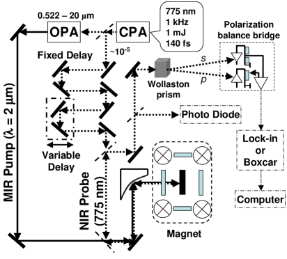
We performed two-color time-resolved MOKE spectroscopy using femtosecond pulses of MIR and NIR radiation. The experimental setup is schematically shown in Figure 1. The source of intense MIR pulses was an optical parametric amplifier (OPA) (Model FS-TOPAS-4/800, Quantronix/Light Conversion) pumped by a Ti:Sapphire-based regenerative amplifier (Model CPA-2010, Clark-MXR, Inc., 7300 West Huron River Drive, Dexter, MI 48130). The OPA was able to produce tunable and intense radiation from 522 nm to 20 m using different mixing crystals. The CPA produced pulses of NIR radiation with a wavelength of 775 nm, a pulse energy of 1 mJ, and a pulse duration of 140 fs at a tunable repetition rate of 50 Hz 1 kHz. We used a very small fraction () of the CPA beam as a probe and the output beam from the OPA tuned to 2 m as the pump. The CPA probe beam went through a computer-controlled variable delay stage in addition to a fixed 2 m long delay stage that equalized the pump and probe beam path lengths to the sample by taking into account the total path length inside of the multi-pass OPA. The two beams were made collinear by a non-polarizing beam splitter (Lambda Research Optics, Inc.), which was 50% reflective to the NIR probe and 50% transmissive to the MIR pump, and then focused by an off-axis parabolic mirror with a six inch focal length onto the sample mounted inside a 10 Tesla superconducting magnet with ZnS cold windows and CaF2 room temperature windows. The reflected NIR probe beam entered a Wollaston prism which spatially separated the - and -components of the probe beam, which we then focused on a balanced bridge system consisting of a Si detector pair. The signal from the balanced detectors, which was proportional to the induced MOKE angle change, was fed into a lock-in amplifier or a boxcar integrator and was recorded by a computer. An additional beam splitter was placed before the Wollaston prism for monitoring the reflectivity of the probe. With this setup, we were able to record the MOKE angle and reflectivity as functions of time delay and magnetic field.
At this pump wavelength (2 m), the photon energy (0.62 eV) was smaller than the band gaps of the GaSb buffer (0.812 eV) and GaAs substrate (1.519 eV) but larger than that of InMnAs ( 0.42 eV), so the pump created carriers only in the InMnAs layer. The beam diameter of the MIR pump at the sample position was measured by pinholes to be 75 m. The maximum MIR pulse energy used in this experiment was 6.3 J, which corresponds to a fluence of 0.45 J/cm2. Using the optical constants of InAs and the thickness of the InMnAs layer, we estimated the maximum density of photocreated carriers to be 7.5 1022 cm-3, which is an extremely large number, especially if we think of the fact that the density of Mn ions is only 1021 cm-3.
The sample studied was an InMnAs/GaSb single heterostructure with a Curie temperature () of 55 K, consisting of a 25 nm thick In0.91Mn0.09As magnetic layer and an 820 nm thick GaSb buffer layer grown on a semi-insulating GaAs (100) substrate. Its room temperature hole density and mobility were 1.1 1019 cm-3 and 323 cm2/Vs, respectively, estimated from Hall measurements. The sample was grown by low temperature molecular beam epitaxy (growth conditions described previouslytom ) and then annealed at 250 ∘C, which increased the by 10 K. hayashi ; potashnik The magnetization easy axis was perpendicular to the epilayer due to the strain-induced structural anisotropy caused by the lattice mismatch between InMnAs and GaSb (InMnAs was under tensile strain). This allowed us to observe ferromagnetic hysteresis loops in the polar Kerr configuration.
3 Experimental Results and Discussion
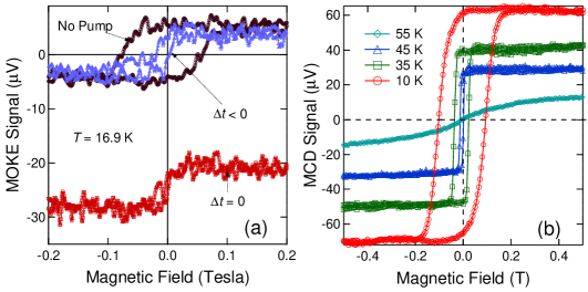
Figure 2(a) shows three magnetic-field-scan data exhibiting ferromagnetic hysteresis loops at a temperature of 16 K, taken under different conditions. The curve labeled ’No Pump’ was taken with the OPA pump beam blocked, while the other two curves were taken under high OPA excitation (fluence 0.2 J/cm2) with a time delay of 0 ps and 7 ps, respectively. It can be seen that at timing zero the loop horizontally collapsed, i.e., the coercivity is almost zero. Note that this curve is not intentionally offset; namely, this vertical shift is a real effect induced by the pump, which exists only for a short time ( 2 ps). As discussed later, we attribute this transient vertical shift of the MOKE signal to the coherent spin polarization of the photo-generated carriers. The negative time delay data also shows similar horizontal shrinkage though it is not as dramatic as the timing zero data. It is important to note that the vertical height (i.e., remanence) of the loops is not much affected by the pump, which excludes simple lattice heating as the cause of the horizontal loop quenching. To support this view more convincingly, we show in Fig. 1(b) CW magnetic circular dichroism data taken for the same sample at 10 K, 35 K, 45 K and 55 K. As can been seen clearly, raising the lattice temperature results in dramatic loop shrinkage both horizontally and vertically. Furthermore, we took time-resolved MOKE data with various combinations of average powers and fluences (data not shown) and found that it is the fluence that determines the degree of collapse, not the average power. For example, data taken at a low repetition rate with a high fluence displayed significant quenching while data taken at a high repetition rate with a low fluence did not show any quenching.
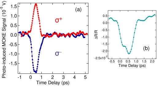
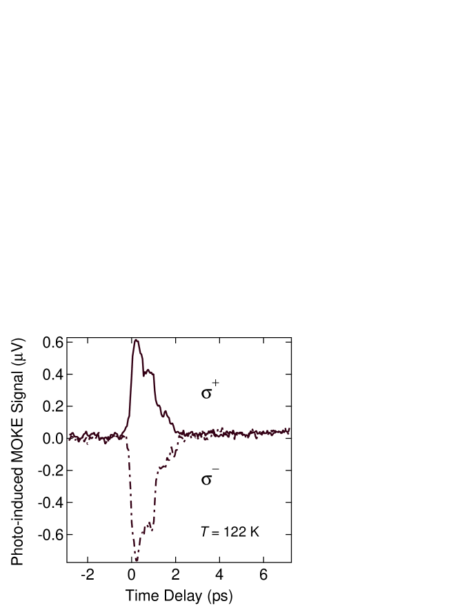
Next we discuss some time-scan data. Figure 3(a) shows two traces representing the pump-induced MOKE signal change versus time delay taken under the excitation of the MIR pump with two opposite senses of circular polarization, i.e., and . As clearly demonstrated here, opposite polarizations result in opposite signs of the photo-induced MOKE change, suggesting that these fast decaying transient MOKE signals are due to the photo-induced coherent carrier spin polarization. The pump-induced reflectivity change as a function of time is shown in Fig. 3(b). One can see a quick disappearance of the induced reflectivity change. In metals, photo-induced transmission or reflection changes can be attributed to electron temperature changes, which show characteristic cooling behavior as the electron system loses it energy to the lattice. In the present semiconductor system, however, the reflectivity change is most likely due to the transient change of the carrier density. The observed fast (¡ 2 ps) decay is consistent with the fast decays typically observed in low temperature grown semiconductors such as those used in terahertz emitters and receivers.smith To further understand the origin of the polarization-dependent ultrafast MOKE change, we did the same measurements at elevated temperatures. As an example, data taken at 122 K is shown in Fig. 4. Here, again, we see opposite signs for and polarization. We attempted to observe photo-induced ferromagnetism by scanning the magnetic field at these high temperatures but did not see any evidence. These facts lead us to believe that these signals are related to the coherent carrier spins. We do not have an explanation for the second peak around 1 ps observed for both polarizations. More detailed temperature dependent measurements are in progress to elucidate this feature.
4 Summary
We have presented results of what we believe to be the first ultrafast optical study of spin/magnetization dynamics in ferromagnetic InMnAs/GaSb heterostructures. Using a novel two-color time-resolved magneto-optical Kerr effect spectroscopy technique, we created transient carriers only within the ferromagnetic InMnAs layer using intense mid-infrared pulses and observed the induced magnetization changes through the Kerr angle of near-infrared probe pulses. Our data shows that magnetic properties, particularly coercivity, can be drastically modified by the intense MIR pulses. We were able to study spin and charge dynamics separately by simultaneously measuring MOKE and reflectivity. Finally, coherent optical spin injection using different senses of circular polarization led to different signs for the net MOKE changes induced by the pump.
Acknowledgements.
This work was supported by the Defense Advanced Research Projects Agency through grant No. MDA972-00-1-0034 and the National Science Foundation through grant No. DMR-0134058 (CAREER). We are grateful to Bruce Brinson and Ben Schmid for their technical assistance.References
- (1) For a review, see, e.g., G. Zhang, W. Hübner, E. Beaurepaire, and J.-Y. Bigot, in Spin Dynamics in Confined Magnetic Structures I, eds. B. Hillebrands and K. Ounadjela (Springer, Berlin, 2002), pp. 245-288.
- (2) E. Beaurepaire, J.-C. Merle, A. Daunois, and J.-Y. Bigot, Phys. Rev. Lett. 76, 4250 (1996).
- (3) J. Hohlfeld, E. Matthias, R. Knorren, and K. H. Bennemann, Phys. Rev. Lett. 78, 4861 (1997).
- (4) A. Scholl, L. Baumgarten, R. Jacquemin, and W. Eberhardt, Phys. Rev. Lett. 79, 5146 (1997).
- (5) M. Aeschlimann, M. Bauer, S. Pawlik, W. Weber, R. Burgermeister, D. Oberli, and H. C. Siegmann, Phys. Rev. Lett. 79, 5158 (1997).
- (6) G. Ju, A. Vertikov, A. V. Nurmikko, C. Canady, G. Xiao, R. F. C. Farrow, and A. Cebollada, Phys. Rev. B 57, R700 (1998).
- (7) E. Beaurepaire, M. Maret, V. Halté, J.-C. Merle, A. Daunois, and J.-Y. Bigot, Phys. Rev. B 58, 12134 (1998).
- (8) J. Güdde, U. Conrad, V. Jähnke, J. Hohlfeld, and E. Matthias, Phys. Rev. B 59, R6608 (1999).
- (9) B. Koopmans, M. van Kampen, J. T. Kohlhepp, and W. J. M. de Jonge, Phys. Rev. Lett. 85, 844 (2000).
- (10) L. Guidoni, E. Beaurepaire, and J.-Y. Bigot, Phys. Rev. Lett. 89, 017401 (2002).
- (11) G. P. Zhang and W. Hübner, Phys. Rev. Lett. 85, 3025 (2000).
- (12) H. Munekata, H. Ohno, S. von Molnar, A. Segmuller, L. L. Chang, and L. Esaki, Phys. Rev. Lett. 63, 1849 (1989); H. Ohno, H. Munekata, T. Penney, S. von Molnar, and L. L. Chang, ibid. 68, 2664 (1992).
- (13) H. Munekata, A. Zaslavsky, P. Fumagalli, and R. J. Gambino, Appl. Phys. Lett. 63, 2929 (1993).
- (14) H. Ohno, A. Shen, F. Matsukura, A. Oiwa, A. Endo, S. Katsumoto, and Y. Iye, Appl. Phys. Lett. 69, 363 (1996).
- (15) See, e.g., H. Akai, Phys. Rev. Lett. 81, 3002 (1998); J. Inoue et al., ibid. 85, 4610 (2000); T. Dietl et al., Science 287, 1019 (2000); J. König et al., Phys. Rev. Lett. 84, 5628 (2000); V. I. Litvinov and V. K. Dugaev, ibid. 86, 5593 (2001); A. Chattopadhyay et al., ibid. 87, 227202 (2001); J. Schliemann and A. H. MacDonald, ibid. 88, 137201 (2002); G. Zaránd and B. Jankó, ibid. 89, 047201 (2002).
- (16) H. Ohno, D. Chiba, F. Matsukura, T. Omiya, E. Abe, T. Dietl, Y. Ohno, and K. Ohtani, Nature 408, 944 (2000).
- (17) S. Koshihara, A. Oiwa, M. Hirasawa, S. Katsumoto, Y. Iye, C. Urano, H. Takagi, and H. Munekata, Phys. Rev. Lett. 78, 4617 (1997).
- (18) T. Slupinski, A. Oiwa, S. Yanagi, and H. Munekata, J. Cryst. Growth 237-239, 1326 (2002).
- (19) T. Hayashi, Y. Hashimoto, S. Katsumoto, and Y. Iye, Appl. Phys. Lett. 78, 1691 (2001).
- (20) S. J. Potashnik, K. C. Ku, S. H. Chun, J. J. Berry, N. Samarth, and P. Schiffer, Appl. Phys. Lett. 79, 1495 (2001).
- (21) F. W. Smith, H. Q. Le, V. Diadiuk, M. A. Hollis, A. R. Calawa, S. Gupta, M. Frankel, D. R. Dykaar, G. A. Mourou, and T. Y. Hsiang, Appl. Phys. Lett. 54, 890 (1989).