Resonant excitation and anti-Stokes luminescence of GaAs single quantum dots
Abstract
We have investigated micro-photoluminescence (-PL) and excitation (-PLE) spectra of a single GaAs/AlGaAs quantum dot grown on a GaAs (411)A surface. We observed sharp resonant lines in both -PL and -PLE spectra, corresponding to the discrete energy levels of the dot. When the sample was excited at one of the resonant lines, resonant luminescence lines appear not only in Stokes side but also in anti-Stokes side. We discuss the possible origins of the anomalus anti-Stokes luminescence.
I Introduction
Semiconductor quantum dots (QDs) or nanocrystals attract much attention because of their unique and interesting optical properties originating from three-dimensional quantum confinement of the electronic states. Single quantum dot spectroscopy has revealed many interesting and important properties of the QDs, such as sharp homogeneous linewidth Gammon , long coherence time Bonadeo , luminescence intermittency Nirmal , and anti-bunched photon emission Michler . In this paper, we report the micro-photoluminescence (-PL) and micro-photoluminescence excitation (-PLE) spectra from a GaAs/AlGaAs quantum dot grown on the (411)A surface of a GaAs substrate. We found anomalous anti-Stokes luminescence that is probably caused by efficient Auger or cascade excitation process of the excitions in a QD.
II Experimental Procedure
Our GaAs quantum dot sample is based on a GaAs/Al0.3Ga0.7As quantum well (QW) on a GaAs (411)A surface, fabricated by a molecular beam epitaxy method Hayashi . Under an appropriate growth condition with relatively small As flux, triangular pyramidal-shaped structures are formed on the surface. Figure 1(a) shows the atomic force microscope (AFM) image of the pyramidal structures. By growing a QW over the pyramid, thicker area of the well is formed on a slope of the pyramid, as shown in Fig. 1(b). The mean thickness of the GaAs layer of the sample used for this experiment was 3 nm. The QW in the region of the pyramidal structure contains a number of localized QD structures, as described later. Since the density of the pyramid is as low as 107cm-2, we can easily resolve the luminescence from a single pyramidal structure using a conventional microscope objective lens.
We used a continuous wave Ti:Sapphire laser as the excitation light source for the -PL and -PLE measurement. The spectral width of the excitation laser was 0.17 meV. The excitation beam was focused by an objective lens (50, N.A.=0.42) onto the sample, which was mounted on the cold-finger of a cryostat and cooled down to 4 K. The luminescence from the sample was collected by the same objective lens. The collected light was dispersed by a triple-grating monochromator (focal length = 64 cm) and then detected by a cooled charge coupled device (CCD) camera. The spectral resolution of the detection system was 0.09 meV.

III Results and Discussion
The -PL spectrum observed from a single pyramidal structure under moderately weak excitation power density (5.5 W/cm2) is shown in Fig. 2. The excitation was made at 1.687 eV, which corresponds to the excitation above the GaAs QW surrounding the pyramidal structure. Several sharp luminescence lines were observed in the -PL spectrum. However, from the PL spectrum, it is difficult to determine the number of QDs and their energy levels corresponding to these lines. In order to observe this, we measured the -PLE spectra detected at four luminescence lines (A to D in Fig. 2), as shown in the upper graph of Fig. 3. For comparison, the -PL spectrum identical to Fig. 2 is also shown in the lower graph of Fig. 3 with the same energy scale as the -PLE spectra, The -PLE spectra have several sharp peaks reflecting the discrete energy levels of the QD. One can see the close similarity between the PLE spectra for A and D, and for B and C. The -PLE spectrum for A has a sharp peak at the energy posision of the PL line D. Also, the -PLE for B has a sharp peak at the PL line C. These findings suggest that the luminescence lines A and D originate from an identical dot, and lines B and C from another dot in a pyramid. Thus, we conclude that there are a number of (at least two) quantum dots in the single pyramidal structure. In addition, for all the PLE spectra, quasi-continuous excitation band appears above 1.594 eV. The continuous PLE band suggests that the quantum dots are surrounded by the two-dimensional states of the adjacent GaAs layer.
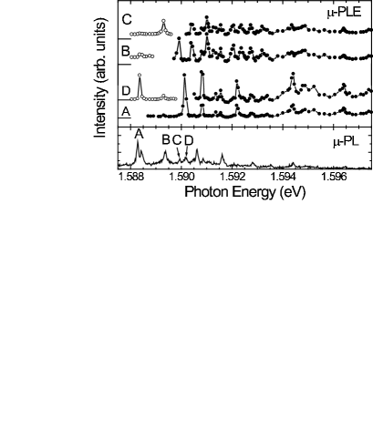

Furthermore, we found that the -PLE spectra for the PL lines D and C have sharp anti-Stokes resonances at the energy positions of the PL lines A and B, respectively, as indicated by the open circles in Fig. 3. The anti-Stokes shifts are 1.9 meV and 0.6 meV for the two cases, respectively. Since the spectra were taken at low temperature (4 K), the anti-Stokes PL cannot be caused by any thermal excitation. Local heating caused by the excitation light was not expected because the spectral position of the luminescence lines did not change noticeably in the range of the excitation intensity (see Fig. 4), although the expected spectral red shift would have been 0.2 meV if the sample temperature had been raised to 20 K (=1.7 meV). To our knowledge, such distinct and anomalous anti-Stokes resonant luminescence has not been reported for any kind of semiconductor QDs or nanocrystals. Figure 4 shows the -PL spectra for various excitation intensities () under resonant excitation at the energy position corresponding to the PL line A. One can see that several PL lines appear on the anti-Stokes sides, including the distinct line at D, which corresponds to the PL line D in Figs. 2 and 3. The energy shift of the anti-Stokes PL from the excitation photon energy ranges more than 8 meV. From the analysis of the excitation-intensity dependence, we found that the intensity of the anti-Stokes PL line at D has almost linear dependence on the excitation intensity under weak or moderate excitation (100 W/cm2 or less). Since the thermal excitation is not expected as mentioned above, the origin of the linear anti-Stokes PL is an open question at present. In contrast, the intensity of PL lines with larger anti-Stokes shift has considerable nonlinear dependence on the excitation intensity. For instance, the intensity of the anti-Stokes PL line indicated by the filled circle in Fig. 4 was found to have quadratic dependence on the excitation intensity. This quadratic excitation-intensity dependence indicates that the anti-Stokes PL line arises when two excitons are created, or two photons are annihilated, in the QD. One possible origin of the anti-Stokes PL is the Auger process between the two excitons in the QD, which often causes luminescence intermittency Nirmal ; Efros . Another possibility is the cascade two-step excitation of the exciton in the QD. Both processes excite the exciton to delocalized or ionized states, from which the electon-hole pair can relax into the initial state of the anti-Stokes luminescence. Furthermore, under high excitation intensity, another PL line (filled square in Fig. 4) appears in the lower energy side. Its intensity also has quadratic dependence on the excitation intensity. This line is attributable to the emission from the biexciton in the QD. The binding energy of the biexciton is 3.4 meV in this case, which is comparable to the reported experimental Brunner and theoretical Tsuchiya values for GaAs QDs. Thus, it is likely that the two-exciton bound state (biexciton) coexists with the Auger or two-step excitation process of the excitons in the QD. The coexistence of the two contradictory processes is an interesting and important problem in discussing the few-particle dynamics in the QD. Further investigation is in progress in order to identify the origin of the anomalous resonant anti-Stokes luminescence.
Acknowledgements
This work was supported by Grant-in-Aid for COE Research (10CE2004) and Grant-in-Aid for Scientific Research from the Ministry of Education, Culture, Sports, Science and Technology of Japan.
References
- (1) D. Gammon, E. S. Snow, B. V. Shanabrook, D.S. Katzer and D. Park, Science 273 (1996) 87.
- (2) N.H. Bonadeo, J. Erland, D. Gammon, D. Park, D.S. Katzer and D.G. Steel, Science 282 (1998) 1473.
- (3) M. Nirmal, B.O. Dabbousi, M.G. Bawendi, J.J. Macklin, J.K. Trautman, T.D. Harris, and L.E. Brus, Nature 383, 802 (1996); Al.L. Efros and M. Rosen, Phys. Rev. Lett. 78 (1997) 1110.
- (4) P. Michler, A. Imamoğlu, M.D. Mason, P.J. Carson, G.F. Strouse and S.K. Buratto, Nature 406 (2000) 968.
- (5) H. Hayashi, S. Shimomura, and S. Hiyamizu, unpublished.
- (6) C. Watatani, K. Edamatsu, T. Itoh, H. Hayashi, S. Shimomura and S. Hiyamizu, Phys. Stat. Sol. (b) 224 (2001) 353.
- (7) Al.L. Efros and M. Rosen, Phys. Rev. Lett. 78 (1997) 1110.
- (8) K. Brunner, G. Abstreiter, G. Bohm, G. Trankle, and G. Weimann, Phys. Rev. Lett. 73 (1994) 1138.
- (9) T. Tsuchiya, Physica E 7 (2000) 470.