Direct experimental verification of applicability of single-site model for angle integrated photoemission of small concentrated Ce compounds
Abstract
Bulk-sensitive high-resolution Ce 4f spectra have been obtained from 3d 4f resonance photoemission measurements on La1-xCexAl2 and La1-xCexRu2 for . The 4f spectra of low-Kondo-temperature () (La,Ce)Al2 are essentially identical except for a slight increase of the Kondo peak with , which is consistent with a known increase of with . In contrast, the 4f spectra of high- (La,Ce)Ru2 show a Kondo-like peak and also a 0.5 eV structure which increases strongly with . The resonance photon-energy dependences of the two contributions are different and the origin of the 0.5 eV structure is still uncertain.
pacs:
Key words: Ce compounds, photoemissionThe single-impurity Anderson model (SIAM) has been used with great success to describe angle integrated 4f photoemission spectra of dense-Kondo Ce compounds, both the low energy Kondo features and the high energy ionization excitations, after proper subtraction of a surface spectrum [1], in spite of claims to the contrary [2]. Theory support for such claims might come from Nozières ‘exhaustion’ idea, that in the simple Kondo lattice there are not enough conduction electrons to screen out the magnetic moments as in the SIAM, essentially because only electrons located within the Kondo temperature () of the Fermi level () can take part in the Kondo screening [3]. The exhaustion problem is thus most serious in the lowest- systems, and would imply a decreased in the lattice system relative to the impurity system. In this work, we have made a direct experimental comparison of 4f spectra of concentrated and dilute Ce compounds by performing bulk-sensitive high-resolution Ce 3d 4f resonant photoemission spectroscopy (RPES) of La1-xCexAl2 and La1-xCexRu2, for which the ’s are 5 K and larger than 1000 K, respectively, in the concentrated limit. They have the same cubic Laves structure in which the number of nearest Ce neighbors of a Ce ion is only four. For , 85% of the Ce ions are completely isolated from other Ce ions if next-nearest Ce-Ce couplings are neglected, so that the 4f spectrum is dominated by the single-site contribution.
Polycrystalline samples of La1-xCexAl2 and La1-xCexRu2 () were prepared by arc melting and characterized by x-ray diffraction. Some elemental Ru impurity was found in the dilute (La,Ce)Ru2 sample. Although the Ru impurity contribution should be removed by the RPES extraction of the Ce 4f spectrum, nonetheless the impurity renders our results for the (La,Ce)Ru2 system to be tentative. Ce 3d 4f RPES was performed at beam line BL25SU of SPring-8 [4]. All samples were fractured to reveal clean surfaces in a vacuum of Torr. The overall energy resolution was about 100 meV and the sample temperature was kept at 20 K.
Figure 1 shows a comparison of the Ce 4f spectra of concentrated and dilute (La,Ce)Al2 obtained from the RPES spectra by a usual method [1]. The sharp two-peak structure near is a generic feature of low- Ce compounds, consisting of the tail of a Kondo resonance (KR) and its spin-orbit replica (SOR) near 0.25 eV. Other structures at higher binding energy are the material-specific ionization features of the hybridization matrix element between Ce 4f and conduction electrons, within the SIAM description. There is perfect agreement of the two spectra except for an overall reduction of the KR and its SOR in the dilute system. The known reduc-
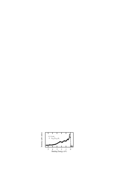
tion of with decreasing Ce concentration [5], due to volume expansion, is opposite to expectations from Nozières ‘exhaustion’ idea but easily explains the small spectral change within a SIAM description, including the stronger intensity reduction of the KR relative to its SOR. The results are a direct experimental confirmation of the applicability of a single-site description of the angle integrated Ce 4f spectrum in this low- system, contrary to objections [2] that have been made.
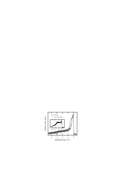
The Ce 4f spectra of concentrated and dilute (La,Ce)Ru2 are presented in Fig. 2. In great contrast to the (La,Ce)Al2 system, the two spectra differ greatly, especially by a 0.5 eV structure in the concentrated system. At present the origin of the 0.5 eV structure is unclear. The difference might well imply the need to go beyond a single-site model in the high- system due to strong intersite couplings arising from large Ce 4f-Ru 4d hybridization [4]. But this conclusion may be premature. As shown in the inset, the off-resonance spectrum has a strong -dependence, that a peaky 0.5 eV structure in the concentrated system is shifted to for the dilute system. Our calculation (not shown) of the 4f spectrum using the Gunnarsson-Schönhammer method [6] and taking to be the off-resonance spectrum, does show the 0.5 eV structure in the concentrated system, though with an intensity that is rather smaller than in the experimental spectrum. The fact that the 0.5 eV structure in CeRu2 is found to become much stronger when the spectrum is obtained at a slightly higher photon energy motivates yet another explanation, that it arises from an incoherent Auger transition, as clearly observed in CeFe2 [7]. Further experimental investigation is now in progress.
This work was supported by the U.S. NSF at the University of Michigan (No. DMR-99-71611), by the U.S. DoE at the University of Michigan (No. DE-FG-02-90ER45416), and by the U.S. NSF at the University of California at San Diego (No. DMR-97-05454).
REFERENCES
- [1] J. W. Allen, et al., Adv. Phys. 35 (1986) 275; D. Malterre, M. Grioni, and Y. Baer, Adv. Phys. 45 (1996) 299; J. W. Allen , et al., J. Appl. Phys. 87 (2000) 6088; A. Sekiyama, et al., J. Phys. Soc. Jpn. 69 (2000) 2771.
- [2] A. J. Arko, et al., in Handbook on the Physics and Chemistry of Rare Earths, Vol. 26, edited by K. A. Gschneidner and L. Eyring (Elsevier Science, B.V., 1999) pp. 265-382.
- [3] Ph. Nozières, Eur. Phys. J. B 6 (1998) 447.
- [4] A. Sekiyama, et al., Nature 403 (2000) 396.
- [5] C. D. Bredl, F. Steglich, and K. D. Schotte, Z. Physik B 29 (1978) 327.
- [6] O. Gunnarsson and K. Schönhammer, Phys. Rev. B 31 (1985) 4815.
- [7] E.-J. Cho and S.-J. Oh, (private communication).