[
Micro-SQUID technique for studying the temperature dependence of switching fields of single nanoparticles
Abstract
An improved micro-SQUID technique is presented allowing us to measure the temperature dependence of the magnetisation switching fields of single nanoparticles well above the critical superconducting temperature of the SQUID. Our first measurements on 3 nm cobalt nanoparticle embedded in a niobium matrix are compared to the Néel Brown model describing the magnetisation reversal by thermal activation over a single anisotropy barrier.
pacs:
Keywords: Stoner-Wohlfarth model, Thermal activation, Nanoparticle, SQUID, Magnetometry]
In order to avoid the complications due to distributions of particles sizes, orientations, etc., which are always present in assemblies of particles [1], single-particle measuring techniques have been developed such as magnetic force microscopy [2] or magnetometry based on micro-Hall probes [3]. Another powerful measurement technique is based on micro-SQUID devices. In this paper we review the an improved micro-SQUID set-up which allow us to detect the magnetisation reversal of single nanoparticles. In respect to previous studies [4], we improved the sensitivity of the micro-SQUID technique by more than two orders of magnitude. Furthermore, we developed a technique to measure the switching fields for all direction of the applied field. Finally, we extended the temperature range to the interval from 30 mK to 30 K. We present these improvements in the following.

In order to measure the magnetisation reversal of a single 3 nm Co nanoparticle (containing about 1000 atoms), we have to couple the magnetic stray field of the nanoparticle with the SQUID loop. In previous studies on larger particles (around 10 nm or larger), it was possible to place the nanoparticle close to the micro-bridge Josephson junctions of the SQUID [4]. This is not sufficient for 3 nm Co nanoparticles due to the small size of the nanoparticle compared with the micro-bridge. Moreover, it is very difficult to protect them efficiently against oxidation. We solved these problems by directly embedding the Co nanoparticles into the micro-bridges (Fig. 1). This was achieved by using a laser vaporisation and inert gas condensation source producing an intense supersonic beam of nanosized Co nanoparticles under ultra-high-vacuum (UHV) conditions. A niobium matrix was simultaneously deposited from a UHV electron gun evaporator leading to continuous films (typical 20 nm thick) with a very low concentration of embedded Co nanoparticles [5]. These films are used to pattern planar microbridge-DC-SQUIDs by electron beam lithography. The desired sensitivity is only achieved for Co-nanoparticles embedded into the micro-bridges where the magnetic flux coupling is high enough. Due to the low concentration of embedded Co-nanoparticles, we have a maximum of 5 non-interacting particles in a micro-bridge (300 nm long and 50 nm wide). We can separately detect the magnetic signal for each nanoparticle. Indeed they are clearly different in intensity and orientation because of the random distribution of the easy magnetisation directions.
The switching field of the magnetisation of a single Co nanoparticle can be measured by using the cold mode method described in detail in Ref. [7]. However, this mode works only in the low temperature regime and for magnetic fields applied in the SQUID plane. We therefore developed the blind mode method [8] allowing us to measure the switching fields for all directions of the applied field by separately driving three orthogonal coils. In addition, the blind mode allowed us to extend the temperature interval from 30 mK to 30 K [6], i.e. to temperatures well above the critical superconducting temperature of the micro-SQUID.
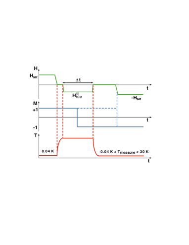
Let us consider Fig. 2 showing schematically the three steps of the blind mode method. The applied magnetic field, the magnetisation state of the nanoparticle, and its temperature are represented as a function of time.
-
1.
Saturation. The magnetisation of the particle is saturated in a given direction (at T = 35 mK).
-
2.
Testing. A test field is applied at a temperature between 35 mK and 30 K and during the time which may or may not cause a magnetisation switching.
-
3.
Probing. After cooling to = 35 mK, the SQUID is switched on and a field is swept in the plane of the SQUID to probe the resulting magnetisation state using the cold mode.
If the SQUID detects a magnetisation jump in step (3), this means that the previously applied test-field was weaker than the switching field for the direction being probed in step (2). The next iteration will then be done with a stronger test field. On the other hand, if the SQUID does not detect any magnetisation jump in step (3), this means that the reversal occurred during step (2). The next interation will then be done with a weaker field. When choosing the new test field with the help of a bisection algorithm, we needed about eight repetitions of the three steps in order to get the switching field with good precision. This method allows us to scan the entire field space.
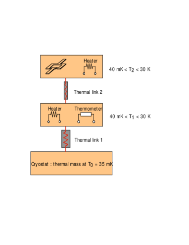
In order to probe the magnetisation reversal at high temperatures, we have developed a two-stage heater shown in the Fig. 3. The upper part of this device is a Si chip containing micro-SQUIDs and a resistor which is used as heating element. The thermal response of this first stage is very fast because it has a small thermal mass. This stage is coupled to a second stage with a much larger thermal mass. It contains another heating element and a thermometer. Finally, the second stage is coupled to the cryostat which has the largest thermal mass. The heat links between the two stages are metallic wires which are adapted so that each stage is a thermal mass for its upper part. The cryostat is the thermal mass of the whole system and stays cold during operation. The purpose of the second stage is to calibrate efficiently the heater of the first stage. With this set-up, our highest temperatures of 30 K was only limited by the cooling time. Below 30 K, we achieved cooling rates of few kelvins per second.
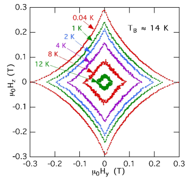
Fig. 4 presents the angular dependence of the switching field of a 3 nm Co nanoparticle measured at different temperatures using the blind mode method. At 0.03 K, the measurement is very close to the standard Stoner–Wohlfarth astroid. For higher temperatures the switching field becomes smaller and smaller. It reaches the origin at about 14 K, yielding the blocking temperature K of the nanoparticle magnetisation. is defined as the temperature for which the waiting time becomes equal to the relaxation time of the particle’s magnetisation at . can be used to estimate the total number of magnetic Co atoms in the nanoparticle. The Néel Brown model leads to , where is the attempt frequency typically between to Hz, Kat is an effective anisotropy energy per atom and is the Boltzmann constant. Using the expression of the switching field at = 0 K and for : (Fig. 4), the atomic moment , = 0.01 s, s, and K, we deduce , which corresponds very well to a 3 nm Co nanoparticle.
In conclusion, we have shown that the micro-SQUID technique combined with the Low Energy nanoparticle Beam Deposition is a powerful method to investigate the magnetic properties of nanosized magnetic particles. In particular, it allows to measure in three dimensions the switching field of individual grains giving access to its magnetic anisotropy. Furthermore, the temperature dependence of the switching field is measurable and allows to probe the magnetisation dynamics.
We thank B. Barbara for his constant support of this research. Part of this work has been supported by DRET, Rhône-Alpes, and the MASSDOTS ESPRIT LTR-Project 32464.
REFERENCES
- [1] J. L. Dormann, D. Fiorani, and E. Tronc, Adv. Chem. Phys. 98, 283 (1997).
- [2] M. Ledermann, S. Schultz, and M. Ozaki, Phys. Rev. Lett. 73, 1986 (1994).
- [3] J.G.S. Lok, A.K. Geim, J.C. Maan, S.V. Dubonos, L. Theil Kuhn, and P.E. Lindelof, Phys. Rev. B 58, 12201 (1998).
- [4] W. Wernsdorfer, E. Bonet Orozco, K. Hasselbach, A. Benoit B. Barbara, N. Demoncy, A. Loiseau, D. Boivin, H. Pascard, and D. Mailly, Phys. Rev. Lett. 78, 1791 (1997); Phys. Rev. Lett. 79, 4014 (1997).
- [5] M. Jamet et al., Phys. Rev. B 62, 493 (2000).
- [6] M. Jamet, W. Wernsdorfer, C. Thirion, D. Mailly, V. Dupuis, P. Mélinon, and A.Pérez, Phys. Rev. Lett. 86, 4676 (2001).
- [7] W. Wernsdorfer, D. Mailly, A. Benoit, J. Appl. Phys. 87, 5094 (2000).
- [8] E. Bonet, W. Wernsdorfer, B. Barbara, A. Benoit, D. Mailly, and A. Thiaville, Phys. Rev. Lett. 83, 4188 (1999).