Protein dynamics with off-lattice
Monte Carlo moves
Abstract
A Monte Carlo method for dynamics simulation of all-atom protein models is introduced, to reach long times not accessible to conventional molecular dynamics. The considered degrees of freedom are the dihedrals at Cα-atoms. Two Monte Carlo moves are used: single rotations about torsion axes, and cooperative rotations in windows of amide planes, changing the conformation globally and locally, respectively. For local moves Jacobians are used to obtain an unbiased distribution of dihedrals. A molecular dynamics energy function adapted to the protein model is employed. A polypeptide is folded into native-like structures by local but not by global moves.
PACS numbers: 87.15.He, 02.70.Lq, 82.20.Wt
Two of the great challenges in theoretical biophysics are the elucidation of the structure function dynamics relationship of proteins [1], and the protein folding problem [2, 3, 4, 5]. Functional processes in proteins are often slow compared to picosecond and subpicosecond dynamics of vibrational degrees of freedom, which are easily accessible by conventional methods of computer simulation. The dynamics of protein folding with a typical time scale from microseconds to seconds is even much slower.
The conventional method of computer simulation of protein dynamics is based on solving Newton’s equations of motion in Cartesian coordinates [6, 7]. This approach is very time consuming for the following reasons: 1. The number of non-bonded atom pair interactions is very large, so that most of the CPU time is spent on their evaluation. 2. It is necessary to follow the fast intramolecular vibrations in all details, which requires an elementary step of propagation in time of typically 1 fs.
There are two important strategies [8] to improve this situation: 1. Reducing the number of non-bonded pair interactions by combining groups of atoms in single interaction centers. The drawback is a loss of detail in the description of the protein. 2. Increasing the elementary time step by eliminating stiff degrees freedom with small amplitudes of motion. These are bond lengths and bond angles, which here are not interesting in their own right, although they may serve as “lubricant” for large conformational changes [9].
Lattice models, and protein models using virtual bonds instead of amide planes are very efficient combinations of both time saving techniques, but they suffer from poor energetic and conformational resolution [10].
In the present approach we use an all-atom protein model where bond lengths and bond angles are fixed and the amide plane is kept planar, so that the only degrees of freedom of the protein backbone are the dihedral angle pairs at the Cα-atoms [11] (Fig. 1). New conformations are generated by moves of two different types. A simple move (SM) is a rotation about a torsion axis. It leads to large displacements of atoms far away from this axis. Thus in a globular protein SM’s often result in structures with overlapping atoms, which are energetically unacceptable. A Window move (WM) is a cooperative rotation in a small window of consecutive amide planes [11] (Fig. 1). WM’s have been employed to prove that a tripeptide can not be cyclic [13] and to generate loop conformations [14]. The finding of WM’s is equivalent to the problem of inverse kinematics for serial manipulators, where the hand of a robot must be oriented and positioned in a specific place [15]. The cooperative motion of several amide planes can be described as a diffusion process. In this sense a chain of WM’s can be interpreted as dynamics evolution.
In a WM the changes of the dihedrals are subject to six constraints which guarantee that the atomic positions of the two parts of the polypeptide outside the window remain fixed [11]. A window can consist of any number of amide planes larger than one. Here only windows of three amide planes are considered, corresponding to eight degrees of freedom (Fig. 1). The first steps in a WM are the pre-rotations, where increments to the dihedrals at one Cα-atom in a window are chosen arbitrarily from a given interval . Possible values of the other six dihedrals are then determined by finding a root of the constraining conditions. For the present geometry of the protein backbone the maximum number of roots found was twelve. On the average a complete set of roots is obtained in less than 10 ms CPU time (SGI R4000), which is negligible in comparison to the time spent on the evaluation of the non-bonded energy.
The distribution of dihedral angles of protein backbone conformations generated by WM’s deviates from a uniform distribution by more than 30% due to the window constraints. More specifically it turns out that this bias concerns four dihedrals out of eight in a window. A corresponding reduced set of four constraints can be used as generalized coordinates which trivially fulfill the window constraints. For the th root of the constraint equations, the Jacobian
| (1) |
accounts for the change of phase space volume when transforming from the to the . The fact that different roots have unequal is the reason for the bias. The bias is eliminated as described in the following. After the pre-rotations have been carried out in a WM, the window constraints are solved and one obtains new window conformations. All conformations belong to a single set of but have different values of and . A weighting among the conformations according to their phase space volumes can be reached by using probabilities that obey . A correct weighting between old and new conformations in the window requires also knowledge of the conformations, which are obtained by solving the window constraints without applying pre-rotations. Consequently a proper weighting is achieved by randomly selecting one of all old and new conformations according to the probability
| (2) |
where . If there are no solutions for the applied pre-rotations, the old conformation is retained. The necessity of the correct weighting of solutions has often been ignored and was first recognized by Dodd et al. [16] in the context of polymer dynamics. The present selection scheme is more efficient than the rejection algorithm used in Ref. [16]. In the two windows at the ends of the polypeptide chain, the dihedral angle changes are randomly chosen from without further constraints. The final decision on acceptance or rejection of a WM is left to the Metropolis criterion [12] with a suitable energy function. With different window positions possible, a chain of WM’s with randomly chosen window positions is called a “scan”. For SM’s the term scan is used in the same spirit, with corresponding to the total number of torsion axes.
Since the protein backbone model has reduced flexibility, a conventional MD energy function cannot be used without modifications. Atomic clashes may occur between atoms separated by only a few torsional degrees of freedom. In a protein model with full flexibility this could be avoided by bond angle bending. In this work these problems are circumvented by letting adjacent amide planes interact only by a two-dimensional potential of the two dihedrals at the Cα-atom connecting the amide planes, which is generated beforehand with CHARMM as described in appendix 2 of ref. [7]. These two-dimensional potentials are residue specific; here only those of glycine and alanine are needed. For all other atom pairs, which are separated by at least four torsional degrees of freedom the CHARMM interactions are used.
In this contribution the folding of an -helix-turn--helix (HTH) structure is simulated. The HTH motif is guided by the structure of ROP [17], an -helical hairpin of 56 residues. In the simulations, only 26 residues are considered, corresponding to residues 18-43 of ROP. The -helical parts are modeled by alanine (A) residues. It is known from MD simulations, that polyalanine forms stable -helices in vacuo [18]. A hydrophobic attraction of the helices is mimicked by specifically increasing the well-depth of the Cβ-Cβ Lennard-Jones interaction from 0.181 to 2.0 kcal/mol between some of the residues (X), corresponding to those residues in ROP which line the interface of the two helices. The well-depth of the X-X interaction is motivated by the measured free energies of transfer of hydrophobic amino acids from water to non-polar solvents (corresponding to the hydrophobic core of proteins), which for leucin or phenylalanin are about 2.0 kcal/mol [19]. The turn region is modeled by five glycines (G), which are the most flexible residues. In total the sequence of the model protein reads AXAAXAAAXXGGGGGXXAAAXAAAXA. N- and C-terminus are blocked with the neutral groups acetamide and N-methyl-amide, respectively, to avoid strong electrostatic interactions. By simulated annealing and subsequent energy minimization the ROP model structure is adapted to the sequence and energy function of the HTH model yielding a root mean square deviation (RMSD) of 1.35 Å for the backbone. The energy of this annealed reference structure (RS) kcal/mol, and its radius of gyration with respect to the Cα-atoms Å correlate well with the values obtained by MC dynamics with WM’s as discussed below.
Four trajectories using WM’s and four using SM’s have been produced (WM and SM). All trajectories start from a -strand conformation, where the chain is almost extended, kcal/mol, and Å. The temperature is 450 K, which in a series of test simulations was found to be low enough for formation of stable conformations but high enough for fast isomerization. In WM simulations dihedrals are allowed to change per move by at most . The global character of SM’s makes atomic clashes more likely, hence we choose for SM’s. In spite of its smaller a SM can change the conformation far more than a WM. For a protein of monomers, the average number of non-bonded energy terms affected by a SM scan and a WM scan is proportional to and , respectively. For a 26-mer the average CPU time per SM scan is therefore about 2.8 times that per WM scan. This difference has been approximately considered by choosing the WM trajectories twice as long as the SM trajectories.
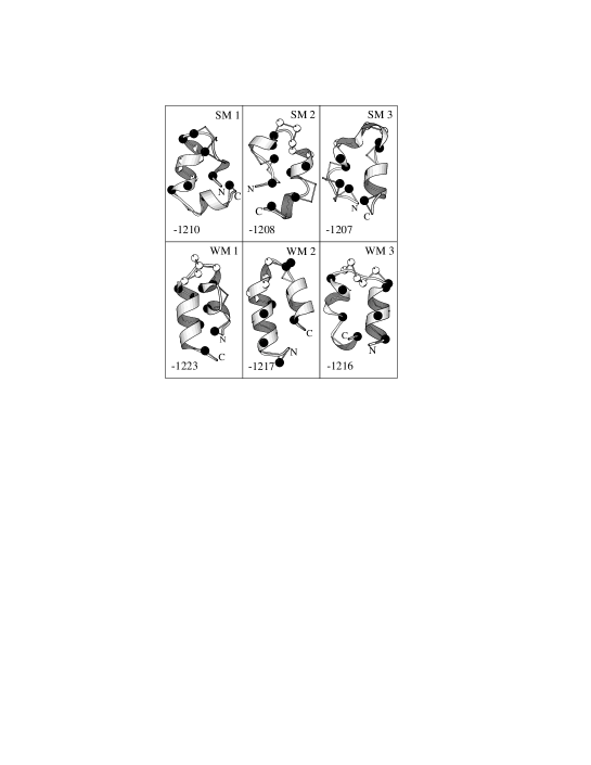
With the exception of SM1, in all SM trajectories the polypeptide collapses within the first few thousand scans into compact random coil type conformations. This is reflected in an abrupt decrease of from 25 Å to less than 7 Å (Fig. 3). After the collapse jumps erratically by up to 0.5 Å. The conformations are thus on the average more compact than the RS. They have low secondary structure content with only a few isolated helix turns. The energy falls very steeply by about 100 kcal/mol within the first 1000 SM scans of SM2-SM4 (Fig. 2). This is due to X-X-contacts and hydrogen bonds, which form instantly but arbitrarily between sequentially distant monomers as the polypeptide becomes kinked. A slower decrease of follows over SM scans. Afterwards only smaller conformational rearrangements take place, accompanied by energy fluctuations about mean values of -1175 kcal/mol (SM2,SM3) and -1150 kcal/mol (SM4). Even after energy minimization none of the conformations with lowest energy of each SM trajectory fulfills kcal/mol (Fig. 4).
SM1 leads to the conformations with the lowest , the highest helix content and largest of all SM simulations. Within the first scans the central helix (Fig. 4) in SM1 forms and falls considerably. The growing of this helix slows down the collapse to a denser conformation. Unfortunately the helix encompasses just the glycines which are known to have low helix propensity.
SM’s require a relative mobility of chain ends. Therefore SM trajectories are often trapped in quasi-cyclical conformations with chain ends linked by strong hydrogen bonds or X-X-contacts. The dropping of acceptance probabilities from more than 0.6 within the first SM scans to typically 0.30 when the conformation has become quasi-cyclic reflects this feature.
In the WM trajectories the folding takes a very different path. Starting from the termini two helices grow towards the center of the polypeptide, which allows to decrease from -1008 kcal/mol to about -1175 kcal/mol within the first scans. It was reported that in MD simulations a 13-mer polyalanine requires several hundred picoseconds to form an -helix [20]. In test simulations of 13-alanine with WM’s helices were formed in several thousand scans. Equating a MD time step with a WM scan, the CPU time for helix formation is 100 times larger for MD than for MC. In WM4 the two helices join after scans in the middle of the chain, which is then a single long helix with average values for and of -1175 kcal/mol and 11.7 Å, respectively. The helix frays at the ends and bends but remains stable. In WM1-WM3 the two helices form a HTH-motif. The turn develops within the first scans as the polypeptide is kinked in the glycine region. The two helices are forced into an anti-parallel alignment by the X-X attractions, leading to a further decrease of to a mean value of -1180 kcal/mol (Fig. 2). The value of drops, too, and fluctuates finally at about Å (Fig. 3). After the formation of the HTH-motifs the conformations continue to rearrange, because they have either imperfect pairing of X-residues (WM2,WM3) or helices with left-handed turns (WM1,WM3). Nevertheless the trajectories WM1-WM3 contain conformations, which after minimization have (Fig. 4), in particular for WM1, kcal/mol, and the RMSD to RS is 1.9 Å. The acceptance probability for WM’s lies at 0.40 and is a product of a probability of 0.66 for the generation of a new conformation and a probability of 0.60 for the acceptance in the Metropolis algorithm.
The Monte Carlo simulations of a model protein have demonstrated that WM’s, which produce gradual and local conformational changes, first lead to a quick formation of secondary (helices) and then a slower development of tertiary (HTH) structure. The same pattern is seen in real folding processes [4, 21]. Conformational reorganization decreases as the simulations proceed, but continues till the end. SM simulations tend to become trapped after a fast initial collapse into compact but disordered conformations, because the global conformational changes of SM’s often require to break several hydrogen bonds and hydrophobic contacts. None of the SM trajectories correlates with RS, whereas three out of four WM trajectories come close to RS. Up to now it was thought that it is not feasible to address the protein folding problem for detailed protein models. The present work shows that with WM’s this problem can be tackled successfully. The method will also be useful for the simulation of polymer models in general.
The authors are grateful to Fredo Sartori for providing the code of the energy function and to the Deutsche Forschungsgemeinschaft for financial support.
References
- [1] R. E. Dickerson and J. Geis, The Structure and Action of Proteins (Harper, New York, 1969); A. Fersht, Enzyme Structure and Mechanism (Freeman, New York, 1984).
- [2] G. Kolata, Science 233, 1037 (1986).
- [3] R. L. Baldwin, Nature 346, 409 (1990).
- [4] S. E. Radford, C. M. Dobson, and P. A. Evans, Nature 358, 302 (1992).
- [5] P. G. Wolynes, J. N. Onuchic, and D. Thirumalai, Science 267, 1619 (1995); K. A. Dill, K. M. Fiebig, and H. S. Chan, Proc. Natl. Acad. Sci. USA 90, 1942 (1993).
- [6] M. Levitt, Ann. Rev. Biophys. Bioeng. 11, 251 (1982).
- [7] B. R. Brooks et al., J. Comp. Chem. 4, 187 (1983).
- [8] W. F. van Gunsteren, in Advances in biomolecular simulations, Vol. 239 of Conference Proceedings, edited by R. Lavery, J. L. Rivail, and J. Smith (American Institute of Physics, New York, 1991), pp. 131–146.
- [9] M. Karplus, Methods in Enzymology 131, 283 (1986).
- [10] M. Levitt and A. Warshel, Nature 253, 694 (1975); J. Skolnick and A. Kolinski, Science 250, 1121 (1991); A. S̆ali, E. Shakhnovich, and M. Karplus, Nature 369, 248 (1994); C. J. Camacho and D. Thirumalai, Phys. Rev. Lett. 71, 2505 (1993).
- [11] E. W. Knapp and A. Irgens-Defregger, J. Comp. Chem. 14, 19 (1993).
- [12] N. Metropolis et al., J. Chem. Phys. 21, 1087 (1953).
- [13] N. Gō and H. A. Scheraga, Macromolecules 3, 178 (1970).
- [14] R. E. Bruccoleri and M. Karplus, Macromolecules 18, 2767 (1985).
- [15] E. J. F. Primrose, Mechanisms and Machine Theory 21, 509 (1986); R. Manseur and K. L. Doty, Int. J. Rob. Res. 8, 75 (1989).
- [16] L. R. Dodd, T. D. Boone, and D. N. Theodorou, Molec. Phys. 78, 961 (1993).
- [17] D. W. Banner, M. Kokkinidis, and D. Tsernoglou, J. Mol. Biol. 196, 657 (1987).
- [18] V. Daggett, P. A. Kollman, and I. D. Kuntz, Biopolymers 31, 1115 (1991).
- [19] C. Chothia, Nature 248, 338 (1974).
- [20] B. Brooks, Chemica Scripta 29A, 165 (1989).
- [21] K. Kuwajima et al., FEBS Lett. 221, 115 (1987).
- [22] P. Kraulis, J. Appl. Cryst. 24, 946 (1991).
Figures