2Department of Biostatistics and Bioinformatics, Duke University
3Departments of Biostatistics and Bioinformatics, Radiology, Electrical and Computer Engineering, and Computer Science, Duke University
*Equal contribution
Are Vision Foundation Models Ready for Out-of-the-Box Medical Image Registration?
Abstract
Foundation models, pre-trained on large image datasets and capable of capturing rich feature representations, have recently shown potential for zero-shot image registration. However, their performance has mostly been tested in the context of rigid or less complex structures, such as the brain or abdominal organs, and it remains unclear whether these models can handle more challenging, deformable anatomy. Breast MRI registration is particularly difficult due to significant anatomical variation between patients, deformation caused by patient positioning, and the presence of thin and complex internal structure of fibroglandular tissue, where accurate alignment is crucial. Whether foundation model-based registration algorithms can address this level of complexity remains an open question. In this study, we provide a comprehensive evaluation of foundation model-based registration algorithms for breast MRI. We assess five pre-trained encoders, including DINO-v2, SAM, MedSAM, SSLSAM, and MedCLIP, across four key breast registration tasks that capture variations in different years and dates, sequences, modalities, and patient disease status (lesion versus no lesion). Our results show that foundation model-based algorithms such as SAM outperform traditional registration baselines for overall breast alignment, especially under large domain shifts, but struggle with capturing fine details of fibroglandular tissue. Interestingly, additional pre-training or fine-tuning on medical or breast-specific images in MedSAM and SSLSAM, does not improve registration performance and may even decrease it in some cases. Further work is needed to understand how domain-specific training influences registration and to explore targeted strategies that improve both global alignment and fine structure accuracy. We also publicly release our code at Github.
Keywords:
Foundation model Breast MRI registration Zero-shot.1 Introduction
Accurate alignment of breast MRI images across longitudinal studies, sequences, or even modalities (e.g., PET-CT) is essential for tracking tumor changes and assisting surgery planning [1]. However, deformable registration of the breast remains highly challenging due to the natural variability and high deformability of both the breast and dense tissue [19, 6]. Most studies still rely on traditional optimization-based registration algorithms [23], such as the symmetric image normalization method (SyN) [2] and Elastix [14]. Several deep learning-based registration methods have recently been proposed, but they typically require large, task-specific training datasets or annotated masks, making them less practical or transferable across different clinical settings [3, 5, 11].
In contrast, vision foundation models (VFMs) have gained attention for their strong generalization ability and zero-shot performance on downstream tasks such as segmentation and classification [24, 18, 9]. One of the few recent efforts, DINO-Reg [26], suggests that the rich semantic embeddings learned by DINO-v2 can be effectively applied to zero-shot registration in cross-domain medical images. Yet, it remains unclear how different pre-training strategies, such as masked autoencoders (MAE), vision-language models (VLMs), or medical domain-specific models like MedSAM, affect registration performance. Moreover, most studies focus on less deformable regions like abdominal organs with larger, more stable structures, while breast registration poses unique challenges with significant shape variation and thin, highly deformable tissue. How foundation model-based algorithms handle such complex anatomy remains poorly understood.
To mitigate this gap, we present the first systematic benchmark of various foundation model encoders for zero-shot breast MRI registration. Our study evaluates different types of pretraining strategies, compares foundation-based methods with other zero-shot approaches, and examines the impact of domain-specific versus natural image pretraining. We conduct comprehensive experiments on a curated breast MRI dataset across multiple challenging scenarios, including multi-time same-sequence registration, multi-sequence registration, cross-modality alignment, and lesion tracking.
The main contributions of our work are:
-
1.
We implement a flexible pipeline for foundation-model-based algorithms supporting different pretrained encoders, and make the code publicly available.
-
2.
We conduct a comprehensive comparison of registration strategies according to the pretraining strategies that were applied to develop the foundation model, including contrastive learning, masked image modeling, and vision-language approaches, across both natural and medical imaging domains.
-
3.
We design a series of breast registration tasks that reflect real-world challenges, covering longitudinal, cross-sequence, lesion-to-non-lesion, and PET/CT-to-MRI alignment.
-
4.
We assess model performance across targets of varying size, anatomical detail, and deformation, including breast, dense tissue, organ, and lesion.
-
5.
We systematically evaluate the strengths and limitations of foundation model-based algorithms compared to other zero-shot approaches.
2 Method
We adopt a training-free registration pipeline that extracts semantic embeddings from input volumes using a pretrained foundation model encoder, followed by deformable registration performed on the embeddings (dimension-reduced) without any additional training or fine-tuning (Sec. 2.1). The pre-trained encoders can be flexibly used as plug-in components. These encoders vary in pre-training strategies (e.g., contrastive learning or masked autoencoding) and in whether they were pre-trained on medical images or natural images (Sec. 2.2).
2.1 Foundation model-based registration
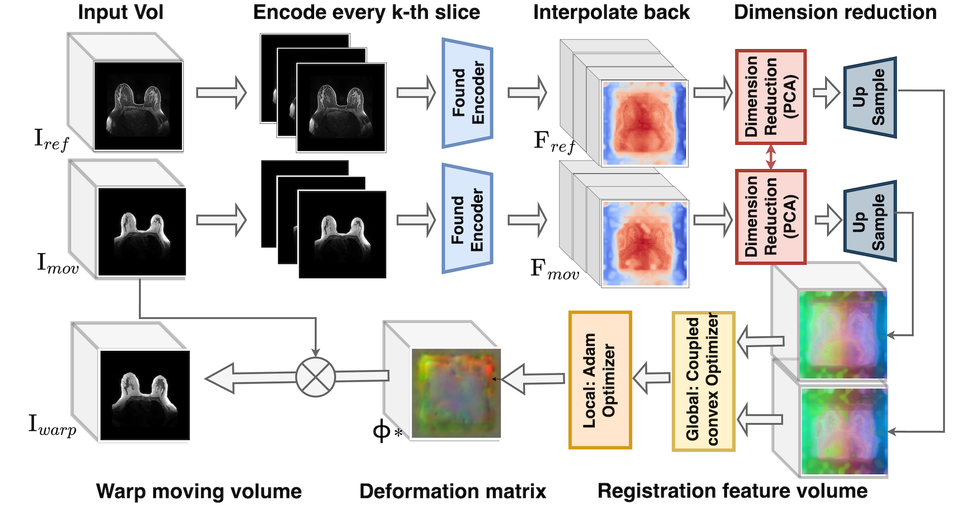
Our training-free, foundation model-based registration pipeline, inspired by the framework of [26], includes three main stages: (1) feature extraction, (2) dimension reduction, and (3) feature-based registration, as shown in Fig. 1.
Feature extraction. Let and denote reference and moving image volumes for registration. Considering that most available pre-trained encoders are for 2D images, we perform feature extraction slice-by-slice along the axial plane. Each volume is treated as a sequence of 2D axial slices, denoted and , where indexes the slice number. To reduce computational cost, we chose to encode every -th slice, where is a hyperparameter that can be selected based on the desired speed of registration. The selected slices are passed through a frozen vision encoder , producing intermediate feature maps:
| (1) |
The features for the skipped slices are then estimated via linear interpolation along the slice axis to form a complete 3D feature volume. When , all slices are encoded and no interpolation is applied. Here, denotes the encoder part of foundation models (e.g., DINO-v2, SAM), typically based on vision transformers (ViTs). For each encoder, the output feature map consists of patch tokens and, optionally, a global CLS token, each of dimension .
Dimension reduction. In the latent space, the feature dimension is typically high (e.g., 256 for SAM and 384 for DINO-v2), which can introduce noise and lead to significant computational cost during registration. To address this, we apply principal component analysis (PCA) to reduce the feature dimensionality for each patch token. Let denote the feature matrix of slice , where is the number of patch tokens. We collect features from all sampled slices of both the reference and moving volumes and concatenate them along the patch dimension, resulting in a joint matrix , where . PCA is then computed on this combined matrix, and the resulting projection is applied slice-wise to both volumes. Let be the number of retained principal components (e.g., ), the reduced feature maps become:
| (2) |
Here, is the projection matrix computed from PCA on . This ensures both volumes are embedded into the same reduced feature space.
To prepare for volumetric registration, we reshape , back to a 3D spatial format. Specifically, each slice-level feature map is first reshaped into a 2D grid of size , where and represent the number of patch tokens along the width and height, respectively. These values depend on the encoder architecture, and does not necessarily equal , as some encoders include a global CLS token in that is excluded prior to reshaping. Stacking the reshaped slices along the -axis results in a 4D volume of size . This intermediate volume is then upsampled to match the original spatial resolution, resulting in a final feature volume of size , which aligns with the input dimensions and serves as input to the registration step.
Feature-based Registration. We follow ConvexAdam [25], adopting a two-stage registration strategy that combines global discrete optimization with instance-level refinement to efficiently align the reduced feature volumes.
In the first stage, we employ a coupled convex discrete optimization framework to obtain an initial deformation field based on the extracted feature volumes. A convex energy function is formulated, which combines the feature similarity term with a regularization term to ensure smooth deformations:
| (3) |
Here, is the dense displacement field, denotes a similarity metric (e.g., sum of squared differences or local cross-correlation), and controls the smoothness constraint. The convex optimization is solved within a discrete search space to efficiently identify a globally optimal deformation.
In the second stage, the deformation field is refined through continuous instance-wise optimization via Adam [12]. Initialized with the displacement field from the first stage, we iteratively update by minimizing the same energy function in a differentiable manner, allowing for sub-voxel accuracy and capturing fine-scale deformations: . This optimization strategy utilizes the robustness of discrete search for global alignment and the flexibility of continuous optimization for local refinement.
2.2 Foundation models
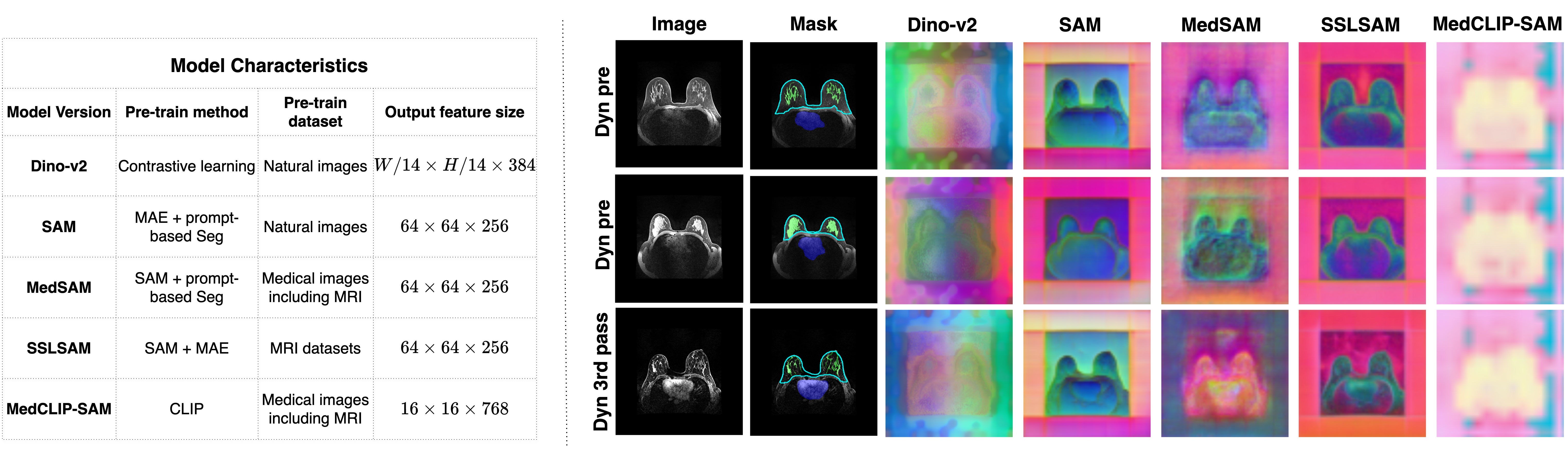
We use five publicly available foundation models as feature extractors, varying in pre-training strategy and domain (natural vs. medical), as shown in Fig. 3.
DINO-v2 [22]: Pre-trained on natural images using a self-supervised contrastive learning strategy (self-distillation without labels). DINO-v2 is known for producing semantically rich features and has been applied in one prior work for cross-domain medical image registration [26].
SAM [13]: Trained on large-scale natural image segmentation datasets using a promptable segmentation objective. The model adopts a ViT-based encoder and was pre-trained with MAE, followed by additional prompt-based segmentation training. SAM has also been widely used for general vision tasks.
MedSAM [17]: A domain-specific version of SAM, fine-tuned on various medical image datasets including MRIs under a prompt-based setting to improve performance on medical image segmentation tasks.
SSLSAM [10]: Fine-tuned SAM on various MRI datasets without labels under the MAE setting. Noticeably, their pre-trained dataset also contains one publicly available breast dataset for dynamic pre-contrast MRIs [16].
MedCLIP-SAM [15]: A vision-language foundation model trained on paired medical images and radiology reports. It follows the same training strategy as MedCLIP, but while MedCLIP was only trained on radiographs, MedCLIP-SAM includes additional MRI data during pretraining.
3 Experiments
3.1 Datasets preparation
Exam and mask collection: For the registration tasks, we collected both internal and external datasets including internal breast MRI exams and external PET-CT/MRI pairs from the BREAST-DIAGNOSIS collection [4]. We include patients with multiple time-stamped exams and pre-contrast axial views, and exclude volumes from patients who underwent mastectomy. For masks, we include three targets for our registration evaluation: breast, breast FGT, and organ. Breast and FGT masks are generated using a 3D V-Net-based model followed by manual correction, while organ and lesion masks are obtained using a nnU-Net-based segmentation model. Full preprocessing and annotation details for exams and masks are provided in Appendix 0.C and Appendix 0.D respectively.
Tasks: In this study, we design four tasks to comprehensively evaluate the registration performance of multiple models. Task 1 involves registering breast MRI scans from different dates and years (longitiduanl exams) with the same image sequence (all images are dynamic pre-contrast). Task 2 focuses on registering longitiduanl breast MRI exams with different image sequences, where the fixed images are set to the dynamic pre-contrast sequence. The category of moving image sequences is shown in Fig. 2 (b). Task 3 explores lesion tracking, where we register the moving image with the lesion to the fixed image without the lesion. This task is designed to evaluate whether the registration model attempts to erase or suppress the lesion in the moving image in order to match the target. For Task 4, we register PET-CT to MRI, which is more challenging due to both the cross-modality nature and the significant breast deformation caused by different patient postures (lying on the back versus facing down).
Evaluation metrics: Tasks 1 to Task 3 are evaluated using the Dice Similarity Coefficient () of three targets: breast, FGT, and organ. For Task 3, we further assess lesion preservation by calculating , which reflects the change in lesion size during registration. For lesion tracking purposes, the ideal goal is to align the breast and dense tissue while preserving the lesion’s physical characteristics, rather than altering its shape or size during the registration process. For Task 5, we qualitatively evaluate the registration performance by visualizing the warped CT and overlaying the PET onto the fixed MRI.
4 Results and Discussion
| Method | Breast | FGT | Organ | Lesion | |
|---|---|---|---|---|---|
| DSC | DSC | DSC | |||
| Task 1: Registration between longitudinal scans within the same MRI sequence | |||||
| Init. | Raw | 49.0 20.6 | 21.2 16.6 | 48.3 21.5 | - |
| Opt. | Affine | 68.2 21.7 | 33.0 21.3 | 60.9 22.8 | - |
| Syn | 80.0 20.0 | 45.4 24.9 | 66.7 23.6 | - | |
| NiftyReg | 81.6 17.9 | 53.8 26.5 | 69.2 24.2 | - | |
| ConvexAdam | 80.8 16.6 | 53.3 23.4 | 64.7 21.3 | - | |
| Found. | DINO-v2 | 81.3 11.7 | 47.4 21.8 | 66.6 19.4 | - |
| SAM | 83.8 14.0 | 48.4 22.5 | 64.4 22.7 | - | |
| MedSAM | 75.5 16.4 | 39.6 23.0 | 64.3 22.8 | - | |
| SSLSAM | 82.1 13.7 | 48.2 24.3 | 67.9 18.2 | - | |
| MedCLIP-SAM | 69.7 13.8 | 33.4 21.5 | 59.1 16.1 | - | |
| Task 2: Registration between longitudinal scans with different MRI sequences | |||||
| Init. | Raw | 44.1 24.4 | 18.7 13.4 | 45.3 21.6 | - |
| Opt. | Affine | 65.8 22.3 | 32.0 18.5 | 61.1 22.7 | - |
| Syn | 76.8 19.1 | 42.8 21.7 | 64.4 21.6 | - | |
| NiftyReg | 73.9 22.2 | 44.3 25.0 | 62.1 22.0 | - | |
| ConvexAdam | 73.4 10.3 | 42.4 31.9 | 41.2 35.4 | - | |
| Found. | DINO-v2 | 80.4 11.4 | 44.5 18.9 | 62.2 18.9 | - |
| SAM | 80.8 16.1 | 43.2 19.8 | 62.5 20.2 | - | |
| MedSAM | 68.3 21.1 | 31.1 18.9 | 59.5 19.9 | - | |
| SSLSAM | 70.8 18.9 | 33.7 20.6 | 56.3 22.1 | - | |
| MedCLIP-SAM | 59.8 18.7 | 18.5 15.9 | 50.6 16.3 | - | |
| Task 3: Registration from lesion-present cases to lesion-absent cases | |||||
| Init. | Raw | 49.4 20.3 | 18.1 14.3 | 40.4 21.9 | 0 (ref) |
| Opt. | Affine | 68.0 15.6 | 31.2 18.2 | 53.8 24.4 | 0 (ref) |
| Syn | 80.5 10.1 | 41.5 21.5 | 57.6 24.8 | 37.7 28.6 | |
| NiftyReg | 81.0 9.9 | 48.4 22.3 | 57.5 25.5 | 48.9 30.3 | |
| ConvexAdam | 78.3 18.9 | 50.3 24.4 | 53.3 27.3 | 60.5 85.5 | |
| Found. | DINO-v2 | 80.5 10.6 | 40.8 18.7 | 55.1 22.0 | 31.3 23.3 |
| SAM | 83.3 13.4 | 39.8 21.9 | 55.9 24.8 | 42.5 43.7 | |
| MedSAM | 73.0 17.6 | 33.5 20.6 | 53.1 24.7 | 120.4 337.5 | |
| SSLSAM | 79.0 15.7 | 37.7 21.8 | 49.3 23.9 | 60.9 41.2 | |
| MedCLIP-SAM | 69.6 17.9 | 26.6 18.9 | 49.0 21.4 | 98.6 150.8 | |
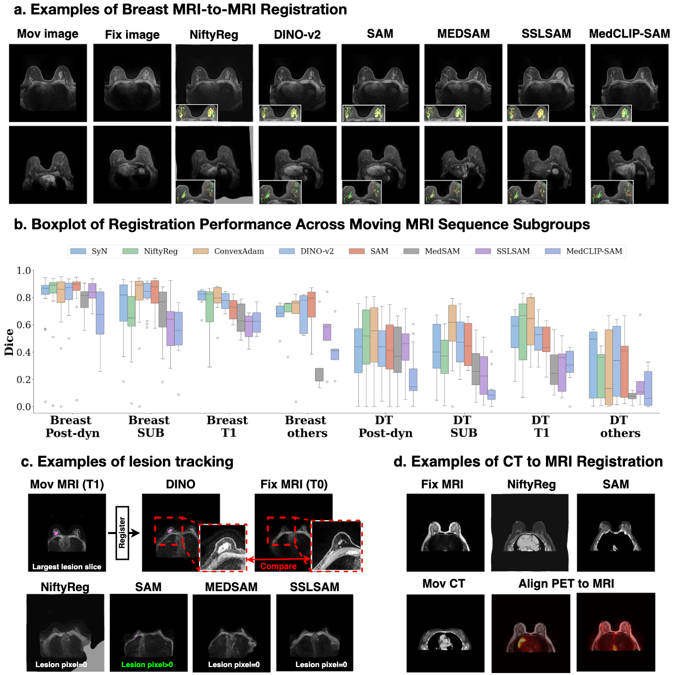
Table 1 shows that foundation models, especially SAM, achieve superior performance in large-structure registration like breast contour. When registering volumes from the same MRI sequence from longitudinal exams, the performance of foundation-based models and optimization-based methods is comparable. However, for cross-sequence registration, SAM noticeably outperforms the best optimization-based method, achieving a Dice score (DSC) of versus . This suggests that features extracted by foundation models are domain-invariant, making them more robust under shifts in image appearance. In contrast, optimization-based methods rely more heavily on image-level similarity.
However, we also observe that foundation model-based algorithms show worse performance in FGT registration for both Task 1 and Task 3, and similar or even worse performance in Task 2. To better understand this, we visualize the feature maps extracted by each model in Fig. 3. The results show that FGT details are barely visible in these feature maps, especially in sparse FGT cases, such as the example in row 3. This limitation is also reflected in the qualitative results in Fig. 2 (a), where, although most models (except MedCLIP-SAM) can register the overall breast contour well, FGT misalignment remains substantial.
For organ registration, although we initially hypothesized that models pre-trained on medical-domain data would perform better due to exposure to similar anatomical structures, all foundation models performed worse than optimization-based methods. Regarding Task 3 (lesion tracking), DINO-v2 achieves the best performance in preserving lesion size, while MedSAM performs poorly.
For Task 4 (PET-CT to MRI registration), the benefits of foundation models become more evident. As shown in Fig. 2 (d), NiftyReg completely fails to align organs between CT and MRI, SAM successfully registers CT to MRI despite significant shape differences, confirming the advantage of foundation models under large domain gaps (e.g., CT to MRI) [26]. This result also suggests potential applications in overlaying PET onto MRI to visualize tumors.
When comparing models trained on natural images versus those trained on medical datasets (such as MedSAM) or breast MRIs (SSLSAM), we observe mixed results. For SSLSAM, there is no clear improvement in Task 1 and a performance drop in Task 2. We suspect this may be due to SSLSAM’s pre-training on only pre-contrast MRIs, leading to domain overfitting. For MedSAM, we observe substantial performance drops across all tasks and targets, likely due to pre-training on medical datasets that excluded breast MRIs. MedCLIP-SAM fails across all tasks, possibly due to the low resolution of feature maps (Fig. 3).
Overall, we do not observe clear benefits from using medical-domain pre-trained models for these breast MRI registration tasks. One possible explanation is that these models, despite being trained on medical data, still use much smaller datasets compared to large-scale natural image pre-training used by models like DINO-v2 or SAM, resulting in less generalizable features. We do not draw definitive conclusions at this stage regarding the effectiveness of medical pre-trained models for zero-shot registration tasks, as new models continue to be proposed [27, 8]. Further exploration and discussion are needed to better understand and improve foundation models for this application.
Conclusion. In this study, we explored whether foundation models can be used for zero-shot registration tasks in the context of breast MRI. We find that models pre-trained on natural images, such as SAM and DINO-v2, can achieve strong performance for large-structure alignment, particularly for breast contour registration. However, these models struggle to preserve fine anatomical details, such as FGT, limiting their application in tasks that require accurate tracking of internal structures. We believe this highlights an important direction for future research, which could develop strategies to better preserve fine-grained information within the feature representations of foundation models.
References
- [1] Alfano, F., Cordero-Grande, L., Ortuño, J.E., García, K.F., García-Sevilla, M., Zamora, O.B., Conde, M.H., Lizarraga, S., Santos, A., Pascau, J., et al.: Breast tumor localization by prone to supine landmark driven registration for surgical planning. IEEE Access 10, 122901–122911 (2022)
- [2] Avants, B.B., Epstein, C.L., Grossman, M., Gee, J.C.: Symmetric diffeomorphic image registration with cross-correlation: evaluating automated labeling of elderly and neurodegenerative brain. Medical image analysis 12(1), 26–41 (2008)
- [3] Balakrishnan, G., Zhao, A., Sabuncu, M.R., Guttag, J., Dalca, A.V.: Voxelmorph: a learning framework for deformable medical image registration. IEEE transactions on medical imaging 38(8), 1788–1800 (2019)
- [4] Bloch, B.N., Jain, A., Jaffe, C.C.: Breast-diagnosis. Data set (2015). https://doi.org/10.7937/K9/TCIA.2015.SDNRQXXR, http://doi.org/10.7937/K9/TCIA.2015.SDNRQXXR, the Cancer Imaging Archive
- [5] Chen, J., Frey, E.C., He, Y., Segars, W.P., Li, Y., Du, Y.: Transmorph: Transformer for unsupervised medical image registration. Medical image analysis 82, 102615 (2022)
- [6] Chen, Y., Gu, H., Dong, H., Li, Q., Chen, Y., Konz, N., Li, L., Mazurowski, M.A.: Guidedmorph: Two-stage deformable registration for breast mri. arXiv preprint arXiv:2505.13414 (2025)
- [7] Chen, Y., Li, L., Gu, H., Dong, H., Nguyen, D.L., Kirk, A.D., Mazurowski, M.A., Hwang, E.S.: Breast density in mri: an ai-based quantification and relationship to assessment in mammography. arXiv preprint arXiv:2504.15192 (2025)
- [8] Dong, H., Chen, Y., Gu, H., Konz, N., Chen, Y., Li, Q., Mazurowski, M.A.: Mri-core: A foundation model for magnetic resonance imaging. arXiv preprint arXiv:2506.12186 (2025)
- [9] Dong, H., Gu, H., Chen, Y., Yang, J., Chen, Y., Mazurowski, M.A.: Segment anything model 2: an application to 2d and 3d medical images. arXiv preprint arXiv:2408.00756 (2024)
- [10] Gu, H., Dong, H., Yang, J., Mazurowski, M.A.: How to build the best medical image segmentation algorithm using foundation models: a comprehensive empirical study with segment anything model. arXiv preprint arXiv:2404.09957 (2024)
- [11] Kim, B., Kim, D.H., Park, S.H., Kim, J., Lee, J.G., Ye, J.C.: Cyclemorph: cycle consistent unsupervised deformable image registration. Medical image analysis 71, 102036 (2021)
- [12] Kingma, D.P., Ba, J.: Adam: A method for stochastic optimization. In: Bengio, Y., LeCun, Y. (eds.) 3rd International Conference on Learning Representations, ICLR 2015, San Diego, CA, USA, May 7-9, 2015, Conference Track Proceedings (2015), http://arxiv.org/abs/1412.6980
- [13] Kirillov, A., Mintun, E., Ravi, N., Mao, H., Rolland, C., Gustafson, L., Xiao, T., Whitehead, S., Berg, A.C., Lo, W.Y., et al.: Segment anything. In: Proceedings of the IEEE/CVF international conference on computer vision. pp. 4015–4026 (2023)
- [14] Klein, S., Staring, M., Murphy, K., Viergever, M.A., Pluim, J.P.: Elastix: a toolbox for intensity-based medical image registration. IEEE transactions on medical imaging 29(1), 196–205 (2009)
- [15] Koleilat, T., Asgariandehkordi, H., Rivaz, H., Xiao, Y.: Medclip-samv2: Towards universal text-driven medical image segmentation (2025), https://arxiv.org/abs/2409.19483
- [16] Lew, C.O., Harouni, M., Kirksey, E.R., Kang, E.J., Dong, H., Gu, H., Grimm, L.J., Walsh, R., Lowell, D.A., Mazurowski, M.A.: A publicly available deep learning model and dataset for segmentation of breast, fibroglandular tissue, and vessels in breast mri. Scientific reports 14(1), 5383 (2024)
- [17] Ma, J., He, Y., Li, F., Han, L., You, C., Wang, B.: Segment anything in medical images. Nature Communications 15(1), 654 (2024)
- [18] Mazurowski, M.A., Dong, H., Gu, H., Yang, J., Konz, N., Zhang, Y.: Segment anything model for medical image analysis: an experimental study. Medical Image Analysis 89, 102918 (2023)
- [19] Mehrabian, H., Richmond, L., Lu, Y., Martel, A.L.: Deformable registration for longitudinal breast mri screening. Journal of digital imaging 31, 718–726 (2018)
- [20] Modat, M., Cash, D.M., Daga, P., Winston, G.P., Duncan, J.S., Ourselin, S.: Global image registration using a symmetric block-matching approach. Journal of medical imaging 1(2), 024003–024003 (2014)
- [21] Modat, M., Ridgway, G.R., Taylor, Z.A., Lehmann, M., Barnes, J., Hawkes, D.J., Fox, N.C., Ourselin, S.: Fast free-form deformation using graphics processing units. Computer methods and programs in biomedicine 98(3), 278–284 (2010)
- [22] Oquab, M., Darcet, T., Moutakanni, T., Vo, H., Szafraniec, M., Khalidov, V., Fernandez, P., Haziza, D., Massa, F., El-Nouby, A., Assran, M., Ballas, N., Galuba, W., Howes, R., Huang, P.Y., Li, S.W., Misra, I., Rabbat, M., Sharma, V., Synnaeve, G., Xu, H., Jegou, H., Mairal, J., Labatut, P., Joulin, A., Bojanowski, P.: Dinov2: Learning robust visual features without supervision (2024), https://arxiv.org/abs/2304.07193
- [23] Ringel, M.J., Richey, W.L., Heiselman, J.S., Luo, M., Meszoely, I.M., Miga, M.I.: Supine magnetic resonance image registration for breast surgery: insights on material mechanics. Journal of Medical Imaging 9(6), 065001–065001 (2022)
- [24] Shi, P., Qiu, J., Abaxi, S.M.D., Wei, H., Lo, F.P.W., Yuan, W.: Generalist vision foundation models for medical imaging: A case study of segment anything model on zero-shot medical segmentation. Diagnostics 13(11), 1947 (2023)
- [25] Siebert, H., Großbröhmer, C., Hansen, L., Heinrich, M.P.: Convexadam: Self-configuring dual-optimisation-based 3d multitask medical image registration. IEEE Transactions on Medical Imaging (2024)
- [26] Song, X., Xu, X., Yan, P.: Dino-reg: General purpose image encoder for training-free multi-modal deformable medical image registration. In: International Conference on Medical Image Computing and Computer-Assisted Intervention. pp. 608–617. Springer (2024)
- [27] Sun, Y., Wang, L., Li, G., Lin, W., Wang, L.: A foundation model for enhancing magnetic resonance images and downstream segmentation, registration and diagnostic tasks. Nature Biomedical Engineering 9(4), 521–538 (2025)
- [28] Tustison, N.J., Cook, P.A., Holbrook, A.J., Johnson, H.J., Muschelli, J., Devenyi, G.A., Duda, J.T., Das, S.R., Cullen, N.C., Gillen, D.L., et al.: The antsx ecosystem for quantitative biological and medical imaging. Scientific reports 11(1), 9068 (2021)
Appendix 0.A Implementation details for each foundation model
For SAM-related models, including SAM, MedSAM, and SSL-SAM, the input slices are resized to . For DINO-v2, the input size is flexible, so we upsample the slices to the largest size that can fit into a single GPU A6000, which is , ensuring the size is divisible by 14 to match the patch size. For MedCLIP-SAM, the input slices are resized to . All inputs are normalized according to the pretraining settings of each model, using the normalization methods provided in their respective codebases. For the dimension reduction step, the size of the extracted feature map for each slice () varies by model, as described in Fig 3. Based on this, we set the number of retained principal components to for SAM-based models, for DINO-v2, and for MedCLIP-SAM. The choice of considers two factors: (1) models with higher original feature dimension require more components to preserve information, and (2) hyperparameters are optimized on one selected reference image pair. For the registration step, we follow the hyperparameters selected by DINO-reg [26].
Appendix 0.B Baseline Methods to Compare
In this study, we provide four optimization-based registration algorithms as baseline methods to evaluate the performance of foundation-model-based approaches. Specifically, we include (1) affine registration implemented using NiftyReg [20] to account for global linear transformations, (2) the Symmetric Normalization (SyN) [2] algorithm from ANTs [28], a widely adopted deformable registration method known for its accuracy in capturing complex non-linear deformations, and (3) NiftyReg-based deformable registration [21], which leverages free-form deformation using cubic B-splines to model local anatomical variations. (4) ConvexAdam MIND [25], compared to other optimization-based registration algorithms, replaces commonly used similarity metrics such as normalized cross correlation (NCC) and mutual information (MI) with the Modality Independent Neighborhood Descriptor (MIND), which captures local structural information and is robust to intensity variations across modalities. The implementation details for these four methods are presented below, respectively.
Affine: For Affine registration, we utilize the reg_aladin tool from the NiftyReg [20] library to align the moving image to the fixed image using a global linear transformation. The resulting affine matrix is applied to breast, dense tissue, and organ masks with nearest-neighbor interpolation.
SyN: SyN-based [2] deformable registration baseline is performed utilizing the antsRegistration tool from the ANTs [28] library. The gradient step size is 0.1, with a total field variance regularization of 3, and mutual information (MI) is used as the similarity metric. A multi-resolution optimization strategy is adopted with four levels of resolution, defined by shrink factors 8 4 2 1 and smoothing sigmas 3 2 1 0. The convergence criteria are set as [100x70x50x20, 1e-6, 10], indicating the maximum number of iterations per level, the convergence threshold, and the number of consecutive steps below the threshold required for convergence.
NiftyReg: reg_f3d tool from the NiftyReg library is utilized to perform the NiftyReg-based deformable registration [21]. We utilize the default hyperparameters in the reg_f3d implementation.
ConvexAdam MIND: The implementation of ConvexAdam follows the official implementation provided by Siebert et al. [25]. For feature extraction, we follow MIND with the Self-Similarity Context (SSC) extension. The neibohood size used for calculating similarity is set as and the dialation size is 2.
Appendix 0.C Exam Collection
For the MRI-to-MRI registration tasks, the data is collected internally from our institution, consisting of exams performed under the bilateral breast MRI protocol, ranging from the year 2014 to the year 2021. We only include patients who have multiple time-stamped exams and at least one pre-contrast axial view volume available. For the PET-CT to MRI registration task, the data is collected externally from BREAST-DIAGNOSIS collection [4], where we select patients who have both PET-CT and MRI scans acquired within a six-month interval. All exams are first resampled into isotropic voxel spacing and then padded into as volume size. For each task, we randomly selected fifty pairs from the pairs that meet the task criteria, with the task details described in a later paragraph Tasks. From these pairs, volumes from patients who had undergone mastectomy were excluded.
Appendix 0.D Mask Collection
We include three targets for our registration evaluation: breast, breast fibroglandular tissue (FGT), and organ. The first two, breast and FGT, are important targets to register for breast-specific tasks, as accurate alignment of both overall breast shape and internal dense tissue is critical for clinical and research use. For the organ label, we suspect that pre-trained networks may perform relatively well, as this type of structure is commonly seen in abdominal MRI or CT datasets that many of these models were originally exposed to. For breast and FGT mask, we employ the breast segmentation model given by the previous study utilizing 3D V-Net architecture [7, 16] followed by the manual correction. The organ and lesion masks are generated using a publicly available breast MRI segmentation model based on nnU-Net, followed by manual correction. The generated masks are visualized in the right-hand side of Fig. 3, in the Mask column.