CH2I2_UV_IR_CEI_SM
Imaging transient molecular configurations in UV-excited diiodomethane
Abstract
Femtosecond structural dynamics of diiodomethane () triggered by ultraviolet (UV) photoabsorption at 290 nm and 330 nm are studied using time-resolved coincident Coulomb explosion imaging driven by a near-infrared probe pulse. We map the dominant single-photon process, the cleavage of the carbon-iodine bond producing rotationally excited radical, identify the contributions of the three-body () dissociation and molecular iodine formation channels, which are primarily driven by the absorption of more than one UV photon, and demonstrate the existence of a weak reaction pathway involving the formation of short-lived transient species resembling iso- geometries with the slightly shorter I-I separation compared to the ground-state . These transient molecular configurations, which can be separated from the other channels by applying a set of conditions on the correlated momenta of three ionic fragments, are formed within approximately 100 fs after the initial photoexcitation and decay within the next 100 fs.
I Introduction
The interaction between light and matter is a common and essential aspect of many fundamental processes in nature[1, 2], including photosynthesis[3, 4, 5], vision[6, 7, 8, 9], vitamin D synthesis[10, 11, 12], DNA repair[13, 14, 15], and various atmospheric reactions[16]. Among the latter, UV-induced photodissociation reactions of halogenated alkanes are significant sources of reactive halogens, which have a considerable impact on environmental and atmospheric chemistry. Iodine, one of the halogens, plays diverse roles in chemistry, serving as a fundamental element in human health and bio-chemistry, a useful catalyst in organic synthesis [17], and a major contributor to the destruction of ozone molecules [18]. Due to its strong absorption of sunlight in a broad range of UV wavelengths, diiodomethane (), a polyhalogenated alkane, is a major source of highly reactive iodine molecules influencing tropospheric chemistry and the marine boundary layer[19].
Numerous studies have been published on the UV-induced photochemistry of , which have reported the primary cleavage of one of the C-I bonds and the formation of and or photoproducts [20, 21, 22]. It has been concluded that and are obtained via photodissociation from the and states, respectively. Due to possible couplings between and higher-lying electronic states, could also be produced via non-adiabatic dissociation from the state [22, 23]. Baughcum et al. [22] reported that the ratio of the yield of and decreases as the excitation wavelength increases, with the quantum yield for production dropping from 0.46 at 248 nm to 0.25 at 308 nm. Using ions generated by a (2+1) resonance enhanced multiphoton ionization process, Xu et al.[23] measured the translational kinetic energy distributions of both and fragments in the wavelength range of 277-305 nm and concluded that the co-fragment is produced with significant internal excitation, with approximately 80% of the total available energy being partitioned into the internal energy of the fragment.
Reid et al.[24, 25] later drew attention to the importance of isomerization of halogenated alkanes into iso-haloalkanes upon UV absorption as a major pathway leading to the production of molecular halogens. The photoisomerization of to iso- has been reported to be an extremely efficient process upon UV absorption in the solution phase and in cages[26, 27, 28]. Since it has been suggested that the dominant isomerization mechanisms in these studies are driven by the interaction with solvent molecules or cage, it is helpful to examine the process in isolated gas-phase molecules, free from such interactions. Borin et al.[29] reported observing photoisomerization of in the gas phase upon 330 nm UV excitation by probing it with femtosecond transient absorption. The delayed rise in the transient absorption of at 380 and 612 nm, approximately 35-90 fs after UV excitation, was attributed to the creation of a short-lived iso-. However, a recent ultrafast electron diffraction (UED) study, where Liu et al.[30] directly mapped the C-I cleavage upon 266 nm excitation, quantified the resulting increase of the C-I and I-I separations, and observed signatures of the rotation of the radical after the C-I bond cleavage, did not mention any observation of iso- formation.
Here, we employ Coulomb Explosion Imaging (CEI), a powerful method for determining the geometric structure of gas-phase molecules [31, 32, 33, 34, 35, 36, 37, 38, 39, 40, 41, 42, 43], to investigate the structural dynamics of upon UV photoabsorption. CEI has been shown to be a useful tool in identifying molecular isomers [35] and conformers [36] of polyhalogenated alkanes and other organic molecules [42], and even imaging the complete structure of halogenated alkanes [38, 39] and ring molecules [37, 40, 42]. When used as a time-resolved method in a pump-probe scheme, laser-induced CEI can measure wave-packets dynamics[44, 45, 46], visualize vibrational motions[47, 48, 46], and image photo-dissociation[49, 50, 51, 52] with high temporal resolution[53]. In particular, CEI provides multidimensional data crucial for identifying any transient structural changes in the molecule upon UV excitation, often with the help of simple classical modeling and computations.
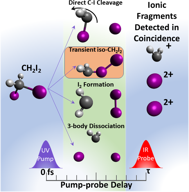
In this work, we investigate the dynamics of diiodomethane upon UV excitation using ionization and Coulomb explosion by an intense strong-field near-infrared (NIR) probe pulse. The primary objective of this time-resolved study is to explore possible signatures of the intramolecular photoisomerization process in , guided by the findings reported by Borin et al. [29]. We analyze the delay-dependent angular correlations between the momenta of the ions, the total kinetic energy release (KER), and the kinetic energies (KE) of individual ionic fragments to differentiate the possible photoisomerization channel from the direct two-body break-up channel that leads to (or ) products. We also identify the three-body () dissociation and the I2 formation channels, which primarily occur after the absorption of multiple UV photons. A schematic of the pump-probe experiment and the different reaction pathways after UV photoexcitation is shown in Fig. 1.
II Methods
II.1 Experimental
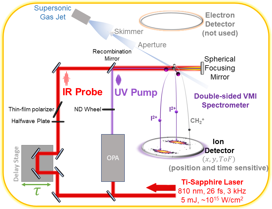
Figure 2 illustrates the experimental setup for the UV-pump and NIR-probe CEI experiment. The femtosecond laser system comprises of a 5-mJ, 3-kHz Coherent Legend Elite DUO Ti:Sapphire laser coupled to a Light Conversion TOPAS Prime optical parametric amplifier (OPA). The output of the Ti:Sapphire laser is split evenly into two independently compressed beam paths: one driving the OPA, and the other providing near-Fourier-transform-limited NIR pulses with a full width at half maximum intensity (FWHM) duration of 26 fs and a bandwidth of 60 nm at a central wavelength of 810 nm. In this experiment, the OPA was set to produce pulses at a central wavelength of 290 nm and 330 nm, with a 5-nm bandwidth, pulse duration of 70 fs (FWHM), and pulse energy up to 15 J. The UV and NIR pulses were recombined using a 45-degree recombination mirror that transmitted the NIR pulses and reflected the UV pulses, and the combined beam was directed collinearly into the vacuum chamber through a 1 mm calcium fluoride window. The delay between NIR and UV pulses was adjusted via a computer-controlled optical delay stage in the NIR arm. Inside the vacuum chamber, the laser pulses are focused using a normal-incidence spherical mirror with 75-mm focal length and UV enhanced aluminium coating.
Diiodomethane, which is liquid at room temperature, is introduced into the vacuum chamber as a supersonic molecular beam expanded through a 30-m nozzle using helium at 3 psig as carrier gas, which is collimated by a skimmer with a 500-m diameter opening. A second differential pumping stage in the molecular beam, separated from the interaction chamber by a 700-m aperture, and a beam dump comprising of two differential pumping stages ensure that the base vacuum in the interaction chamber stays around mbar when the molecular beam is operating.
Ions produced by the interaction between the focused laser pulses and the molecular beam are collected using a double-sided velocity map imaging (VMI) spectrometer. This apparatus is similar to the one employed in the CAMP end-station for free-electron laser experiments [54, 55], while using position-sensitive delay-line detectors (Roentdek DLD80 and HEX80) for the coincident detection of multiple electrons and ions, as described for a similar setup by Ablikim et al. [56]. For the experiment described here, only the ion detector is used. The three-dimensional momentum vectors are obtained from the ions’ time of flight and hit positions as described by Lam et al.[42] and Ablikim et al. [56]. From these momentum vectors, we determine the KER and the angular correlations between the ionic momenta of all the coincidence channels that are of interest.
II.2 Coulomb explosion simulation
To guide the search for a possible isomer and identify the different reaction channels, we performed classical Coulomb explosion simulations for the molecular geometries corresponding to the equilibrium geometry, the iso-, and the C-I bond cleavage pathway including rotation of the intermediate. The simulations are based on solving Newton’s equations of motion for three-point charges located at the positions of the carbon and iodine atoms. To match the experimental observables, the fragment was kept intact. It is treated as a point particle in the simulations. Furthermore, the ionization and fragmentation process induced by the probe pulse was assumed to be instantaneous (i.e., instantaneously breaking both C-I bonds and resulting in a charge q=1 on the carbon atom and q=2 on each of the two iodine atoms), and the repulsion between the three fragments was treated as purely Coulombic. From prior experience, the calculations based on these assumptions typically overestimate the resulting KER but reproduce the experimentally observed momentum correlations [57, 58, 59, 60, 35, 36, 38, 42].
The optimized structures of the molecule in the equilibrium geometry of the neutral electronic ground state and the isomer geometry were taken from Borin et al. [29]. For each case, an ensemble of 5000 geometries was generated by randomly varying the initial atomic positions within a radius of 0.1 Å around the equilibrium geometries and the initial velocities by up to a.u., respectively. These parameters were chosen empirically in order to approximately match the width of the fragment kinetic energy and momentum distributions observed in the experiment. In the simulation for the dissociation channel, a rotational period of 300 fs (obtained experimentally) was used for the intermediate, and the translational KEs used for the ion pairs were obtained from the experimental asymptotic KER for the respective channels.
III Results and discussions
While the main objective of this work is to investigate the transient photoisomerization of diiodomethane (-) after UV photoabsorption, the majority of the photoexcited molecules undergo direct dissociation following C-I bond cleavage, and the first task is therefore to identify the signatures of these competing reaction pathways. The intense NIR probe pulse ionizes both, the unpumped and the photoexcited molecules, resulting in multiple channels with different final charge states and different ionic fragments (see Figs. S1 - S4 of the Supplementary Material (SM) for ion time-of-flight (ToF) mass spectra and multi-ion coincidence spectra). The following discussion focuses on the analysis of the coincidence channel because, as discussed in detail in[61], this breakup channel contains minimal contribution from sequential fragmentation, which is prominent for channels with lower total charge states (see Fig. S5 and Fig. S6 of the SM), where it complicates the analysis. In Fig. 3a, the ion yield of this coincident channel is plotted as a function of the KER and the pump-probe delay.


Three distinct features are visible in this plot: (i) a horizontal band in the KER range between 30 and 40 eV, which corresponds to the Coulomb explosion of bound (i.e. non-dissociating) molecules that may or may not have absorbed a UV photon and that were five-fold ionized by the probe pulse; (ii) a curved feature whose KER decreases with increasing pump-probe delay and which emerges from the horizontal band near time-zero, reaching a KER of approximately 15-18 eV at a delay of 2 ps and 9-14 eV at a delay of 13.5 ps (see Fig. 3b); and (iii) another curved feature whose KER decreases even faster than that of feature (ii), reaching a KER of approximately 8-10 eV at a delay of 2 ps and 1-5 eV at a delay of 13.5 ps, at which point the KER no longer changes with delay. Features (ii) and (iii) originate from the molecules that dissociated upon UV photoabsorption, with feature (ii) corresponding to two-body dissociations into and or and , while feature (iii) corresponds to direct three-body dissociation by the UV pulse into , I and I. While these assignments can be made with good confidence based on the asymptotic KERs of both channels, further insights and confirmation are obtained when inspecting the angular correlation between the momentum vectors of the ionic fragments[61].
Figure 4a shows the coincidence ion yield at the asymptotic delay of 13.5 ps as a function of the KER and the angle between the momentum vectors of two iodine dications. The most intense feature at a KER of 30 - 40 eV (with a smaller side-peak between 20 - 30 eV) and relatively well defined (, ) angle centered around 150∘ - marked by the black ellipse in Fig. 4a - stems from the Coulomb explosion of bound molecules in or near their equilibrium geometry, as confirmed by the data taken without the UV pulse present (see Fig. S6 in the SM). At a KER of approximately 15 eV (red ellipse), an angularly broad feature spanning 60-180 degrees in the (, ) angle can be attributed to C-I cleavage and dissociation into + induced by a single-UV-photon absorption. The broad spread in angle results from high rotational excitation of the fragment due to the torque imparted from the C-I bond cleavage, as also observed in other dihalomethanes [62, 63, 64, 65, 66, 67].
Two additional features, each of them with approximately half the number of events as in the red ellipse discussed above, are prominent in Fig. 4. First, a rather localized spot peaked at a KER slightly below 20 eV and an (, ) angle close to 180° (green ellipse) can be uniquely attributed to formation after UV excitation (i.e., dissociation into + ) since both the KER and the back-to-back emission of the two iodine ions is consistent with a Coulomb explosion of . Second, a clearly separated contribution at low KER (blue ellipse) is attributed to the UV-induced three-body dissociation into + I + I. Both of these features result mainly from multi-photon excitation by the UV pulse. This assignment is based on (i) the energy required to trigger three-body dissociation (4.8 - 5 eV [68, 69]), (ii) earlier experimental work that reported rather low quantum yield of elimination upon single-photon excitation [68], and (iii) on the observed dependence on the UV power. A detailed analysis of the UV power dependence of the different channels for pump wavelengths of 290 nm and 330 nm is presented in Fig. S12 of the SM.
The results of the classical Coulomb explosion simulations for the neutral ground-state geometry and the iso- equilibrium geometry are shown in Fig. 4b, along with the simulation results for the , and dissociation channels at 13.5 ps pump-probe delay. Apart from overestimating the measured KER, most likely due to the assumption of a purely Coulombic potential, the simulated results qualitatively match the experimental observations for all channels, validating the use of these simulations in identifying the observables corresponding to the possible formation of the isomer.
Although the simulations show a subtle difference in the KER of the equilibrium geometry and the isomer, this distinction can be expected to be much less pronounced in the experiment, where a significant spread in the KER is observed even when no UV pulse is present. However, the simulations also predict that the Coulomb explosion of the isomer will lead to some ion yield at larger angles between the (, ) momenta than those realized by the Coulomb explosion of the equilibrium geometry. In the experiment, essentially no events are observed with (, ) angles larger than 160° when no UV pulse is present (see Fig. S6 in the SM), but a transient signal with high KER is observed in this region for pump probe delays between 0 and 250 fs (see Supplementary Movie 1). By selecting only those events in the experimental data where the (, ) angle is larger than 160°, we can thus almost completely suppress the contribution from molecules in the equilibrium geometry.

However, from the experimental data, it is evident that the three dissociation pathways - (i) -I two-body dissociation with rotation, (ii) -I-I three-body dissociation and (iii) molecular formation - could also contribute to the region with the (, ) angle larger than 160° (at least at large delays), as seen in Fig. 4a. Contributions from these pathways need to be filtered out to identify possible signatures of isomerization to -I. The simulations in Fig. 5 show that after Coulomb explosion, the sum of the KEs of the two fragments is larger for the iso- molecules than that of the molecules that undergo direct dissociation into and I via C-I cleavage. Furthermore, in the cases of three-body dissociation or molecular iodine formation, the KE of the methyl ion decreases rapidly with pump-probe delay as the fragment quickly moves away from the two iodine atoms or the molecule. Therefore, the sum of the KEs of the two fragments as well as the KE of the fragment can serve as additional parameters to discriminate the different channels.
Since it is very likely that any isomer-like geometries, were they to be formed as predicted by Borin et al., would be visited quickly due to the large amount of internal energy deposited into the molecule by the UV photon, we will concentrate our search on the smaller delays below 1 ps, where the isomer-geometry should appear as an additional contribution in the region corresponding to the bound molecules. Note that we have, nonetheless, also searched at larger pump-probe delays but have not found any statistically significant indications of isomer-like geometries at larger delays.

In Fig. 6, the KE distribution of the methyl cation () is shown as a function of the pump-probe delay up to 800 fs and only for those events where the (, ) angle is larger than 160°. Based on the reasoning outlined in the previous paragraph, we can conclude that the events with quickly decreasing KE, below the blue line in Fig. 6, correspond to either -I-I three-body dissociation or molecular iodine formation. The events above the blue line at 13 eV mainly correspond to dissociation of into and (with subsequent rotation of the fragment) as well as any possible formation of iso-, with only spurious contributions from the three-body dissociation and molecular iodine formation pathways at small delays below 200 fs.
Next, we select only those events with kinetic energy of ionic fragment above 13 eV from Fig. 6 and plot the sum of the KEs of the two iodine fragments as a function of pump-probe delay in Fig. 7(a). The plot reveals two contributions: an intense feature with a sum KE between 10 and 17 eV (marked by the red rectangle), and a much weaker feature at a higher sum KE, marked by the green rectangle, which contains approximately 7% of the events in the intense feature. The projection in panel (b) reveals that these two contributions (blue line) are shifted toward lower and higher energies, respectively, compared to the sum KE of the unpumped molecules (red dotted line). The integrated yield inside the green region of interest as a function of the pump-probe delay is shown as green line in Fig. 7(c). Based on the simulations shown in Fig. 5(b), we assign the events with the higher sum KE to molecular geometries resembling the iso- structure predicted by Borin et al., which have a slightly smaller I-I distance than geometries resulting from the dissociation of . Fig. 7(c) shows that the occurrence of these structures peaks at a delay of approximately 120 fs and quickly decays again on a similar time scale. Analysis of the pump-probe data recorded at a pump wavelength of 330 nm, following the exact same steps as described above (see section II in SM), yields the blue dashed line in Fig. 7(c), which shows the same behavior as the data at 290 nm except for possibly a subtle shift towards slightly longer delays, which is at the borderline of statistical significance for the present data set.


Additional information on the geometry of the transient species can be obtained by considering the energy sharing between the two detected fragments, as shown in Fig. 8. Symmetric geometries, such as the original geometry, or any molecules excited to symmetric vibrational modes, would lead to equal energy sharing, whereas strongly asymmetric geometries such as the predicted iso- species, would lead to a pronounced asymmetry in the energy sharing. From Fig. 8(a), it is evident that all the events surviving both the filters - large angle and higher energy, have asymmetric energy sharing between the fragments, excluding, for example, the possibility that any of these events stem from a symmetric formation channel. In contrast, symmetric geometries contribute strongly to the coincidence yield with low energy, as shown in Fig. 8(b), corroborating our previous conclusions that many of those events are due to formation.
At this point, we would like to emphasize that although detailed analysis of the CEI data clearly shows the transient formation of isomer-like geometries that are consistent with previous literature [29], the CEI experiment alone is not able to confirm this as an "isomerization" process, nor can the CEI experiment identify the electronic state of the molecule when it assumes these isomer-like geometries.
Whether or not the process observed and reported here can or should be referred to as "isomerization" cannot be answered by the experimental data alone and also depends on the exact definition of "isomerization" one chooses to use. Given the short time that the molecules spend in the isomer-like geometry, which is less than the timescale of the rotation of the fragment that is produced by direct C-I bond cleavage, the process might be better described as a transient passage through isomer-like geometries rather than an actual trapping in the potential well of the isomer, but these terminologies and interpretations strongly rely on careful theoretical modeling, which is beyond the scope of the present paper.
IV Conclusion
We have applied time-resolved three-body Coulomb explosion imaging to study the UV-induced (290-nm and 330-nm) photochemical dynamics in gas-phase . In addition to the primary dissociation into via direct cleavage of the C-I bond, the formation of molecular , and the three-body dissociation into (where the latter two processes are attributed to the absorption of more than one UV photon), we also observe the transient molecular configurations resembling iso- geometries, which are formed within approximately 100 fs after the photoexcitation and completely decay within an additional 100 fs. Within the uncertainties of the experimental data, the relative yield, formation and decay time of the transient iso- products appear to be similar at both excitation wavelengths employed in this study, suggesting that their formation does not depend strongly on the excitation wavelength.
Acknowledgements.
We gratefully acknowledge the technical staff of the J.R. Macdonald Laboratory for their excellent support of the experiments; former PhD students Dr. Seyyed Javad Robatjazi and Dr. Shashank Pathak for commissioning the double-sided VMI apparatus; and Prof. Brett Esry for insightful discussions throughout the project. This work was supported by the Chemical Sciences, Geosciences, and Biosciences Division, Office of Basic Energy Sciences, Office of Science, US Department of Energy, grants no. DE-FG02-86ER13491 and DE-SC0020276 (S.B.), and by the National Science Foundation grant no. PHYS-2409365 (A.S.V.).Conflict of Interest
The authors have no conflicts to disclose.
References
- Kamat et al. [2015] P. V. Kamat, G. S. Schatz, G. Scholes, and T. Zwier, “Photons, physical chemistry, and the year of light - A virtual issue,” Journal of Physical Chemistry Letters 6, 1420–1422 (2015).
- Glusac [2016] K. Glusac, “What has light ever done for chemistry?” Nature Chemistry 2016 8:8 8, 734–735 (2016).
- Govindjee and Govindjee [1974] Govindjee and R. Govindjee, “The absorption of light in photosynthesis,” Scientific American 231, 68–82 (1974).
- Renger [2011] G. Renger, “The light reactions of photosynthesis,” Naturwissenschaften 98, 1305–1319 (2011).
- Zubik et al. [2013] M. Zubik, R. Luchowski, M. Puzio, E. Janik, J. Bednarska, W. Grudzinski, and W. I. Gruszecki, “The negative feedback molecular mechanism which regulates excitation level in the plant photosynthetic complex LHCII: Towards identification of the energy dissipative state,” Biochimica et Biophysica Acta (BBA)-Bioenergetics 1827, 355–364 (2013).
- Schoenlein et al. [1991] R. W. Schoenlein, L. A. Peteanu, R. A. Mathies, and C. V. Shank, “The first step in vision: Femtosecond isomerization of rhodopsin,” Science 254, 412–415 (1991).
- Peteanu et al. [1993] L. A. Peteanu, R. W. Schoenlein, Q. Wang, R. A. Mathies, and C. V. Shank, “The first step in vision occurs in femtoseconds: complete blue and red spectral studies.” Proceedings of the National Academy of Sciences 90, 11762–11766 (1993).
- Wang et al. [1994] Q. Wang, R. W. Schoenlein, L. A. Peteanu, R. A. Mathies, and C. V. Shank, “Vibrationally Coherent Photochemistry in the Femtosecond Primary Event of Vision,” Science 266, 422–424 (1994).
- Polli et al. [2010] D. Polli, P. Altoè, O. Weingart, K. M. Spillane, C. Manzoni, D. Brida, G. Tomasello, G. Orlandi, P. Kukura, R. A. Mathies, M. Garavelli, and G. Cerullo, “Conical intersection dynamics of the primary photoisomerization event in vision,” Nature 467, 440–443 (2010).
- Holick et al. [1980] M. F. Holick, J. A. Maclaughlin, M. B. Clark, S. A. Holick, J. T. Potts, R. R. Anderson, I. H. Blank, J. A. Parrish, and P. Elias, “Photosynthesis of previtamin in human skin and the physiologic consequences,” Science 210, 203–205 (1980).
- Maclaughlin, Anderson, and Holick [1982] J. A. Maclaughlin, R. R. Anderson, and M. F. Holick, “Spectral character of sunlight modulates photosynthesis of previtamin and its photoisomers in human skin,” Science 216, 1001–1003 (1982).
- Holick [1988] M. F. Holick, “Skin: Site of the Synthesis of Vitamin D and a Target Tissue for the Active Form,1,25-Dihydroxy vitamin ,” Annals of the New York Academy of Sciences 548, 14–26 (1988).
- Sinha and Häder [2002] R. P. Sinha and D.-P. Häder, “UV-induced DNA damage and repair: a review,” Photochemical & Photobiological Sciences 1, 225–236 (2002).
- Essen and Klar [2006] L. O. Essen and T. Klar, “Light-driven DNA repair by photolyases,” Cellular and Molecular Life Sciences 63, 1266–1277 (2006).
- Gustavsson, Improta, and Markovitsi [2010] T. Gustavsson, R. Improta, and D. Markovitsi, “DNA/RNA: Building Blocks of Life Under UV Irradiation,” J. Phys. Chem. Lett 17, 48 (2010).
- Keller-Rudek et al. [2013] H. Keller-Rudek, G. K. Moortgat, R. Sander, and R. Sörensen, “The MPI-Mainz UV/VIS Spectral Atlas of Gaseous Molecules of Atmospheric Interest,” Earth System Science Data 5, 365–373 (2013).
- Schomburg and Köhrle [2008] L. Schomburg and J. Köhrle, “On the importance of selenium and iodine metabolism for thyroid hormone biosynthesis and human health,” Mol. Nutr. Food Res 52, 1235–1246 (2008).
- Koenig et al. [2021] T. K. Koenig, R. Volkamer, E. C. Apel, J. F. Bresch, C. A. Cuevas, B. Dix, E. W. Eloranta, R. P. Fernandez, S. R. Hall, R. S. Hornbrook, R. B. Pierce, J. M. Reeves, A. Saiz-Lopez, and K. Ullmann, “Ozone depletion due to dust release of iodine in the free troposphere,” Science Advances 7, 6544 (2021).
- Carpenter [2003] L. J. Carpenter, “Iodine in the Marine Boundary Layer,” Chemical Reviews 103, 4953–4962 (2003).
- Kawasaki, Lee, and Bersohn [1975] M. Kawasaki, S. J. Lee, and R. Bersohn, “Photodissociation of molecular beams of methylene iodide and iodoform,” The Journal of Chemical Physics 63, 809–814 (1975).
- Kroger, Demou, and Riley [1976] P. M. Kroger, P. C. Demou, and S. J. Riley, “Polyhalide photofragment spectra. I. Two-photon two-step photodissociation of methylene iodide,” The Journal of Chemical Physics 65, 1823–1834 (1976).
- Baughcum and Leone [1980] S. L. Baughcum and S. R. Leone, “Photofragmentation infrared emission studies of vibrationally excited free radicals and ,” The Journal of Chemical Physics 72, 6531–6545 (1980).
- Xu et al. [2002] H. Xu, Y. Guo, S. Liu, X. Ma, D. Dai, and G. Sha, “Photodissociation dynamics of CH2l2 molecules in the ultraviolet range studied by ion imaging,” Journal of Chemical Physics 117, 5722–5729 (2002).
- Kalume, George, and Reid [2010] A. Kalume, L. George, and S. A. Reid, “Isomerization as a key path to molecular products in the gas-phase decomposition of halons,” Journal of Physical Chemistry Letters 1, 3090–3095 (2010).
- Reid [2014] S. A. Reid, “When isomerisation is electron transfer: the intriguing story of the iso-halocarbons,” International Reviews in Physical Chemistry 33, 341–370 (2014).
- Tarnovsky et al. [2004] A. N. Tarnovsky, V. Sundstro, E. Åkesson, and T. 1rn Pascher, “Photochemistry of Diiodomethane in Solution Studied by Femtosecond and Nanosecond Laser Photolysis. Formation and Dark Reactions of the Isomer Photoproduct and Its Role in Cyclopropanation of Olefins,” The Journal of Physical Chemistry A 108, 237–249 (2004).
- Panman et al. [2020] M. R. Panman, E. Biasin, O. Berntsson, M. Hermann, S. Niebling, A. J. Hughes, J. Kübel, K. Atkovska, E. Gustavsson, A. Nimmrich, A. O. Dohn, M. Laursen, D. B. Zederkof, A. Honarfar, K. Tono, T. Katayama, S. Owada, T. B. Van Driel, K. Kjaer, M. M. Nielsen, J. Davidsson, J. Uhlig, K. Haldrup, J. S. Hub, and S. Westenhoff, “Observing the structural evolution in the photodissociation of diiodomethane with femtosecond solution x-ray scattering,” Physical Review Letters 125, 226001 (2020).
- Kim et al. [2019] T. Kim, H. S. Kim, Y. Lee, K. H. Kim, H. Kim, and H. Ihee, “Fate of transient isomer of : Mechanism and origin of ionic photoproducts formation unveiled by time-resolved x-ray liquidography,” The Journal of Chemical Physics 150, 224201 (2019).
- Borin et al. [2016] V. A. Borin, S. M. Matveev, D. S. Budkina, P. Z. El-Khoury, and A. N. Tarnovsky, “Direct photoisomerization of vs in the gas phase: a joint 50 fs experimental and multireference resonance-theoretical study,” Physical Chemistry Chemical Physics 18, 28883–28892 (2016).
- Liu et al. [2020] Y. Liu, S. L. Horton, J. Yang, J. P. F. Nunes, X. Shen, T. J. Wolf, R. Forbes, C. Cheng, B. Moore, M. Centurion, K. Hegazy, R. Li, M. F. Lin, A. Stolow, P. Hockett, T. Rozgonyi, P. Marquetand, X. Wang, and T. Weinacht, “Spectroscopic and Structural Probing of Excited-State Molecular Dynamics with Time-Resolved Photoelectron Spectroscopy and Ultrafast Electron Diffraction,” Physical Review X 10, 21016 (2020).
- Vager, Naaman, and Kanter [1989] Z. Vager, R. Naaman, and E. P. Kanter, “Coulomb Explosion Imaging of Small Molecules,” Science 244, 426–431 (1989).
- Kella et al. [1993] D. Kella, M. Algranati, H. Feldman, O. Heber, H. Kovner, E. Malkin, E. Miklazky, R. Naaman, D. Zaifman, J. Zaifman, and Z. Vager, “A system for Coulomb explosion imaging of small molecules at the Weizmann Institute,” Nuclear Instruments and Methods in Physics Research Section A: Accelerators, Spectrometers, Detectors and Associated Equipment 329, 440–452 (1993).
- Cornaggia [2009] C. Cornaggia, “Ultrafast Coulomb explosion imaging of molecules,” Laser Physics 19, 1660–1670 (2009).
- Corrales et al. [2018] M. E. Corrales, J. Gonzaíez-Va, R. de Nalda, and L. Ban, “Coulomb Explosion Imaging for the Visualization of a Conical Intersection,” The Journal of Physical Chemistry Letters 10, 138–143 (2018).
- Ablikim et al. [2016] U. Ablikim, C. Bomme, H. Xiong, E. Savelyev, R. Obaid, B. Kaderiya, S. Augustin, K. Schnorr, I. Dumitriu, T. Osipov, R. Bilodeau, D. Kilcoyne, V. Kumarappan, A. Rudenko, N. Berrah, and D. Rolles, “Identification of absolute geometries of cis and trans molecular isomers by Coulomb Explosion Imaging,” Scientific reports 6, 38202 (2016).
- Pathak et al. [2020] S. Pathak, R. Obaid, S. Bhattacharyya, J. Bürger, X. Li, J. Tross, T. Severt, B. Davis, R. C. Bilodeau, C. A. Trallero-Herrero, A. Rudenko, N. Berrah, and D. Rolles, “Differentiating and quantifying gas-phase conformational isomers using coulomb explosion imaging,” J. Phys. Chem. Lett. 11, 10205–10211 (2020).
- Boll et al. [2022] R. Boll, J. M. Schäfer, B. Richard, K. Fehre, G. Kastirke, Z. Jurek, M. S. Schöffler, M. M. Abdullah, N. Anders, T. M. Baumann, S. Eckart, B. Erk, A. De Fanis, R. Dörner, S. Grundmann, P. Grychtol, A. Hartung, M. Hofmann, M. Ilchen, L. Inhester, C. Janke, R. Jin, M. Kircher, K. Kubicek, M. Kunitski, X. Li, T. Mazza, S. Meister, N. Melzer, J. Montano, V. Music, G. Nalin, Y. Ovcharenko, C. Passow, A. Pier, N. Rennhack, J. Rist, D. E. Rivas, D. Rolles, I. Schlichting, L. P. H. Schmidt, P. Schmidt, J. Siebert, N. Strenger, D. Trabert, F. Trinter, I. Vela-Perez, R. Wagner, P. Walter, M. Weller, P. Ziolkowski, S.-K. Son, A. Rudenko, M. Meyer, R. Santra, and T. Jahnke, “X-ray multiphoton-induced Coulomb explosion images complex single molecules,” Nature Physics 18, 423–428 (2022).
- Bhattacharyya et al. [2022] S. Bhattacharyya, K. Borne, F. Ziaee, S. Pathak, E. Wang, A. S. Venkatachalam, X. Li, N. Marshall, K. D. Carnes, C. W. Fehrenbach, T. Severt, I. Ben-Itzhak, A. Rudenko, and D. Rolles, “Strong-Field-Induced Coulomb Explosion Imaging of Tribromomethane,” J. Phys. Chem. Lett 13, 22 (2022).
- Li et al. [2022] X. Li, A. Rudenko, M. S. Schöffler, N. Anders, T. M. Baumann, S. Eckart, B. Erk, A. De Fanis, K. Fehre, R. Dörner, L. Foucar, S. Grundmann, P. Grychtol, A. Hartung, M. Hofmann, M. Ilchen, C. Janke, G. Kastirke, M. Kircher, K. Kubicek, M. Kunitski, T. Mazza, S. Meister, N. Melzer, J. Montano, V. Music, G. Nalin, Y. Ovcharenko, C. Passow, A. Pier, N. Rennhack, J. Rist, D. E. Rivas, I. Schlichting, L. P. H. Schmidt, P. Schmidt, J. Siebert, N. Strenger, D. Trabert, F. Trinter, I. Vela-Perez, R. Wagner, P. Walter, M. Weller, P. Ziolkowski, A. Czasch, D. Rolles, M. Meyer, T. Jahnke, and R. Boll, “Coulomb explosion imaging of small polyatomic molecules with ultrashort x-ray pulses,” Physical Review Research 4, 013029 (2022).
- Lam et al. [2023] H. V. S. Lam, A. S. Venkatachalam, S. Bhattacharyya, E. Wang, K. Borne, K. Chen, A. Rudenko, and D. Rolles, “Coulomb explosion imaging: a robust method for distinguishing molecular structures and tracking structural changes in photochemical reactions,” in Ultrafast Nonlinear Imaging and Spectroscopy XI, Vol. 12681, edited by Z. Liu, D. Psaltis, and K. Shi, International Society for Optics and Photonics (SPIE, 2023) p. 1268108.
- Wang et al. [2023] E. Wang, S. Bhattacharyya, K. Chen, K. Borne, F. Ziaee, S. Pathak, H. V. S. Lam, A. S. Venkatachalam, X. Chen, R. Boll, T. Jahnke, A. Rudenko, and D. Rolles, “Time-resolved coulomb explosion imaging unveils ultrafast ring opening of furan,” (2023), arXiv:2311.05099 [physics.chem-ph] .
- Lam et al. [2024] H. V. S. Lam, A. S. Venkatachalam, S. Bhattacharyya, K. Chen, K. Borne, E. Wang, R. Boll, T. Jahnke, V. Kumarappan, A. Rudenko, and D. Rolles, “Differentiating Three-Dimensional Molecular Structures Using Laser-Induced Coulomb Explosion Imaging,” Physical Review Letters 132, 123201 (2024).
- Jahnke et al. [2025] T. Jahnke, S. Mai, S. Bhattacharyya, K. Chen, R. Boll, M. E. Castellani, S. Dold, U. Frühling, A. E. Green, M. Ilchen, R. Ingle, G. Kastirke, H. V. S. Lam, F. Lever, D. Mayer, T. Mazza, T. Mullins, Y. Ovcharenko, B. Senfftleben, F. Trinter, Atia-Tul-Noor, S. Usenko, A. S. Venkatachalam, A. Rudenko, D. Rolles, M. Meyer, H. Ibrahim, and M. Gühr, “Direct observation of ultrafast symmetry reduction during internal conversion of 2-thiouracil using coulomb explosion imaging,” Nature Communications 16, 2074 (2025).
- Stapelfeldt, Constant, and Corkum [1995] H. Stapelfeldt, E. Constant, and P. B. Corkum, “Wave Packet Structure and Dynamics Measured by Coulomb Explosion,” Physical Review Letters 74, 3780–3783 (1995).
- Lam et al. [2020] H. V. S. Lam, S. Yarlagadda, A. Venkatachalam, T. N. Wangjam, R. K. Kushawaha, C. Cheng, P. Svihra, A. Nomerotski, T. Weinacht, D. Rolles, and V. Kumarappan, “Angle-dependent strong-field ionization and fragmentation of carbon dioxide measured using rotational wave packets,” Phys. Rev. A 102, 043119 (2020).
- Lam et al. [2025] H. V. S. Lam, V.-H. Hoang, A. S. Venkatachalam, S. Bhattacharyya, K. Chen, S. Jacob, S. Kudagama, T. T. Nguyen, D. Rolles, U. Thumm, A. Rudenko, and V. Kumarappan, “Simultaneous imaging of vibrational, rotational, and electronic wave-packet dynamics in a triatomic molecule,” Phys. Rev. A , – (2025).
- Stapelfeldt et al. [1998] H. Stapelfeldt, E. Constant, H. Sakai, and P. B. Corkum, “Time-resolved Coulomb explosion imaging: A method to measure structure and dynamics of molecular nuclear wave packets,” Physical Review A 58, 426–433 (1998).
- Ergler et al. [2006] T. Ergler, A. Rudenko, B. Feuerstein, K. Zrost, C. D. Schröter, R. Moshammer, and J. Ullrich, “Ultrafast mapping of nuclear wave packets using time-resolved Coulomb explosion imaging,” Journal of Physics B: Atomic, Molecular and Optical Physics 39, S493–S501 (2006).
- Bocharova et al. [2011] I. A. Bocharova, A. S. Alnaser, U. Thumm, T. Niederhausen, D. Ray, C. L. Cocke, and I. V. Litvinyuk, “Time-resolved Coulomb-explosion imaging of nuclear wave-packet dynamics induced in diatomic molecules by intense few-cycle laser pulses,” Physical Review A 83, 013417 (2011).
- Amini et al. [2018] K. Amini, E. Savelyev, F. Brauße, N. Berrah, C. Bomme, M. Brouard, M. Burt, L. Christensen, S. Düsterer, B. Erk, H. Höppner, T. Kierspel, F. Krecinic, A. Lauer, J. W. L. Lee, M. Müller, E. Müller, T. Mullins, H. Redlin, N. Schirmel, J. Thøgersen, S. Techert, S. Toleikis, R. Treusch, S. Trippel, A. Ulmer, C. Vallance, J. Wiese, P. Johnsson, J. Küpper, A. Rudenko, A. Rouzée, H. Stapelfeldt, D. Rolles, and R. Boll, “Photodissociation of aligned I and molecules probed with time-resolved Coulomb explosion imaging by site-selective extreme ultraviolet ionization,” Structural Dynamics 5, 14301 (2018).
- Ziaee et al. [2023] F. Ziaee, K. Borne, R. Forbes, K. Raju, Y. Malakar, B. Kaderiya, T. Severt, I. Ben-Itzhak, A. Rudenko, and D. Rolles, “Single-and multi-photon-induced ultraviolet excitation and photodissociation of probed by coincident ion momentum imaging †,” Phys. Chem. Chem. Phys 25, 9999 (2023).
- Bhattacharyya et al. [2024] S. Bhattacharyya, E. Wang, K. Borne, K. Chen, A. S. Venkatachalam, H. V. S. Lam, F. Ziaee, S. Pathak, A. Khmelnitskiy, K. D. Carnes, C. W. Fehrenbach, I. Ben-Itzhak, A. Rudenko, and D. Rolles, “Delayed dissociation and transient isomerization during the ultrafast photodissociation of the tribromomethane cation,” The Journal of Physical Chemistry Letters 15, 12188–12196 (2024), pMID: 39622006.
- Légaré et al. [2005a] F. Légaré, K. F. Lee, I. V. Litvinyuk, P. W. Dooley, A. D. Bandrauk, D. M. Villeneuve, and P. B. Corkum, “Imaging the time-dependent structure of a molecule as it undergoes dynamics,” Physical Review A 72, 052717 (2005a).
- Rolles et al. [2014] D. Rolles, R. Boll, M. Adolph, A. Aquila, C. Bostedt, J. D. Bozek, H. N. Chapman, R. Coffee, N. Coppola, P. Decleva, T. Delmas, S. W. Epp, B. Erk, F. Filsinger, L. Foucar, L. Gumprecht, A. Hömke, T. Gorkhover, L. Holmegaard, P. Johnsson, C. Kaiser, F. Krasniqi, K. U. Kühnel, J. Maurer, M. Messerschmidt, R. Moshammer, W. Quevedo, I. Rajkovic, A. Rouzée, B. Rudek, I. Schlichting, C. Schmidt, S. Schorb, C. D. Schröter, J. Schulz, H. Stapelfeldt, M. Stener, S. Stern, S. Techert, J. Thøgersen, M. J. Vrakking, A. Rudenko, J. Küpper, and J. Ullrich, “Femtosecond x-ray photoelectron diffraction on gas-phase dibromobenzene molecules,” Journal of Physics B: Atomic, Molecular and Optical Physics 47 (2014), 10.1088/0953-4075/47/12/124035.
- Erk et al. [2018] B. Erk, J. P. Müller, C. Bomme, R. Boll, G. Brenner, H. N. Chapman, J. Correa, S. Düsterer, S. Dziarzhytski, S. Eisebitt, H. Graafsma, S. Grunewald, L. Gumprecht, R. Hartmann, G. Hauser, B. Keitel, C. von Korff Schmising, M. Kuhlmann, B. Manschwetus, L. Mercadier, E. Müller, C. Passow, E. Plönjes, D. Ramm, D. Rompotis, A. Rudenko, D. Rupp, M. Sauppe, F. Siewert, D. Schlosser, L. Strüder, A. Swiderski, S. Techert, K. Tiedtke, T. Tilp, R. Treusch, I. Schlichting, J. Ullrich, R. Moshammer, T. Möller, and D. Rolles, “CAMP@FLASH: an end-station for imaging, electron- and ion-spectroscopy, and pump–probe experiments at the FLASH free-electron laser,” Journal of Synchrotron Radiation 25, 1529–1540 (2018).
- Ablikim et al. [2019] U. Ablikim, C. Bomme, T. Osipov, H. Xiong, R. Obaid, R. C. Bilodeau, N. G. Kling, I. Dumitriu, S. Augustin, S. Pathak, K. Schnorr, D. Kilcoyne, N. Berrah, and D. Rolles, “A coincidence velocity map imaging spectrometer for ions and high-energy electrons to study inner-shell photoionization of gas-phase molecules,” Rev. Sci. Instrum 90, 55103 (2019).
- Lezius et al. [2002] M. Lezius, . V. Blanchet, M. Y. Ivanov, A. Stolow, and . V. Blanchet, “Polyatomic molecules in strong laser fields: Nonadiabatic multielectron dynamics,” J. Chem. Phys 117, 1575–1588 (2002).
- Légaré et al. [2005b] F. Légaré, K. F. Lee, I. V. Litvinyuk, P. W. Dooley, S. S. Wesolowski, P. R. Bunker, P. Dombi, F. Krausz, A. D. Bandrauk, D. M. Villeneuve, and P. B. Corkum, “Laser Coulomb-explosion imaging of small molecules,” Physical Review A - Atomic, Molecular, and Optical Physics 71, 013415 (2005b).
- Liu et al. [2007] H. Liu, Z. Yang, Z. Gao, and Z. Tang, “Ionization and dissociation of in intense laser field,” J. Chem. Phys 126, 44316 (2007).
- Corrales et al. [2012] M. E. Corrales, G. Gitzinger, J. Us Gonz Alez-V Azquez, V. Loriot, R. De Nalda, and L. Ba˜, “Velocity Map Imaging and Theoretical Study of the Coulomb Explosion of under Intense Femtosecond IR Pulses,” J. Phys. Chem. A 116, 2669–2677 (2012).
- Kaderiya [2021] B. Kaderiya, Imaging Photo-Induced Dynamics in Halomethane Molecules with Coincident Ion Momentum Spectroscopy, Ph.d. dissertation, Kansas State University, Manhattan, Kansas, USA (2021), advisor: Dr. Artem Rudenko.
- Murillo-Sánchez et al. [2018] M. L. Murillo-Sánchez, S. Marggi Poullain, J. J. Bajo, M. E. Corrales, J. González-Vázquez, I. R. Solá, and L. Bañares, “Halogen-atom effect on the ultrafast photodissociation dynamics of the dihalomethanes and ,” Physical Chemistry Chemical Physics 20, 20766–20778 (2018).
- Allum et al. [2018] F. Allum, M. Burt, K. Amini, R. Boll, H. Köckert, P. K. Olshin, S. Bari, C. Bomme, F. Brauße, B. Cunha de Miranda, S. Düsterer, B. Erk, M. Géléoc, R. Geneaux, A. S. Gentleman, G. Goldsztejn, R. Guillemin, D. M. P. Holland, I. Ismail, P. Johnsson, L. Journel, J. Küpper, J. Lahl, J. W. L. Lee, S. Maclot, S. R. Mackenzie, B. Manschwetus, A. S. Mereshchenko, R. Mason, J. Palaudoux, M. N. Piancastelli, F. Penent, D. Rompotis, A. Rouzée, T. Ruchon, A. Rudenko, E. Savelyev, M. Simon, N. Schirmel, H. Stapelfeldt, S. Techert, O. Travnikova, S. Trippel, J. G. Underwood, C. Vallance, J. Wiese, F. Ziaee, M. Brouard, T. Marchenko, and D. Rolles, “Coulomb explosion imaging of ch3i and ch2cli photodissociation dynamics,” The Journal of Chemical Physics 149, 204313 (2018).
- Marggi Poullain et al. [2018] S. Marggi Poullain, a. V. David Chicharro, E. Navarro, L. Rubio-Lago, J. Gonzá lez Vá zquez, and L. Bañ ares, “Photodissociation dynamics of bromoiodomethane from the first and second absorption bands. A combined velocity map and slice imaging study,” Phys. Chem. Chem. Phys 20, 3490 (2018).
- Köckert et al. [2022] H. Köckert, J. W. Lee, F. Allum, K. Amini, S. Bari, C. Bomme, F. Brauße, M. Brouard, M. Burt, B. Cunha De Miranda, S. Düsterer, P. Eng-Johnsson, B. Erk, M. Géléoc, R. Geneaux, A. S. Gentleman, R. Guillemin, G. Goldsztejn, D. M. Holland, I. Ismail, L. Journel, T. Kierspel, J. Küpper, J. Lahl, S. R. Mackenzie, S. Maclot, B. Manschwetus, A. S. Mereshchenko, T. Mullins, P. K. Olshin, J. Palaudoux, F. Penent, M. N. Piancastelli, D. Rompotis, A. Rouzée, T. Ruchon, A. Rudenko, N. Schirmel, M. Simon, S. Techert, O. Travnikova, S. Trippel, C. Vallance, E. Wang, J. Wiese, F. Ziaee, T. Marchenko, D. Rolles, and R. Boll, “UV-induced dissociation of probed by intense femtosecond XUV pulses,” Journal of Physics B: Atomic, Molecular and Optical Physics 55 (2022), 10.1088/1361-6455/AC489D.
- Recio et al. [2022] P. Recio, J. Cachón, L. Rubio-Lago, D. V. Chicharro, A. Zanchet, P. Limão-Vieira, N. De Oliveira, P. C. Samartzis, S. Marggi Poullain, and L. Bañares, “Imaging the Photodissociation Dynamics and Fragment Alignment of at 193 nm,” Journal of Physical Chemistry A 126, 8404–8422 (2022).
- Walmsley et al. [2023] T. Walmsley, J. Unwin, F. Allum, S. Bari, R. Boll, K. Borne, M. Brouard, P. Bucksbaum, N. Ekanayake, B. Erk, R. Forbes, A. J. Howard, P. Eng-Johnsson, J. W. Lee, Z. Liu, B. Manschwetus, R. Mason, C. Passow, J. Peschel, D. Rivas, D. Rolles, A. Rörig, A. Rouzée, C. Vallance, F. Ziaee, and M. Burt, “Characterizing the multi-dimensional reaction dynamics of dihalomethanes using XUV-induced Coulomb explosion imaging,” Journal of Chemical Physics 159, 144302 (2023).
- Chen et al. [2011] S. Y. Chen, P. Y. Tsai, H. C. Lin, C. C. Wu, K. C. Lin, B. J. Sun, and A. H. Chang, “I2 molecular elimination in single-photon dissociation of CH2I2 at 248 nm by using cavity ring-down absorption spectroscopy,” Journal of Chemical Physics 134 (2011), 10.1063/1.3523571.
- Toulson et al. [2016] B. W. Toulson, J. P. Alaniz, J. G. Hill, and C. Murray, “Near-UV photodissociation dynamics of CH2I2,” Physical Chemistry Chemical Physics 18, 11091–11103 (2016).