ConnectomeDiffuser: Generative AI Enables Brain Network Construction from Diffusion Tensor Imaging
Abstract
Brain network analysis plays a crucial role in diagnosing and monitoring neurodegenerative disorders such as Alzheimer’s disease (AD). Existing approaches for constructing structural brain networks from diffusion tensor imaging (DTI) often rely on specialized toolkits that suffer from inherent limitations: operator subjectivity, labor-intensive workflows, and restricted capacity to capture complex topological features and disease-specific biomarkers. To overcome these challenges and advance computational neuroimaging instrumentation, ConnectomeDiffuser is proposed as a novel diffusion-based framework for automated end-to-end brain network construction from DTI. The proposed model combines three key components: (1) a Template Network that extracts topological features from 3D DTI scans using Riemannian geometric principles, (2) a diffusion model that generates comprehensive brain networks with enhanced topological fidelity, and (3) a Graph Convolutional Network classifier that incorporates disease-specific markers to improve diagnostic accuracy. ConnectomeDiffuser demonstrates superior performance by capturing a broader range of structural connectivity and pathology-related information, enabling more sensitive analysis of individual variations in brain networks. Experimental validation on datasets representing two distinct neurodegenerative conditions demonstrates significant performance improvements over other brain network methods. This work contributes to the advancement of instrumentation in the context of neurological disorders, providing clinicians and researchers with a robust, generalizable measurement framework that facilitates more accurate diagnosis, deeper mechanistic understanding, and improved therapeutic monitoring of neurodegenerative diseases such as AD.
Index Terms:
Brain network construction, diffusion tensor imaging, diffusion model, graph convolution network.I Introduction
Advances in neuroimaging technologies have revolutionized the diagnosis and understanding of neurodegenerative diseases such as Alzheimer’s disease (AD). The growing of medical imaging methods such as Functional Magnetic Resonance Imaging (fMRI) and other neuroimaging modalities has provided unprecedented insights into brain morphology and pathophysiology [1, 2, 3]. Learning-based computational imaging has transformed multiple facets of medical image analysis, including lesion detection [4, 5], tissue segmentation [6], image reconstruction [7, 8, 9, 10], super-resolution enhancement [11, 12, 13, 14], and diagnostic decision support [15, 16, 17]. Similar transformer-based approaches have shown success in other imaging domains [18, 19, 20]. Despite these advances, the extraction and interpretation of high-dimensional patterns within neuroimaging data remain challenges, particularly for neurodegenerative conditions characterized by subtle and progressive alterations in brain structure and function. Similar challenges exist in other medical domains, such as identifying biomarkers for osteoporosis through integrated genomic approaches [21].
The human brain represents an intricate network of interconnected neurons, requiring precise coordination across anatomically distinct regions for optimal cognitive function. Conceptualizing the brain as a network structure has emerged as a transformative approach in neuroimaging [22, 23], offering a more accurate representation of brain features. This network-based framework models the brain as a graph where vertices represent regions of interest (ROIs) and edges signify the interactions between these regions. The two primary forms of connectivity—functional connectivity (FC) and structural connectivity (SC)—have demonstrated exceptional sensitivity in detecting disease-specific alterations that can not be detected by conventional medical imaging [24, 25]. However, current methodologies for constructing brain networks frequently depend on specialized preprocessing toolkits such as PANDA [26], which suffer from inherent limitations: procedural complexity, inconsistent outcomes due to parameter variability, and susceptibility to processing errors introduced by manual intervention. These shortcomings compromise the reliability of brain network analyses for neurological disorders and underscore the necessity for more robust, automated, and generalizable approaches.
To overcome these limitations, ConnectomeDiffuser is proposed as a comprehensive end-to-end framework that automates and refines the process of converting neuroimaging data into brain network representations. Designed specifically for brain disorder characterization, our framework integrates three components: a Template Network, a diffusion-based generative model, and a Graph Convolutional Network (GCN) classifier, collectively forming a robust pipeline for brain network construction and analysis.
The Template Network forms the foundation of our framework, extracting geometrically informed graph-structured features from three-dimensional Diffusion Tensor Imaging (DTI) data. This component employs the Anatomical Automatic Labeling (AAL) template [27] to ensure anatomical precision while capturing complex non-linear fiber tract relationships. Drawing inspiration from Physics-Informed Neural Networks (PINNs) [28], the Template Network incorporates self-attention mechanisms to effectively model the intricate spatial relationships between neuronal pathways.
Building upon these extracted features, the diffusion-based model generates comprehensive brain networks that incorporate disease-specific characteristics. This generative component demonstrates superior robustness and fidelity in producing biologically plausible graph topologies compared to alternative generative approaches such as Generative Adversarial Networks (GANs) [29, 30] and Variational Autoencoders (VAEs) [31]. The resulting connectivity matrices accurately represent the complex interrelationships between brain regions while preserving pathologically relevant patterns.
The GCN classifier completes our framework, analyzing the generated graph representations to capture both local and global connectivity patterns. This component’s adaptability to diverse topological structures enables accurate categorization of different brain disorders based on their brain network abnormalities. The integration of the Template Network with the GCN classifier provides an unprecedented detailed characterization of brain connectivity, enhancing diagnostic accuracy for neurological disorders such as AD.
Our main contributions are as follows:
-
1.
The ConnectomeDiffuser is proposed as an end-to-end method to identify the biomarkers for neurological disorders by transforming neuroimaging data into graph-based brain connectivity representations.
-
2.
The Template Network is proposed to generate anatomically informed brain feature matrices using Riemannian geometric principles.
-
3.
The Brain Diffusive Loss is proposed to precisely quantify and minimize topological discrepancies in the generated networks, significantly enhancing the model’s ability to identify the complex connectivity patterns characteristic of various neurodegenerative diseases.
II Related Work
II-A Brain Network Analysis
Graph Neural Networks (GNNs) have emerged as powerful computational tools for modeling brain networks, primarily due to their inherent capacity to capture complex, high-order interactions among regions of interest (ROIs). Among various GNN architectures, Graph Convolutional Networks (GCNs) have gained particular prominence in neuroimaging analysis. Parisot et al.[32] and Ktena et al.[33] demonstrated GCNs’ effectiveness in detecting aberrant neuronal connections associated with Alzheimer’s disease (AD), establishing their utility for neurological disorder characterization. Zuo et al.[34] leveraged generative AI approaches to enable causality analysis for mild cognitive impairment, further advancing the application of AI in understanding neurodegenerative disease progression.
Recent advances have further extended GNN applications in brain network modeling. Pan et al.[35] introduced the Decoupling Generative Adversarial Network (DecGAN), which effectively identifies abnormal neural circuits implicated in AD pathophysiology. Their subsequent work on CT-GAN [36] leveraged cross-modal Transformer architectures to integrate functional connectivity from resting-state fMRI with structural connectivity derived from DTI, enhancing the comprehensiveness of brain network representations. Kong et al.[37] developed the Structural Brain-Network Generative Model (SBGM), employing an adversarial learning framework to generate structural brain networks directly from neuroimaging data, reducing reliance on conventional processing pipelines. Gong et al.[38] introduced spatial attention recurrent networks with feature selection for identifying addiction-related brain networks, demonstrating the versatility of neural network approaches in characterizing various neurological conditions. More recently, Jing et al.[39] proposed Graph Diffusion Reconstruction Network for identifying addiction-related brain networks, highlighting the emerging integration of diffusion techniques with graph-based brain network analysis.
Despite these significant advancements, current GCN-based methodologies face persistent challenges in fully capturing the intricate topological characteristics of brain networks. Many approaches inadequately incorporate disease-specific information and biomarkers, limiting their translational potential in computational neuroimaging for clinical diagnosis of neurological disorders. Our work addresses these limitations by adopting GCN as a specialized classifier for extracting disease-related embeddings while enhancing its capabilities through strategic integration with a Template Network and a diffusion-based generative model.
II-B Diffusion Models
Diffusion models [40] represent a class of probabilistic generative frameworks designed to learn data distributions by iteratively reducing noise in normally distributed data. These models have demonstrated exceptional promise across diverse medical imaging applications. Wolleb et al.[41] pioneered a weakly supervised approach for anomaly detection using Denoising Diffusion Implicit Models, generating highly detailed anomaly maps without requiring complex training regimens.
In neuroimaging specifically, Pinaya et al.[42] established the clinical viability of diffusion models, while Chung et al.[43] developed a sophisticated score-based diffusion model for three-dimensional medical image reconstruction. Their approach integrates denoising score matching to train time-dependent score functions with an iterative inference process that alternates between numerical Stochastic Differential Equation (SDE) solvers and data consistency steps. Gonzalez-Jimenez et al.[44] extended these concepts to dermatological applications, employing an unsupervised diffusion model to generate log-likelihood gradient maps that highlight pathological regions indicative of eczema.
The synthesis of medical images represents another domain where diffusion models have demonstrated considerable utility. Han et al.[45] developed a coarse-to-fine Positron Emission Tomography (PET) reconstruction framework incorporating diffusion techniques, achieving superior sample quality and higher log-likelihood scores compared to conventional GAN-based approaches. Yao et al.[46] specifically applied conditional diffusion models for Alzheimer’s disease prediction through data augmentation, demonstrating the potential of diffusion-based approaches in neurodegenerative disease analysis. The Latent Diffusion Model (LDM) [47], an advanced generative architecture capable of capturing complex statistical characteristics and data structures, has found applications in clinical imaging. Wang et al.[48] exemplified this through InverseSR, an LDM-based methodology for enhancing the resolution of low-resolution functional MRI scans.
While diffusion-based approaches have demonstrated considerable promise across various medical imaging tasks, their application to the generalized construction of brain networks for neurological disorders remains largely unexplored. Current methodologies frequently lack the capacity to effectively incorporate disease-specific information and often inadequately capture the complex topological characteristics inherent to brain networks. Our work leverages the superior generative capabilities of diffusion models specifically for brain network construction, aiming to provide robust stability while addressing the limitations of existing approaches. Through the strategic integration of a Template Network and a GCN classifier, our proposed model enables the generalized construction of brain networks with enhanced fidelity, ultimately contributing to improved diagnosis and understanding of neurological disorders.
III Method
III-A Overview
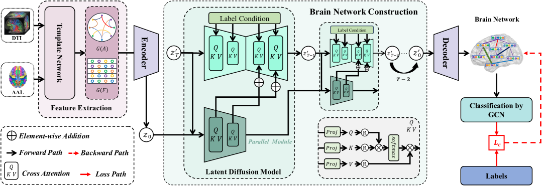
The proposed ConnectomeDiffuser framework comprises three principal components: the Template Network, a Latent Diffusion model, and a Graph Convolutional Network (GCN) classifier. As illustrated in Figure 1, the processing pipeline begins with the registration of Diffusion Tensor Imaging (DTI) data to the Anatomical Automatic Labeling (AAL) template, followed by feature extraction via the Template Network. Subsequently, these extracted features are processed through the ConnectomeDiffuser to generate a comprehensive brain network representation. Finally, the GCN classifier analyzes the vertex structure of the brain features to perform diagnostic categorization.
III-B Anatomical Automatic Labeling Template
The input to our model consists of Diffusion Tensor Imaging (DTI) data, denoted as , representing a multidimensional array of diffusion-weighted measurements that characterize water molecule diffusion properties within cerebral tissue. Our objective is to transform this input into a brain network representation , where and denote the edges and vertices of the graph, respectively. Essentially, we aim to establish a complex non-linear mapping function that translates neuroimaging data into a topologically meaningful graph structure.
To facilitate precise anatomical localization within the DTI data, we integrate the AAL template, which partitions the brain into 90 distinct neuroanatomical regions. This standardized parcellation scheme provides a consistent reference framework, enabling systematic alignment of DTI measurements with specific brain structures across subjects.
The registration process encompasses three sequential operations: (1) spatial normalization of DTI data to the template coordinate space, (2) identification of brain regions according to the AAL template labels, and (3) extraction of region-specific DTI information through element-wise multiplication between the template mask and the DTI data. This procedure yields an initial anatomical matrix , where , encapsulating a high-dimensional feature representation for each of the 90 brain regions.
The region-specific DTI features extracted through this registration process serve as the foundation for subsequent processing by the Template Network, which further refines and transforms these features into a more informative representation of brain connectivity patterns.
III-C Template Network
The Template Network is designed to extract meaningful graph-structured features from the region-specific DTI data. While conventional approaches might employ standard convolutional neural networks, we implement a geometric approach that more effectively captures the complex fiber structures in the brain.
The brain’s white matter pathways can be effectively modeled as geodesics (shortest paths) on a Riemannian manifold, which is a mathematical space where distances are measured along curved paths [49, 50, 51]. This approach is particularly suitable for DTI data, as it naturally represents the directional diffusion of water molecules along fiber tracts.
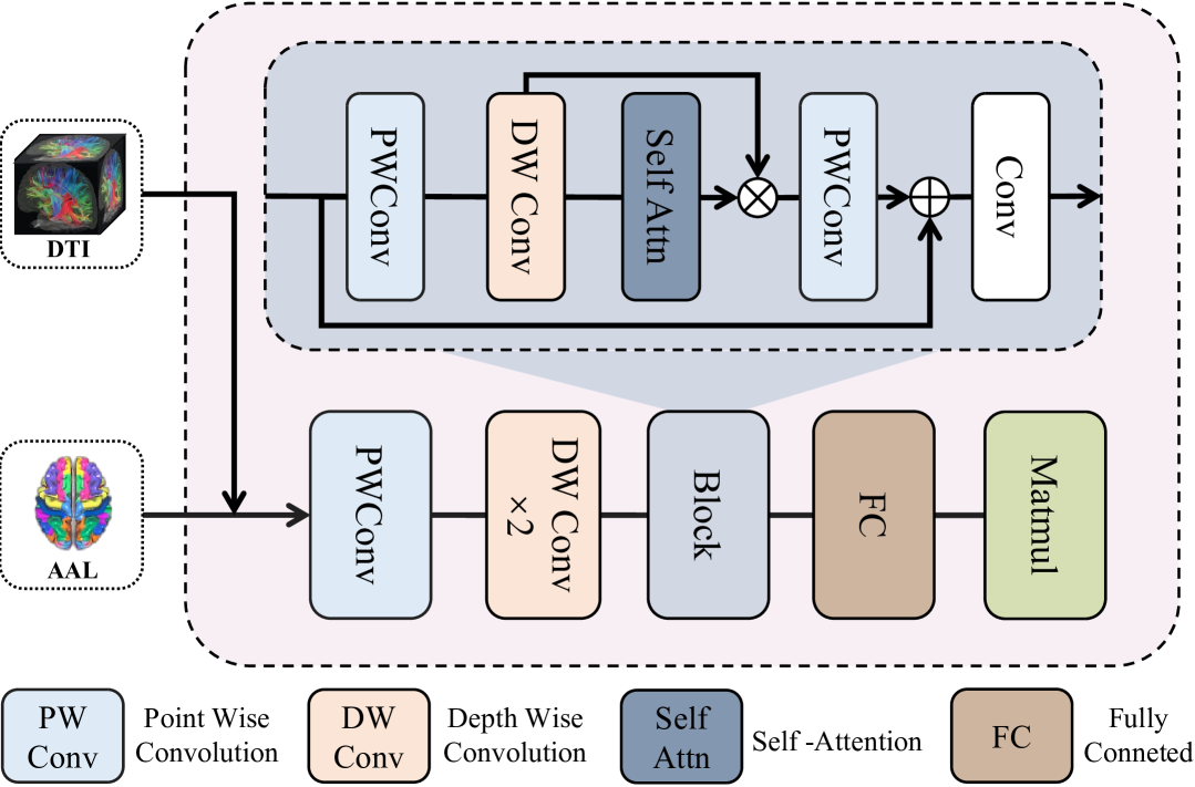
As depicted in Figure 2, the Template Network processes the registered DTI data through a series of specialized convolutional blocks and self-attention mechanisms. The network architecture consists of pointwise and depthwise convolutional layers for local feature processing, self-attention blocks to capture long-range dependencies, and fully connected layers to generate the final brain feature matrix.
The network transforms the initial anatomical matrix into two key outputs: (1) a brain feature matrix , where each of the 90 brain regions is represented by an 80-dimensional feature vector, and (2) an initial adjacency matrix representing preliminary connectivity between brain regions.
To preserve the geometric properties of brain fiber structures, we model the output as a Riemannian metric, which is a symmetric positive-definite (SPD) matrix. This matrix encodes distances on the manifold and is constructed using eigenvalue decomposition:
| (1) |
where is a rotation matrix that aligns with the principal diffusion directions, and is a diagonal matrix with positive entries representing diffusion magnitudes. The parameters of and are learned from the data.
The network is optimized to minimize the discrepancy between predicted geodesics (fiber pathways) and actual fiber orientations observed in the DTI data, effectively learning the underlying geometric structure of the brain’s white matter.
III-D ConnectomeDiffuser
Diffusion Models [40] are probabilistic generative models that learn data distributions by iteratively denoising normally distributed variables through a fixed-length Markov Chain of length . For image generation, certain diffusion models [52, 53] employ a reweighted variant of the variational lower bound, inspired by denoising score-matching models [54], to capture complex patterns and generate high-quality outputs.
The diffusion model comprises an encoder and a decoder that focus on the semantic content of the data while optimizing computational resources. The brain feature matrix is encoded by into a latent representation . The decoder subsequently reconstructs the brain network from this latent representation, yielding , where . The encoder downsamples the input by a factor , with . The latent representation is processed by the diffusion module, and the decoder transforms the output into a brain network .
In the diffusion process, Gaussian noise with variance is added to the encoded latent representation at time to produce the noisy latent representation:
| (2) |
where , and . As increases, approximates a standard Gaussian distribution.
During the diffusion process, we employ a U-Net [55] denoiser comprising an encoder, a middle block, and a decoder. The noisy latent variable is combined with the encoder output and processed through the model to obtain a clean latent representation :
| (3) |
where represents conditioning information and denotes the brain feature matrix.
To enhance adaptability, the U-Net is combined with cross-attention mechanisms [56]. A domain-specific encoder projects the input to an intermediate representation , which is integrated into the U-Net’s intermediate layers via cross-attention:
| (4) |
where represents a flattened intermediate representation of the U-Net implementing , and , , and are learnable projection matrices [57].
We further enhance the diffusion model with a parallel module that shares the same architecture as the U-Net’s middle block. This module processes disease-specific conditional information concurrently with the latent representation, enhancing the model’s ability to generate condition-consistent brain networks. The outputs of this parallel module are integrated into the U-Net through adaptive convolutional layers at each scale.
During inference, the model samples from a standard normal distribution and iteratively applies the reverse diffusion process to obtain . The decoder then transforms this latent representation into the final brain network .
III-E GCN Classifier
The proposed GCN classifier integrates a Graph Convolutional Network layer [58] with a fully connected layer. The GCN layer aggregates structural characteristics from each vertex’s neighborhood, enabling the model to capture both local and global connectivity patterns within the brain network.
The GCN follows a layer-wise propagation rule:
| (5) |
where represents the adjacency matrix with added self-connections, denotes the identity matrix, is a layer-specific trainable weight matrix, and is the degree matrix. The activation function is the ReLU function, defined as . The matrix represents the activations in the layer, with being the initial input features.
The fully connected layer classifies the entire graph based on the features extracted by the GCN layer. While the classifier’s primary function is categorization, it also serves to guide the generation process by providing classification signals that inform the diffusion model.
By leveraging the GCN architecture, our model effectively captures the complex relationships between brain regions and their connectivity patterns, facilitating comprehensive analysis of structural alterations associated with various neurological conditions.
III-F Brain Diffusive Loss
The Brain Diffusive Loss () is a composite loss function that guides the learning process by incorporating information from multiple components of our model. It consists of three essential components:
1. Feature Extraction Loss (): This component ensures accurate registration and feature extraction from the DTI data:
| (6) |
where represents the Riemannian metric learned by the Template Network, are vector fields corresponding to fiber directions, is the covariant derivative, measuring how vector fields change along geodesics, is a scaling factor, is a regularization term that ensures smoothness, is a weighting parameter.
2. Latent Diffusion Loss (): This component guides the diffusion model to generate realistic brain networks:
| (7) |
where represents the encoded DTI features, represents the disease labels, is a noise vector sampled from a standard Gaussian, is a time step in the diffusion process, is the noisy latent variable at time , and are learnable functions.
3. Classification Loss (): This component ensures the generated brain networks contain disease-relevant information:
| (8) |
where is the number of samples, is the true label distribution, is the predicted label distribution.
The total Brain Diffusive Loss is the sum of these three components:
| (9) |
By simultaneously optimizing these three aspects, our model learns to generate anatomically accurate, realistic, and diagnostically informative brain networks that effectively capture the complex structural characteristics associated with various neurological disorders.
IV Experiment
IV-A Data Preparation and Preprocessing
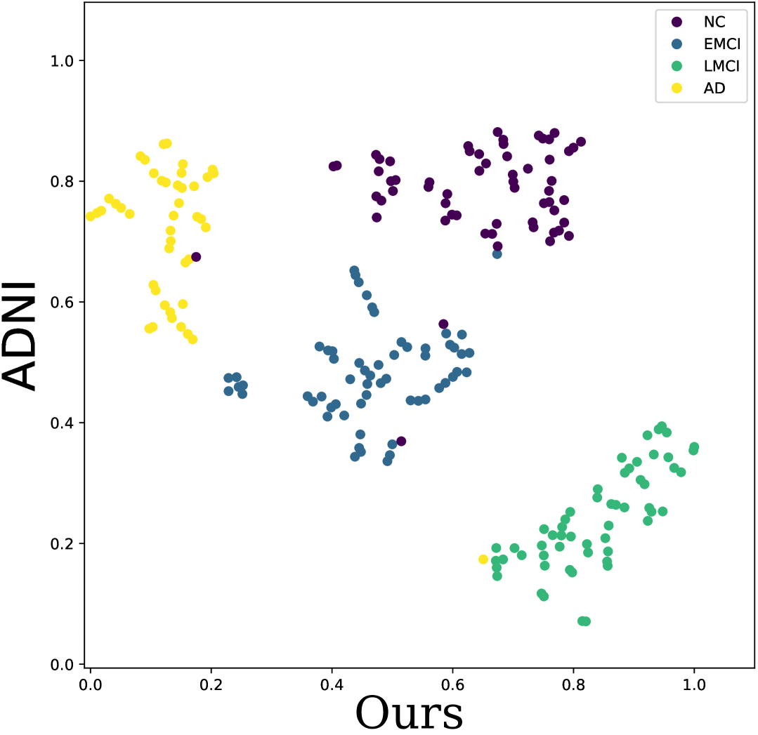
We evaluated the proposed ConnectomeDiffuser framework on two well-established neuroimaging datasets: the Alzheimer’s Disease Neuroimaging Initiative (ADNI) [59] and the Autism Brain Imaging Data Exchange (ABIDE) [60]. These datasets represent distinct neurodegenerative and neurodevelopmental conditions, allowing us to assess the generalizability of our approach.
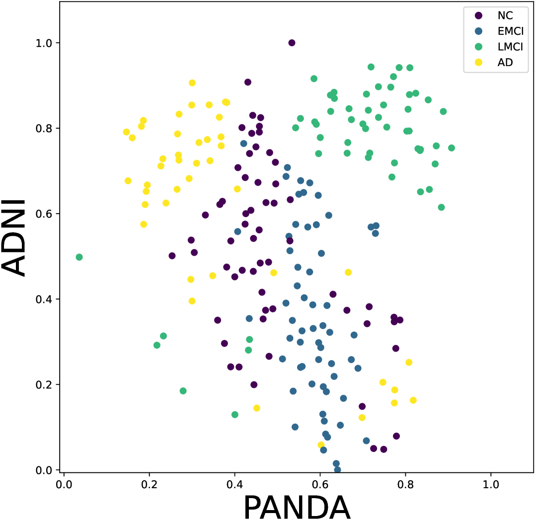
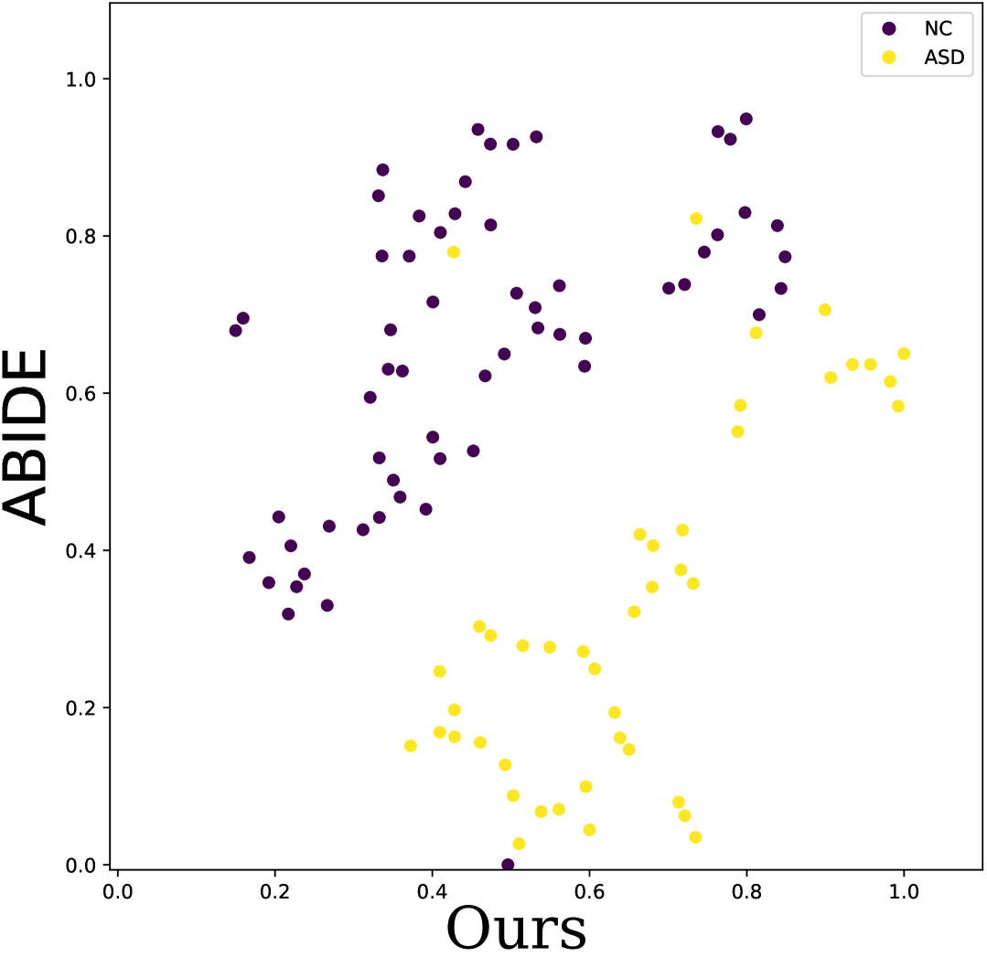
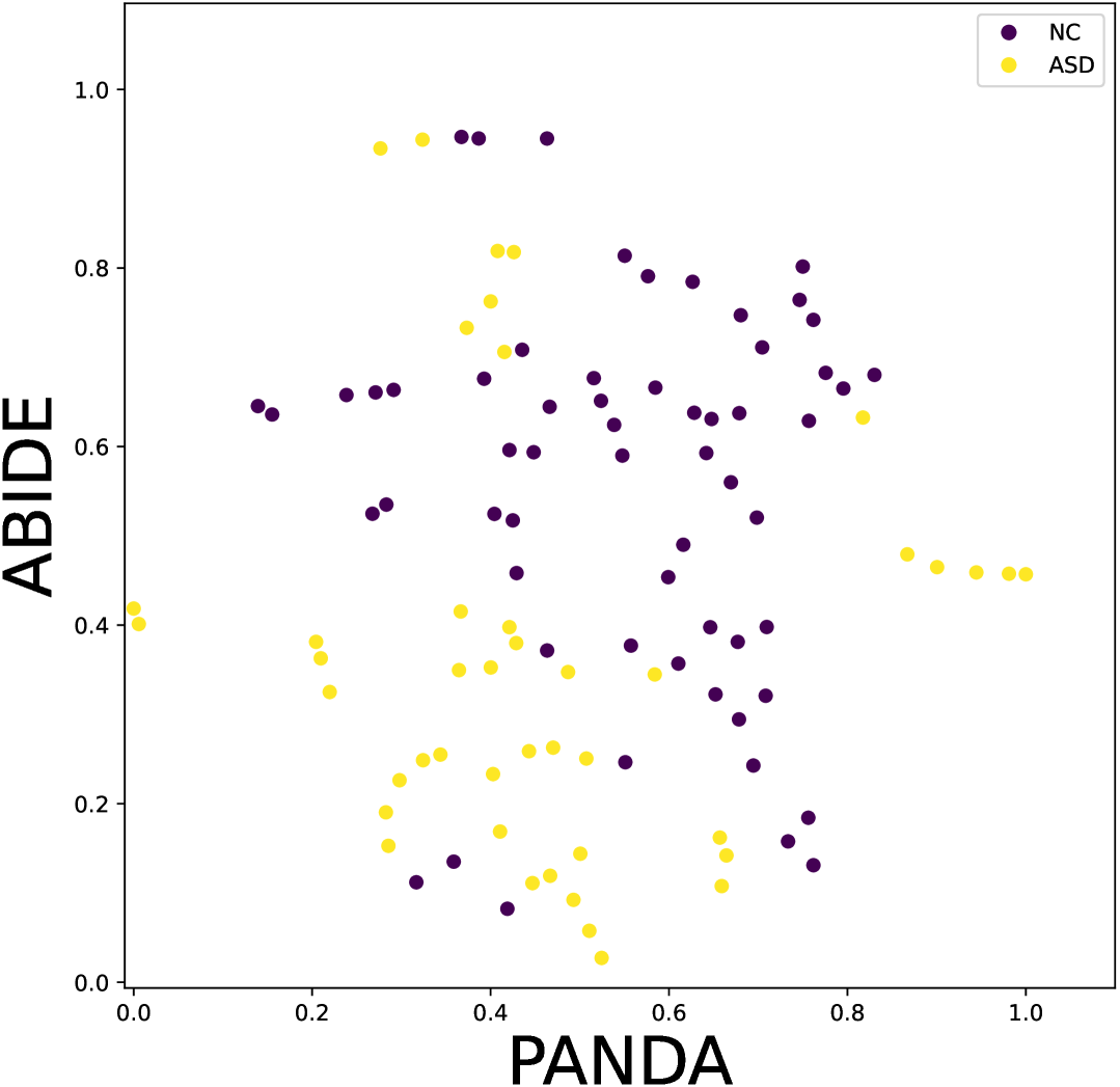
The ADNI dataset (Table I) comprises 300 subjects stratified into four clinical categories: normal controls (NC, ), early mild cognitive impairment (EMCI, ), late mild cognitive impairment (LMCI, ), and Alzheimer’s disease (AD, ). This stratification enables the analysis of progressive neurodegeneration across the AD continuum. The ABIDE dataset (Table II) includes 160 participants, evenly distributed between individuals with autism spectrum disorder (ASD, ) and matched neurotypical controls (NC, ).
Prior to analysis, all diffusion tensor imaging (DTI) data underwent rigorous preprocessing using the Pipeline for Analyzing braiN Diffusion imAges (PANDA) toolkit [26]. The preprocessing pipeline encompassed: (1) skull stripping to eliminate non-brain tissue, (2) motion correction to compensate for subject movement, (3) eddy current correction to mitigate gradient-induced distortions, and (4) tensor model fitting using a least-squares approach to compute diffusion metrics. This standardized preprocessing workflow ensured data consistency while minimizing artifacts that could potentially confound subsequent analyses.
We employed a stratified data partitioning strategy to maintain class distribution across training and testing sets. For the ADNI dataset, we allocated 220 subjects for model training and reserved 80 subjects (20 from each group) for testing. Similarly, the ABIDE dataset was divided into 148 subjects for training and 16 subjects (8 from each group) for testing. To enhance statistical reliability, all experiments were conducted using five-fold cross-validation, thereby mitigating potential biases from any particular data split.
| Group | NC | EMCI | LMCI | AD |
|---|---|---|---|---|
| Number | 80 | 80 | 76 | 64 |
| Gender | 38M/42F | 41M/39F | 36M/40F | 31M/33F |
| Age | 74.1 6.4 | 75.4 7.1 | 75.1 4.6 | 76.2 5.6 |
| Group | NC | ASD |
|---|---|---|
| Number | 80 | 80 |
| Gender | 65M/15F | 70M/10F |
| Age | 13.2 3.2 | 12.9 2.7 |
IV-B Implementation Details
We implemented the ConnectomeDiffuser framework using PyTorch and trained the model on dual NVIDIA V100 GPUs. The optimization process employed the AdamW optimizer with default parameters, a learning rate of , and a batch size of 4. All models were trained for 300 epochs to ensure convergence.
The diffusion model component utilized a downsampling factor of 4 in the encoder and 100 diffusion steps, striking an optimal balance between computational efficiency and network generation quality. The U-Net architecture for denoising comprised a symmetric encoder-decoder structure with four resolution levels. Each level in the downsampling path contained two convolutional layers followed by downsampling, with the number of filters progressively increasing from 64 to 512 (specifically, 64, 128, 256, and 512 filters). The middle block incorporated two convolutional layers with 512 filters augmented by self-attention mechanisms to capture global context. The upsampling path mirrored this architecture in reverse, replacing downsampling operations with upsampling layers. All convolutional layers employed a kernel size of 3×3, instance normalization for stable training, and SiLU (Sigmoid Linear Unit) activation functions to introduce non-linearity.
IV-C Evaluation Metrics
To comprehensively assess the efficacy of ConnectomeDiffuser in brain network construction and neurological disorder classification, we employed four classification metrics:
-
1.
Accuracy (ACC): This metric provides the proportion of true results among the total number of cases examined.
-
2.
Precision (PRE): This metric measures the proportion of true positive identifications among all positive identifications made by the model.
-
3.
Recall (REC): This metric assesses the proportion of actual positives correctly identified by the model.
-
4.
F1 Score (F1): This metric is the harmonic mean of precision and recall.
These metrics collectively provide a multifaceted evaluation of classification performance, enabling a robust comparison between ConnectomeDiffuser and alternative approaches.
IV-D Classification Performance
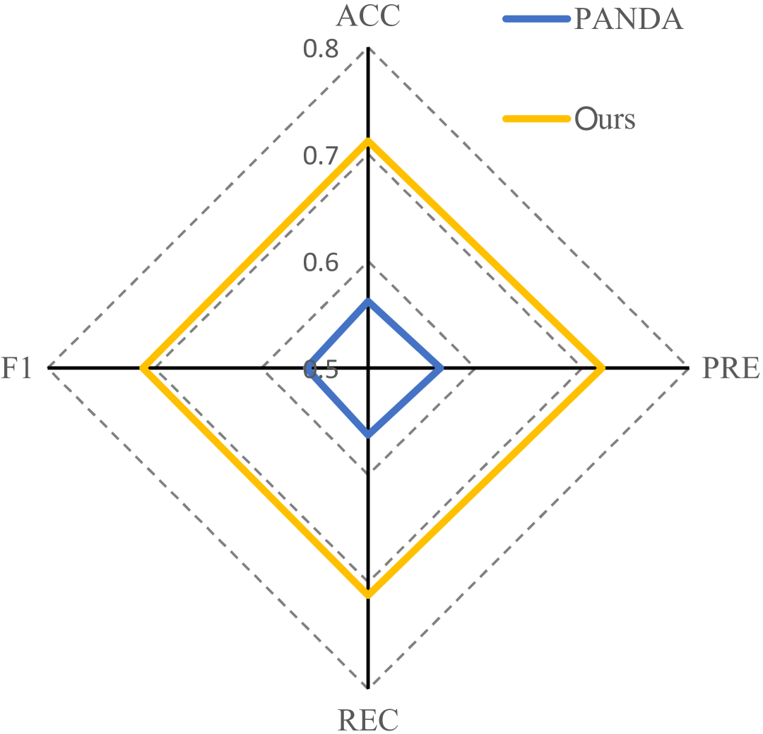
Table III and Figure 3 present a comprehensive comparison of ConnectomeDiffuser against several state-of-the-art methods for brain network construction and neurological disorder classification. Our proposed framework consistently outperformed competing approaches across both datasets, achieving superior accuracy (71.25% for ADNI, 87.50% for ABIDE), precision (71.84% and 87.50%), recall (71.25% and 87.50%), and F1 scores (71.09% and 87.50%). These results demonstrate ConnectomeDiffuser’s enhanced capability for identifying distinctive biomarkers associated with neurodegenerative and neurodevelopmental disorders.
The performance improvement over the PANDA toolkit is particularly noteworthy, with absolute accuracy gains of 15.00% for ADNI and 18.75% for ABIDE. This substantial enhancement underscores the effectiveness of our end-to-end approach in capturing complex topological features and disease-specific markers directly from DTI data. The t-SNE visualization in Fig. 4 further illustrates ConnectomeDiffuser’s superior discriminative capability, revealing more distinct clusters corresponding to different diagnostic categories compared to PANDA-generated networks.
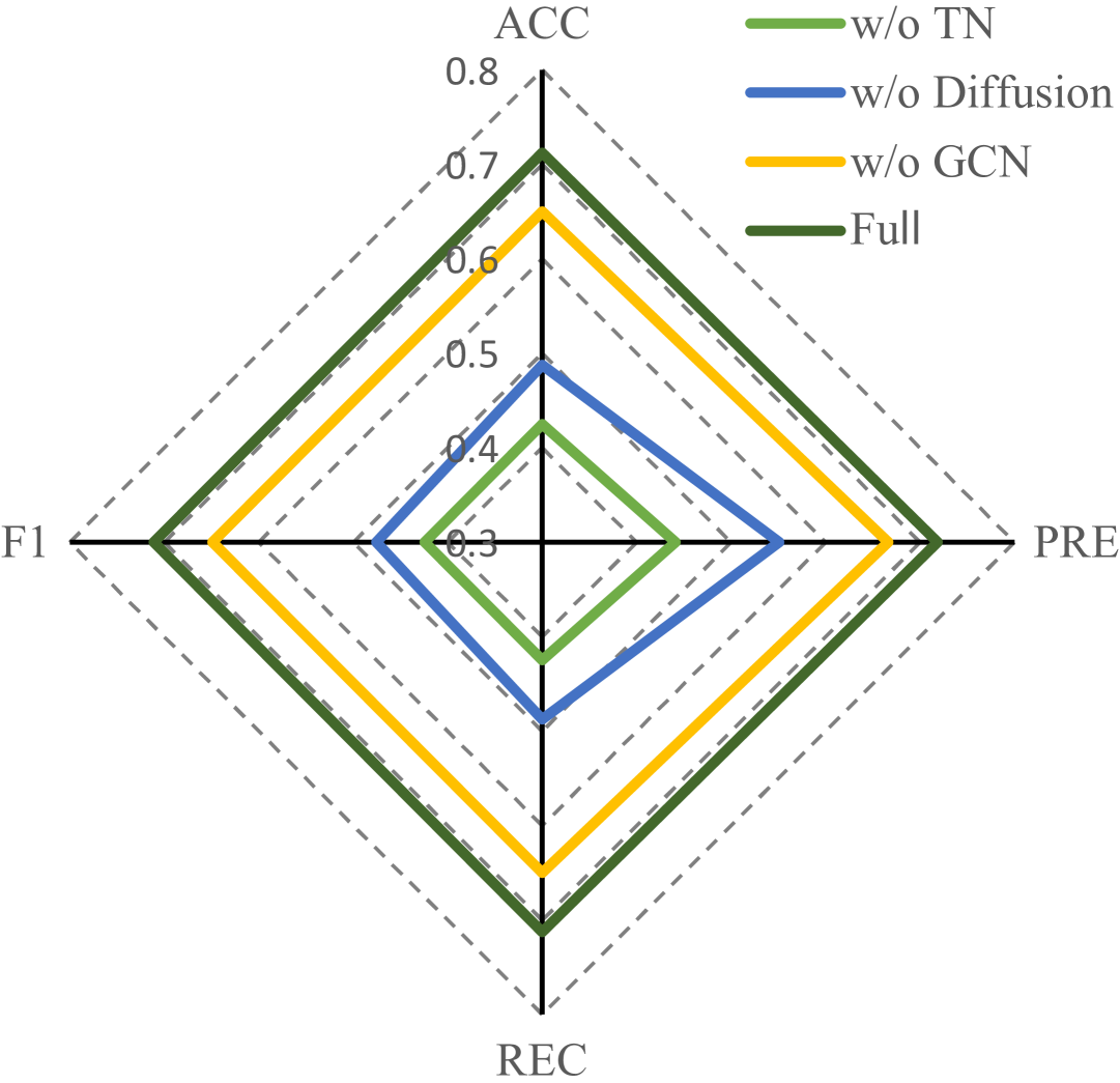
The ablation study, detailed in Table IV, Table V and Figure 3, is conducted by the following manner:
-
1.
Remove Template Network (TN) and AAL atlas template, use normal 3D CNN instead
-
2.
Remove diffusion model and replace with Variational Autoencoder (VAE)
-
3.
Remove GCN classifier and replace with normal linear layer
-
4.
The impact of different numbers of GCN layer
-
5.
The effect of different diffusion steps
Results demonstrate that removing the AAL template and Template Network significantly impairs the model’s ability to extract meaningful topological features from brain imaging data, leading to decreased performance. The diffusion model proves superior to the VAE in generative capabilities, capturing more disease-relevant information. While the Graph Convolutional Network (GCN) classifier has a relatively modest impact on overall model performance, its removal still results in reduced classification accuracy. Regarding GCN architecture, increasing the number of layers initially improves prediction accuracy through better capture of both local and global graph structures. However, performance deteriorates beyond an optimal point due to the over-smoothing effect, where node representations become homogeneous. Prediction accuracy improves with more diffusion steps as the model better approximates the underlying data distribution, with 100 steps providing an optimal balance between computational cost and performance.

Dataset Method ACC(%) PRE(%) REC(%) F1(%) ADNI ConnectomeDiffuser 71.25 71.84 71.25 71.09 SBGM 60.00 62.50 50.00 55.56 DecGAN 60.00 58.33 70.00 63.64 Brain Diffuser 65.00 60.00 66.67 63.16 Sang et al. 50.00 57.14 66.67 61.54 PANDA 56.25 56.75 56.25 55.80 ABIDE ConnectomeDiffuser 87.50 87.50 87.50 87.50 SBGM 62.50 50.00 66.67 57.14 DecGAN 75.00 83.33 62.50 71.43 Brain Diffuser 75.00 66.67 85.71 75.00 Sang et al. 50.00 40.00 66.67 50.00 PANDA 68.75 75.00 66.66 70.59
TN Diffusion GCN ACC(%) PRE(%) REC(%) F1(%) ✖ ✔ ✔ ✔ 42.50 44.13 42.50 42.43 ✔ ✖ ✔ ✔ 63.75 55.26 63.64 59.15 ✔ ✔ ✖ ✔ 48.75 55.09 48.75 47.54 ✔ ✔ ✔ ✖ 65.00 66.65 65.00 64.90 ✔ ✔ ✔ ✔ 71.25 71.84 71.25 71.09
GCN layers Diffusion steps ACC(%) PRE(%) REC(%) F1(%) 2 100 65.00 59.46 62.86 61.11 3 100 71.25 71.84 71.25 71.09 4 100 68.75 65.79 67.57 66.67 3 10 55.00 36.84 53.85 43.75 3 50 60.00 47.37 60.00 52.94
NC vs. EMCI
EMCI vs. LMCI
LMCI vs. AD
NC vs. EMCI
EMCI vs. LMCI
LMCI vs. AD
IV-E Structural Brain Network Connectivity Analysis of Alzhemier’s Disease
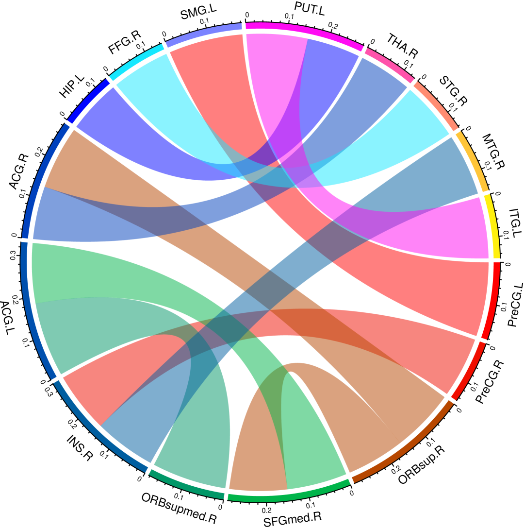
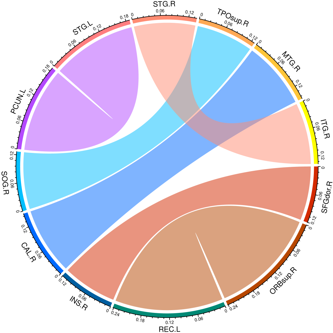
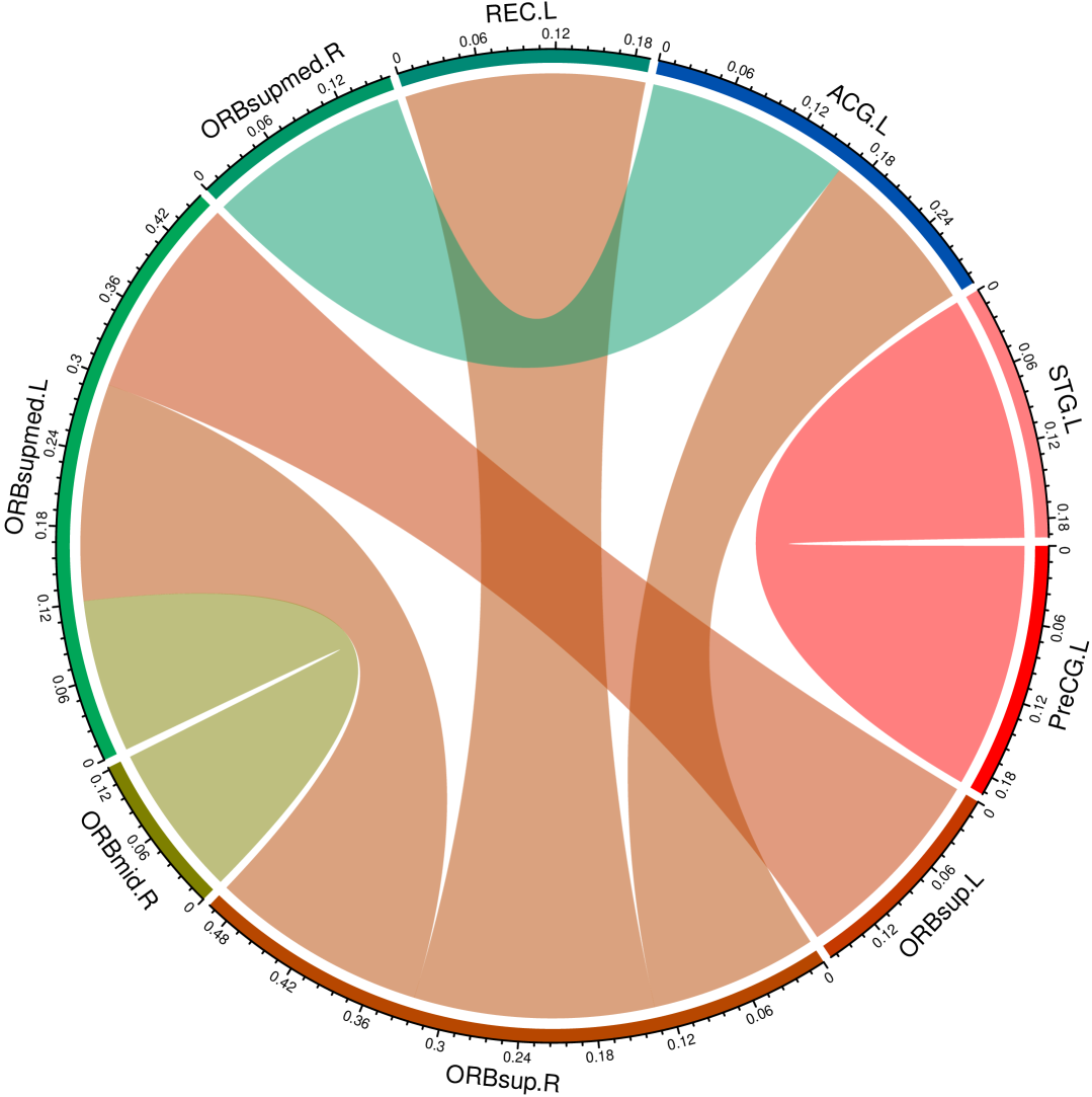
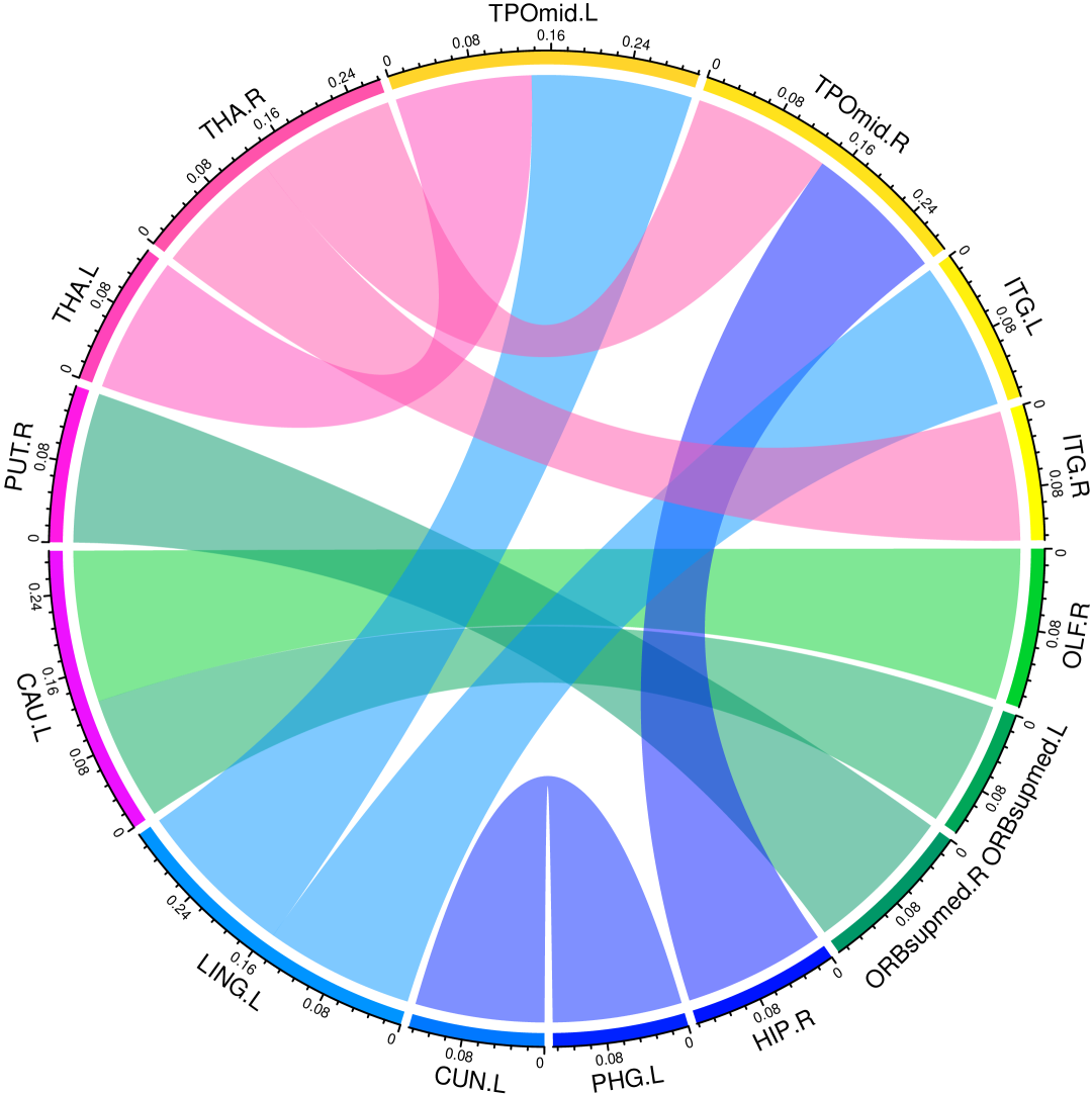
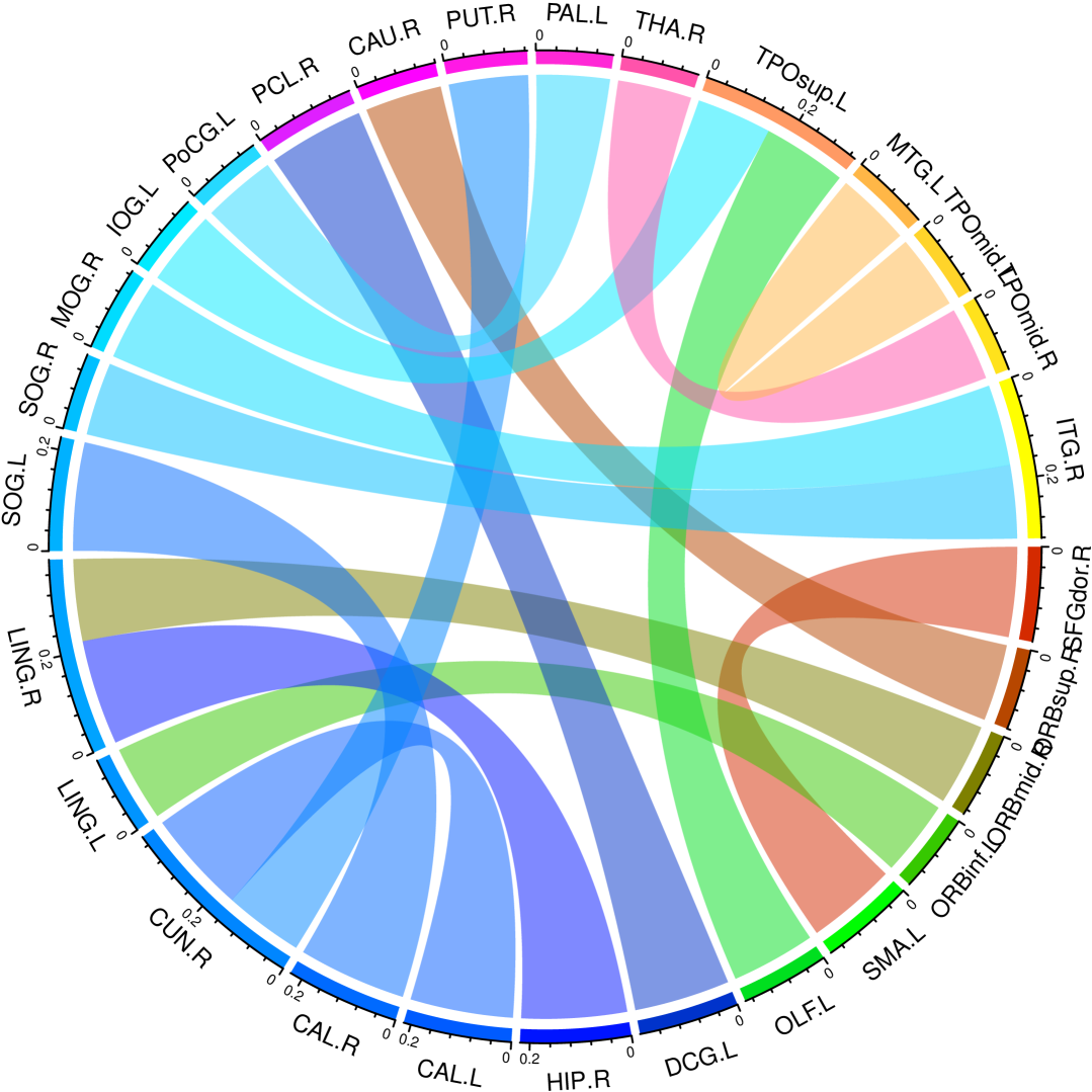
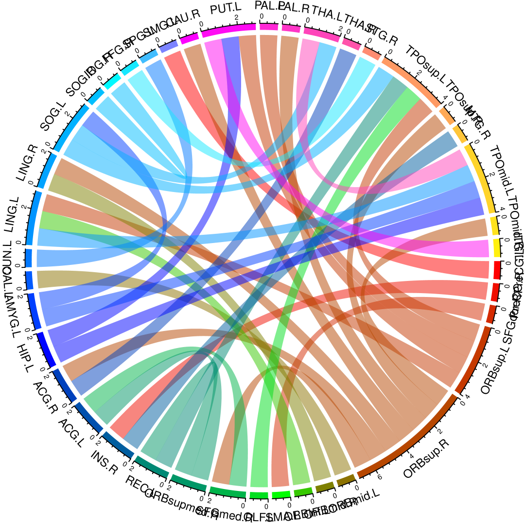
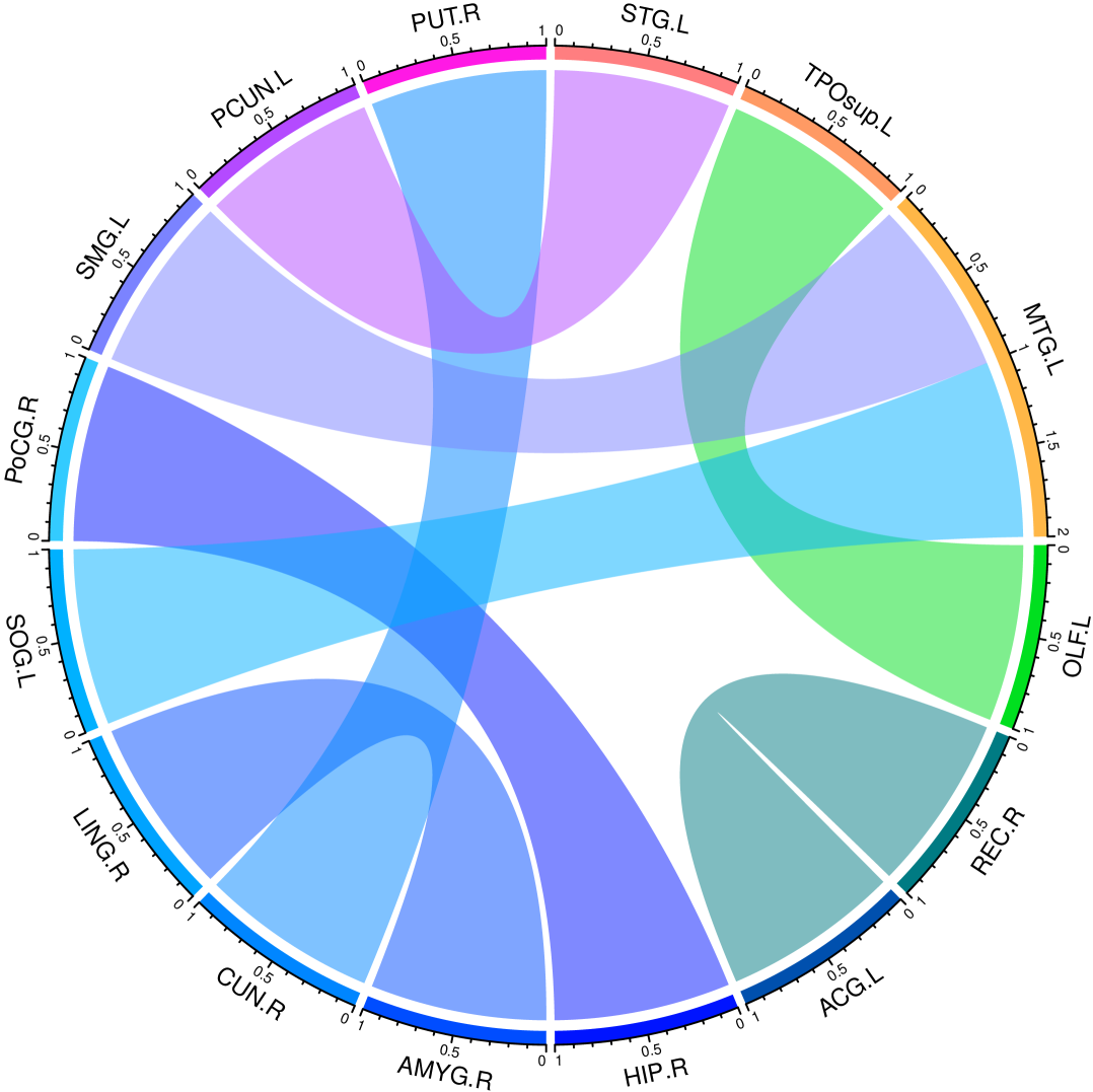
We leveraged ConnectomeDiffuser to investigate the progressive alterations in brain connectivity patterns across the Alzheimer’s disease stages. For each subject in the ADNI test set, we generated structural brain networks and subsequently employed two-sample t-tests to identify statistically significant connectivity differences (p 0.05) between diagnostic groups. Connections failing to meet this significance threshold were considered disease-irrelevant and subsequently nullified.
To elucidate the subtle connectivity alterations associated with disease progression, we constructed average brain networks for each diagnostic category and computed inter-group differences. These differential patterns were then correlated with the abnormal regions identified via statistical testing, revealing distinct patterns of connectivity enhancement and deterioration across disease stages, as visualized in Figure 5.
Our analysis revealed a striking imbalance between increased and decreased connectivity as AD progresses, with connectivity reductions becoming increasingly predominant at more advanced disease stages. This pattern aligns with the neuropathological trajectory of AD, characterized by progressive neuronal loss and brain tissue atrophy that systematically disrupts structural connections between brain regions. Figure 6 illustrates that the magnitude of neural connectivity alterations diminishes as the disease advances from EMCI to LMCI to AD, suggesting that major connectivity disruptions occur early in the disease process, with fewer additional changes as neurodegeneration progresses to advanced stages.
Interestingly, we observed selective increases in connectivity among specific brain regions despite the overall trend toward reduced connectivity. This phenomenon likely represents compensatory mechanisms whereby the brain recruits alternative neural pathways to maintain cognitive function despite accumulating neuropathology, particularly in earlier disease stages.
Increased connectivity
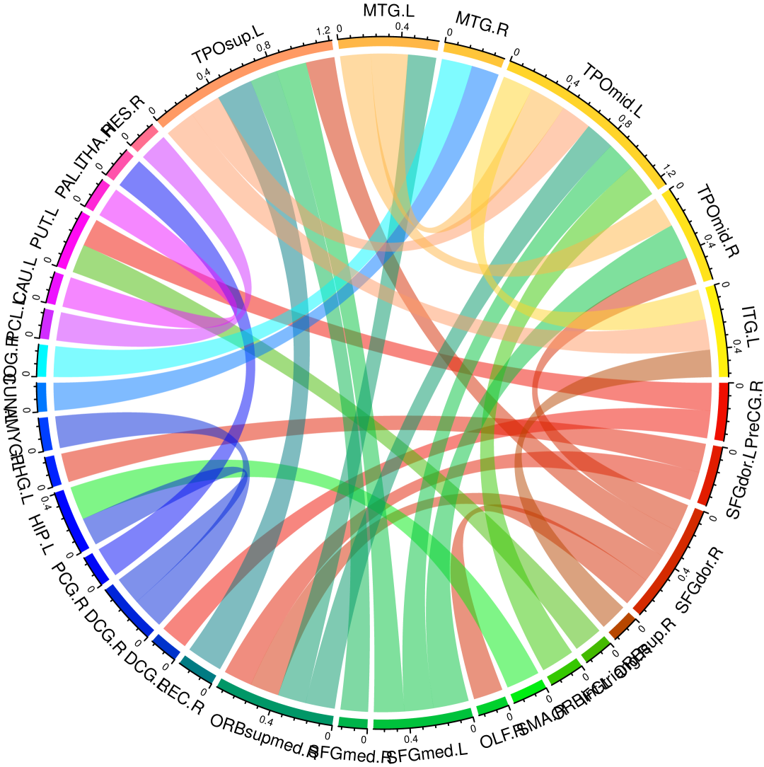
Decreased connectivity
IV-F Structural Brain Network Connectivity Analysis of Autism Spectrum Disorder
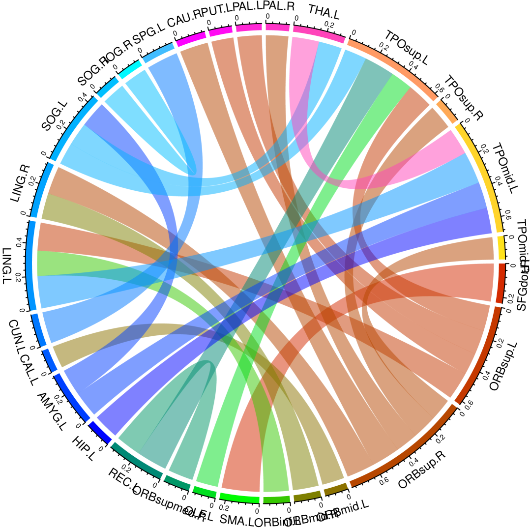
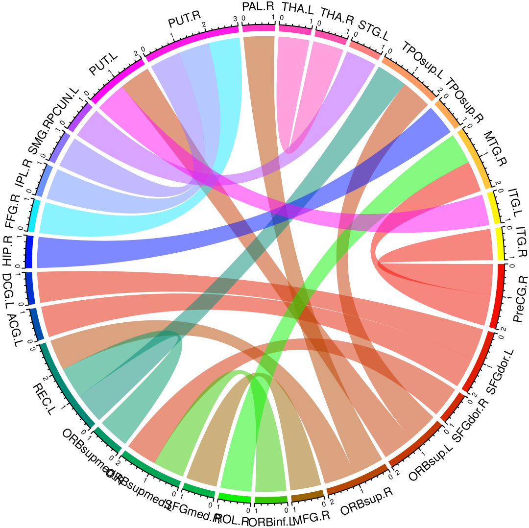
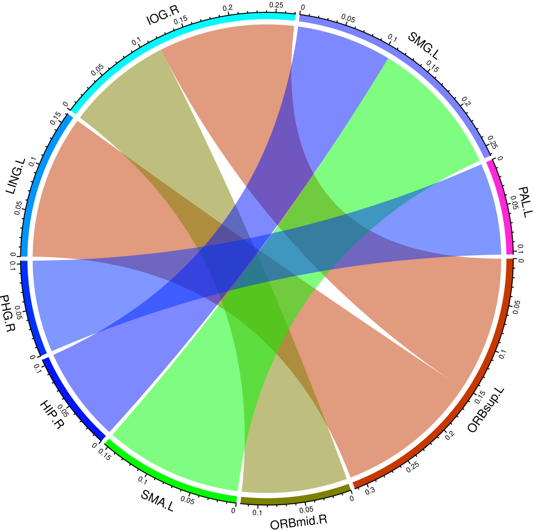
We extended our connectivity analysis methodology to the ABIDE dataset, examining structural brain network differences between individuals with ASD and matched neurotypical controls. Following network construction using ConnectomeDiffuser, we applied the same statistical method to identify significant connectivity alterations between groups.
In contrast to the predominantly decreasing connectivity observed in AD progression, ASD demonstrated a more balanced pattern of connectivity changes, with comparable numbers of significantly increased and decreased connections (Figure 7). This distinct pattern suggests that ASD involves specific reconfiguration of structural connectivity rather than wholesale connectivity reduction, consistent with contemporary theories characterizing ASD as a disorder of brain connectivity.
The more balanced connectivity alterations in ASD may also reflect neurodevelopmental adaptations, particularly given the adolescent age range of the ABIDE cohort. During this period of heightened neuroplasticity, compensatory mechanisms may establish long-range connections to counterbalance local connectivity disruptions characteristic of ASD, potentially facilitating the development of alternative cognitive strategies.
IV-G Advanced Analysis of Structural Brain Network Construction Efficacy
Beyond classification performance, we evaluated ConnectomeDiffuser’s practical utility by assessing its computational efficiency and output reproducibility compared to the conventional PANDA toolkit. Table VI presents a comparative analysis focusing on processing time and result consistency.
The PANDA toolkit requires extensive manual parameter calibration and complex computational procedures for structural brain network construction. The reported processing time (900 seconds per subject) encompasses both computational operations and necessary manual interventions, including parameter tuning and data preparation. This substantial time investment varies considerably (900-1500 seconds) depending on operator expertise, introducing potential workflow inefficiencies.
In contrast, ConnectomeDiffuser streamlines this process through its end-to-end design, requiring merely 2.5 seconds per subject for inference following the one-time offline training phase. This represents a 360-fold reduction in processing time, dramatically enhancing throughput for large-scale neuroimaging studies and clinical applications.
Importantly, ConnectomeDiffuser’s data-driven approach eliminates the need for subjective parameter tuning, mitigating human bias and enhancing result objectivity. We quantitatively assessed result consistency using the Intra-Class Correlation (ICC) coefficient [61, 62], which measures the reliability and reproducibility of measurements. ConnectomeDiffuser demonstrated superior ICC values (0.76 for ADNI, 0.68 for ABIDE) compared to PANDA (0.73 and 0.64, respectively), confirming its enhanced reproducibility. This improvement is particularly valuable in clinical settings, where consistent and reliable results are paramount for accurate diagnosis and treatment planning.
| Dataset | Method | Time(s) | Intra-Class Correlation |
|---|---|---|---|
| ADNI | PANDA | 900 | 0.73 |
| ConnectomeDiffuser | 2.5 | 0.76 | |
| ABIDE | PANDA | 900 | 0.64 |
| ConnectomeDiffuser | 2.5 | 0.68 |
IV-H Limitations
Despite its compelling performance, ConnectomeDiffuser has several limitations worth acknowledging. First, the framework relies on the AAL atlas as a standardized template, which may not optimally capture individual anatomical variations. While this approach facilitates cross-subject comparability, it potentially obscures subject-specific anatomical nuances that could be clinically relevant.
Second, the current implementation focuses exclusively on structural brain networks derived from DTI. This single modality approach does not capitalize on the complementary information available from functional connectivity, which has demonstrated significant value in characterizing neurological disorders. Integrating functional connectivity data could potentially enhance diagnostic precision and provide more comprehensive insights into disease mechanisms.
These limitations present promising directions for future research, including the incorporation of subject-specific anatomical templates and multimodal integration strategies to further enhance the clinical utility of ConnectomeDiffuser.
V Conclusion
In this study, we introduce ConnectomeDiffuser, an innovative end-to-end model for the construction of individualized structural brain networks derived from DTI. Utilizing the AAL atlas as a structural guide, we harness a Template Network to effectively capture topological features. ConnectomeDiffuser then synthesizes these features, along with disease-specific labels from a classifier, to produce the brain networks. Our experimental evaluations, conducted on the ADNI and ABIDE datasets, reveal that ConnectomeDiffuser surpasses existing methods in terms of its analytical capabilities and predictive accuracy regarding the progression of various brain disorders. A significant finding of our model is the tendency for a reduction in brain connectivity as diseases progress. This insight has the potential to contribute to a deeper understanding of the pathophysiological mechanisms of brain disorders and may facilitate the identification of early biomarkers critical for targeted therapeutic interventions.
References
- [1] S. Wang, Y. Hu, Y. Shen, and H. Li, “Classification of diffusion tensor metrics for the diagnosis of a myelopathic cord using machine learning,” International journal of neural systems, vol. 28, no. 02, p. 1750036, 2018.
- [2] H. Wang, Y. Shen, S. Wang, T. Xiao, L. Deng, X. Wang, and X. Zhao, “Ensemble of 3d densely connected convolutional network for diagnosis of mild cognitive impairment and alzheimer’s disease,” Neurocomputing, vol. 333, pp. 145–156, 2019.
- [3] B. Lei, E. Liang, M. Yang, P. Yang, F. Zhou, E.-L. Tan, Y. Lei, C.-M. Liu, T. Wang, X. Xiao et al., “Predicting clinical scores for alzheimer’s disease based on joint and deep learning,” Expert Systems with Applications, vol. 187, p. 115966, 2022.
- [4] Y. Liu, Z. Xiuxu, H. Feng, and Y. Yan, “Abnormality detection in neurodegenerative disease analysis based on topological nonlinear and dynamic modelling of gait,” Journal of Integration Technology, vol. 11, no. 4, p. 92, 2022. doi: 10.12146/j.issn.2095-3135.20211215001 .
- [5] S. Wang, Y. Shen, W. Chen, T. Xiao, and J. Hu, “Automatic recognition of mild cognitive impairment from mri images using expedited convolutional neural networks,” in International Conference on Artificial Neural Networks. Springer, 2017, pp. 373–380.
- [6] Y. Zhu, Y. Chen, C. qingling, and T. Chen, “Isa-dunet: Inception spatial-attention dense u-net for head and neck tumor segmentation in pet-ct,” Journal of Integration Technology, vol. 12, no. 3, p. 94, 2023. doi: 10.12146/j.issn.2095-3135.20230119001 .
- [7] B. Hu, C. Zhan, B. Tang, B. Wang, B. Lei, and S.-Q. Wang, “3-d brain reconstruction by hierarchical shape-perception network from a single incomplete image,” IEEE Transactions on Neural Networks and Learning Systems, vol. 35, no. 10, pp. 13 271–13 283, 2024.
- [8] S. Hu, W. Yu, Z. Chen, and S. Wang, “Medical image reconstruction using generative adversarial network for alzheimer disease assessment with class-imbalance problem,” in 2020 IEEE 6th International Conference on Computer and Communications (ICCC). IEEE, 2020, pp. 1323–1327.
- [9] B. Li, P. Zhang, Y. Cao, L. Sun, J. Feng, Y. Zhang, Q. Yang, Y. Li, and Z. Liu, “Aivus: guidewire artifacts inpainting for intravascular ultrasound imaging with united spatiotemporal aggregation learning,” IEEE Transactions on Computational Imaging, vol. 8, pp. 679–692, 2022.
- [10] V. A. Kelkar, S. Bhadra, and M. A. Anastasio, “Compressible latent-space invertible networks for generative model-constrained image reconstruction,” IEEE Transactions on Computational Imaging, vol. 7, pp. 209–223, 2021.
- [11] S. You, B. Lei, S. Wang, C. K. Chui, A. C. Cheung, Y. Liu, M. Gan, G. Wu, and Y. Shen, “Fine perceptive gans for brain mr image super-resolution in wavelet domain,” IEEE transactions on neural networks and learning systems, vol. 34, no. 11, pp. 8802–8814, 2022.
- [12] X. Chen, Y. Qiu, W. Liang, G. Zhou, and S. Xie, “Spatial information regularized tensor decomposition framework for super-resolution reconstruction of medical mri and radiographs,” IEEE Transactions on Computational Imaging, vol. 8, pp. 865–878, 2022.
- [13] S. Wang, T. Zhou, Y. Shen, Y. Li, G. Huang, and Y. Hu, “Generative ai enables eeg super-resolution via spatio-temporal adaptive diffusion learning,” IEEE Transactions on Consumer Electronics, 2025. doi: 10.1109/TCE.2025.3528438 .
- [14] S. You, Y. Shen, G. Wu, and S. Wang, “Brain mr images super-resolution with the consistent features,” in Proceedings of the 2022 14th International Conference on Machine Learning and Computing, 2022, pp. 501–506.
- [15] S. Wang, X. Wang, Y. Shen, B. He, X. Zhao, P. W.-H. Cheung, J. P. Y. Cheung, K. D.-K. Luk, and Y. Hu, “An ensemble-based densely-connected deep learning system for assessment of skeletal maturity,” IEEE Transactions on Systems, Man, and Cybernetics: Systems, vol. 52, no. 1, pp. 426–437, 2020.
- [16] X. Chen, W. Yao, Y. Li, D. Liang, H. Zheng, S. Shakil, S. Wang, and T. Sun, “Ig-gcn: Empowering e-health services for alzheimer’s disease prediction,” IEEE Transactions on Consumer Electronics, vol. 70, no. 4, pp. 6442–6451, 2024.
- [17] S. Wang, W. Yu, Z. Chen et al., “Smart diagnosis assistance method to solve results of inaccurate classification of image, and terminal based on medical images,” mar 2025, uS Patent 12,254,684.
- [18] W. Chen, Y. Lei, S. Luo, Z. Zhou, M. Li, and C.-M. Pun, “Uwformer: Underwater image enhancement via a semi-supervised multi-scale transformer,” in 2024 International Joint Conference on Neural Networks (IJCNN). IEEE, 2024, pp. 1–8.
- [19] X. Guo, X. Chen, S. Wang, and C.-M. Pun, “Underwater image restoration through a prior guided hybrid sense approach and extensive benchmark analysis,” IEEE Transactions on Circuits and Systems for Video Technology, 2025.
- [20] Y. Dong, X. Chen, Y. Shen, M. K.-P. Ng, T. Qian, and S. Wang, “Multi-modal mood reader: Pre-trained model empowers cross-subject emotion recognition,” in International Conference on Neural Computing for Advanced Applications. Springer Nature Singapore Singapore, 2024, pp. 178–192.
- [21] M. Zhu, P. Yin, F. Hu, J. Jiang, L. Yin, Y. Li, and S. Wang, “Integrating genome-wide association and transcriptome prediction model identifies novel target genes for osteoporosis,” Osteoporosis International, vol. 32, no. 12, pp. 2493–2503, 2021.
- [22] S. Li, Q. Zhu, L. Sun, K. Ma, Y. Ji, S. Qi, and D. Zhang, “Multi-modal brain network fusion for intelligent diagnostic devices,” IEEE Transactions on Consumer Electronics, pp. 1–1, 2025. doi: 10.1109/TCE.2025.3563691 .
- [23] A. Shehzad, D. Zhang, S. Yu, S. Abid, D. K. Kumar, and F. Xia, “Multiscale graph transformer for brain disorder diagnosis,” IEEE Transactions on Consumer Electronics, pp. 1–1, 2025. doi: 10.1109/TCE.2025.3542794 .
- [24] E. Jeon, E. Kang, J. Lee, J. Lee, T.-E. Kam, and H.-I. Suk, “Enriched representation learning in resting-state fmri for early mci diagnosis,” in International Conference on Medical Image Computing and Computer-Assisted Intervention. Springer, 2020, pp. 397–406.
- [25] C. Jing, C. Gong, Z. Chen, B. Lei, and S. Wang, “Ta-gan: transformer-driven addiction-perception generative adversarial network,” Neural Computing and Applications, vol. 35, no. 13, pp. 9579–9591, 2023.
- [26] Z. Cui, S. Zhong, P. Xu, Y. He, and G. Gong, “Panda: a pipeline toolbox for analyzing brain diffusion images,” Frontiers in human neuroscience, vol. 7, p. 42, 2013.
- [27] R. S. Desikan, F. Ségonne, B. Fischl, B. T. Quinn, B. C. Dickerson, D. Blacker, R. L. Buckner, A. M. Dale, R. P. Maguire, B. T. Hyman et al., “An automated labeling system for subdividing the human cerebral cortex on mri scans into gyral based regions of interest,” Neuroimage, vol. 31, no. 3, pp. 968–980, 2006.
- [28] M. Raissi, P. Perdikaris, and G. E. Karniadakis, “Physics-informed neural networks: A deep learning framework for solving forward and inverse problems involving nonlinear partial differential equations,” Journal of Computational physics, vol. 378, pp. 686–707, 2019.
- [29] I. Goodfellow, J. Pouget-Abadie, M. Mirza, B. Xu, D. Warde-Farley, S. Ozair, A. Courville, and Y. Bengio, “Generative adversarial nets,” in Advances in Neural Information Processing Systems, 2014, pp. 2672–2680.
- [30] S. Wang, S. Yanyan, and W. Zhang, “Enhanced generative adversarial network and target sample recognition method,” nov 2024, uS Patent 12,154,036.
- [31] D. P. Kingma and M. Welling, “Auto-encoding variational bayes,” in International Conference on Learning Representations, 2014.
- [32] S. Parisot, S. I. Ktena, E. Ferrante, M. Lee, R. Guerrero, B. Glocker, and D. Rueckert, “Disease prediction using graph convolutional networks: application to autism spectrum disorder and alzheimer’s disease,” Medical image analysis, vol. 48, pp. 117–130, 2018.
- [33] S. I. Ktena, S. Parisot, E. Ferrante, M. Rajchl, M. Lee, B. Glocker, and D. Rueckert, “Distance metric learning using graph convolutional networks: Application to functional brain networks,” in International Conference on Medical Image Computing and Computer-Assisted Intervention. Springer, 2017, pp. 469–477.
- [34] Q. Zuo, L. Chen, Y. Shen, M. K.-P. Ng, B. Lei, and S. Wang, “Bdht: generative ai enables causality analysis for mild cognitive impairment,” IEEE Transactions on Automation Science and Engineering, 2024.
- [35] J. Pan, Q. Zuo, B. Wang, C. P. Chen, B. Lei, and S. Wang, “Decgan: Decoupling generative adversarial network for detecting abnormal neural circuits in alzheimer’s disease,” IEEE Transactions on Artificial Intelligence, vol. 5, no. 10, pp. 5050–5063, 2024.
- [36] J. Pan, C. Jing, Q. Zuo, M. Nieuwoudt, and S. Wang, “Cross-modal transformer gan: a brain structure-function deep fusing framework for alzheimer’s disease,” in International Conference on Brain Inspired Cognitive Systems. Springer, 2023, pp. 82–92.
- [37] H. Kong, J. Pan, Y. Shen, and S. Wang, “Adversarial learning based structural brain-network generative model for analyzing mild cognitive impairment,” in Chinese Conference on Pattern Recognition and Computer Vision. Springer, 2022, pp. 361–375.
- [38] C. Gong, X. Chen, B. Mughal, and S. Wang, “Addictive brain-network identification by spatial attention recurrent network with feature selection,” Brain Informatics, vol. 10, no. 1, p. 2, 2023.
- [39] C. Jing, H. Kuai, H. Matsumoto, T. Yamaguchi, I. Y. Liao, and S. Wang, “Addiction-related brain networks identification via graph diffusion reconstruction network,” Brain Informatics, vol. 11, no. 1, p. 1, 2024.
- [40] J. Sohl-Dickstein, E. Weiss, N. Maheswaranathan, and S. Ganguli, “Deep unsupervised learning using nonequilibrium thermodynamics,” in International conference on machine learning. PMLR, 2015, pp. 2256–2265.
- [41] J. Wolleb, F. Bieder, R. Sandkühler, and P. C. Cattin, “Diffusion models for medical anomaly detection,” in International Conference on Medical Image Computing and Computer-Assisted Intervention, 2022, pp. 35–45.
- [42] H. Chung and J.-C. Ye, “Score-based diffusion models for accelerated mri,” Medical image analysis, vol. 80, p. 102479, 2021.
- [43] J. Jo, S. Lee, and S. J. Hwang, “Score-based generative modeling of graphs via the system of stochastic differential equations,” in International conference on machine learning. PMLR, 2022, pp. 10 362–10 383.
- [44] A. Gonzalez-Jimenez, S. Lionetti, M. Pouly, and A. A. Navarini, “Sano: Score-based diffusion model for anomaly localization in dermatology,” in Proceedings of the IEEE/CVF Conference on Computer Vision and Pattern Recognition, 2023, pp. 2987–2993.
- [45] Z. Han, Y. Wang, L. Zhou, P. Wang, B. Yan, J. Zhou, Y. Wang, and D. Shen, “Contrastive diffusion model with auxiliary guidance for coarse-to-fine pet reconstruction,” in International Conference on Medical Image Computing and Computer-Assisted Intervention. Springer, 2023, pp. 239–249.
- [46] W. Yao, Y. Shen, F. Nicolls, and S.-Q. Wang, “Conditional diffusion model-based data augmentation for alzheimer’s prediction,” in International Conference on Neural Computing for Advanced Applications. Springer Nature Singapore Singapore, 2023, pp. 33–46.
- [47] R. Rombach, A. Blattmann, D. Lorenz, P. Esser, and B. Ommer, “High-resolution image synthesis with latent diffusion models,” in CVPR, 2022, pp. 10 684–10 695.
- [48] J. Wang, J. Levman, W. H. L. Pinaya, P.-D. Tudosiu, M. J. Cardoso, and R. Marinescu, “Inversesr: 3d brain mri super-resolution using a latent diffusion model,” in International Conference on Medical Image Computing and Computer-Assisted Intervention. Springer, 2023, pp. 438–447.
- [49] T. Bihonegn, S. Kaushik, A. Bansal, L. Vojtíšek, and J. Slovák, “Geodesic fiber tracking in white matter using activation function,” Computer Methods and Programs in Biomedicine, vol. 208, p. 106283, 2021.
- [50] T. T. Bihonegn, A. Bansal, J. Slovák, and S. Kaushik, “4th order tensors for multi-fiber resolution and segmentation in white matter,” in Proceedings of the 2020 7th International Conference on Biomedical and Bioinformatics Engineering, 2020, pp. 36–42.
- [51] L. O’Donnell, S. Haker, and C.-F. Westin, “New approaches to estimation of white matter connectivity in diffusion tensor mri: Elliptic pdes and geodesics in a tensor-warped space,” in International Conference on Medical Image Computing and Computer-Assisted Intervention. Springer, 2002, pp. 459–466.
- [52] P. Dhariwal and A. Nichol, “Diffusion models beat gans on image synthesis,” in Advances in Neural Information Processing Systems, 2021, pp. 8780–8794.
- [53] J. Ho, A. Jain, and P. Abbeel, “Denoising diffusion probabilistic models,” in Advances in Neural Information Processing Systems, 2020, pp. 6840–6851.
- [54] Y. Song, J. Sohl-Dickstein, D. P. Kingma, A. Kumar, S. Ermon, and B. Poole, “Score-based generative modeling through stochastic differential equations,” in International Conference on Learning Representations, 2021.
- [55] O. Ronneberger, P. Fischer, and T. Brox, “U-net: Convolutional networks for biomedical image segmentation,” in International Conference on Medical Image Computing and Computer-Assisted Intervention. Springer, 2015, pp. 234–241.
- [56] A. Vaswani, N. Shazeer, N. Parmar, J. Uszkoreit, L. Jones, A. N. Gomez, Ł. Kaiser, and I. Polosukhin, “Attention is all you need,” in Advances in Neural Information Processing Systems, 2017, pp. 5998–6008.
- [57] A. Jaegle, F. Gimeno, A. Brock, O. Vinyals, A. Zisserman, and J. Carreira, “Perceiver: General perception with iterative attention,” in International conference on machine learning. PMLR, 2021, pp. 4651–4664.
- [58] T. N. Kipf and M. Welling, “Semi-supervised classification with graph convolutional networks,” in International Conference on Learning Representations, 2017.
- [59] M. W. Weiner, D. P. Veitch, P. S. Aisen, L. A. Beckett, N. J. Cairns, R. C. Green, D. Harvey, C. R. Jack Jr, W. Jagust, J. C. Morris et al., “The alzheimer’s disease neuroimaging initiative 3: Continued innovation for clinical trial improvement,” Alzheimer’s & Dementia, vol. 13, no. 5, pp. 561–571, 2017.
- [60] A. Di Martino, C.-G. Yan, Q. Li, E. Denio, F. X. Castellanos, K. Alaerts, J. S. Anderson, M. Assaf, S. Y. Bookheimer, M. Dapretto et al., “The autism brain imaging data exchange: towards a large-scale evaluation of the intrinsic brain architecture in autism,” Molecular psychiatry, vol. 19, no. 6, pp. 659–667, 2014.
- [61] Y. Hu, Q. Li, K. Qiao, X. Zhang, B. Chen, and Z. Yang, “Phipipe: a multi-modal mri data processing pipeline with test–retest reliability and predicative validity assessments,” Human Brain Mapping, vol. 44, no. 5, pp. 2062–2084, 2023.
- [62] Y. Zong, Q. Zuo, M. K.-P. Ng, B. Lei, and S. Wang, “A new brain network construction paradigm for brain disorder via diffusion-based graph contrastive learning,” IEEE Transactions on Pattern Analysis and Machine Intelligence, 2024. doi: 10.1109/TPAMI.2024.3442811 .