A generalisable head MRI defacing pipeline: Evaluation on 2,566 meningioma scans
Abstract
Reliable MRI defacing techniques to safeguard patient privacy while preserving brain anatomy are critical for research collaboration. Existing methods often struggle with incomplete defacing or degradation of brain tissue regions. We present a robust, generalisable defacing pipeline for high-resolution MRI that integrates atlas-based registration with brain masking. Our method was evaluated on 2,566 heterogeneous clinical scans for meningioma and achieved 99.92% success rate (2,564/2,566) upon visual inspection. Excellent anatomical preservation is demonstrated with a Dice similarity coefficient of between brain masks automatically extracted from the original and defaced volumes. Source code at https://github.com/cai4cai/defacing_pipeline.
0.0.1 Purpose
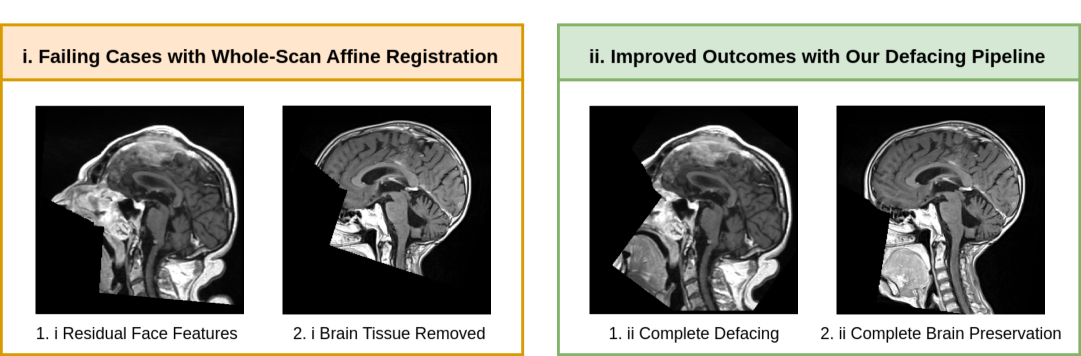
The availability of reliable defacing techniques to protect patient identity is increasingly important for cross-institutional research collaborations. Existing methods include: 1) registration-based approaches using predefined atlases such as AFNI Refacer [2], mri_reface [7], and PyDeface [9]; 2) geometry-based techniques including QuickShear [10]; and 3) model-based frameworks such as DeepDefacer [5]. While widely adopted, registration-based methods can fail to achieve accurate alignment, leaving facial features or removing brain tissue. QuickShear provides a geometry-driven alternative by using a brain mask to compute the convex hull on a middle slice, from which a defacing plane is derived. However, this approach does not guarantee full preservation of brain tissue, limiting its reliability. Additionally, model-based techniques may lack generalisability across different sequences or pathological cases. Preliminary experiments illustrated in Figure 1 with currently available tools on a large-scale clinical dataset highlighted the need for an alternative with higher success rate.
To address limitations of widely available tools, this study presents the development of a robust, generalisable defacing pipeline for high-resolution MRI data. The proposed method combines atlas-based registration with brain masking to reliably remove facial features while preserving brain anatomy. It maintains the native image space and voxel dimensions, ensuring the defaced data remain suitable for downstream quantitative analyses.
0.0.2 Material and methods
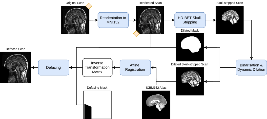
The proposed open-source pipeline [1] illustrated in Figure 2 comprises the following stages: (1) reorientation using fslreorient2std [11] to match the orientation of the MNI152 template; (2) tight skull-stripping using HD-BET [4]; (3) binarisation of the tightly skull-stripped images to obtain a binary brain mask; (4) morphological dilation of the brain mask with a safety margin of 7 mm to preserve relevant features near the brain boundary such as potential convexity meningiomas; (5) generation of a loosely skull-stripped image by applying the dilated brain mask to the original image; (6) affine registration of the loosely skull-stripped image to a skull-stripped T1-weighted ICBM152 reference template [8, 3] using mutual information as the similarity metric; (7) application of the inverse transformation matrix to a defacing mask, followed by resampling to generate an initial registered defacing mask; (8) union of the initial registered defacing mask and the dilated brain mask to generate a brain-safe defacing mask; and (9) generation of the final defaced output.
The pipeline was evaluated on a large, heterogeneous dataset comprising 4,932 MRIs obtained from King’s College Hospital from 105 newly diagnosed meningioma patients, across 453 scanning sessions between January 2012 and November 2020. Ethical approval was acquired from the NHS Health Research Authority Research Ethics Committee (REC Ref 22/NS/0160). After excluding localisers, diffusion, perfusion, angiography, and other non-structural sequences, 2,566 scans were retained for defacing. The dataset included a wide range of scanners, acquisition protocols, image resolutions, and anatomical coverage.
A biomedical engineer with 3 years of experience performed quality control of all scans to confirm complete removal of identifiable facial features (mouth, nose, eyes) and absence of alteration of the brain. As an additional automated measure to evaluate whether the defacing algorithm successfully removed facial features without compromising brain tissue, the Dice Similarity Coefficient (DSC) was calculated between the brain masks produced by HD-BET from 1) the original and 2) defaced scans. Similarly, anatomical label propagation was conducted on the original and defaced images using a single-atlas approach with the CerebrA atlas [6]. Subsequently, DSCs were computed for the resulting segmentations to evaluate the impact of defacing on the accuracy of anatomical labelling.
0.0.3 Results
Quality control revealed a 99.92% success rate. Out of the 2,566 scans, only two demonstrated suboptimal defacing, with residual facial features. Both failures were fat-only images acquired using the Turbo Spin Echo (TSE) Dixon sequence [12]. We hypothesise that, although HD-BET successfully segmented the brain region in these cases, the imaging characteristics affected the affine registration stage, where mutual information was used as the similarity metric, resulting in less accurate alignment than for other sequences.
The average DSC obtained from comparing the binary brain masks produced by HD-BET for the original and defaced images was with a standard deviation of . This near-perfect overlap confirms that the defacing process reliably preserved brain tissue and further demonstrates that models such as HD-BET can accurately identify and segment the brain post-defacing. From the single-atlas label propagation, DSCs across scans for individual labels were consistently high, with the best-performing label achieving an average of , and the lowest-performing one obtaining . These results indicate that the defacing pipeline preserves brain regions sufficiently for consistent brain mask generation and anatomical labelling accuracy using HD-BET and single-atlas propagation.
0.0.4 Conclusion
Integrating skull-stripping and registration using this proved fundamental to the success of our defacing pipeline as shown in Figure 1. This strategy ensures that registration is performed relative to the brain itself while the accompanying brain mask reliably prevents inadvertent removal of brain tissue during defacing. The resulting pipeline enables secure, multi-centre sharing of sensitive neuroimaging data while retaining anatomical integrity for downstream applications. Future work will evaluate the preservation of individual brain structures through multi-atlas label propagation.
0.0.5 Acknowledgements
LGFM is supported by the EPSRC CDT in Smart Medical Imaging [EP/S022104/1].
References
- [1] CAI4CAI: cai4cai/defacing_pipeline: Pipeline for automatic defacing with manual correction of failed cases (2024), https://github.com/cai4cai/defacing_pipeline, accessed: 2024-05-14
- [2] Cox, R.W.: AFNI: Software for analysis and visualization of functional magnetic resonance neuroimages. Computers and Biomedical Research 29, 162–173 (1996)
- [3] Fonov, V., Evans, A.C., Botteron, K., Almli, C.R., McKinstry, R.C., Collins, D.L., Group, B.D.C., et al.: Unbiased average age-appropriate atlases for pediatric studies. Neuroimage 54(1), 313–327 (2011)
- [4] Isensee, F., Schell, M., Tursunova, I., Brugnara, G., Bonekamp, D., Neuberger, U., Wick, A., Schlemmer, H.P., Heiland, S., Wick, W., Bendszus, M., Maier-Hein, K.H., Kickingereder, P.: Automated brain extraction of multi-sequence mri using artificial neural networks. Human Brain Mapping 40, 4952–4964 (2019)
- [5] Khazane, A., Hoachuck, J., Gorgolewski, K.J., Poldrack, R.A.: DeepDefacer: Automatic removal of facial features via U-Net image segmentation. arXiv preprint arXiv:2205.15536 (May 2022)
- [6] Manera, A.L., Dadar, M., Fonov, V., Collins, D.L.: CerebrA, registration and manual label correction of Mindboggle-101 atlas for MNI-ICBM152 template. Scientific Data 7, 1–9 (2020)
- [7] Medical Image Handling group (MIH): mih/mridefacer: Helper to aid de-identification of MRI images (3D or 4D). https://github.com/mih/mridefacer, accessed: 2025-05-14
- [8] NIST-Lab: ICBM 152 nonlinear atlases (2009) – NIST (2009), https://nist.mni.mcgill.ca/icbm-152-nonlinear-atlases-2009/, accessed: 2024-05-14
- [9] Poldrack Lab: poldracklab/pydeface at v2.0.0. https://github.com/poldracklab/pydeface/tree/v2.0.0, accessed: 2025-05-14
- [10] Schimke, N., Kuehler, M., Hale, J.: Preserving privacy in structural neuroimages. In: Lecture Notes in Computer Science, vol. 6818, pp. 301–308. Springer, Berlin, Heidelberg (2011)
- [11] Smith, S.M., Jenkinson, M., Woolrich, M.W., Beckmann, C.F., Behrens, T.E.J., Johansen-Berg, H., Bannister, P.R., Luca, M.D., Drobnjak, I., Flitney, D.E., Niazy, R.K., Saunders, J., Vickers, J., Zhang, Y., Stefano, N.D., Brady, J.M., Matthews, P.M.: Advances in functional and structural mr image analysis and implementation as FSL. NeuroImage 23(Suppl 1), S208–S219 (2004)
- [12] Wang, D., Li, X., Wu, X., Ugurbil, K.: Improving time efficiency for T2-weighted fat-water imaging by using multiband simultaneous multi-slice accelerated TSE dixon. In: Proceedings of the International Society for Magnetic Resonance in Medicine (ISMRM). p. 3262 (2016), accessed: 2024-05-14