Ab initio X-ray Near-Edge Spectroscopy of Sodium-Based Multi-Alkali Antimonides
Abstract
Multi-alkali antimonides (MAAs) are promising materials for vacuum electron sources. While sodium-based MAAs have demonstrated superior characteristics for ultrabright electron sources, their synthesis remains challenging, often resulting in mixed stoichiometries and polycrystalline domains. To address this complexity and guide the characterization of experimentally grown photocathodes, we present a comprehensive theoretical study of the X-ray near-edge spectroscopy (XANES) of four ternary MAAs: cubic Na2KSb and hexagonal NaK2Sb, representing the experimentally known phase of each stoichiometry, as well as hexagonal Na2KSb and cubic NaK2Sb, two computationally predicted polymorphs. Employing state-of-the-art ab initio methods based on all-electron density-functional theory and the solution of the Bethe-Salpeter equation (BSE), we compute and analyze the XANES at the sodium and potassium K-edges, potassium L2,3-edge, and antimony K and L2-edges. Our analysis reveals distinct spectral fingerprints for the experimentally known phases, cubic Na2KSb and hexagonal NaK2Sb, particularly at the sodium K-edge and potassium L2,3-edge, providing useful indications for their identification in complex samples. We further investigate the role of excitonic effects by comparing BSE spectra and their counterparts obtained in the independent-particle approximation, highlighting their significant influence on the near-edge features, especially for shallower core levels. Our findings offer a useful theoretical benchmark for the experimental characterization and diagnostics of sodium-based MAA photocathodes.
I Introduction
Multi-alkali antimonides (MAAs) are an established class of semiconducting materials for vacuum electron sources [1, 2, 3]. Thanks to their favorable properties, including a narrow band gap, low electron affinity, and sensitivity to visible light, cesium antimonide photocathodes have been investigated for decades [4, 5, 6, 7, 8]. After the first seminal studies of the past century [9, 10, 11, 12], they are still the subject of intensive experimental [13, 14, 15, 16, 17, 18, 19] and computational research [20, 21, 22, 23, 24, 25, 26, 27, 28, 29, 30]. More recently, Na-based MAAs have demonstrated superior characteristics for ultrabright electron sources, such as near-infrared optical response [31] and enhanced thermal emittance compared to other MAAs [32, 33]. The experimental studies exploring these materials have been complemented by ab initio investigations [34, 35, 36, 37, 38, 39], confirming the near-infrared absorption threshold of these and their favorable characteristics as photocathodes.
Despite these encouraging results, the growth of Na-based MAA photocathodes remains challenging [40, 41], and samples often exhibit coexisting stoichiometries and polycrystalline domains [40, 42]. Because of this uncertainty, it is essential to develop efficient characterization techniques that reveal the fingerprints of each compound. X-ray spectroscopy is particularly suited for this purpose as it provides information about the sample structure and stoichiometry as well as on the local atomic environment [43, 44, 45, 46, 47, 48, 49, 50]. Recent advances in X-ray spectroscopy techniques [51, 52] have been accompanied by the refinement of ab initio methods that are capable of describing the excitation process explicitly taking into account electron-hole interactions [53, 54, 55, 56, 57]. Solving the Bethe-Salpeter equation (BSE) on top of all-electron density-functional theory (DFT) [58, 53, 54] is currently considered the state-of-the-art approach to compute X-ray absorption spectra from first principles.
In this work, we employ DFT and BSE to investigate X-ray near-edge spectra (XANES) of four ternary MAAs with chemical compositions Na2KSb and NaK2Sb with either hexagonal or cubic lattice. Cubic Na2KSb and hexagonal NaK2Sb represent the experimentally known phases of the respective crystals [5] while hexagonal Na2KSb and cubic NaK2Sb are computationally predicted polymorphs that have been characterized computationally in recent work [39]. We compute the XANES of all four materials, exploring excitations from different core levels experimentally accessible by hard or soft X-rays. We compare the spectral features of the different compounds, highlighting the fingerprints that enable distinguishing one phase and/or stoichiometry from the other. We additionally discuss the excitonic character of the resonances through the comparison between BSE spectra and their counterparts computed in the so-called independent-particle approximation (IPA), where electron-hole Coulomb interactions are neglected. This assessment offers insight into the characteristics of the core-level excitations and provides additional information on the electronic structure of these crystals. Our results offer a valuable tool for the diagnostics and characterization of Na-based MAA photocathodes.
II Methods
II.1 Theoretical Background
The results presented in this work are obtained in the framework of all-electron DFT [59] and MBPT [54]. After solving the Kohn-Sham equations [60], we compute the XANES from the BSE [53, 54] mapped into the eigenvalue problem
| (1) |
where the eigenvalues are the excitation energies, while the eigenvectors contain information about the character and composition of the excitations. They both enter the imaginary part of the macroscopic dielectric tensor,
| (2) |
where is the unit cell volume, is the angular frequency of the incoming photon, and the are the transition coefficients between initially occupied core levels () and unoccupied conduction states ():
| (3) |
The denominator of Eq. (3) includes KS energies instead of quasi-particle energies. This simplification is justified by the substantial difference in the corrections needed for core and conduction level energies. While efficient developments for core electrons [61] have been proposed recently, sometimes even combined with the BSE [62], their application goes beyond the scope of this work. Given the lack of experimental data to establish a reliable scissors shift, we align all calculated spectra to zero at the IPA onset.
The BSE Hamiltonian appearing in Eq. (1),
| (4) |
includes the diagonal component, , which accounts for vertical transitions between core and conduction states, the repulsive exchange interaction between electron and holes, , multiplied by 2 upon spin-degeneracy, and the direct term, , which embeds the statically screened electron-hole Coulomb potential. By neglecting the last two terms, we obtain , which delivers IPA spectra retaining the main features of the electronic density of states projected on the unoccupied target states. When electron-hole correlations are negligible, it represents a good approximation for the XANES [63, 64, 65]. Comparing BSE and IPA spectra reveals excitonic effects [66, 67], which can be sizeable even in the XANES of inorganic crystals, of the order of 1 eV [45, 68, 69, 26, 70].
II.2 Computational Details
All calculations are performed with exciting, a full-potential all-electron code implementing the linearized augmented plane waves and local orbitals method for DFT and MBPT [59, 54]. The explicit treatment of core electrons makes this software ideally suited to address the central problem of this work. To ensure convergence, we set the muffin-tin radius of both Na and K atoms to 2.0 bohr and that of Sb to 2.2 bohr. The plane-wave basis set cutoff is fixed to 8.0 for the cubic crystals and 8.5 for the hexagonal ones, see Ref. [59] for the definitions of these parameters. The exchange-correlation potential of DFT is approximated using the spin-unpolarized Perdew-Burke-Ernzerhof functional for solids (PBEsol) [71]. The Brillouin zones of cubic and hexagonal crystals are sampled with and -meshes, respectively.
The BSE is solved in the Tamm-Dancoff approximation using a -shifted k-mesh with points for the cubic crystals and for the hexagonal ones. The screened Coulomb interaction is computed in the random phase approximation, including 100 empty states. An energy cutoff of 1.5 Ha is taken for the local fields. The BSE is solved with 10 (12) empty states for the cubic (hexagonal) crystals. A Lorentzian broadening of is used for plotting all spectra.
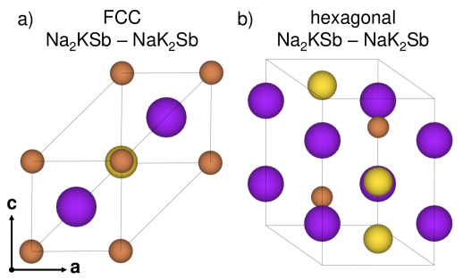
The materials considered in this work are taken from the Open Quantum Materials Database (OQMD) [73], structure numbers: 7813090 for hexagonal Na2KSb (hNa2KSb), 42945 for cubic Na2KSb (cNa2KSb), 1245926 for cubic NaK2Sb (cNaK2Sb), and 7751654 for hexagonal NaK2Sb (hNaK2Sb). The cubic phases of both Na2KSb and NaK2Sb are face-centred cubic Bravais lattices with Sb atoms at Wyckoff position and alkali species at and , see Fig. 1a. In the hexagonal crystals, the Sb atoms occupy the and Wyckoff positions while alkali atoms are at , , and , see Fig. 1b. The initial structures taken from OQMD are subsequently relaxed with exciting, using the standard force minimization procedure of DFT combined with volume optimization based on the Birch-Murnaghan fit [74, 75]. Further details on this procedure can be found in Ref. [39].
III Results
In the following, we analyze the XANES computed for the four MAA crystals considered in this work. In Sec. III.1, we examine excitations from the sodium K-edge (1 core electrons), targeting unoccupied electrons with -orbital character. In Sec. III.2, we inspect spectra computed from the potassium K-edge, whereby the 1 electrons of this element are excited to unoccupied -states. In Sec. III.3, we discuss excitations from the potassium L2,3-edge, corresponding to transitions from the 2 core electrons of this element to unoccupied levels bearing and character. In Sec. III.4, we investigate the XANES computed from the antimony K-edge, i.e., excitations from Sb 1 electrons to unoccupied bands with Sb character. Finally, in Sec. III.5, we analyze the spectra obtained by exciting antimony 2 core electrons to unoccupied states with Sb and character. Due to the large spin-orbit splitting (several tens of eV) in the Sb 2 shell, we consider only the 2 component of the XANES (L2-edge), which yields equivalent signatures, albeit with different oscillator strength, as expected, than the L3-edge [76].
III.1 Sodium K-edge
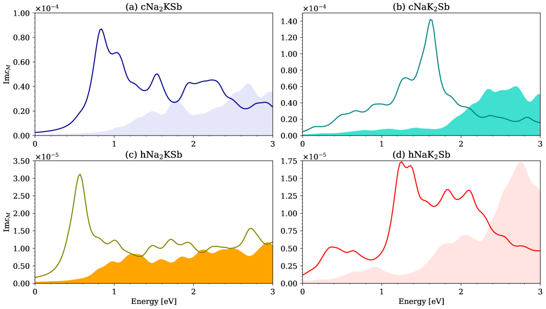
We begin our analysis with the inspection of the sodium K-edge spectra (Fig. 2), which reveal notable similarities between the Na2KSb phases (Fig. 2a,c). Both spectra exhibit a high-intensity peak at low energies, with the maximum at 0.9 eV and 0.6 eV above the IPA onset for cNa2KSb and hNa2KSb, respectively. The excitonic nature of this peak is evident from the comparison with the IPA spectra, which reflect the weak contribution of Na -states to the lowest unoccupied electronic states (Fig. S1 in the Supporting Information). This oscillator strength enhancement and a red shift of almost 1 eV are characteristic signatures of strong electron-hole correlations. Two pairs of degenerate excitons appear below the IPA onset of cNa2KSb, with binding energies of 229 meV and 59 meV (see Table S1). In hNa2KSb, a single pair appears 70 meV below the IPA onset. All these transitions are dark, a consequence of the negligible contribution of Na -states to the conduction band minima of these materials (Fig. S1) [36, 39].
The BSE spectra of the NaK2Sb compounds (Fig. 2b,d) are dominated by an intense peak at approximately 1.5 eV (cNaK2Sb) and 1.2 eV (hNaK2Sb) above the IPA onset. Comparison with the respective IPA spectra confirms the excitonic character of these features, whereby electron-hole interactions are responsible for spectral weight enhancement at low energies and for a red shift of the order of 1 eV. At lowest energies, the NaK2Sb spectra are characterized by a broad set of excitations with weak oscillator strength. In cNaK2Sb, two pairs of degenerate excitons appear below the IPA onset, with binding energies of 574 meV and 55 meV (Table S1). In analogy with the Na2KSb compounds, their oscillator strength is negligible due to the minimal contribution of the Na -states to the bottom of the conduction band [39], see Fig. S1. In hNaK2Sb, which corresponds to the experimentally resolved phase of this crystal, two degenerate dark excitons appear below the IPA onset, with a binding energy of 131 meV (Table S1).
The similarities between the spectra of the materials with equal stoichiometry and different crystal structure are expected to facilitate the identification of specific compositions in polycrystalline samples. Likewise, the distinct spectral features of the experimentally resolved crystals, cNa2KSb and hNaK2Sb (Fig. 2a,d), will promote their recognition in mixed samples.
III.2 Potassium K-edge
In analogy to the Na K-edge XANES, the results obtained for the cubic and hexagonal phases of Na2KSb are very similar, see Fig. 3a,c. Both spectra are characterized by a broad absorption onset with several maxima covering the first 3 eV above the IPA onset. The oscillator strength in this region is enhanced by electron-hole correlations, although the total spectral weight remains weak due to the small contributions of K -states to the bottom of the conduction region of both materials [36, 39], see Fig. S2. Two degenerate dark excitations appear below the IPA onset. Their binding energy is 111 meV in cNa2KSb and 60 meV in hNa2KSb, see Table S1.
The XANES of the computationally predicted cNaK2Sb crystal is again characterized by broad excitations at low energies (Fig. 3b). However, the oscillator strength of the first peak is weaker than in cNa2KSb and the BSE spectrum retains the overall spectral shape of its IPA counterpart, confirming the less prominent role of excitonic effects. On the other hand, the spectrum of hNaK2Sb is dominated by a sharp excitonic peak at the onset (Fig. 3d), stemming from the broad feature in the IPA spectrum between approximately 1.2 and 2.2 eV, reflecting the contributions from K -states to the PDOS (Fig. S2). The comparison between the sharp excitonic resonance in the BSE result and the energetically distributed oscillator strength in the IPA spectrum allows assigning the former a binding energy of approximately 800 meV. The sharp excitonic maximum has a shoulder at higher energies and is followed by a broader region with non-negligible oscillator strength, red-shifted by about 1 eV from its IPA counterpart (Fig. 3d). The presence of this prominent excitonic feature in the XANES of hNaK2Sb makes this experimentally resolved phase well detectable in polycrystalline samples.
III.3 Potassium L2,3-edge
We continue our analysis with the XANES computed from the potassium L2,3-edge. The result obtained for the experimentally known cubic phase of Na2KSb is dominated by two intense peaks at the onset (Fig. 4a), which offer a valuable spectroscopic signature to identify this phase in a polycrystalline sample. The excitonic nature of these maxima is evident from the comparison with the IPA spectrum, which features weak oscillator strength distributed up to almost 3.5 eV (Fig. 4a), following the contributions of K -electrons in the unoccupied region of this material (Fig. S3). The replicas of these peaks, appearing approximately 3 eV above the first ones, stem from transitions from 2 electrons and, as expected, their oscillator strength is approximately of those generated by excitations from the 2 core states.
The XANES of the computationally predicted cubic NaK2Sb is characterized by very weak peaks at the onset and by a strong maximum at 2 eV (Fig. 4b). The replica of this feature is visible at about 5 eV, but it would be hardly detectable in experiments due to the relatively large oscillator strength of overlapping excitations between 3 and 4 eV. The spectrum of the other computationally predicted compound, hNa2KSb, is again characterized by excitations of increasing strength at the onset up to approximately 2 eV (Fig. 4c). The spectral shape in the BSE result reflects the IPA spectrum, suggesting that electron-hole interactions red shift the excitations by about 1 eV and enhance their oscillator strength toward lower energies. In this case, the high-energy replica of the first excitations is not clearly visible, due to other bright excitations appearing in the same energy range.
The XANES computed for hNaK2Sb, the other experimentally known phase, is dominated by a sharp peak slightly about 1 eV, see Fig. 4d. The excitonic nature of this feature is testified not only by its very strong oscillator strength but also by the absence of a direct counterpart in the IPA spectrum. A replica of this intense maximum is visible around 4 eV, i.e., about 3 eV above the first one, with an oscillator strength approximately of the lower-energy resonance, as expected. Interestingly, a weaker but equally sharp peak is visible a few tens of meV above the onset. Its higher-energy replica is not visible, again due to overlap with other bright states. All compounds are characterized by four degenerate excitations below the IPA onset. Their binding energies are on the order of 60 meV in both cNaK2Sb and hNa2KSb, while they are almost twice as large in the experimentally known phases cNa2KSb and hNaK2Sb (Table S1).
The peculiar spectral characteristics of cNa2KSb and hNaK2Sb make these two experimentally resolved phases expectedly well detectable in polycrystalline samples. In particular, the striking differences between the two spectra in Fig. 4a and Fig. 4d, namely the presence of two low-energy sharp peaks in the former and a single intense low-energy maximum in the latter, is expected to promote the identification of either compound in a mixed sample.
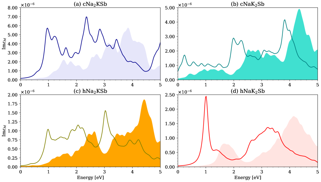
III.4 Antimony -edge
The XANES calculated from the Sb K-edge exhibit pronounced similarities for each considered stoichiometry. The cubic and hexagonal phases of Na2KSb are characterized by relatively weak oscillator strength at the onset and larger spectral weight at higher energies, around 1 eV (Fig. 5a,c). In the BSE spectrum of cNa2KSb (Fig. 5a), the lowest-energy region is dominated by four relatively broad maxima mimicking the features of the IPA spectrum and of the Sb -orbital contributions to the conduction region (Fig. S4). In the spectrum of the computationally predicted hexagonal phase of Na2KSb, the oscillator strength at low energies is focused across a narrow range of approximately 1.5 eV (Fig. 5c). The first weak peak appears around 0.6 eV in both the BSE and IPA spectra, suggesting an almost negligible influence of electron-hole correlations. It is followed by a broad distribution of bright excitations with the most intense maximum at 2 eV. The spectral features characterizing the BSE result are almost identically reproduced in the IPA spectrum, only shifted by about 1 eV. This finding suggests that excitonic effects in this material manifest themselves in the above-mentioned sizeable red shift of excitation energies but do not alter the spectral shape. In both Na2KSb polymorphs, two degenerate excitations are found below the IPA onset. Both of them are dark, being two orders of magnitude weaker than the strongest resonances. Their binding energy is 105 meV in cNa2KSb and 59 meV in hNa2KSb, see Table S1. The systematically lower intensity of excitations from the Sb K-edge compared to those analyzed above (e.g., from the K L2,3-edge) is ascribed to the negligible contribution of Sb states to the bottom of the conduction region of both materials (Fig. S4) [36, 39].
The XANES of both NaK2Sb phases are characterized by similar spectral fingerprints, which are in turn quite different from those of Na2KSb. In the computationally predicted cubic polymorph, the absorption from the Sb K-edge is dominated by two broad regions (1 eV each) of intense excitations, see Fig. 5b, both red-shifted by about 200 meV compared to the IPA results. While the first manifold almost doubles its oscillator strength upon inclusion of electron-hole correlations, this effect is less pronounced for the higher-energy maxima. The spectrum of the experimentally resolved hexagonal structure of NaK2Sb is dominated by an intense peak at about 1 eV (Fig. 5d). This strong excitation is preceded by a weak shoulder at approximately 0.5 eV. Higher-energy excitations have about half the oscillator strength of the aforementioned intense peak and form a broad band spanning almost 2 eV. The peaks in the BSE spectrum mirror those computed from the IPA, and the red shift associated with their binding energy is on the order of 800 meV. However, only the intensity of the first resonance is significantly affected by the inclusion of electron-hole correlations. The sharp resonance in the spectrum of hNaK2Sb represents a relevant signature to identify this structure in a polycrystalline sample, although the large broadening expected for excitations from such a deep edge (30 keV [77]) likely overshadows these features during measurements.
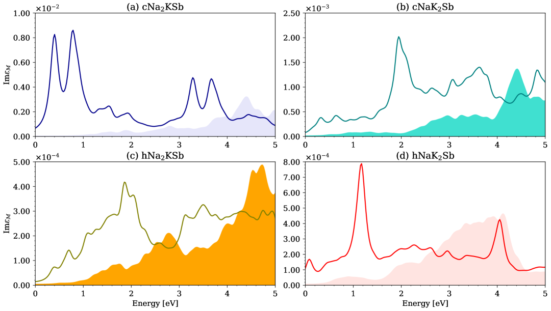
III.5 Antimony L2-edge
We conclude our analysis by inspecting the XANES computed from Sb L2-edge. Target states for the excited Sb 2 electrons are unoccupied bands with Sb and character. As extensively discussed in Ref. 36 for the experimentally resolved phases cNa2KSb and hNaK2Sb, and in Ref. 39 for the computationally predicted polymorphs hNa2KSb and cNaK2Sb, Sb electrons contribute to the lowest unoccupied region of all materials while Sb states appear only at higher energies (see also Fig. S5). The spectra of the two Na2KSb polymorphs exhibit similar features, see Fig. 6a,c. They are both characterized by weak transitions at the onset and by stronger peaks above 1 eV. The spectral features from the BSE are present already in the IPA spectra, only with weaker oscillator strength (about for the first manifold of excitations up to approximately 2.2 eV) and an energy shift of about 700 meV in cNa2KSb and 400 meV in hNa2KSb.
The NaK2Sb spectra display weak excitonic effects, with red shifts of the order of 200 meV in cNaK2Sb (Fig. 6b) and 600 meV in hNaK2Sb (Fig. 6d), and an oscillator strength of the first excitations only marginally amplified by electron-hole correlations. While the XANES of cNaK2Sb hosts very weak excitations up to about 3 eV, the one of hNaK2Sb exhibits a weak but distinguishable absorption band between 0.3 and 1 eV, and a stronger one between 2 and 3 eV. It is hard to expect that these features will enable the identification of specific stoichiometries and/or crystal structures in mixed samples. All compounds exhibit a doubly degenerate excitation below the IPA onset. Its oscillator strength is at least one order of magnitude lower than the transitions giving rise to the visible peaks in the XANES, while its binding energy is of the order of 60 meV in the two computationally predicted compounds (cNaK2Sb and hNa2KSb) and 120 meV in the experimental phases (cNa2KSb and hNaK2Sb), see Table S1.
IV Summary, Discussion, and Conclusions
In summary, we investigated from first-principles many-body theory the XANES of four Na-based ternary alkali antimonide phases considering excitations from sodium, potassium, and antimony K-edge, potassium L2,3-edge, and antimony L2-edge. In this analysis, we included both the experimentally resolved cubic polymorph of Na2KSb and hexagonal phase of NaK2Sb [5, 36], as well as the computationally predicted cubic NaK2Sb and hexagonal Na2KSb [39]. By comparing the results obtained for all compounds, we identified the spectral fingerprints that can enable their identification in polycrystalline samples embedding different stoichiometries. From a fundamental viewpoint, we addressed the role of electron-hole correlations to gain further insight into the electronic structure of each material.
The intense peak dominating the onset of the Na K-edge spectra of both phases of Na2KSb is expected to provide a valuable tool to distinguish this composition from NaK2Sbwhile in the potassium K-edge XANES, a sharp excitonic peak in the spectrum of hNaK2Sb will facilitate the identification of this crystal in a mixed sample. The potassium L2,3-edge spectra of the two experimental phases, cNa2KSb and hNaK2Sb, are dominated by clear spectral signatures promoting their recognition in a polycrystalline sample. In particular, the spectrum of cNa2KSb hosts two resonances at the onset with strong intensity and almost equal oscillator strength, while the XANES of hNaK2Sb features a distinct strong peak about 1 eV from the onset. In contrast, the spectra of two computationally predicted phases are characterized by weak excitations at the onset, growing in intensity with increasing energies, and forming broad absorption bands. The XANES computed from Sb core electrons offer very few signatures for identification of specific crystal structures and stoichiometries. All Sb K-edge spectra feature broad excitations at the onset, except for hNaK2Sb and, to a lesser extent, its cubic counterpart, characterized by an intense peak around 1 eV. However, the large broadening associated with such an energetically deep excitation is expected to overshadow these features and to prevent any direct fingerprinting of these compounds. The scenario is even less favorable for the XANES from the Sb L2-edge. In this case, all spectra exhibit broad absorption bands that are hardly distinguishable from each other in a mixed sample.
Comparing the spectra computed from the BSE and in the IPA reveals the influence of excitonic effects. They are most pronounced in the XANES from Na K-edge, where they give rise to a large redistribution of oscillator strength to the lowest-energy excitations in Na2KSb and at higher energies (1 eV) in NaK2Sb. In all materials, these excitations undergo a red shift larger than 0.5 eV compared to the IPA. In the spectra computed from the potassium K-edge, excitonic effects remain strong but are less relevant than from the Na K-edge. Binding energies are on the order of 200 meV, and the oscillator strength enhancement of low-energy excitations is weakly pronounced except for hNaK2Sb. Stronger excitonic effects are found in the potassium L2,3-edge spectra of all considered crystals, especially in the experimental phases cNa2KSb and hNaK2Sb, where they manifest themselves through strong resonances at lowest energies (cNa2KSb) and around 1 eV (hNaK2Sb). In the XANES from the Sb K- and L2-edges, excitonic effects are least pronounced: binding energies do not exceed 200 meV and the oscillator strength enhancement at lowest energies is negligible. These results confirm previous findings [53, 65], suggesting a systematic reduction of electron-hole correlation strength with increasing energy of the excited core electrons. This behavior is consistent with physical intuition, whereby transitions from deeper states are subject to a larger screening generated by a larger number of semi-core and valence electrons separating the initial core state of the transition to the final one in the conduction band. It is also worth noting that in the considered MAAs, many-body effects are more pronounced in X-ray spectra than in optical spectra [36, 39], as predicted by Spicer in 1967 [78].
In conclusion, our study provides valuable indications for the X-ray spectroscopic characterization of MAA photocathodes, where different stoichiometries and crystal structures often coexist. Our results suggest that excitations from the Na K-edge and the potassium L2,3-edge are mostly suited to identify different compositions and crystal structures, while XANES from the potassium K-edge can reveal the presence of hexagonal NaK2Sb, featuring a distinct sharp excitonic resonance. On the contrary, X-ray absorption from Sb K- and L2-edge does not provide any clear fingerprints to identify specific stoichiometries and crystal structures, also given the larger broadening associated with these deep core-level transitions. The absence of experimental references on these compounds prevents a direct comparison between our computational results and measurements, but we are confident that our comprehensive study will stimulate corresponding experiments validating our predictions.
Acknowledgments
This work was funded by the German Research Foundation (DFG), Project No. 490940284, DAAD (Program RISE), the German Federal Ministry of Education and Research (Professorinnenprogramm III), and the State of Lower Saxony (Professorinnen für Niedersachsen). Computational resources were provided by the HPC cluster ROSA at the University of Oldenburg, funded by the DFG (project number INST 184/225-1 FUGG) and by the Ministry of Science and Culture of the Lower Saxony State.
Data availability statement
The data that support the findings of this article are openly available at the following Zenodo link: https://doi.org/10.5281/zenodo.15297231.
Supporting Information
In the Supporting Information, the orbital-resolved contributions to the unoccupied density of states of the considered materials are provided (Fig. S1-S5) along with a table reporting the binding energies of the excitons below the independent-particle onset (Table S1).
References
- Dowell et al. [2010] D. Dowell, I. Bazarov, B. Dunham, K. Harkay, C. Hernandez-Garcia, R. Legg, H. Padmore, T. Rao, J. Smedley, and W. Wan, Cathode r&d for future light sources, Nucl. Instrum. Methods Phys. Res. A 622, 685 (2010).
- Musumeci et al. [2018] P. Musumeci, J. Giner Navarro, J. Rosenzweig, L. Cultrera, I. Bazarov, J. Maxson, S. Karkare, and H. Padmore, Advances in bright electron sources, Nucl. Instrum. Methods Phys. Res. A 907, 209 (2018), advances in Instrumentation and Experimental Methods (Special Issue in Honour of Kai Siegbahn).
- Schmeißer et al. [2018] M. A. H. Schmeißer, S. Mistry, H. Kirschner, S. Schubert, A. Jankowiak, T. Kamps, and J. Kühn, Towards the operation of cs-k-sb photocathodes in superconducting rf photoinjectors, Phys. Rev. Accel. Beams 21, 113401 (2018).
- Jack and Wachtel [1957] K. H. Jack and M. Wachtel, The characterization and crystal structure of caesium antimonide, a photo-electric surface material, Proc. R. Soc. London A 239, 46 (1957).
- McCarroll [1960] W. McCarroll, Phases in the photoelectric sodium-potassium-antimony system, Journal of Physics and Chemistry of Solids 16, 30 (1960).
- Michelato et al. [1994] P. Michelato, P. Gallina, and C. Pagani, Alkali photocathode development for superconducting rf guns, Nucl. Instrum. Methods Phys. Res. A 340, 176 (1994).
- Di Bona et al. [1997] A. Di Bona, F. Sabary, S. Joly, P. Michelato, D. Sertore, C. Pagani, and S. Valeri, Development, operation and analysis of bialkali antimonide photocathodes for high-brightness photo-injectors, Nucl. Instrum. Methods Phys. Res. A 385, 385 (1997).
- Ding et al. [2017a] Z. Ding, S. Karkare, J. Feng, D. Filippetto, M. Johnson, S. Virostek, F. Sannibale, J. Nasiatka, M. Gaowei, J. Sinsheimer, et al., Temperature-dependent quantum efficiency degradation of k-cs-sb bialkali antimonide photocathodes grown by a triple-element codeposition method, Phys. Rev. ST Accel. Beams 20, 113401 (2017a).
- Sommer [1955] A. H. Sommer, New photoemissive cathodes of high sensitivity, Rev. Sci. Instrum. 26, 725 (1955).
- Spicer [1958] W. E. Spicer, Photoemissive, photoconductive, and optical absorption studies of alkali-antimony compounds, Phys. Rev. 112, 114 (1958).
- Nathan and Mee [1967] R. Nathan and C. H. B. Mee, Photoelectric and related properties of the Potassium—Antimony—Caesium photocathode, Int. J. Electron. 23, 349 (1967).
- Ghosh and Varma [1978] C. Ghosh and B. P. Varma, Preparation and study of properties of a few alkali antimonide photocathodes, J. Appl. Phys. 49, 4549 (1978).
- Mamun et al. [2017] M. Mamun, M. Hernandez-Flores, E. Morales, C. Hernandez-Garcia, and M. Poelker, Temperature dependence of alkali-antimonide photocathodes: Evaluation at cryogenic temperatures, Phys. Rev. ST Accel. Beams 20, 103403 (2017).
- Ding et al. [2017b] Z. Ding, M. Gaowei, J. Sinsheimer, J. Xie, S. Schubert, H. Padmore, E. Muller, and J. Smedley, In-situ synchrotron x-ray characterization of k2cssb photocathode grown by ternary co-evaporation, J. Appl. Phys. 121, 055305 (2017b), https://pubs.aip.org/aip/jap/article-pdf/doi/10.1063/1.4975113/13272316/055305_1_online.pdf .
- Dai et al. [2020] J. Dai, Y. Ding, C. Ruan, X. Xu, and H. Liu, High photocurrent density and continuous electron emission characterization of a multi-alkali antimonide photocathode, Electronics 9, 1991 (2020).
- Panuganti et al. [2021] H. Panuganti, E. Chevallay, V. Fedosseev, and M. Himmerlich, Synthesis, surface chemical analysis, lifetime studies and degradation mechanisms of cs-k-sb photocathodes, Nucl. Instrum. Methods Phys. Res. A 986, 164724 (2021).
- Pavlenko et al. [2022] V. Pavlenko, J. Smedley, A. Scheinker, R. L. Fleming, A. Alexander, M. A. Hoffbauer, and N. A. Moody, Stoichiometry control and automated growth of alkali antimonide photocathode films by molecular beam deposition, Appl. Phys. Lett. 120, 091901 (2022), https://pubs.aip.org/aip/apl/article-pdf/doi/10.1063/5.0080948/16476606/091901_1_online.pdf .
- Kachwala et al. [2023] A. Kachwala, P. Saha, P. Bhattacharyya, E. Montgomery, O. Chubenko, and S. Karkare, Demonstration of thermal limit mean transverse energy from cesium antimonide photocathodes, Appl. Phys. Lett. 123, 044106 (2023), https://pubs.aip.org/aip/apl/article-pdf/doi/10.1063/5.0159924/18063298/044106_1_5.0159924.pdf .
- Guo et al. [2025] L. Guo, K. Shiohara, H. Yamaguchi, G. Wang, Y. Okabe, M. Nakatake, S. Takakura, M. Yamamoto, S. Ogawa, and Y. Takashima, Improved robustness of sequentially deposited potassium cesium antimonide photocathodes achieved by increasing the potassium content towards theoretical stoichiometry, Sci. Rep. 15, 2900 (2025).
- Guo [14] S.-D. Guo, Electronic structures and elastic properties of X3Sb (X = Li, K, Cs) from the first-principles calculations, Mater. Res. Express 1, 015906 (14).
- Kalarasse et al. [2010] L. Kalarasse, B. Bennecer, F. Kalarasse, and S. Djeroud, Pressure effect on the electronic and optical properties of the alkali antimonide semiconductors cs3sb, kcs2sb, csk2sb and k3sb: Ab initio study, J. Phys. Chem. Solids 71, 1732 (2010).
- Xie et al. [2016] H. Xie, I. Ben-Zvi, T. Rao, T. Xin, and E. Wang, Experimental measurements and theoretical model of the cryogenic performance of bialkali photocathode and characterization with monte carlo simulation, Phys. Rev. ST Accel. Beams 19, 103401 (2016).
- Gupta et al. [2017] P. Gupta, L. Cultrera, and I. Bazarov, Monte carlo simulations of electron photoemission from cesium antimonide, J. Appl. Phys. 121, 215702 (2017), https://pubs.aip.org/aip/jap/article-pdf/doi/10.1063/1.4984263/14852891/215702_1_online.pdf .
- Cocchi et al. [2018] C. Cocchi, S. Mistry, M. Schmeißer, J. Kühn, and T. Kamps, First-principles many-body study of the electronic and optical properties of csk2sb, a semiconducting material for ultra-bright electron sources, Journal of Physics: Condensed Matter 31, 014002 (2018).
- Cocchi et al. [2019] C. Cocchi, S. Mistry, M. Schmeißer, R. Amador, J. Kühn, and T. Kamps, Electronic structure and core electron fingerprints of caesium-based multi-alkali antimonides for ultra-bright electron sources, Sci. Rep. 9, 18276 (2019).
- Cocchi [2020] C. Cocchi, X-ray absorption fingerprints from cs atoms in cs3sb, Phys. Status Solidi (RRL) 14, 2000194 (2020).
- Antoniuk et al. [2020] E. R. Antoniuk, Y. Yue, Y. Zhou, P. Schindler, W. A. Schroeder, B. Dunham, P. Pianetta, T. Vecchione, and E. J. Reed, Generalizable density functional theory based photoemission model for the accelerated development of photocathodes and other photoemissive devices, Phys. Rev. B 101, 235447 (2020).
- Wu and Ganose [2023] R. Wu and A. M. Ganose, Relativistic electronic structure and photovoltaic performance of k 2 cssb, J. Mater. Chem. A 11, 21636 (2023).
- Sharma et al. [2023] G. Sharma, M. Sajjad, and N. Singh, Impressive electronic and thermal transports in csk2sb: A thermoelectric perspective, ACS Applied Energy Materials 6, 11179 (2023), https://doi.org/10.1021/acsaem.3c02024 .
- Santana-Andreo et al. [2024] J. Santana-Andreo, H.-D. Saßnick, and C. Cocchi, Thermodynamic stability and vibrational properties of multi-alkali antimonides, Journal of Physics: Materials 7, 035004 (2024).
- Mohanty et al. [2023] S. K. Mohanty, M. Krasilnikov, A. Oppelt, F. Stephan, D. Sertore, L. Monaco, C. Pagani, and W. Hillert, Development and characterization of multi-alkali antimonide photocathodes for high-brightness rf photoinjectors, Micromachines 14, 1182 (2023).
- Cultrera et al. [2016] L. Cultrera, C. Gulliford, A. Bartnik, H. Lee, and I. Bazarov, Ultra low emittance electron beams from multi-alkali antimonide photocathode operated with infrared light, Appl. Phys. Lett. 108, 134105 (2016), https://pubs.aip.org/aip/apl/article-pdf/doi/10.1063/1.4945091/14478751/134105_1_online.pdf .
- Motta and Schönert [2005] D. Motta and S. Schönert, Optical properties of bialkali photocathodes, Nucl. Instrum. Methods Phys. Res. A 539, 217 (2005).
- Yalameha et al. [2018] S. Yalameha, Z. Nourbakhsh, and A. Vaez, Hydrostatic strain-induced topological phase of kna2sb, 468, 279 (2018).
- Khan et al. [2021] Z. Khan, G. Murtaza, A. A. Khan, A. Laref, N. A. Kattan, and M. Haneef, Different physical properties of bi-alkali pnictogen compounds using density functional theory, Int. J. Energy Res. 45, 7703 (2021).
- Amador et al. [2021] R. Amador, H.-D. Saßnick, and C. Cocchi, Electronic structure and optical properties of na2ksb and nak2sb from first-principles many-body theory, J. Phys. Condens. Matter. 33, 365502 (2021).
- Yue et al. [2022] T. Yue, P. Sui, Y. Zhao, J. Ni, S. Meng, and Z. Dai, Theoretical prediction of mechanics, transport, and thermoelectric properties of full heusler compounds na 2 ksb and x 2 cssb (x= k, rb), Phys. Rev. B 105, 184304 (2022).
- Schier et al. [2024] R. Schier, D. Guo, H.-D. Saßnick, and C. Cocchi, Stability and electronic properties of k-sb and na-sb binary crystals from high-throughput ab initio calculations, Adv. Theory Simul. , 2400680 (2024).
- Xu et al. [2025] C. Xu, R. Schier, and C. Cocchi, Electronic and optical properties of computationally predicted na-k-sb crystals, Electronic Structure 7, https://doi.org/10.1088/2516-1075/adaab8 (2025).
- Rozhkov et al. [2024] S. Rozhkov, V. Bakin, V. Rusetsky, D. Kustov, V. Golyashov, A. Demin, H. Scheibler, V. Alperovich, and O. Tereshchenko, interface engineering for high-efficiency photocathodes, Phys. Rev. Appl. 22, 024008 (2024).
- Dube et al. [2025] J. Dube, J. Kühn, C. Wang, S. Mistry, G. Klemz, A. Galdi, and T. Kamps, Triple evaporation of bialkali antimonide photocathodes and photoemission characterization at the photex experiment, arXiv preprint arXiv:2503.03573 (2025).
- Rusetsky et al. [2022] V. S. Rusetsky, V. A. Golyashov, S. V. Eremeev, D. A. Kustov, I. P. Rusinov, T. S. Shamirzaev, A. V. Mironov, A. Y. Demin, and O. E. Tereshchenko, New spin-polarized electron source based on alkali antimonide photocathode, Phys. Rev. Lett. 129, 166802 (2022).
- Branci et al. [2000] C. Branci, M. Womes, P. Lippens, J. Olivier-Fourcade, and J. Jumas, Use of x-ray absorption spectra as a “fingerprint” of the local environment in complex chalcogenides, J. Solid State Chem. 150, 363 (2000).
- Cocchi et al. [2016] C. Cocchi, H. Zschiesche, D. Nabok, A. Mogilatenko, M. Albrecht, Z. Galazka, H. Kirmse, C. Draxl, and C. T. Koch, Atomic signatures of local environment from core-level spectroscopy in -ga 2 o 3, Phys. Rev. B 94, 075147 (2016).
- Vorwerk et al. [2018] C. Vorwerk, C. Hartmann, C. Cocchi, G. Sadoughi, S. N. Habisreutinger, R. Félix, R. G. Wilks, H. J. Snaith, M. Bär, and C. Draxl, Exciton-dominated core-level absorption spectra of hybrid organic–inorganic lead halide perovskites, J. Phys. Chem. Lett. 9, 1852 (2018).
- Fratesi et al. [2018] G. Fratesi, S. Achilli, N. Manini, G. Onida, A. Baby, A. Ravikumar, A. Ugolotti, G. P. Brivio, A. Milani, and C. S. Casari, Fingerprints of sp1 hybridized c in the near-edge x-ray absorption spectra of surface-grown materials, Materials 11, 2556 (2018).
- Wu et al. [2021] M. Wu, X. Huang, K. Zhang, S. Hu, L. Chen, H.-Q. Wang, and J. Kang, Determination of the crystal field and nature of x-ray linear dichroism for co-o with local octahedral, tetrahedral, and tetragonal symmetries, Phys. Rev. B 104, 075109 (2021).
- Olovsson and Magnuson [2022] W. Olovsson and M. Magnuson, Rhombohedral and turbostratic boron nitride polytypes investigated by x-ray absorption spectroscopy, J. Phys. Chem. C 126, 21101 (2022).
- Wibowo et al. [2023] R. E. Wibowo, R. Garcia-Diez, M. van der Merwe, D. Duarte-Ruiz, Y. Ha, R. Félix, A. Efimenko, T. Bystron, M. Prokop, R. G. Wilks, et al., Core-level spectroscopy with hard and soft x-rays on phosphorus-containing compounds for energy conversion and storage, J. Phys. Chem. C 127, 20582 (2023).
- Zimina et al. [2024] A. Zimina, A. Léon, and R. Steininger, Chemical bonding effects in sc compounds studied using x-ray absorption and x-ray photoelectron spectroscopies, Phys. Chem. Chem. Phys. 26, 2613 (2024).
- Greczynski et al. [2023] G. Greczynski, R. T. Haasch, N. Hellgren, E. Lewin, and L. Hultman, X-ray photoelectron spectroscopy of thin films, 3, 40 (2023).
- Greczynski and Hultman [2025] G. Greczynski and L. Hultman, Binding energy referencing in x-ray photoelectron spectroscopy, Nat. Rev. Mater. 10, 62 (2025).
- Vorwerk et al. [2017] C. Vorwerk, C. Cocchi, and C. Draxl, Addressing electron-hole correlation in core excitations of solids: An all-electron many-body approach from first principles, Phys. Rev. B 95, 155121 (2017).
- Vorwerk et al. [2019] C. Vorwerk, B. Aurich, C. Cocchi, and C. Draxl, Bethe–salpeter equation for absorption and scattering spectroscopy: implementation in the exciting code, Electron. Struct. 1, 037001 (2019).
- Benfatto et al. [2021] M. Benfatto, S. Della Longa, E. Pace, G. Chillemi, C. Padrin, C. R. Natoli, and N. Sanna, Mxan: A new program for ab-initio structural quantitative analysis of xanes experiments, Comput. Phys. Commun. 265, 107992 (2021).
- Besley [2021] N. A. Besley, Modeling of the spectroscopy of core electrons with density functional theory, Wiley Interdiscip. Rev. Comput. Mol. Sci. 11, e1527 (2021).
- Vorwerk et al. [2022] C. Vorwerk, F. Sottile, and C. Draxl, All-electron many-body approach to resonant inelastic x-ray scattering, Phys. Chem. Chem. Phys. 24, 17439 (2022).
- Olovsson et al. [2009] W. Olovsson, I. Tanaka, T. Mizoguchi, P. Puschnig, and C. Ambrosch-Draxl, All-electron bethe-salpeter calculations for shallow-core x-ray absorption near-edge structures, Phys. Rev. B 79, 041102 (2009).
- Gulans et al. [2014] A. Gulans, S. Kontur, C. Meisenbichler, D. Nabok, P. Pavone, S. Rigamonti, S. Sagmeister, U. Werner, and C. Draxl, exciting: a full-potential all-electron package implementing density-functional theory and many-body perturbation theory, J. Phys. Condens. Matter. 26, 363202 (2014).
- Kohn and Sham [1965] W. Kohn and L. J. Sham, Self-consistent equations including exchange and correlation effects, Phys. Rev. 140, A1133 (1965).
- Golze et al. [2020] D. Golze, L. Keller, and P. Rinke, Accurate absolute and relative core-level binding energies from gw, J. Phys. Chem. Lett. 11, 1840 (2020).
- Yao et al. [2022] Y. Yao, D. Golze, P. Rinke, V. Blum, and Y. Kanai, All-electron bse@ gw method for k-edge core electron excitation energies, J. Chem. Theory. Comput. 18, 1569 (2022).
- Betti et al. [2012] M. G. Betti, P. Gargiani, C. Mariani, S. Turchini, N. Zema, S. Fortuna, A. Calzolari, and S. Fabris, Formation of hybrid electronic states in fepc chains mediated by the au (110) surface, J. Phys. Chem. C 116, 8657 (2012).
- Timrov et al. [2020] I. Timrov, P. Agrawal, X. Zhang, S. Erat, R. Liu, A. Braun, M. Cococcioni, M. Calandra, N. Marzari, and D. Passerone, Electronic structure of pristine and ni-substituted la fe o 3 from near edge x-ray absorption fine structure experiments and first-principles simulations, Phys. Rev. Res. 2, 033265 (2020).
- Duarte-Ruiz and Cocchi [2022] D. Duarte-Ruiz and C. Cocchi, First-principles core spectroscopy of licoo2 and coo2, J. Phys. Chem. C 126, 10949 (2022).
- Cocchi and Draxl [2015a] C. Cocchi and C. Draxl, Optical spectra from molecules to crystals: Insight from many-body perturbation theory, Phys. Rev. B 92, 205126 (2015a).
- Cocchi and Draxl [2015b] C. Cocchi and C. Draxl, Bound excitons and many-body effects in x-ray absorption spectra of azobenzene-functionalized self-assembled monolayers, Phys. Rev. B 92, 205105 (2015b).
- Abdolmaleki and Dadsetani [2018] A. Abdolmaleki and M. Dadsetani, Excitonic effects in the k and l2, 3 edges spectra of bulk and monolayer black phosphorus from first-principles, J. Electron Spectrosc. Relat. Phenom. 223, 1 (2018).
- Woicik et al. [2018] J. Woicik, C. Weiland, A. Rumaiz, M. Brumbach, N. Quackenbush, J. Ablett, and E. Shirley, Revealing excitonic processes and chemical bonding in mo s 2 by x-ray spectroscopy, Phys. Rev. B 98, 115149 (2018).
- Urquiza et al. [2023] M. L. Urquiza, M. Gatti, and F. Sottile, Pseudopotential bethe-salpeter calculations for shallow-core x-ray absorption near-edge structures: Excitonic effects in -al 2 o 3, Phys. Rev. B 107, 205148 (2023).
- Perdew et al. [2008] J. P. Perdew, A. Ruzsinszky, G. I. Csonka, O. A. Vydrov, G. E. Scuseria, L. A. Constantin, X. Zhou, and K. Burke, Restoring the density-gradient expansion for exchange in solids and surfaces, Phys. Rev. Lett. 100, 136406 (2008).
- Momma and Izumi [2011] K. Momma and F. Izumi, VESTA3 for three-dimensional visualization of crystal, volumetric and morphology data, J. Appl. Cryst. 44, 1272 (2011).
- Saal et al. [2013] J. E. Saal, S. Kirklin, M. Aykol, B. Meredig, and C. Wolverton, Materials Design and Discovery with High-Throughput Density Functional Theory: The Open Quantum Materials Database (OQMD), JOM 65, 1501 (2013).
- Birch [1947] F. Birch, Finite elastic strain of cubic crystals, Phys. Rev. 71, 809 (1947).
- Murnaghan [1944] F. D. Murnaghan, The compressibility of media under extreme pressures, Proc. Natl. Acad. Sci. USA 30, 244 (1944).
- Thole and van der Laan [1988] B. T. Thole and G. van der Laan, Branching ratio in x-ray absorption spectroscopy, Phys. Rev. B 38, 3158 (1988).
- Deslattes et al. [2003] R. D. Deslattes, E. G. Kessler Jr, P. Indelicato, L. De Billy, E. Lindroth, and J. Anton, X-ray transition energies: new approach to a comprehensive evaluation, Rev. Mod. Phys. 75, 35 (2003).
- Spicer [1967] W. E. Spicer, Possible non-one-electron effects in the fundamental optical excitation spectra of certain crystalline solids and their effect on photoemission, Phys. Rev. 154, 385 (1967).