Energetics and dynamics of membrane necks in particle wrapping
Abstract
Transport of microscopic objects across biological membranes usually involves membrane deformation to enclose the object followed by detachment of the engulfed particle. However, in artificial membranes, this last topological remodeling step is in many cases not spontaneous due to the elastic stability of the neck structure formed upon complete particle wrapping. In this work, we use optical trapping to induce the wrapping of a non-adhesive microsphere by the membrane of a giant lipid vesicle and investigate the energetics and dynamics of the resulting neck structure. We find that neck formation occurs as a result of membrane shape energy minimization under the application of external force. Remarkably, increasing membrane tension could reopen the neck and reverse the wrapping process. This process shows a clear hysteresis and a degree of reversibility. Neck cleavage and particle detachment into the vesicle’s interior could not be triggered in the range of our optical forces. Systematic studies on the thermal dynamics of wrapped particles allowed to establish that diffusion properties of the system are in agreement with a coupling of the particle motion with the neck structure, modeled as a solid inclusion within the membrane. Interestingly, the wrapped-particle dynamics exhibited a tension dependency, which can be described as the sum of several drag contributions.
I INTRODUCTION
Wrapping and budding events constitute a crucial step of many transport processes across biological membranes [1, 2, 3]. These membrane remodeling processes involve the formation of a narrow neck structure connecting the bud to the mother membrane, which eventually undergoes fission to create a daughter vesicle [4, 5]. Extensive efforts in the last couple of decades have unraveled the physical principles of adhesion-driven wrapping of micro- and nanoparticles by model lipid membranes and vesicles. Theoretical [6, 7, 8, 9, 10, 11, 12] and experimental studies have established phase diagrams for adhesion-driven wrapping [13, 14, 15]. The neck shape and associated energetics was not considered in depth, the process being primarily dominated by adhesion energy.
A different regime of interaction arises when the particle and membrane have no particular affinity, yet the microparticle is capable of deforming the membrane thanks to an external or active self-propulsion force. A thorough understanding of the principles underlying such force-driven wrapping processes is still lacking, despite its relevance to systems involving biological microswimmers like bacteria [16, 17] and artificial ones like active Janus colloids [18, 19, 20]. Recent experiments have demonstrated that external forces, such as those provided by optical tweezers, can lead to stable fully wrapped configurations, even in the absence of significant adhesion energy [21, 22]. In this case, the stability of the fully wrapped state crucially depends on membrane shape energy, in particular at the neck, and membrane properties such as spontaneous curvature and tension are expected to have a dramatic influence on the wrapping stability.
Additionally, in force-driven wrapping, due to the absence of strong attractive forces, the distances between the membrane and particle surfaces can be significantly larger than the few nanometer distance expected for the adhesive particle case. This difference impacts the particle dynamics on the membrane. A relatively large gap distance could lead to very different particle translational drag as compared to the ones measured for engulfed adhesive particles [23, 24, 25, 26]. Recent experiments for adhesive particles spontaneously wrapped by vesicle membranes show that the particle translational drag depends not only on the membrane surface viscosity but also on the wrapping degree and
the finite distance between the particle and the membrane [26, 27]. Still, it is not clear how the drag is affected by the presence of the neck and to which extent it depends on the geometry of the system. While some experimental results were satisfactorily interpreted using models considering an inclusion either with a thickness comparable to the membrane [28] or protruding into the bulk fluid nearby [29, 30, 31], no predictions so far account for the exact geometry of a membrane neck connecting the mother membrane and wrapped particle. One of the reasons why such predictions are difficult to establish is because of the unknown detailed shape of the neck (due to the limited resolution when using optical methods), which should give the area of the membrane whose motion is coupled to the particle and predict the dominating dissipation modes. Moreover, additional confinement effects such as the presence of solid wall close to the membrane-particle system are also expected to impact the drag experienced by the particle.
Here, we investigate the stability and dynamics of membrane necks formed during microsphere engulfment by a lipid vesicle, driven by optical forces. Our model system features giant unilamellar vesicles (GUVs) with a small negative membrane spontaneous curvature induced by a sugar asymmetry across the lipid bilayer [32, 22, 33] and tunable tension using a micropipette setup. We start by systematically varying the membrane tension, thereby probing the elastic stability of wrapping-induced neck structures. Next, we explore how wrapping affects the Brownian motion of the wrapped particle. Varying the particle size and the membrane tension, we identify the origins of enhanced dissipation in this system, which can be affected by the presence of a nearby solid wall. Finally, we evidence a dependence of the wrapped particle mobility on membrane tension, where an increase in membrane tension leads to a decreased mobility, and discuss the origin of this phenomenon.
II MATERIALS AND METHODS
Chemicals and microparticles. The phospholipids used are 1-palmitoyl-2-oleoyl-sn-glycero-3-phosphocholine (POPC) and fluorescent 1,2-dioleoyl-sn-glycero-3-phosphoethanolamine-N-(7-nitro-2-1,3-benzoxadiazol-4-yl) (NBD-PE) lipids purchased from Avanti Polar Lipids (Alabaster, AL, USA). Solid polyvinyl alcohol (PVA, molecular weight 145 000), chloroform, sucrose, D-glucose were obtained from Sigma-Aldrich (St. Louis, MO, USA). All chemicals were used as received. Commercial spherical SiO2 particles were purchased from microParticles GmbH (Berlin, Germany) with radii and and nominal size coefficient of variation CV . Diluted particle solutions were prepared from the highly concentrated mother dispersion (5% (w/v) aqueous suspension) by performing a tenfold dilution in a 0.5 mL Eppendorf tube. The particles were subsequently thoroughly cleaned by performing 3 cycles of successive centrifugation, removal of the supernatant, and redispersion in fresh MilliQ water.
Lipid vesicles formation. Giant unilamellar vesicles (GUVs) were prepared using the PVA (polyvinyl alcohol) gel-assisted method as described in [34]. A 5% (w/v) PVA gel was prepared by dissolving PVA in MilliQ water, then uniformly spread (200 L) onto cylindrical wells of radius and depth machined in a polytetrafluoroethylene (PTFE) plate and dried at 80°C for 45 minutes. Subsequently, 5 L of a 99:1 molar mixture of POPC and NBD-PE lipids in chloroform (1 g/L) was deposited on the dried PVA gel, followed by vacuum desiccation for 15 minutes. The lipid-coated gel was hydrated with 200 L of 150 mM sucrose solution and sealed for 2–3 hours to promote vesicle growth. The vesicles were collected and sedimented in a 150 mM glucose solution ( of vesicles in sucrose transferred in an Eppendorf tube containing 1 mL 150 mM glucose solution). The density mismatch between the internal sucrose and the external sucrose-glucose solution allowed gentle sedimentation without deforming the vesicles.
Micropipette fabrication and implementation. The capillaries used to fabricate micropipettes are borosilicate glass capillaries of inner diameter 0.58 mm and 1 mm outer diameter (Ref. GC100-10 Harvard Apparatus). Pulling of micropipettes was performed using the Sutter Instruments Co. P-97 Pipette Puller. Microforging of the pulled capillaries was done using a homemade microforge setup consisting in a heated tungsten filament on which sodium tetraborate decahydrate was deposited. Microforging process resulted in the clean cutting of the cylindrical micropipette tip with the desired inner diameter suitable for GUVs manipulation . When the micropipette is carefully filled without introducing air bubbles, it can be connected to the hydrostatic setup using a PTFE tube. This setup allows hydrostatic pressure to be applied via the height difference between the water tank and the sample, which can be precisely controlled using a micrometric screw.
Optical trapping and microscopy. The setup used for simultaneous imaging and optical trapping in this work is a modified OTKB Modular Optical tweezers (Thorlabs Inc., USA). It involves a 976 nm single mode laser diode whose temperature is precisely controlled by a TEC controller and thermistor to ensure a stable power output of the laser, guaranteeing a constant trapping force. A 100X objective (Plan Fluorite Oil Immersion Objective, 1.3 Numerical aperture, 0.16 mm working distance, Nikon) is used both to trap the particles and image the sample using a camera. A mercury light source (Nikon C-HGFI Intensilight) with a blue filter is used as an excitation source for the fluorescent NBD probes in the sample. A translation stage (NanoMax 300 3-axis MAX311D, Thorlabs Inc.) connected to a set of piezo controllers (K-cubes KPC101, Thorlabs Inc.) was used in combination with an arbitrary waveform generator (Agilent 33521A) for controlled and precise displacement of the sample relative to the optical trap.
Acquisition and particle tracking. We used a Hamamatsu Orca Flash-4 CMOS camera with a maximum detector size of 2048×2048 pixels (/pix with the 100x objective) and a high temporal resolution of up to 1000 fps. The center of mass (, ) of particles was tracked using the open source software Blender’s motion tracking feature [35], which employs the Kanade-Lucas-Tomasi algorithm, optical flow techniques, and a Kalman Filter. Manual input and refinement tools ensured precise and reliable 2D tracking, even in challenging bright-field microscopy conditions or for particles with changing aspect due to position fluctuations around the focus plane.
Sample cell preparation. The sample cell consists in two glass coverslips (Menzel Gläser, Germany) separated by a self-adhesive silicone isolator purchased from Grace Bio-labs (OR, USA) containing a volume of solution . The silicone isolator presents a mm wide slit to allow the micropipette to access the sample. In order to obtain deflated vesicles, one disperses a volume of concentrated vesicle solution in an open observation cell containing a volume of matching concentration glucose solution and leave the sample open for the solvent to partially evaporate. Over the course of time, the evaporation of water will generate an osmotic imbalance, which leads to a slow deflation of the vesicles. A volume of diluted particles solution is also added to the sample. After about one hour, the sample is sealed (i.e. the top coverslip is put into place) to slow down the evaporation and allow correct optical visualization. Experiments are carried out in this configuration. Note that when using a micropipette, a small slit to allow insertion of the micropipette remains, making the sample not completely sealed.
III RESULTS AND DISCUSSION
III.1 NECK FORMATION
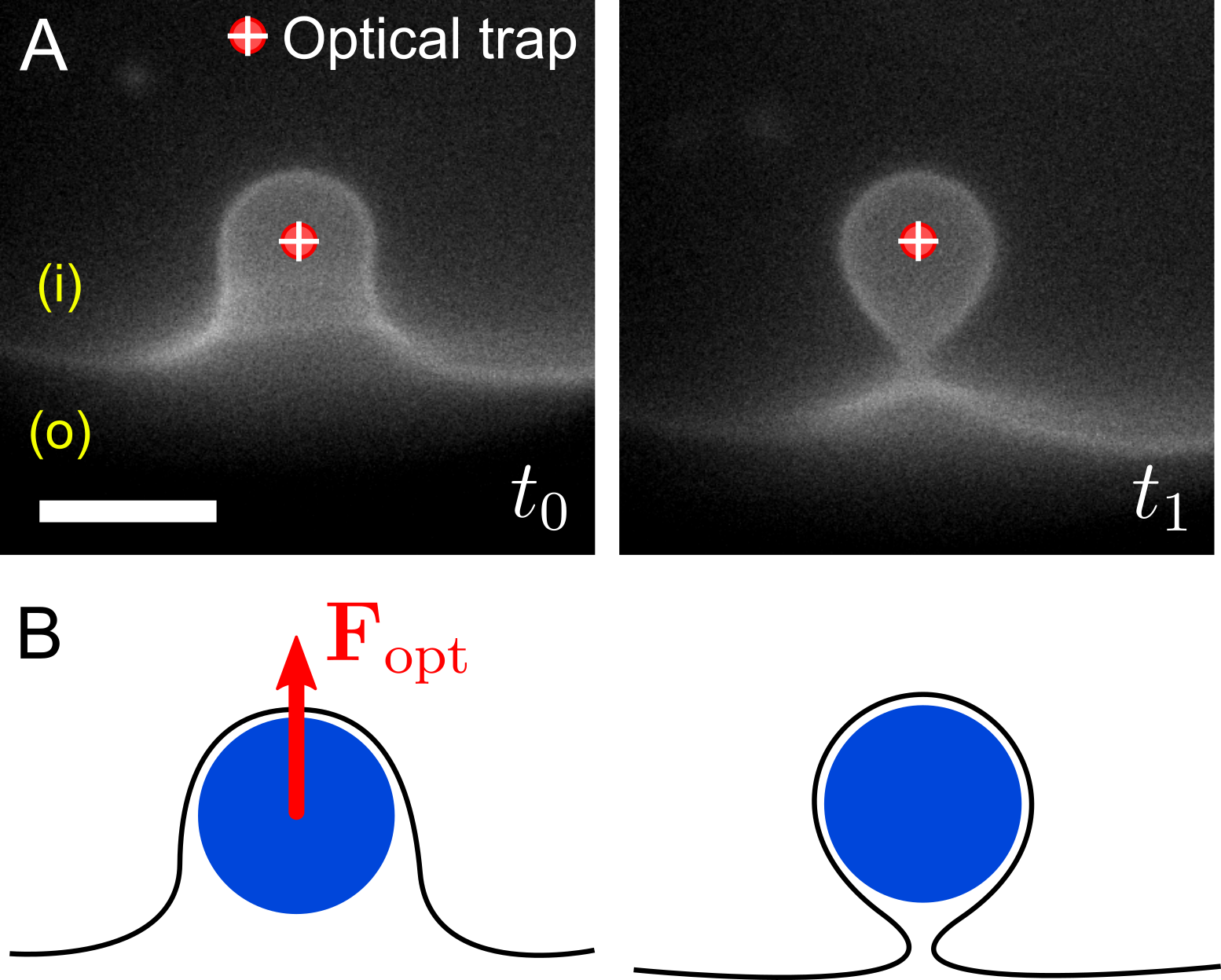
To experimentally investigate the energetics of wrapping-induced membrane necks, we study a model system consisting of a floppy () POPC giant unilamellar vesicle (enclosing a sucrose solution and suspended in a glucose solution) and a spherical SiO2 microparticle. By imposing a relative motion between an optically trapped SiO2 microsphere and a floppy GUV, we can bring the particle from a non-wrapping state to a stable full-wrapping state thanks to the force of the optical tweezers deforming the membrane [22], as shown in Figure 1 for a particle (see also Supp. movie S1_MOV.avi). The fluorescence microscopy snapshots in Figure 1A together with the schematic representation in Figure 1B allow to visualize the neck closure process leading to the nucleation of an inward-pointing membrane bud containing the particle. This structure is always stable on the timescale of an experiment (up to several hours) even when the optical trap is switched off.
In contrast with adhesion-driven particle wrapping processes, the stability of the final full wrapping state in the forced particle wrapping experiment reported here cannot be understood considering only a competition between local membrane deformations (associated to the membrane segment bound to the particle) and adhesion [13, 14]. If very weakly attractive particle-membrane interactions can exist in the case of SiO2 and POPC lipids, typical values for the adhesive energy per unit area are expected to be small with [36, 20]. It is therefore readily evident that the energetic costs of local deformations induced by a spherical particle in the final fully wrapped state [6] outweigh the negative (favorable) adhesion energy contribution. This holds even in a low membrane tension regime and considering the existence of a weak negative membrane spontaneous curvature (), which is expected in our system due to the glucose/sucrose asymmetry across the membrane [22, 20]. Indeed, we have:
| (1) |
where is the bending rigidity, the membrane spontaneous curvature and corresponds to the excess membrane area that was pulled to the wrapping site and contributing to the adhesion free energy in the full engulfment state. This area exactly corresponds to the particle surface area considering the ideal case of a perfectly spherical final bud matching the particle size connected to a flat membrane by an infinitesimally small neck. In reality, the radius of the membrane bud enclosing the particle depends on the water gap separating the particle and the membrane, which is expected to be at least several tens of nanometers for nonadhesive particles. Still, Eq. 1 predicts an unfavorable full wrapping state, due to the interplay between bound membrane segment deformations and adhesion. Hence, the total free energy change between the non-wrapping and full wrapping states. It follows that the observed metastability should be provided by a local minimum of the full membrane shape energy. Bistability between non-wrapping and fully wrapped states was indeed predicted in the presence of a negative spontaneous curvature by full membrane shape energy calculations [37]. The bistability then results from the energetically costly shapes adopted by the unbound membrane segment for intermediate wrapping states, leading to an energy barrier for wrapping. This barrier is overcome by supplying external energy through optical tweezers, enabling the system to reach a metastable fully wrapped state.
Within the framework of spontaneous curvature elasticity theory, stability relations for membrane necks were derived by parameterizing the generic shape of vesicle-bud complexes as two hemispheres connected by two unduloid-shaped segments forming a neck of radius . It can be shown that the bending energy of such a shape for a membrane with a spontaneous curvature has the form [38, 10, 39]:
| (2) |
where is the energy associated to the shape far from the neck and the so-called closed neck curvature defined from the mean curvatures of adjacent segments of the mother vesicle and membrane bud with curvature radii and , respectively. It follows that for the neck structure to be stable, the free energy must increase with increasing , leading to the stability criterion [38]:
| (3) |
Calculating for our case in the approximation m and considering a size dispersion of the vesicles yields . Eq. 3 in turn implies as a lower bound for the spontaneous curvature absolute value in our system for it to be able to stabilize the formed neck. These values are consistent with spontaneous curvature values inferred in our experimental system [22] arising from a glucose/sucrose asymmetry across the lipid membrane [32]. The constrictions force at the neck are apparently not large enough to lead to spontaneous cleavage of the neck, contrarily to what was recently observed in the case of vesicles exposed to His-tagged proteins [40]. Note that while this stability criterion seems to agree with our observations of neck stability for floppy vesicles, it does not take into account any influence of the membrane tension . Indeed, the latter could have a dramatic effect on the stability of neck, leading either to a re-opening or to a cleavage of the neck (fission). The influence of tension will be investigated in the next section.
III.2 NECK OPENING
Starting from the fully wrapped state, we aim at investigating the stability of the neck structure, thereby exploring whether particle endocytosis or particle unwrapping can be triggered.
By optical tweezers, further forcing the particle towards the GUV center of mass leads to the nucleation of a membrane tube which never undergoes fission to complete particle endocytosis [22]. Inverting the relative motion direction between the wrapped particle and the GUV (distance between the particle and GUV center of mass is being increased), the particle escapes from the optical trap without membrane unwrapping in the accessible range of optical forces of our setup (see Supp. movie S1_MOV.avi). This suggests that the neck formation introduces, if not an irreversibility, a hysteresis in the wrapping-unwrapping process.
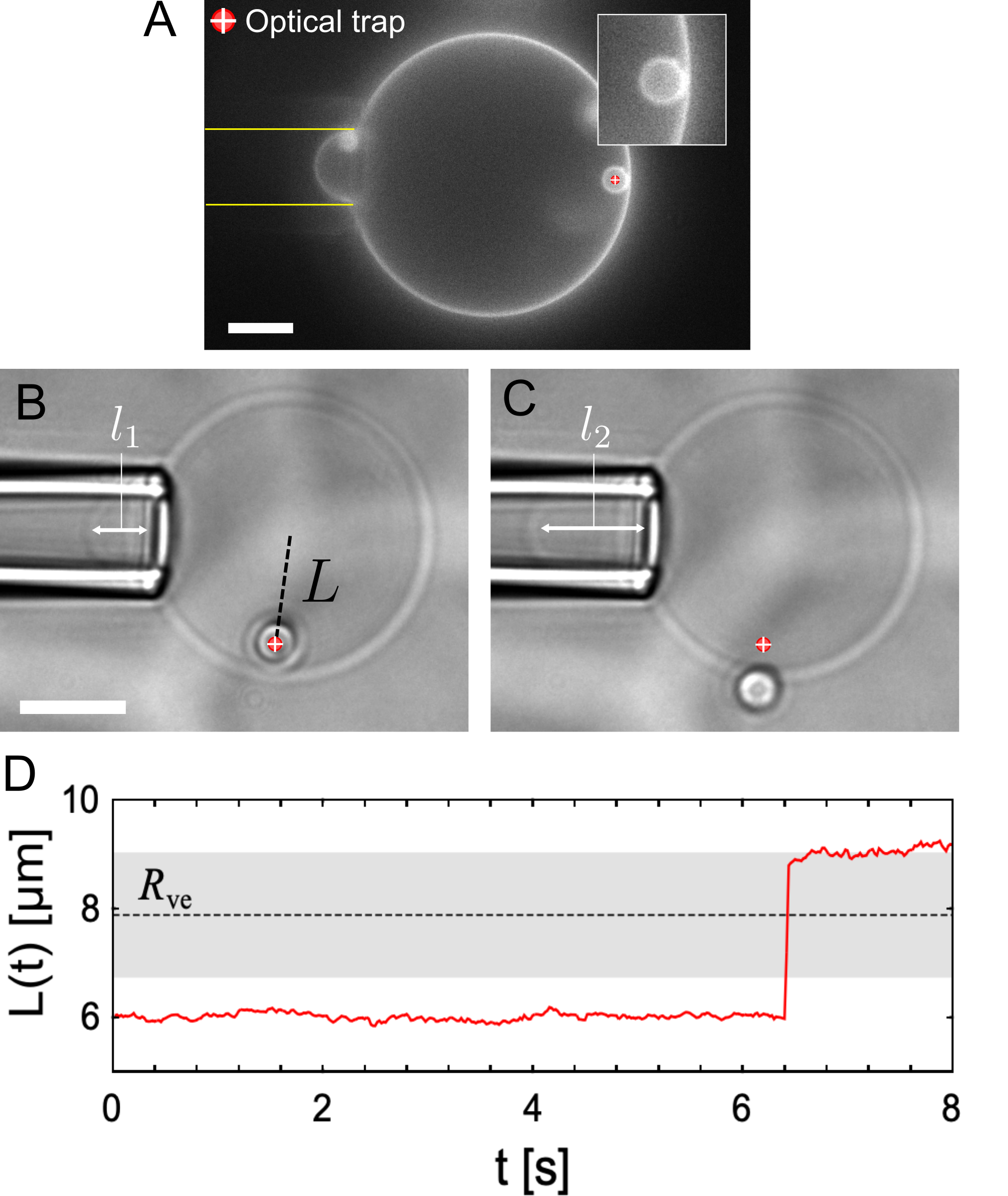
Next, we use a micropipette to probe the influence of membrane tension on the neck by putting under tension the vesicle in which a particle was wrapped by the membrane. A fluorescent microscopy image of a typical experiment is shown in Figure2A, where the wrapped particle is optically trapped with weak trapping stiffness at the vesicle equator to image both the particle and the aspirated membrane segment. The experimental protocol involves stepwise increments of the pressure difference in the micropipette using a hydrostatic device, which translates into membrane tension through Laplace’s law. At each pressure step (i.e., membrane tension increase), we allow 1 minute for equilibration. In the meantime, the particle is maintained at the vesicle equator using a weak optical trap. There are two possible outcomes for such experiments. The first is depicted in Figure 2B-C (and can be seen in Supp. movie S2_MOV.avi), where the neck opens at a critical tension , resulting in a sudden radial displacement of the particle from the intravesicular space to the exterior. This displacement is evidenced by a stepwise increase in the particle-GUV center-of-mass distance , as shown in the plot in Figure 2D, transitioning from to in less than 100 ms. This corresponds to a lower bound for the particle escape velocity . Simultaneously, an apparent area increase is observed, corresponding to the increase in length of the cylindrical portion of the aspirated tongue (see Figure2B-C for tongue lengths and definition). This area increase represents the surface area that previously wrapped the engulfed particle, confirming the opening of the membrane neck and the particle’s release. We verify this by noting that . Such an event is referred to as a neck opening event, triggered by an increase in membrane tension.
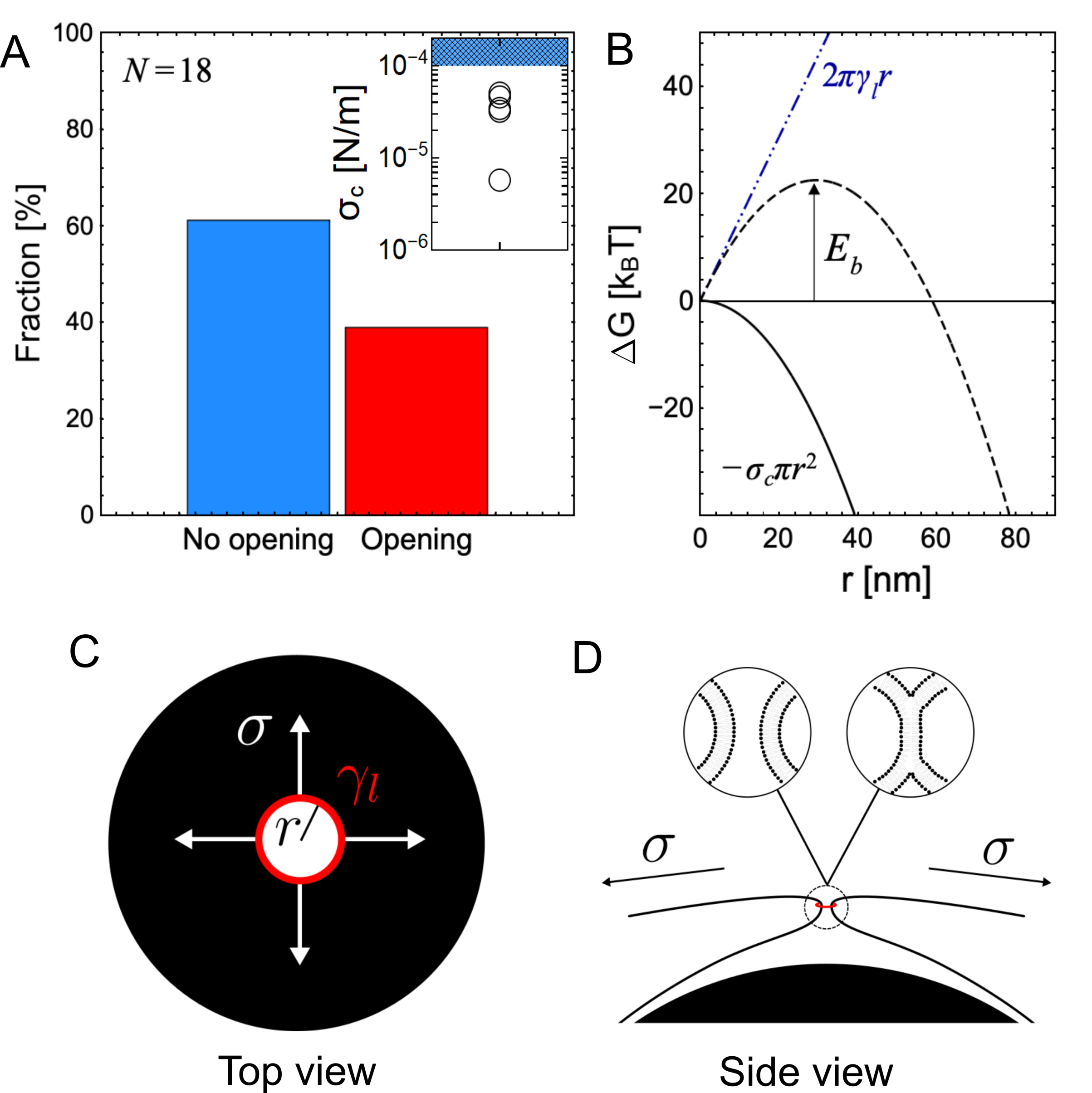
It occurs % of the observed cases. A critical tension could be measured, and the mean value is reported in the inset of Figure 3A and reads .
The other outcome in our experiments is that the particle remains wrapped and no neck opening event occur in the range of pressure our setup allows to explore Pa, i.e. . Data on the neck opening statistics for experiments are shown in Figure 3A, showing that in more than half of the cases ( 61 %), we could not observe an opening of the neck for the range of tensions accessible, pointing to a critical tension for neck opening .
We note here that the stability criterion given by Eq.3 does not account for any influence of the membrane tension . However, we just demonstrated experimentally that increasing the tension can in some cases lead to an unstable neck structure. In order to account for the contribution of tension, we make an analogy between our geometry of wrapped particle and treat the opening of the neck analogously to the opening of a pore, where the membrane tension promotes the opening such that the change in total energy of the neck as a function of the neck radius is plotted in Fig 3B and reads [41]:
| (4) |
where resists the wrapping and originates from Eq. 2 where is a line tension analogous to the one defined for pores, see schematics in Figure 3C-D. From Eq. 4, the competition between the tension and bending energies lead to the existence of an energy barrier for neck opening . In our experiments when an opening was reported, the opening was spontaneous at the critical tension, meaning it was activated only by thermal energy such that , see plot in Figure 3B. Following this reasoning, we obtain pN and not too large as compared to using the experimentally measured critical tension , as shown in Figure 3B. pN implies that should be of the order of m-1 for the cases when neck opening could be triggered by the applied tensions. Using the experimental bound m-1, this yields m-1, which is in the expected range of . These considerations point to the fact that the energetics of the neck in our system is well described in some cases by a competition of tension and curvature energy. However, for the cases when a tension of was not sufficient to trigger the opening of the neck (i.e. % of the cases), it either means that the membranes in those cases possessed much larger spontaneous curvatures, or that other mechanisms than spontaneous curvature-induced constriction forces were at play. In particular, additional costs for opening are to be expected if a rearrangement of the lipids occurred at the neck, and a stalk or partial fusion of the membrane was initiated [42], as suggested by the schematics in Figure 3D.
III.3 WRAPPED PARTICLE DYNAMICS
In the fully wrapped particle state, optical microscopy techniques do not allow to visualize the neck structure, given its small size (see e.g. fluorescence microscopy image in Figure 2A). In order to gain insight on the properties of the neck, we study the dynamics of the wrapped particle, which should carry information on the neck structure and dynamics. To do so, we perform a set of particle tracking experiments on the Brownian motion of \ceSiO2 particles with three different radii (, and ) at an acquisition frequency close to 1 kHz (996 frames per second) in the free and fully wrapped situations. The so-called free case refers to the situation where a particle diffuses close to the substrate (gravitational force confines the particle in a 2-dimensional plane close to the solid surface) in the absence of a lipid membrane in proximity, whereas the wrapped case refers to the situation where the particle was previously fully wrapped in a lipid vesicle with optical tweezers, as described in Sec. III.1. Representative trajectories for free (in red) and wrapped (in blue) particles are shown in Figure 4A-B, with a total duration of the trajectory of s ( points).
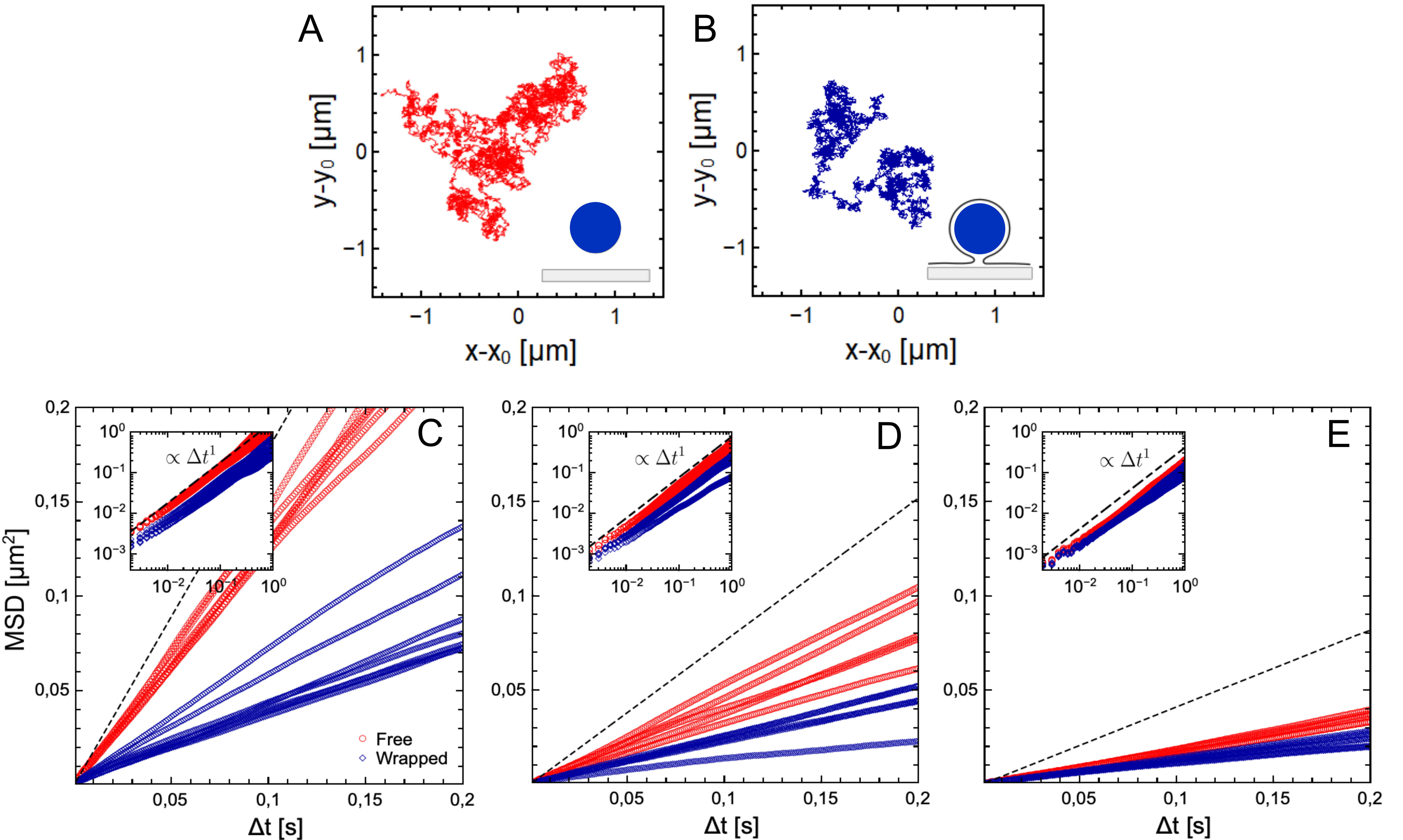
We plot the experimental two-dimensional mean squared displacement (MSD = ) curves associated to several wrapped and free trajectories ( trajectories for each scenario) in Figure 4C-E. It appears that for all particle sizes, the MSD shows a purely diffusive behavior MSD for s, as shown in linear and log-log scale representation in inset. This confirms that the presence of a neck or tube does not introduce any elasticity at the considered timescales and agrees with the picture of a neck diffusing in the fluid lipid bilayer which only contributes as an additional dissipation of the thermal energy. Indeed, the diffusion appears systematically slowed down, i.e. linear MSD has a smaller slope, for the wrapped particle cases (blue diamonds) comparatively to the free cases (red circle), for all particle sizes.
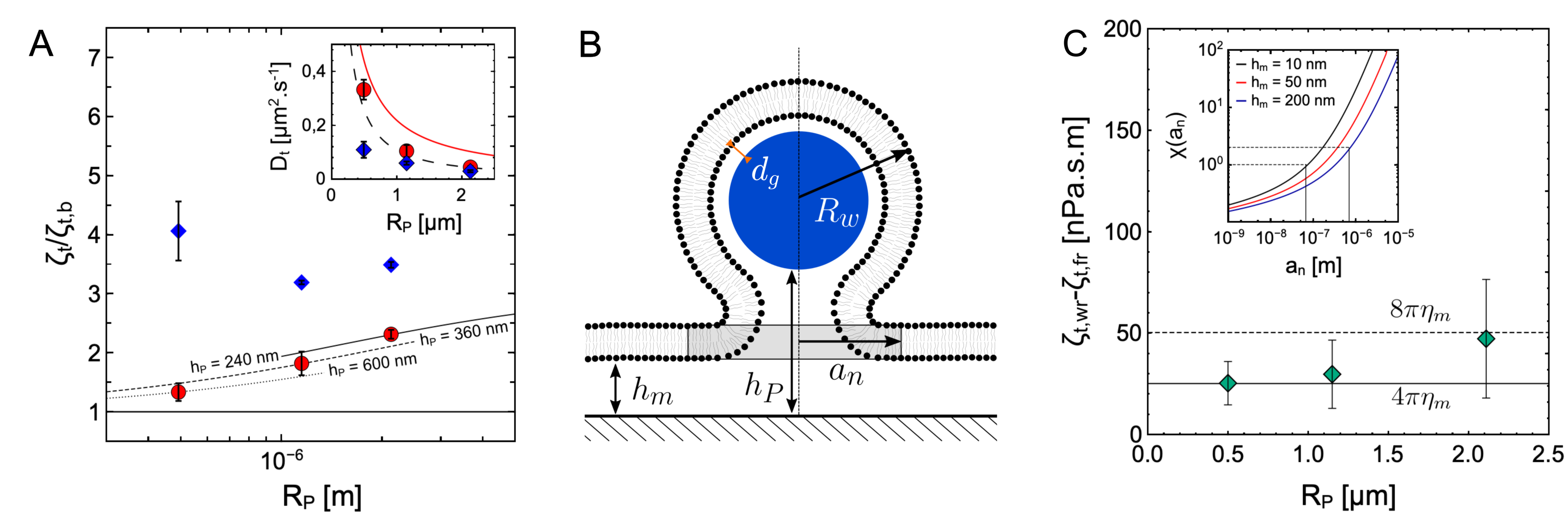
To quantify this slowed-down diffusion, we extract the translational friction coefficient from individual trajectories by performing linear fits of the experimental MSD curves with . We plot the average reduced friction as a function of particle size for both scenarios in Figure 5A, with the theoretical prediction for the friction coefficient in a bulk fluid from Stokes’ law. In inset of Figure 5A, the diffusion coefficients are also plotted. It appears that the experimental values of both in the free and wrapped cases are systematically greater than such that for all particle sizes. This increased friction is due to the presence of the underlying substrate imposing no-slip boundary conditions, leading to a particle-wall distance -dependent (see Figure 5B) drag increase factor such that . Far from any interface, for Stokes’ law to be recovered. In the presence of a solid wall, an expression for is given by the approximate solution to the Stokes equation derived by Faxén [43, 44]. The reduced drag for gap distances and nm, were calculated [43, 44] and plotted in Figure 5B. These reduced drags can describe the friction experienced by free particles (red disks) with radii = 600 nm), = 360 nm) and = 240 nm), respectively. Such decreasing particle-wall distances with increasing particle size in the absence of lipid membrane agree with an equilibrium particle-wall distance determined by the balance of electrostatic and gravitational forces, as discussed in Supp. A. The monotonic increase of the reduced drag with increasing for free particles is therefore a result of the monotonic decrease of the particle-wall distance with .
For the wrapped particle case (blue diamonds in Figure5A), we measure a 3 to 4-fold translational drag increase from the bulk prediction value . One can also note that in this case, is not monotonic with . To rationalize the magnitude of this drag increase, we start by accounting either (i) for a change in particle hydrodynamic radius of the wrapped particle if remains unvaried or (ii) to a change of the particle-wall distance if the radius remains the same (see Figure 5B for definition of and ). Doing so, we start by neglecting potential effects of the membrane viscosity. To describe our data, the hydrodynamic radius should strongly increase: . This result is in contrast to fluorescence microscopy images of the wrapped particles, which provide a higher bound for such that . Indeed, a small volume of fluid is enclosed in the membrane bud with the particle upon wrapping leading to the membrane bud radius being larger than the bare particle radius. However, it can be not larger than by more than a few hundreds of nanometers. Concerning the influence of the wrapping membrane on the equilibrium particle-wall distance , it is hard to determine whether the wrapping has an effect for example on the electrostatic repulsion between the particle and the substrate, and in turn on . Still, the presence of two bilayers between the particle surface and the solid wall in this geometry (see Figure 5B) allows to define a lower bound nm suggesting a higher bound value according to Fàxen and the expression for lubrication theory valid at such small gap distances [44]. Within this framework, considering the limiting scenario where and nm yields for the reduced translational drag:
| (5) |
which could be one interpretation of the drag increase matching our measurements. However, this reasoning fails to capture the non-monotonic size-dependence of with , suggesting a significant influence of the wrapping membrane and neck structure on the drag.
Now, we consider the fact that the connection between the wrapped particle and the membrane, in the form a neck, introduces an additional dissipation associated to the membrane 2-dimensional viscosity. The motion of the wrapped particle is indeed now coupled to the motion of this connecting structure within the lipid membrane, which experiences a drag expected to depend on the membrane viscosity . While no model exists accounting for our complex geometry of a wrapped particle connected to a lipid membrane moving close to a wall, we can compare our results with some existing models describing ideal situations. In particular, we model the neck structure connecting the wrapped particle to the lipid membrane as a solid cylindrical inclusion with radius and account for the dissipations associated to its translational motion in the fluid lipid bilayer.
The translational drag on a cylindrical inclusion of radius and thickness comparable to the fluid membrane in which it is embedded was modelled by Saffman and Delbrück for a membrane with two dimensional viscosity [28], and was later modified by Evans and Sackmann [45] to account for the presence of a solid substrate closeby. This last model can be used in our case to estimate the additional contribution to the drag from a neck of radius that has to diffuse with the wrapped particle in the plane of the mother vesicle membrane (see scheme Figure 5B). In the case of a thin lubricating layer of thickness between the membrane and the underlying solid substrate, the drag force exerted on the translating disk is proportional to its velocity with the friction coefficient [45]
| (6) |
where is the dimensionless radius in the lubrication approximation and and are first and zero order modified Bessel functions of the second kind. Assuming that the wrapped particle-neck complex diffuses as a single solid object, we can sum the drag exerted on the disk inclusion with the modified Stokes drag from Eq. 5, which yields:
| (7) |
Here, the second term on the right hand side is proportional to the the Boussinesq number = describing the relative importance of membrane viscosity contributions as compared to bulk viscosity, and introduces a dependency. The presence of this term could explain the larger measured drag for the smallest particle radius and the non-monotonic dependence with . Indeed, we showed that the first term on the right hand side increases monotonically with due to the increase of as the particle comes close to the substrate for larger particle size.
In order to confront these assumptions quantitatively, we plot in Figure 5C the difference of the experimentally measured drags as a function of (we subtract the measured drag value in the free case to the measured value for the wrapped case for each particle size). Doing so, we expect to isolate the contribution associated to the diffusion of the equivalent inclusion in the membrane (see Eq. 7). It appears that the difference is almost constant with a small increase with increasing radius considering the error bars, and for our range of particle size and using a typical value for POPC lipid membrane viscosity Pa.s.m [46]. In other terms, our results agree with an almost constant contribution of given by Eq. 6, with . Note that in our system, while we do not know the radius of the equivalent disk that has to diffuse within the membrane nor do we know the membrane distance , we can can assume and as they are tabulated properties of the bulk fluid and membrane, respectively. Hence, by plotting for reasonable values of in inset of Figure 5C, we find that implies . Such values for the equivalent disk radius are realistic, despite being larger than the expected geometrical radius of the membrane neck considered so far (e.g. in Section III.2, where a closed neck would imply nm). Other dissipative contributions could exist which could lead to the overestimation of the equivalent radius in this simple model of a particle coupled to a disk inclusion in a membrane, and these contributions will be investigated in the last section.
III.4 TENSION-DEPENDENT DISSIPATION
In the previous section, particle size was used as an experimentally tunable parameter to investigate the dissipative mechanisms in the specific geometry of an engulfed colloid connected to a floppy vesicle. Here, we explore the influence of a membrane property that already proved to have an influence on the neck in Section III.2: the membrane tension .
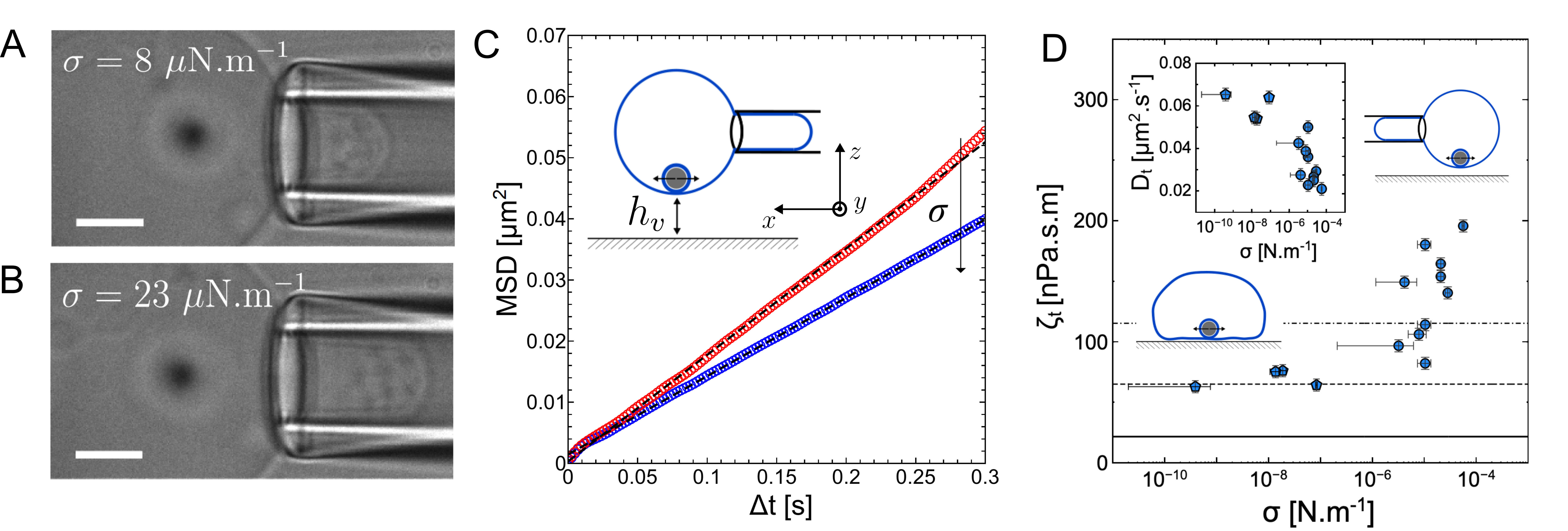
To do so, the experimental protocol again consists in performing long acquisitions at high frame rate of both the vesicle aspirated in the micropipette and the engulfed microparticle, as shown in Figure 6A-B. Due to the geometry and sedimentation of the particle, it is not possible to have both the particle and the aspirated vesicle segment in focus. We choose to keep the image focus on the aspirated membrane segment as it allows to make sure that the aspiration pressure is properly applied. The particle being out of focus can still be efficiently tracked with our tracking routine, and we do not expect it to introduce inaccuracies in our measurements of the particle position. Additionnally, geometrical corrections associated to the fact that the particle diffuses on a sphere (the vesicle, see sketch in Figure 6C) and not in a plane can be disregarded as the particle only explores a small region of space close to the bottom of the vesicle , such that .
In Figure 6C, we report the one-dimensional MSD along of an engulfed particle upon increasing the membrane tension, corresponding to the images in Figure 6A-B. In a single acquisition of 30000 images acquired at 419 fps, the tension applied with the micropipette is increased step-wise from to . The red curve shows the MSD for the particle motion during the first 10000 frames and the blue curve for the last 10000 frames (the motion is not analyzed during 10000-20000 frames as it corresponds to the time for the membrane to reach the higher equilibrium tension). It clearly appears that the slope of the MSD curve for lower tension is larger than for the higher tension case. Linear fits up to s allow to extract and for the low and high membrane tension cases, respectively. MSD along is similar to the MSD along but shows oscillations arising from an external noise, see Supp. B. Such a relative increase in drag coefficient with increasing tension was systematically measured when reproducing this experiment in this geometry, also when using optical tweezers, see Supp. C.
In order to extend the range of tensions probed, we measured the diffusion coefficient from MSD curves for particles engulfed in non-stressed floppy vesicles ( N.m-1). The corresponding tension then is not the one applied with the micropipette, but the one measured from the tube force which contains information on the membrane tension. In Figure 6D, we plot the measured friction coefficient (and corresponding diffusion coefficient in inset) for these non-stressed low tension vesicles (pentagons) together with values measured from experiments where the membrane tension was controlled with the micropipette. Note that in the low tension regime (pentagons), the vesicle-substrate distance (defined in Figure 6C) can not be controlled as the vesicle is not manipulated with the micropipette. A significant contribution of the particle-wall proximity is expected, as already discussed previously. However, despite this contribution which is not present for higher tension as the vesicles are further away from the substrate, we can still observe a decrease in mobility for higher membrane tensions.
Quantitatively, the highest measured drag experienced by engulfed particles in the most tense vesicle is , which is approximately three times the drag measured for wrapped particles in floppy vesicles. Since none of the models introduced so far to interpret our drag measurements reveal an explicit dependence on the membrane tension, we will now discuss the possible origin of such high values of the measured drag for large .
Deviations from the tension independent Saffman-Delbrück model [28, 47] were recently reported [48, 49, 50, 51, 52]. In particular, the influence of membrane tension on the mobility of curvature-inducing proteins was evidenced and interpreted as an effect of the influence of tension on local protein-induced membrane deformations [50]. However, in this work, the sum of the considered contributions was shown to lead to the opposite dependency with tension, namely a decrease of the translational drag for increasing tension.
In our system, increasing tension leads to a change in the membrane deformation regimes. The bendocapillary length of the membrane varies from 3 µm at = 10-8 N.m-1 to 30 nm at = 10-4 N.m-1. Hence, the deformation induced by a particle with corresponds to a transition form a regime dominated by the bending to a regime dominated by the tension [50]. The radius of the disk effectively diffusing within the membrane and attached to the wrapped particle can be regarded as the immobile part of the membrane subjected to significant curvature changes due to either the wrapping on the particle or to the formation of the neck.
Using the same analysis as in the previous section, a value of the disk radius gives a value of yielding the value of measured on tense vesicles (Figure 6D).
As before, such large disk radius comparable to the particle radius is not only related to the neck size but to the immobile part of the membrane which is connected to the particle in its dynamics.
In our geometry, a tension increase translates into a decrease of the distance between the mother vesicle membrane and the lipid bilayer wrapping the membrane. Indeed, for the low membrane tension regime, the interbilayer distance is expected to be large due to large membrane fluctuations and associated steric repulsion. A reasonable lower bound approximation for the interbilayer distance in this case would be the amplitude of the bilayer undulations at low tension nm [53]. Higher membrane tension leads to suppression of these small wavenumber thermal undulations. In the absence of other repulsive forces such as electrostatics (lipids are zwitterionic), the interbilayer distance for high membrane tension can reach values as small as a few nanometers, until the hydration force prevents further thinning of the water gap. When interbilayer distance becomes small, shearing of the mother membrane upon translational motion of the wrapped particle may occur far from the neck location and starts only at relatively large interbilayer distances. However, the magnitude of these contributions in the case of a microparticle close to a tense membrane were reported to be 8% greater at most from the bulk value at smallest accessible distances from the membrane using photonic force microscopy [54].
It is worth noting that the high drag values measured for large membrane tensions correspond to cases where the neck did not open (no opening cases), suggesting a higher spontaneous curvature of the membrane, as indicated by the results in section III.2. While no existing model explicitly predicts a dependence of membrane spontaneous curvature on the drag experienced by inclusions, its influence cannot be ruled out. Spontaneous curvature may directly affect either the size of the equivalent disk associated with the translating particle, which interacts with the membrane, or the membrane’s effective viscosity.
Finally, an alternative interpretation of the high particle drag experienced at high tensions can be made considering that the leaflets constituting the membrane start to slide past each other due to the strongly curved membrane segments at the neck. This results into a intermonolayer friction contribution that becomes significant [50]. The intermonolayer friction coefficient in POPC membranes was measured to be of the order of Pa s m-1 [55, 56]. Typical associated friction contributions in our case of a membrane neck are expected to be of the order of where nm2 is a typical value of the contributing membrane area. Although it is not evident on which lengthscales such dissipation modes would extend, it appears that, taking the typical area associated to the neck nm2 leads to a friction that could explain our friction data at high membrane tension Figure 6D.
IV CONCLUSION
We demonstrated that nonadhesive microparticles can achieve stable wrapping by low-tension vesicle membranes under an external force, provided the membranes exhibit a small negative spontaneous curvature. The stability of the neck structure, crucial for maintaining the fully wrapped state in floppy GUVs, was explained using spontaneous curvature elasticity models. Through micropipette experiments, we assessed the reversibility of the force-driven wrapping process and quantified the membrane tension required to reopen the neck. The observed variability in this tension likely stems from differences in spontaneous membrane curvature or lipid reorganization at the neck. Furthermore, we revealed that wrapped particles experience additional dissipation due to the coupling between their motion and the neck structure within the bilayer. Comparing experimental data for different particle sizes with theoretical models highlighted dissipative mechanisms tied to neck translation within the fluid membrane. Notably, we identified a strong dependence of particle mobility on the membrane’s tension. This contrasts with prior reports for curvature-inducing proteins, which showed decreasing drag with increasing tension. Some potential dissipation pathways were identified, including effects involving intermonolayer friction. However, the results highlight the need for further theoretical and numerical efforts to fully account for this specific geometry. These findings offer key insights into the physical principles governing the wrapping of particles by lipid vesicles or cells under directional forces or particle self-propulsion.
Acknowledgements.
We acknowledge fundings from the Ecole Doctorale de Physique et Chimie-Physique from Université de Strasbourg and ANR EDEM (ANR-21-CE06-0042), and Andre Schroder for the help in implementing the micropipette setup.Supporting material
Movies S1_MOV.avi, S2_MOV.avi as well as a document containing 3 figures constitute the supplementary material of this work.
Author contributions
F.F.: conceptualization, data curation, formal analysis, investigation, methodology, writing – original draft, writing – review & editing. P.M.: investigation, methodology, writing – review & editing. A. S.: conceptualization, funding acquisition, investigation, methodology, project administration, resources, validation, writing – review & editing.
References
- Alberts et al. [2022] B. Alberts, R. Heald, A. D. Johnson, D. Morgan, M. Raff, K. Roberts, and P. Walter, Molecular Biology of the Cell, 7th ed. (W. W. Norton & Company, New York, NY, 2022).
- Bonifacino and Glick [2004] J. S. Bonifacino and B. S. Glick, The mechanisms of vesicle budding and fusion, cell 116, 153 (2004).
- Garoff et al. [1998] H. Garoff, R. Hewson, and D.-J. E. Opstelten, Virus maturation by budding, Microbiology and Molecular Biology Reviews 62, 1171 (1998).
- Schmid [1997] S. L. Schmid, Clathrin-coated vesicle formation and protein sorting:an integrated process, Annual review of biochemistry 66, 511 (1997).
- Kozlovsky and Kozlov [2003] Y. Kozlovsky and M. M. Kozlov, Membrane fission: Model for intermediate structures, Biophysical journal 85, 85 (2003).
- Deserno [2004] M. Deserno, Elastic deformation of a fluid membrane upon colloid binding, Physical Review E - Statistical, Nonlinear, and Soft Matter Physics 69, 10.1103/PhysRevE.69.031903 (2004).
- Bahrami et al. [2014] A. H. Bahrami, M. Raatz, J. Agudo-Canalejo, R. Michel, E. M. Curtis, C. K. Hall, M. Gradzielski, R. Lipowsky, and T. R. Weikl, Wrapping of nanoparticles by membranes, Advances in Colloid and Interface Science 208, 214 (2014).
- Bahrami et al. [2016] A. H. Bahrami, R. Lipowsky, and T. R. Weikl, The role of membrane curvature for the wrapping of nanoparticles, Soft Matter 12, 581 (2016).
- Lipowsky [2012] R. Lipowsky, Spontaneous tubulation of membranes and vesicles reveals membrane tension generated by spontaneous curvature, Faraday Discussions 161, 305 (2012).
- Agudo-Canalejo and Lipowsky [2015] J. Agudo-Canalejo and R. Lipowsky, Critical particle sizes for the engulfment of nanoparticles by membranes and vesicles with bilayer asymmetry, ACS Nano 9, 3704 (2015).
- Yu et al. [2018] Q. Yu, S. Othman, S. Dasgupta, T. Auth, and G. Gompper, Nanoparticle wrapping at small non-spherical vesicles: curvatures at play, Nanoscale 10, 6445 (2018).
- Lipowsky and Döbereiner [1998] R. Lipowsky and H.-G. Döbereiner, Vesicles in contact with nanoparticles and colloids, Europhysics Letters (EPL) 43, 219 (1998).
- Spanke et al. [2020] H. T. Spanke, R. W. Style, C. François-Martin, M. Feofilova, M. Eisentraut, H. Kress, J. Agudo-Canalejo, and E. R. Dufresne, Wrapping of microparticles by floppy lipid vesicles, Phys. Rev. Lett. 10.1103/PhysRevLett.125.198102 (2020).
- Spanke et al. [2022] H. T. Spanke, J. Agudo-Canalejo, D. Tran, R. W. Style, and E. R. Dufresne, Dynamics of spontaneous wrapping of microparticles by floppy lipid membranes, Phys. Rev. Res. 4, 023080 (2022).
- van der Ham et al. [2024] S. van der Ham, J. Agudo-Canalejo, and H. R. Vutukuri, Role of shape in particle-lipid membrane interactions: From surfing to full engulfment, ACS Nano 18, 10407 (2024).
- Weddle and Agaisse [2018] E. Weddle and H. Agaisse, Principles of intracellular bacterial pathogen spread from cell to cell, PLOS Pathogens 14, 1 (2018).
- Robbins et al. [1999] J. R. Robbins, A. I. Barth, H. Marquis, E. L. de Hostos, W. J. Nelson, and J. A. Theriot, Listeria monocytogenes Exploits Normal Host Cell Processes to Spread from Cell to Cell, Journal of Cell Biology 146, 1333 (1999).
- Xiao et al. [2022] K. Xiao, R. Ma, and C.-X. Wu, Force-induced wrapping phase transition in activated cellular uptake, Physical Review E 106, 10.1103/physreve.106.044411 (2022).
- Vutukuri et al. [2020] H. R. Vutukuri, M. Hoore, C. Abaurrea-Velasco, L. van Buren, A. Dutto, T. Auth, D. A. Fedosov, G. Gompper, and J. Vermant, Active particles induce large shape deformations in giant lipid vesicles, Nature 586, 52 (2020).
- Fessler et al. [2024] F. Fessler, M. Wittmann, J. Simmchen, and A. Stocco, Autonomous engulfment of active colloids by giant lipid vesicles, Soft Matter 20, 5904 (2024).
- Meinel et al. [2014] A. Meinel, B. Tränkle, W. Römer, and A. Rohrbach, Induced phagocytic particle uptake into a giant unilamellar vesicle, Soft Matter 10, 3667 (2014).
- Fessler et al. [2023] F. Fessler, V. Sharma, P. Muller, and A. Stocco, Entry of microparticles into giant lipid vesicles by optical tweezers, Phys. Rev. E 107, L052601 (2023).
- Velikov et al. [1997] K. Velikov, C. Dietrich, A. Hadjiisky, K. Danov, and B. Pouligny, Motion of a massive microsphere bound to a spherical vesicle, Europhysics Letters 40, 405 (1997).
- Dimova et al. [1999] R. Dimova, C. Dietrich, A. Hadjiisky, K. Danov, and B. Pouligny, Falling ball viscosimetry of giant vesicle membranes: Finite-size effects, Eur. Phys. J. B 12, 589 (1999).
- Shigyou et al. [2016] K. Shigyou, K. H. Nagai, and T. Hamada, Lateral diffusion of a submicrometer particle on a lipid bilayer membrane, Langmuir 32, 13771 (2016).
- van der Wel et al. [2017] C. van der Wel, D. Heinrich, and D. J. Kraft, Microparticle assembly pathways on lipid membranes, Biophysical Journal 113, 1037 (2017).
- Marque et al. [2025] C. Marque, G. D’Avino, D. Larobina, A. Michel, A. Abou-Hassan, and A. Stocco, Diffusion of a single colloid on the surface of a giant vesicle and a droplet, Phys. Rev. E 111, 025411 (2025).
- Saffman and Delbrück [1975] P. G. Saffman and M. Delbrück, Brownian motion in biological membranes, Proceedings of the National Academy of Sciences 72, 3111 (1975).
- Danov et al. [1995] K. Danov, R. Aust, F. Durst, and U. Lange, Influence of the surface viscosity on the hydrodynamic resistance and surface diffusivity of a large brownian particle, Journal of Colloid and Interface Science 175, 36 (1995).
- Danov et al. [2000] K. D. Danov, R. Dimova, and B. Pouligny, Viscous drag of a solid sphere straddling a spherical or flat surface, Physics of fluids (1994) 12, 2711 (2000).
- Fischer et al. [2006] T. M. Fischer, P. Dhar, and P. Heinig, The viscous drag of spheres and filaments moving in membranes or monolayers, Journal of Fluid Mechanics 558, 451 (2006).
- Döbereiner et al. [1999] H. G. Döbereiner, O. Selchow, and R. Lipowsky, Spontaneous curvature of fluid vesicles induced by trans-bilayer sugar asymmetry, European Biophysics Journal 28, 174 (1999).
- Bhatia et al. [2020] T. Bhatia, S. Christ, J. Steinkühler, R. Dimova, and R. Lipowsky, Simple sugars shape giant vesicles into multispheres with many membrane necks, Soft Matter 16, 1246 (2020).
- Weinberger et al. [2013] A. Weinberger, F. C. Tsai, G. H. Koenderink, T. F. Schmidt, R. Itri, W. Meier, T. Schmatko, A. Schröder, and C. Marques, Gel-assisted formation of giant unilamellar vesicles, Biophysical Journal 105, 154 (2013).
- Community [2018] B. O. Community, Blender - a 3D modelling and rendering package, Blender Foundation, Stichting Blender Foundation, Amsterdam (2018).
- Gruhn et al. [2007] T. Gruhn, T. Franke, R. Dimova, and R. Lipowsky, Novel method for measuring the adhesion energy of vesicles, Langmuir 23, 5423 (2007).
- Agudo-Canalejo [2021] J. Agudo-Canalejo, Particle engulfment by strongly asymmetric membranes with area reservoirs, Soft Matter 17, 298 (2021).
- Lipowsky [2022] R. Lipowsky, Remodeling of membrane shape and topology by curvature elasticity and membrane tension, Advanced Biology 6, 10.1002/adbi.202101020 (2022).
- Agudo-Canalejo and Lipowsky [2016] J. Agudo-Canalejo and R. Lipowsky, Stabilization of membrane necks by adhesive particles, substrate surfaces, and constriction forces, Soft Matter 12, 8155 (2016).
- Steinkühler et al. [2020] J. Steinkühler, R. L. Knorr, Z. Zhao, T. Bhatia, S. M. Bartelt, S. Wegner, R. Dimova, and R. Lipowsky, Controlled division of cell-sized vesicles by low densities of membrane-bound proteins, Nature communications 11, 905 (2020).
- Litster [1975] J. Litster, Stability of lipid bilayers and red blood cell membranes, Physics Letters A 53, 193 (1975).
- Smirnova and Müller [2021] Y. Smirnova and M. Müller, How does curvature affect the free-energy barrier of stalk formation? small vesicles vs apposing, planar membranes, European Biophysics Journal 50, 253 (2021).
- Faxen [1923] H. Faxen, Die bewegung einer starren kugel langs der achse eines mit zaner flusigkeit gefullten rohres, Arkiv. Mat. Astron. Fys 17, 1 (1923).
- Brenner [1961] H. Brenner, The slow motion of a sphere through a viscous fluid towards a plane surface, Chemical Engineering Science 16, 242 (1961).
- Evans and Sackmann [1988] E. Evans and E. Sackmann, Translational and rotational drag coefficients for a disk moving in a liquid membrane associated with a rigid substrate, Journal of Fluid Mechanics 194, 553 (1988).
- Faizi et al. [2022] H. A. Faizi, A. Tsui, R. Dimova, and P. M. Vlahovska, Bending rigidity, capacitance, and shear viscosity of giant vesicle membranes prepared by spontaneous swelling, electroformation, gel-assisted, and phase transfer methods: A comparative study, Langmuir 38, 10548 (2022).
- Stone and Ajdari [1998] H. A. Stone and A. Ajdari, Hydrodynamics of particles embedded in a flat surfactant layer overlying a subphase of finite depth, J. Fluid Mech 369, 151 (1998).
- Domanov et al. [2011] Y. A. Domanov, S. Aimon, G. E. S. Toombes, M. Renner, F. Quemeneur, A. Triller, M. S. Turner, and P. Bassereau, Mobility in geometrically confined membranes, Proceedings of the National Academy of Sciences 108, 12605 (2011).
- Gambin et al. [2006] Y. Gambin, R. Lopez-Esparza, M. Reffay, E. Sierecki, N. S. Gov, M. Genest, R. S. Hodges, and W. Urbach, Lateral mobility of proteins in liquid membranes revisited, Proceedings of the National Academy of Sciences 103 (2006).
- Quemeneur et al. [2014] F. Quemeneur, J. K. Sigurdsson, M. Renner, P. J. Atzberger, P. Bassereau, and D. Lacoste, Shape matters in protein mobility within membranes, Proceedings of the National Academy of Sciences of the United States of America 111, 5083 (2014).
- Hughes et al. [1981] B. D. Hughes, B. A. Pailthorpe, and L. R. White, The translational and rotational drag on a cylinder moving in a membrane, Journal of fluid mechanics 110, 349 (1981).
- Naji et al. [2007] A. Naji, A. J. Levine, and P. Pincus, Corrections to the saffman-delbrück mobility for membrane bound proteins, Biophysical journal 93, L49 (2007).
- Bernard et al. [2000] A. L. Bernard, M. A. Guedeau-Boudeville, L. Jullien, and J. M. D. Meglio, Strong adhesion of giant vesicles on surfaces: dynamics and permeability, Langmuir 16, 6809 (2000).
- Jünger et al. [2015] F. Jünger, F. Kohler, A. Meinel, T. Meyer, R. Nitschke, B. Erhard, and A. Rohrbach, Measuring local viscosities near plasma membranes of living cells with photonic force microscopy, Biophysical Journal 109, 869 (2015).
- Rodríguez-García et al. [2009] R. Rodríguez-García, L. R. Arriaga, M. Mell, L. H. Moleiro, I. López-Montero, and F. Monroy, Bimodal spectrum for the curvature fluctuations of bilayer vesicles: Pure bending plus hybrid curvature-dilation modes, Phys. Rev. Lett. 102, 128101 (2009).
- Arriaga et al. [2010] L. Arriaga, R. Rodriguez-Garcia, I. López-Montero, B. Farago, T. Hellweg, and F. Monroy, Dissipative curvature fluctuations in bilayer vesicles: Coexistence of pure-bending and hybrid curvature-compression modes, The European Physical Journal E 31, 105 (2010).