Towards All-in-One Medical Image Re-Identification
Abstract
Medical image re-identification (MedReID) is under-explored so far, despite its critical applications in personalized healthcare and privacy protection. In this paper, we introduce a thorough benchmark and a unified model for this problem. First, to handle various medical modalities, we propose a novel Continuous Modality-based Parameter Adapter (ComPA). ComPA condenses medical content into a continuous modality representation and dynamically adjusts the modality-agnostic model with modality-specific parameters at runtime. This allows a single model to adaptively learn and process diverse modality data. Furthermore, we integrate medical priors into our model by aligning it with a bag of pre-trained medical foundation models, in terms of the differential features. Compared to single-image feature, modeling the inter-image difference better fits the re-identification problem, which involves discriminating multiple images. We evaluate the proposed model against 25 foundation models and 8 large multi-modal language models across 11 image datasets, demonstrating consistently superior performance. Additionally, we deploy the proposed MedReID technique to two real-world applications, i.e., history-augmented personalized diagnosis and medical privacy protection. Codes and model is available at https://github.com/tianyuan168326/All-in-One-MedReID-Pytorch.
1 Introduction
Medical images [88], such as X-ray images and Computed Tomography (CT) scans, are essential for diagnosing and monitoring various health conditions. Up to 2020, images have accounted for about 90% of all medical data [114].
Despite the large-scale data advanced the computer-aided diagnosis tasks [80, 6], its privacy concern [62] is also serious. It is urgent to (1) efficiently manage patient historical images for personalized healthcare [44, 2, 58] and (2) effectively protect privacy before images are shared [22, 41, 30]. We argue that both sides call for the medical image re-identification (MedReID) technique.
 |
As for historical image management, traditional methods [52, 68] manually pre-link images to patient metadata (e.g., name, medical record numbers), and retrieve images by querying the system with the metadata. However, the links are not always complete and accurate, especially when data are stored in different Picture Archiving and Communication System (PACS) platforms. This requires the MedReID technique to retrieve personal images from poorly organized data, providing accurate historical evidence for disease diagnosis [37].
As for medical image privacy protection, current methods only remove explicit information, such as the patient name [38]. However, some works [56, 21] have found that the identifiable visual information within the images can also breach privacy. A robust MedReID model can detect the identity-related regions of the image. By post-processing these regions, the images become unidentifiable, thereby enhancing their safety before data sharing.
Despite the importance of the MedReID problem, there are only few works investigating this. Fukuta et al. [20] and Singh et al. [67] exploit the low-level features for identifying fundus images. Packhäuser et al. [56] leverages neural networks to identify chest X-ray images. However, all these approaches are designed for one specific modality. They can not enjoy the mutual enhancement from various-modality data sources. Moreover, these models are with less medical priors, which limits their generalization.
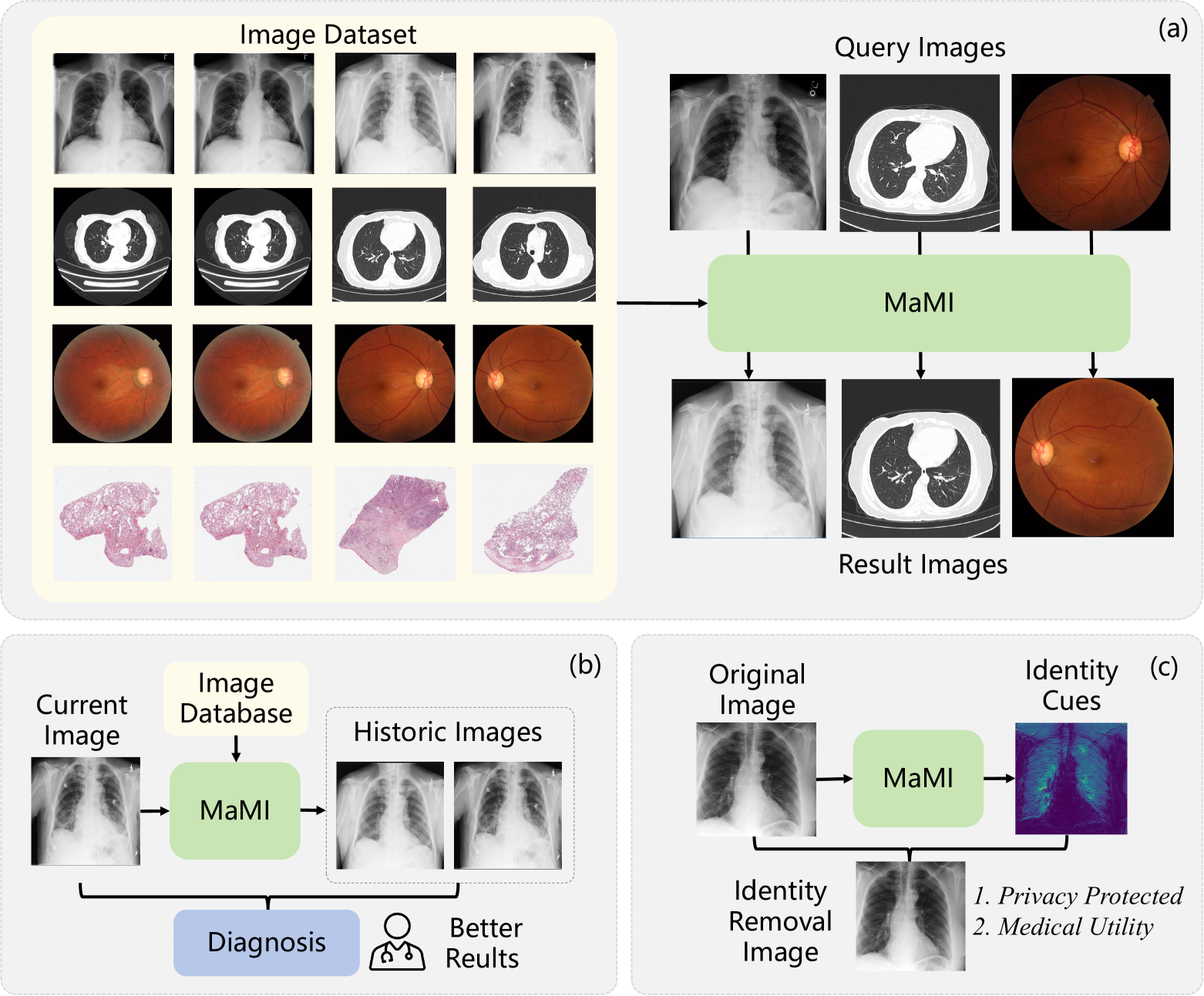
In this paper, we introduce a unified MedReID model, termed Modality-adaptive Medical Identifier (MaMI). To handle heterogeneous data from various modalities, MaMI introduces a Continuous Modality-based Parameter Adapter (ComPA). ComPA adapts a modality-agnostic model to modality-specific models at runtime. Given an input image, ComPA generates a continuous modality context, which dynamically produces modality-specific parameters. These parameters are then used to adjust the modality-agnostic model, enabling accurate re-identification of diverse medical modalities with a single model.
Furthermore, we integrate medical priors into our model by aligning it with pre-trained medical foundation models (MFMs), in terms of the inter-image key feature differences. The key features are obtained by attending to the local features using a group of learnable modality-specific query tokens. Compared to the single-image feature, the inter-image differences are more consistent with the ReID, which targets discriminating the identity relation of multiple images.
We compare our model, MaMI, against 25 foundation models and 8 large multi-modal language models across 11 medical image datasets, encompassing a wide range of modalities and body organs, establishing a thorough benchmark for the MedReID problem. Our model consistently outperforms the others. Additionally, we deploy our approach in real-world applications. First, historical data-augmented diagnosis, i.e., MaMI retrieves personalized historical patient data from unorganized datasets, significantly enhancing the accuracy of current medical examinations. Second, privacy protection, i.e., MaMI detects subtle visual cues that reveal patient identity and removes them from images before data sharing, ensuring privacy while preserving medical utility. Our contributions are:
-
•
We propose the first all-in-one medical re-identification model, termed MaMI, capable of re-identifying medical images of various modalities using a single model. We build a thorough and fair benchmark for this novel problem.
-
•
We propose a novel Continuous modality-based Parameter Adapter, which dynamically produces modality-specific parameters, and enables the model to adaptively re-identify different modalities.
-
•
Our model inherits the medical priors from medical foundation models, while adapting them to the ReID problem by inter-image difference modeling.
-
•
We showcase that MaMI can benefit real-world medical applications, e.g., history-augmented healthcare and medical privacy protection.
2 Related Work
Medical Image Re-Identification (MedReID). Numerous medical models focus on automatically diagnosing medical images [90, 6] or retrieving the images by disease features [17, 39]. There are few works focusing on the MedReID problem. Heinrich et al. [29] utilized low-level image descriptors such as Sobel [109] to detect patient identity from head CT images. Packhäuser et al. [56] and Ganz et al. [21] re-identify patients from chest X-ray and histopathology images, respectively. However, all these approaches are limited to a single modality and cannot benefit from large-scale data of various modalities.
Object Re-Identification. Most approaches [98] focus on identifying faces [110, 15, 59, 78], persons [31, 49, 27, 95, 19, 9, 84, 50, 100, 42, 33, 28], animals [64, 36], and vehicles [111, 40, 113]. However, there are few methods dedicated to medical images.
Medical Foundation Models. Early, there are amounts of dedicated models for independent tasks, such as video recognition [18, 75, 72, 76, 12, 77, 73, 70], low-level image processing [104, 106, 107, 108, 93, 99, 71, 74], and medical image analysis [7, 69, 8] tasks. Later, foundation models [43, 55] are becoming more and more popular, due to their strong generalization capability and strong performance. Recently, numerous medical foundation models, such as X-ray models [83, 97, 81], fundus image models [115][66], and CT models [25, 91], have been continuously proposed. We are the first to adapt their medical priors to the MedReID problem.
Medical Image Domain Adaptation. Medical image domain adaptation addresses domain shifts in imaging data, improving model generalization across different clinical settings [101, 23, 105]. However, these methods mainly focus on diagnosis tasks, how to devise a highly generalizable medical ReID model is left blank.
3 Approach
3.1 Overview
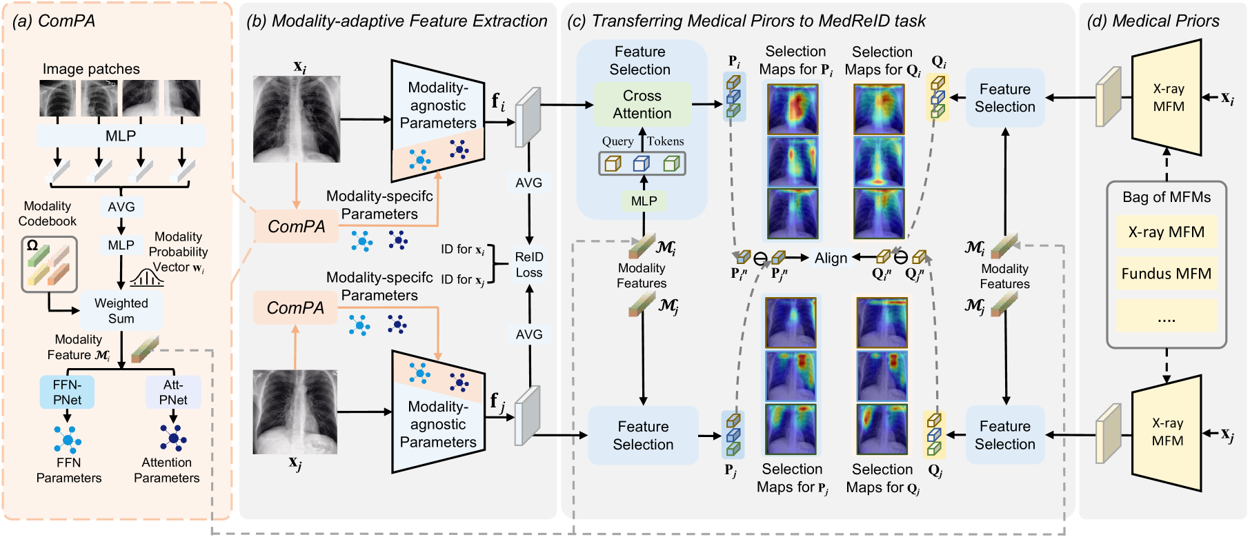
As outlined in Figure 2, we introduce two key ideas to enable a single model to identify various-modality medical images, in an all-in-one manner. First, we achieve modality-adaptive feature extraction, by upgrading a modality-agnostic model to a modality-specific model at runtime. Second, we optimize the model to focus more on medically-relevant regions, by transferring the medical priors within medical foundation models to the MedReID task.
3.2 Modality-Adaptive Feature Extraction
We leverage a typical Transformer network, ViT-Base [16], as the backbone for feature extraction. ViT consists of several attention blocks and feed-forward networks (FFNs). During runtime, we dynamically adjust the network to cater to the current input image.
Motivation. We try to fine-tune a pre-trained ViT model, namely, CLIP [63], towards the MedReID task with two strategies, (1) Single-modality, which separately fine-tunes a specialized model for each modality, and (2) Multiple-modality, which combines the data of all modalities and fine-tunes a unified model. The results are shown in Table 1. Compared to single-modality, the multiple-modality strategy shows improvement in eye fundus modality (76.88% 82.48%), while demonstrating a decrease in X-ray modality (94.21% 92.30%). This indicates that using combined data to learn a unified model benefits some modalities due to more training data, while also limiting the upper bound of some other modalities. We argue that the reason is that, naively putting multiple modalities into a single model, mostly learns the modality-agnostic knowledge, neglecting the modality-specific knowledge.
| Method | X-ray (%) | Fundus (%) |
| CLIP baseline | 33.10 | 41.14 |
| Single-modality | 94.21 | 76.88 |
| Multiple-modality | 92.30 | 82.48 |
| Continuous-modality (Ours) | 96.89 | 85.71 |
Continuous modality-based Parameter Adapter (ComPA). To address the above challenge, we propose the ComPA to amend the modality-agnostic model with input-modality-specific model parameters, as shown in Figure 2 (b). This effectively decouples the learning of modality-agnostic and modality-specific knowledge.
Rather than employing categorical modality labels, such as 0/1/2 for X-ray/Fundus/CT, ComPA introduces a novel continuous modality representation to handle the modality specificity, as shown in Figure 2 (a). Specifically, given an input image , where and denote its spatial scales, we convert each patch into local modality contexts by a Multilayer Perceptron (MLP), which are averaged to obtain the global modality context.
To improve generalization for images outside the training domain, instead of directly employing the above un-constrained modality context, we constrain the underlying modality representations to be derived from a set of basis centers. Specifically, another MLP transforms the global context into a modality probability vector , where denotes the number of all pseudo modalities. Note that this number significantly exceeds the typical number of medical modalities, such as CT and X-ray, due to the diverse imaging styles within a single modality class. For example, variations in X-ray machines and settings can result in numerous imaging styles [5]. is then used to compute a weighted sum of learnable modality bases , producing the ultimate continuous modality feature . is randomly initialized and learned with other components in an end-to-end manner.
Given , two MLPs, named Att-PNet and FFN-PNet, generate parameters for the attention and FFN layers of the ViT model, respectively. Nevertheless, directly predicting these parameters would require an infeasibly large number of parameters. For instance, for a ViT-Base model with 86M parameters, given the dimension of is 768, the last layer of the above two PNets would include M G parameters, which is intractable. To mitigate this issue, we predict low-rank parameters [32], instead of the full parameters. Meanwhile, we implement the last linear layer of two PNets in a group-wise manner [94], for further reducing the parameter number and computational cost.
The above-generated parameters are merged into the modality-agnostic network in a layer-wise manner. Following LORA [32], we expand the generated low-rank parameters to match the shape of the ViT layers and add them to the corresponding layer parameters.
Our approach shares similarities with recent Mixture-of-Expert (MOE)-LORA paradigms [92, 96], which dynamically weights a series of LORA modules. However, there are two fundamental differences. Goal Difference: We aim to perceive the input medical image modality by operating on low-level patch features, whereas MOE-LORA methods utilize high-level semantic features to select different LORAs for various semantic tasks. Mechanism Difference: MOE-LORA weights a series of LORA modules fixed in runtime, while our approach directly generates LORA parameters at runtime. This makes our approach fitting the current input image more precisely.
Feature Extraction. The input image is fed into the above merged network to produce the feature , where and denote the feature resolution. is then averaged into the global identity feature for identity comparison. For multi-slice modalities, such as CT/MRI scans, we extract feature maps from each slice in the scan and further average them as the scan-wise feature. While more advanced inter-slice operations [57][45] could be employed, we opt for the average operation to maintain the simplicity and efficiency.
3.3 Learning Rich Medical Priors from MFMs
Motivation. With the MedReID loss alone as the learning objective, the model may be biased towards the trivial textures, such as machine noises. In contrast, medical foundation models (MFMs) pre-trained on massive medical images focus on anatomical characteristics, which is more related to the patient intrinsic identity. This motivates us to transfer the rich medical priors within MFMs to our model.
Considering that local features contain more fine-grained information than global features, we use the local feature map of MFMs to guide our model. Furthermore, to close the domain gap between the pre-training task of MFMs and our MedReID task, we propose two strategies, (1) selecting the identity-related key features from the local features, (2) learning the inter-image differential features, instead of the single-image features, as shown in Figure 2 (c).
Key Feature Selection. Given the modality feature of the image , we use a three-layer MLP to map it into query tokens , where denotes the query number. The above tokens are modality-specific, enabling flexible handling of key structures in different modalities. For example, key features for Chest X-ray images include ribbon shape, heart size, and clavicle shape, while key features for fundus images include optic disc shape and vessel patterns, etc.
For the th query token , we calculate its attention map with the image feature map ,
| (1) |
where the feature dimension is 768, denotes a linear transformation. Then, attentively pools the feature map , producing the th key feature , where denote the spatial position index. For the features from the MFM, we first choose the MFM from the MFM sets, based on the modality of . Then, the th key feature is selected in a similar manner, denoted as .
Feature Difference Alignment. Considering that the MedReID task requires modeling the subtle differences between different images, we propose to align the inter-image feature difference from our model to those of MFM, instead of directly aligning singe-image feature. Given two medical images, and , after performing the above feature selection procedures, the th key features from our model are denoted as and , while those from the MFM are denoted as and . Then, we could use a simple subtraction operation to calculate the th feature differences, which are given by and , respectively, for our model and the MFM, respectively. Then, we adopt the contrastive loss to align the above features,
| (2) |
where
| (3) |
where denotes negative samples, which include non-th feature differences of the image pair (,), as well as all feature differences from other image pairs. denotes the temperature, which is set to 0.07, following MoCo [10].
3.4 Framework Training
To enable our model to discriminate the medical images from different patients, while also of rich medical priors, we adopt the following loss function:
| (4) |
where is the cross-entropy loss for patient ID classification, denotes the triplet loss with soft margin, following [27]. denotes the balancing weight.
4 Experiments
4.1 Model Details
Implementation Details. During training, we apply random flipping, random cropping, random erasing [112], and random slice sampling for data augmentation. Specifically, random flipping involves horizontal and vertical flips, while random cropping randomly crops the patches of size 224 224 from the original image. Random slice sampling denotes randomly selecting 8 slices of the CT scans. For each training batch, all images belong to the same modality. is set to 0.01. The rank number of the generated parameters is set to 16. The group number of the last linear layer of FFN-PNet and Att-PNet is set to 64. At test time, we resize the shorter side of the images to 256 and then center-crop the middle 224224 region. For multiple-slice scans, we uniformly sample 8 slices. The initial learning rate is set to 1e-5 and is gradually decayed with the cosine annealing strategy [48]. The total number of training steps is 300,000. The mini-batch size is 196 for single-image medical imaging, while 24 for multiple-slice medical sequences. We utilize the AdamW optimizer [47] implemented in PyTorch [60] with CUDA support. The values of and are set to 0.9 and 0.999, respectively. The weight decay is set to 0.05. The entire training process takes about two days on a machine equipped with four NVIDIA RTX 4090 GPUs.
4.2 Datasets
Training and Internal Validation Sets. We re-organize the public datasets with multiple images per patient, excluding those with less than two images, to ensure each patient has at least one query and target images for re-identification. The re-organized datasets include, (1) 111333 X-ray images from MIMIC-X [38]. (2) 2460 lung CT scans from CCII [102]. (3) 211 abdominal CT scans from HCC-TACE [53]. and (4) 35126 eye fundus images EyePACS (5) 6068 eye fundus images from ODIR [1]. (6) 542 histopathology images from LUAD [13]. The train/validation splitting protocols and dataset details are provided in the supplementary material.
External Validation Sets. We also evaluate our model on six external validation sets, the results of which can reflect the model’s generalization capability. (1) To build external X-ray set, we sample 6569 images of 1000 patients from Chest-X [85]. (2) To build abdominal CT set, we sample 239 CT scans of 70 patients from KIRC [3]. (3) As another abdominal CT set, we sample 194 CT scans of 56 patients from LIHC [3]. It is worth mentioning that a little proportional of LIHC contains the MRI images. (4) To build brain MRI set, we use all 55 MRI scans of 20 patients from OASIS2 [51]. (5) To build eye fundus image set, we use 700 fundus images of 350 patients from Mess2 [14]. (6) As another eye fundus image set, we use all 521 images of 144 patients from GRAPE [34].
| Method | Dataset | |||||||||||
| MIMIC-X | Chest-X | CCII | HCC-TACE | KIRC | LIHC | OASIS2 | Mess2 | ODIR | GRAPE | LUAD | ||
| X-Ray | X-Ray | Lung-CT | Ab-CT | Ab-CT | Ab-CT | Br-MRI | Fundus | Fundus | Fundus | Histo | ||
| Visual Foundation Models | ||||||||||||
| ImageNet-Sup [16] | 34.10 | 39.90 | 84.04 | 50.00 | 47.14 | 26.78 | 47.99 | 47.14 | 32.70 | 44.30 | 29.13 | |
| MoCoV3 [10] | 45.10 | 46.50 | 84.79 | 45.24 | 46.43 | 30.36 | 50.00 | 56.86 | 42.26 | 59.93 | 47.24 | |
| DINOv2 [54] | 36.40 | 37.60 | 91.52 | 50.00 | 42.86 | 28.57 | 46.00 | 36.00 | 23.72 | 41.37 | 42.52 | |
| BEITv2 [61] | 35.10 | 35.30 | 89.53 | 52.38 | 25.00 | 30.36 | 70.00 | 52.00 | 37.89 | 54.40 | 45.67 | |
| CAE [11] | 36.20 | 32.40 | 71.32 | 45.24 | 28.57 | 21.43 | 40.00 | 41.43 | 28.34 | 50.16 | 47.24 | |
| MAE [26] | 23.80 | 23.10 | 68.33 | 35.71 | 32.14 | 23.21 | 15.99 | 47.14 | 23.15 | 33.55 | 30.71 | |
| MaskFeat [89] | 9.20 | 11.60 | 19.95 | 28.57 | 17.86 | 14.29 | 10.00 | 20.00 | 8.32 | 16.94 | 14.17 | |
| MoCoV3† | 84.20 | 64.00 | 92.52 | 71.43 | 46.43 | 33.93 | 56.00 | 70.99 | 65.90 | 67.43 | 51.97 | |
| MAE† | 88.20 | 68.60 | 93.27 | 76.19 | 57.14 | 41.07 | 60.00 | 72.57 | 61.12 | 60.91 | 45.67 | |
| Visual-Language Foundation Models | ||||||||||||
| Align [35] | 0.40 | 0.90 | 43.39 | 4.76 | 17.86 | 12.50 | 0.00 | 13.71 | 3.38 | 7.82 | 5.51 | |
| BLIP [46] | 3.10 | 4.80 | 79.05 | 21.42 | 25.00 | 14.29 | 10.00 | 33.43 | 10.54 | 17.59 | 24.41 | |
| CLIP [63] | 33.10 | 31.60 | 93.02 | 45.24 | 35.71 | 28.57 | 68.00 | 41.14 | 30.15 | 50.81 | 46.46 | |
| CLIP† | 92.30 | 73.00 | 93.52 | 69.05 | 57.14 | 51.79 | 68.00 | 73.71 | 66.06 | 60.52 | 40.94 | |
| Object ReID Model | ||||||||||||
| TransReID [27] | 29.30 | 33.90 | 88.78 | 33.33 | 39.29 | 26.79 | 69.99 | 42.29 | 30.89 | 36.81 | 30.71 | |
| TransReID† | 86.80 | 68.60 | 93.52 | 80.95 | 47.14 | 39.29 | 64.00 | 74.00 | 65.52 | 60.36 | 54.33 | |
| Medical Foundation Models | ||||||||||||
| BioMedClip [103] | 25.20 | 24.00 | 82.04 | 40.48 | 32.14 | 26.79 | 32.00 | 23.14 | 19.44 | 27.68 | 33.07 | |
| RetFound [115] | 12.10 | 15.00 | 61.85 | 35.71 | 39.29 | 16.07 | 15.99 | 53.71 | 28.83 | 35.50 | 25.98 | |
| CT-CLIP [25] | 3.80 | 5.30 | 87.03 | 9.52 | 33.14 | 13.51 | 5.99 | 33.14 | 17.79 | 16.61 | 16.54 | |
| Med-Unic [81] | 48.70 | 44.90 | 77.06 | 33.33 | 32.14 | 25.00 | 23.99 | 27.71 | 21.75 | 35.83 | 28.35 | |
| BioMedClip† | 20.10 | 19.00 | 83.04 | 52.38 | 25.00 | 26.79 | 36.00 | 28.57 | 18.62 | 27.69 | 42.52 | |
| RetFound† | 54.80 | 42.80 | 92.27 | 66.67 | 28.57 | 35.71 | 50.00 | 74.14 | 66.70 | 61.10 | 37.80 | |
| CT-CLIP† | 19.70 | 19.70 | 94.04 | 47.62 | 21.43 | 28.57 | 37.99 | 29.71 | 19.93 | 29.97 | 42.52 | |
| Med-Unic† | 92.90 | 74.30 | 69.08 | 57.14 | 39.29 | 35.71 | 41.99 | 24.57 | 16.39 | 22.15 | 25.20 | |
| Modality-specialized MedReID Models | ||||||||||||
| Packhäuser et al. [56] | 92.42 | 88.21 | 68.63 | 45.24 | 35.7 | 32.11 | 36.02 | 23.74 | 15.18 | 23.77 | 29.19 | |
| Ganz et al. [21] | 11.40 | 11.90 | 53.62 | 33.33 | 39.29 | 33.93 | 28.00 | 27.43 | 22.65 | 25.08 | 56.76 | |
| All-in-One MedReID Models | ||||||||||||
| Ours | 96.89 | 91.49 | 95.01 | 88.09 | 82.68 | 76.82 | 85.00 | 85.71 | 71.34 | 71.00 | 68.75 | |
4.3 Results
MedReID Benchmark. As shown in Table 2, we evaluate various visual foundation models, visual-language foundation models, Person-ReID model, medical foundation models, and single-modality MedReID models. To fully release their potential, we fine-tune some representative models using our training datasets, ensuring a fair comparison.
For visual foundation models, contrastive learning approaches like MoCoV3 and DINOv2 achieve decent performance, with accuracies of 84.79% and 91.52% on the CCII (Lung-CT) dataset, respectively. In contrast, masked learning models such as MAE and MaskFeat perform much worse, achieving only 68.33% and 19.95% on the same dataset. However, after fine-tuning for the MedReID task, MAE† outperforms MoCoV3† on most datasets. These findings align with previous research [54, 26], i.e., contrastive features are more linearly separable when being directly deployed, while MAE-style models excel after adaptation due to their more powerful representations.
For visual-language foundation models, CLIP consistently outperforms other methods by a substantial margin, achieving 93.02% accuracy on CCII and 70.00% on OASIS2. In contrast, Align and BLIP perform much worse, with accuracies below 20% on OASIS2. These results highlight that CLIP, trained on approximately one billion image-text pairs, learns highly generalizable visual features. After further tuning, the fine-tuned CLIP† shows another significant improvement, surpassing both MoCoV3† and MAE† models by a large margin. For example, on the Chest-X dataset, CLIP†, MoCoV3†, and MAE† achieve, 73.00%, 64.00%, and 68.60%, respectively.
Person ReID method TransReID has generally produced suboptimal results when applied to medical images, largely attributed to the substantial domain gap between person images and medical images. After fine-tuning, TransReID† improves somewhat, but still lags far behind CLIP†.
For medical foundation models, BioMedClip performs much inferior to CLIP, due to the smaller training dataset PMC-15M. Specialized models like Med-UniC achieve decent performance in their training modality, such as 48.70% accuracy on X-ray images, but perform poorly on other modalities like fundus and CT. This is similar to CT-CLIP and RetFound. After fine-tuning, CT-CLIP†, RetFound†, and Med-UniC† show a further performance boost on the modalities consistent with their pre-training dataset, demonstrating that their pre-trained medical priors are beneficial for the ReID task, but perform unsatisfactorily on other modalities. For example, RetFound† achieves 74.14% on Mess2 (fundus), outperforming the strong CLIP†, but only 42.80% on Chest-X (X-ray).
Single-modality MedReID methods [56, 21] fail to generalize to the modalities out of the training scope. For instance, the X-ray ReID model [56] attains 92.42% accuracy on MIMIC-X (X-ray) but only 15.18% on ODIR (fundus). In contrast, we outperform them by a large margin, due to learning and combining identity cues from several diverse-modality training sources. Additionally, we surpass fine-tuned medical foundation models, such as RetFound† and Med-UniC†, by inheriting and adapting their medical priors to the MedReID problem. Our approach also surpasses various visual foundation models, achieving state-of-the-art performance across all modalities and datasets.
We benchmark eight large visual-language model on the medical ReID task. The results are detailed in the supplementary material. Our approach also demonstrates obvious superiority, achieving 98.80% accuracy on Chest-X, while QWen-VL-Max and GPT-4o only achieves 76.80% and 62.50%, respectively.
Finally, we study the cross-modality capability of our model. We evaluate models on a licensed private dataset of 1814 respiratory patients with paired Chest X-ray and CT images. Our all-in-one model learns to associate patient ID across modalities, achieving 87.28% accuracy (Tab. R1), outperforming single-modality-only models. This suggests the all-in-one paradigm benefits cross-modality ReID. The fine-tuning further improves the result to 94.38%.
| MaMI(X-ray only) | MaMI(CT only) | MaMI(Ours) | MaMI(Tuned) |
| 76.42% | 78.19% | 87.28% | 94.38% |
Application I: Longitudinal Personalized Healthcare. Further, we consider a realistic scenario where patients’ past medical images are not under good management. Given the current image, we use MaMI to retrieve relevant historical images and combine them with the current image for diagnosis. Notably, only the images themselves are utilized, without any historical labels. To integrate features from multiple historical images, we employ a simple MLP.
 |
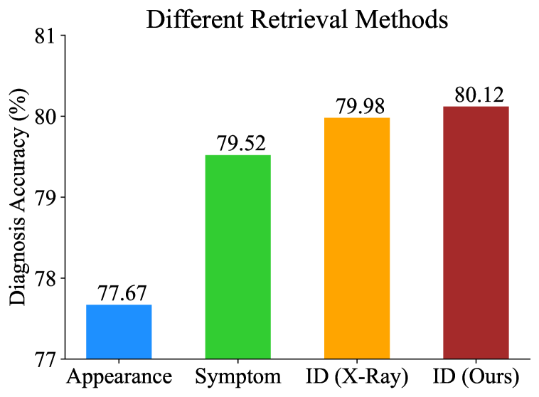 |
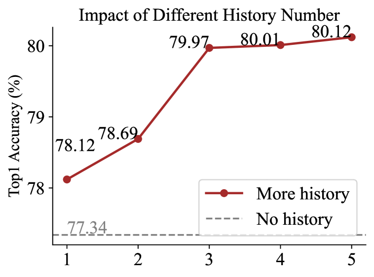
As shown in Figure 3 left, the enhancement through historical image retrieval boosts diagnostic accuracy, due to more longitudinal observations. Specifically, when retrieving five historical images, the accuracy increases from 77.34% to 80.12%, a gain of 2.78%. This demonstrates that MaMI can effectively enhance clinical utility by retrieving relevant historical data from unstructured archives. We further compare different image retrieve approaches, as shown in Figure 3 right. Our approach consistently outperforms the appearance(DINOv2 [54])-based, symptom(Med-Unic [81])-based, and X-ray-specialized ReID (Packhäuser et al. [56]) methods.
| MaMI | MAE† | CLIP† | Med-Unic† | [56] | |
| Original | 91.49% | 68.60% | 73.00% | 74.30% | 88.21% |
| Protected | 21.23% | 14.52% | 11.86% | 13.94% | 15.68% |
Application II: Privacy Protection. We adopt a simple U-Net [65] to predict identity-related visual cues and remove them from the original images. The training objective is to minimize the identity similarity distance between the identity-removed image and the original image, while maximizing their medical feature similarity. Details are in the supplementary material. The identity-removal U-Net is trained on the MIMIC-X dataset and evaluated on the Chest-X dataset. As shown in Table 4, the protected images resist re-identification attacks from various ReID models. We also train disease classification models on both the original and privacy-protected datasets. The accuracies are 81.24% and 80.67%, respectively, indicating that the privacy-protected images preserve the data utility well.
4.4 Model Analysis
Framework-level Ablation Study. As shown in Table 5, the baseline model M, which naively fine-tunes the CLIP model on our multi-modality training dataset, results in the poorest performance. Introducing the modality-adaptive component ComPA, the resulting M achieves substantial gains on various modalities, i.e., 4.31% and 11.91% gains on MIMIC-X (X-ray) and HCC-TACE (CT), due to handling inter-modality heterogeneity.
| Model | ComPA | MFMs | Internal Validation | External Validation | ||
| MIMIC-X | HCC-TACE | Chest-X | GRAPE | |||
| M | ✗ | ✗ | 92.29 | 69.04 | 86.21 | 66.51 |
| M | ✓ | ✗ | 96.60 | 80.95 | 89.35 | 69.45 |
| M | ✓ | ✓ | 96.89 | 88.09 | 91.49 | 71.00 |
Further alignment with Medical Foundation Models (MFMs) to enrich the model’s medical prior, resulting in M, yields additional performance gains, especially in data-scarce situations. On the HCC-TACE dataset, which contains only 127 training samples, performance increases from 80.95% to 88.09%. This demonstrates that MFMs mitigate the data scarcity issue common in medical imaging. On external datasets (Chest-X and GRAVE), M surpasses M by 2.14% and 1.55%, respectively. The good results on external validation datasets highlight the generalizability of features derived from MFMs.
In summary, both ComPA and MFM alignment are crucial. The ComPA improves overall performance on various modalities, while MFM alignment mitigates the data-scarcity problem and enhances generalization capability.
Ablation Study on ComPA. We further investigate if all designs within ComPA are necessary. As shown in Table 6, without considering any modality specificity, the baseline model M achieves 92.36% and 86.42% on MIMIC-X and Chest-X datasets, respectively.
| Model | Adaptive | Modality | Codebook | MIMIC-X | Chest-X |
| M | ✗ | - | - | 92.36% | 86.42% |
| M | ✗ | Discrete | - | 94.67% | 87.87% |
| M | ✓ | Continuous | ✗ | 96.78% | 90.12% |
| M | ✓ | Continuous | ✓ | 96.89% | 91.49% |
With the discrete modality labels, such as X-ray as 0, Lung CT as 1, Abdominal CT as 2, etc, as the input condition, the produced M substantially improves upon M by 1.45% on Chest-X, proving that modality information is critical for a unified MedReID model. Further introducing instance-adaptive continuous modality design, M surpasses M by another 2.11% on MIMIC-X, indicating that the continuous design better captures data nuances. The introduction of codebook design leads to further improvements, particularly on the external validation set Chest-X (+1.37%). This suggests that the codebook enhances the model’s out-of-domain generalizability.
 |
 |
| Rank | 8 | 16 | 32 |
| Chest-X | 85.56% | 91.49% | 91.50% |
| Group | 32 | 64 | 128 |
| Chest-X | 91.45% | 91.49% | 90.52% |
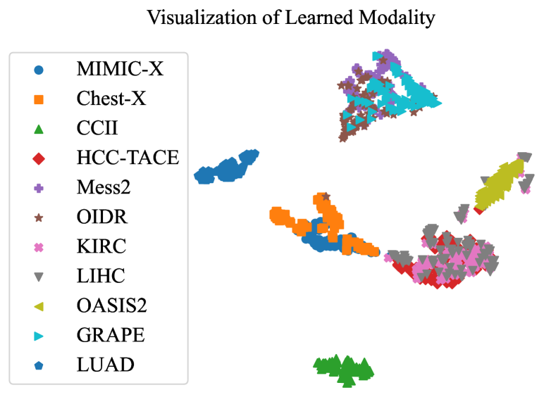
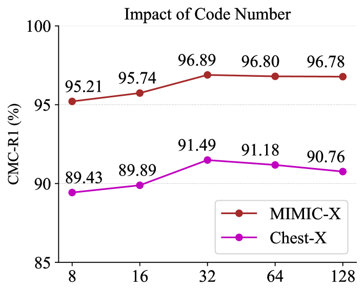
Next, we visualize the learned instance-adaptive modality features by t-SNE[79]. Figure 4 left shows a clear separation between different modalities (MIMIC-X and Mess2), while the same modality datasets (Mess2 and OIDR) cluster closely. We observe that LIHC contains some outliers, as a small proportion of LIHC cases are abdominal MRI scans instead of CT scans. Notably, our model autonomously groups OASIS2 MRI images, despite not training with the brain-MRI data, underscoring the high robustness of our modality representation. Then, we train different variant models by tuning the codebook size. As shown in Figure 4 right, a small code size such as 8 severely reduces performance on all datasets (96.89% 95.21% for MIMIC-X and 91.49% 89.43% for Chest-X), while a large codebook size such as 128 mainly degrades the model generalizability, i.e., 91.49% 90.76% on external Chest-X.
We further investigate the impact of other ComPA hyper-parameters. As shown in Table 7, a small rank constrains model representation, while ranks larger than 16 lead to performance saturation and increased computational cost. For group number, performance is stable at 32 and 64 but degrades at 128 due to much-reduced parameters of FFN- and Att-PNet. For , our model achieves very similar performance for 0.1 and 0.01 (91.32% v.s. 91.49% on Chest-X), but inferior performance 89.82% for 0.001, due to the too loose medical prior regularization.
Learning Strategy of Medical Priors. As shown in Table 8, compared to the baseline model M (no medical priors), introducing global medical priors (M) yields minimal gains, as global features fail to capture subtle identity information. Naive local priors (M) marginally surpass M by 0.23%. After the modality-specific feature selection operation, feature semantics is significantly improved, reflected by a substantial gain of 0.92%.
Replacing single-image feature alignment with inter-image feature relation alignment, where the relation feature is obtained by concatenating the features from different images and feeding them into a three-layer MLP, further boosts performance by 1.20% in M. Finally, substituting the MLP with a subtraction operation in M enforces the model’s focus on subtle image differences, achieving a final performance of 91.49%. This proves that modeling the inter-image relationship is crucial for the ReID problem, regardless of the specific relationship operator. Both the MLP and our subtraction operation achieve good results.
| Model | Feature | Inter-Image | Relation Operator | Chest-X |
| M | - | - | - | 88.54% |
| M | Global | - | - | 88.87% |
| M | Local | ✗ | - | 89.10% |
| M | Selected | ✗ | - | 90.02% |
| M | Selected | ✓ | MLP | 91.22% |
| M | Selected | ✓ | Subtraction | 91.49% |
Model Complexity. With batch size 128, the inference time of our model is 151.56 ms on a machine with an NVIDIA 4090 GPU, compared to 141.21 ms for the vanilla ViT-Base. The ComPA module only additionally consumes 10ms, as it primarily consists of several simple MLPs to compute modality-specific parameters. Given its brought substantial result gains, this minor increase in latency is justified. Further, the MFM alignment procedure incurs no inference cost, as it only regularizes the training procedure.
5 Conclusion
In this paper, we have introduced a comprehensive benchmark and a unified model for a novel MedReID problem, covering a wide range of medical modalities. We have proposed a modality-adaptive architecture to enable a single model to handle diverse medical modalities at runtime. Additionally, we integrate medical priors into our model by exploiting the pre-trained medical foundation models. Our model substantially outperforms all previous approaches.
Acknowledgment This work was supported by Shanghai Artificial Intelligence Laboratory, National Natural Science Foundation of China (Grant No.72293585, No.72293580, No.62225112), the Fundamental Research Funds for the Central Universities, National Key R&D Program of China 2021YFE0206700, Shanghai Municipal Science and Technology Major Project (2021SHZDZX0102), and STCSM 22DZ2229005.
References
- [1] Peking university international competition on ocular disease intelligent recognition (odir-2019). https://odir2019.grandchallenge.org/. Accessed: 2022-02-10.
- Aerts [2016] Hugo JWL Aerts. The potential of radiomic-based phenotyping in precision medicine: a review. JAMA oncology, 2(12):1636–1642, 2016.
- Akin et al. [2016] O Akin, P Elnajjar, M Heller, R Jarosz, BJ Erickson, S Kirk, Y Lee, MW Linehan, R Gautam, R Vikram, et al. The cancer genome atlas kidney renal clear cell carcinoma collection (tcga-kirc)(version 3). the cancer imaging archive, 2016.
- Bolle et al. [2005] Ruud M Bolle, Jonathan H Connell, Sharath Pankanti, Nalini K Ratha, and Andrew W Senior. The relation between the roc curve and the cmc. In Fourth IEEE workshop on automatic identification advanced technologies (AutoID’05), pages 15–20. IEEE, 2005.
- Bushberg and Boone [2011] Jerrold T Bushberg and John M Boone. The essential physics of medical imaging. Lippincott Williams & Wilkins, 2011.
- Cai et al. [2020] Lei Cai, Jingyang Gao, and Di Zhao. A review of the application of deep learning in medical image classification and segmentation. Annals of translational medicine, 8(11), 2020.
- Cao et al. [2022] Hu Cao, Yueyue Wang, Joy Chen, Dongsheng Jiang, Xiaopeng Zhang, Qi Tian, and Manning Wang. Swin-unet: Unet-like pure transformer for medical image segmentation. In European conference on computer vision, pages 205–218. Springer, 2022.
- Chen et al. [2024a] Haiwen Chen, Zhiyuan Qu, Yuan Tian, Ning Jiang, Yuan Qin, Jie Gao, Ruoyan Zhang, Yanning Ma, Zuolin Jin, and Guangtao Zhai. A cross-temporal multimodal fusion system based on deep learning for orthodontic monitoring. Computers in Biology and Medicine, 180:109025, 2024a.
- Chen et al. [2023] Weihua Chen, Xianzhe Xu, Jian Jia, Hao Luo, Yaohua Wang, Fan Wang, Rong Jin, and Xiuyu Sun. Beyond appearance: a semantic controllable self-supervised learning framework for human-centric visual tasks. In Proceedings of the IEEE/CVF conference on computer vision and pattern recognition, pages 15050–15061, 2023.
- Chen et al. [2021] Xinlei Chen, Saining Xie, and Kaiming He. An empirical study of training self-supervised vision transformers. In Proceedings of the IEEE/CVF international conference on computer vision, pages 9640–9649, 2021.
- Chen et al. [2024b] Xiaokang Chen, Mingyu Ding, Xiaodi Wang, Ying Xin, Shentong Mo, Yunhao Wang, Shumin Han, Ping Luo, Gang Zeng, and Jingdong Wang. Context autoencoder for self-supervised representation learning. International Journal of Computer Vision, 132(1):208–223, 2024b.
- Chen et al. [2024c] Zijian Chen, Wei Sun, Yuan Tian, Jun Jia, Zicheng Zhang, Wang Jiarui, Ru Huang, Xiongkuo Min, Guangtao Zhai, and Wenjun Zhang. Gaia: Rethinking action quality assessment for ai-generated videos. Advances in Neural Information Processing Systems, 37:40111–40144, 2024c.
- [13] National Cancer Institute Clinical Proteomic Tumor Analysis Consortium et al. The clinical proteomic tumor analysis consortium lung adenocarcinoma collection (cptac-luad)(version 12). the cancer imaging archive website.
- Decencière et al. [2014] Etienne Decencière, Xiwei Zhang, Guy Cazuguel, Bruno Lay, Béatrice Cochener, Caroline Trone, Philippe Gain, John-Richard Ordóñez-Varela, Pascale Massin, Ali Erginay, et al. Feedback on a publicly distributed image database: the messidor database. Image Analysis & Stereology, pages 231–234, 2014.
- Deng et al. [2019] Jiankang Deng, Jia Guo, Niannan Xue, and Stefanos Zafeiriou. Arcface: Additive angular margin loss for deep face recognition. In Proceedings of the IEEE/CVF conference on computer vision and pattern recognition, pages 4690–4699, 2019.
- Dosovitskiy [2020] Alexey Dosovitskiy. An image is worth 16x16 words: Transformers for image recognition at scale. arXiv preprint arXiv:2010.11929, 2020.
- Fang et al. [2021] Jiansheng Fang, Huazhu Fu, and Jiang Liu. Deep triplet hashing network for case-based medical image retrieval. Medical image analysis, 69:101981, 2021.
- Feichtenhofer et al. [2019] Christoph Feichtenhofer, Haoqi Fan, Jitendra Malik, and Kaiming He. Slowfast networks for video recognition. In Proceedings of the IEEE/CVF international conference on computer vision, pages 6202–6211, 2019.
- Fu et al. [2021] Dengpan Fu, Dongdong Chen, Jianmin Bao, Hao Yang, Lu Yuan, Lei Zhang, Houqiang Li, and Dong Chen. Unsupervised pre-training for person re-identification. In Proceedings of the IEEE/CVF conference on computer vision and pattern recognition, pages 14750–14759, 2021.
- Fukuta et al. [2008] Keisuke Fukuta, Toshiaki Nakagawa, Yoshinori Hayashi, Yuji Hatanaka, Takeshi Hara, and Hiroshi Fujita. Personal identification based on blood vessels of retinal fundus images. In Medical Imaging 2008: Image Processing, pages 630–638. SPIE, 2008.
- Ganz et al. [2025] Jonathan Ganz, Jonas Ammeling, Samir Jabari, Katharina Breininger, and Marc Aubreville. Re-identification from histopathology images. Medical Image Analysis, 99:103335, 2025.
- Giakoumaki et al. [2006] Aggeliki Giakoumaki, Sotiris Pavlopoulos, and Dimitris Koutsouris. Secure and efficient health data management through multiple watermarking on medical images. Medical and Biological Engineering and Computing, 44:619–631, 2006.
- Guan and Liu [2021] Hao Guan and Mingxia Liu. Domain adaptation for medical image analysis: a survey. IEEE Transactions on Biomedical Engineering, 69(3):1173–1185, 2021.
- Hamamci [2024] Ibrahim Ethem Hamamci. CT-CLIP: Contrastive learning of image and text representations for computed tomography. https://github.com/ibrahimethemhamamci/CT-CLIP, 2024.
- Hamamci et al. [2024] Ibrahim Ethem Hamamci, Sezgin Er, Furkan Almas, Ayse Gulnihan Simsek, Sevval Nil Esirgun, Irem Dogan, Muhammed Furkan Dasdelen, Bastian Wittmann, Enis Simsar, Mehmet Simsar, et al. A foundation model utilizing chest ct volumes and radiology reports for supervised-level zero-shot detection of abnormalities. arXiv preprint arXiv:2403.17834, 2024.
- He et al. [2022] Kaiming He, Xinlei Chen, Saining Xie, Yanghao Li, Piotr Dollár, and Ross Girshick. Masked autoencoders are scalable vision learners. In Proceedings of the IEEE/CVF conference on computer vision and pattern recognition, pages 16000–16009, 2022.
- He et al. [2021] Shuting He, Hao Luo, Pichao Wang, Fan Wang, Hao Li, and Wei Jiang. Transreid: Transformer-based object re-identification. In Proceedings of the IEEE/CVF international conference on computer vision, pages 15013–15022, 2021.
- He et al. [2024] Weizhen He, Yiheng Deng, Shixiang Tang, Qihao Chen, Qingsong Xie, Yizhou Wang, Lei Bai, Feng Zhu, Rui Zhao, Wanli Ouyang, et al. Instruct-reid: A multi-purpose person re-identification task with instructions. In Proceedings of the IEEE/CVF Conference on Computer Vision and Pattern Recognition, pages 17521–17531, 2024.
- Heinrich [2024] Andreas Heinrich. Automatic personal identification using a single ct image. European Radiology, pages 1–12, 2024.
- [30] Mattias P Heinrich and Lasse Hansen. Implicit neural compression for privacy preserving medical image sharing. In Medical Imaging with Deep Learning.
- Hermans et al. [2017] Alexander Hermans, Lucas Beyer, and Bastian Leibe. In defense of the triplet loss for person re-identification. arXiv preprint arXiv:1703.07737, 2017.
- Hu et al. [2021] Edward J Hu, Yelong Shen, Phillip Wallis, Zeyuan Allen-Zhu, Yuanzhi Li, Shean Wang, Lu Wang, and Weizhu Chen. Lora: Low-rank adaptation of large language models. arXiv preprint arXiv:2106.09685, 2021.
- Hu et al. [2024] Hezhen Hu, Xiaoyi Dong, Jianmin Bao, Dongdong Chen, Lu Yuan, Dong Chen, and Houqiang Li. Personmae: Person re-identification pre-training with masked autoencoders. IEEE Transactions on Multimedia, 2024.
- Huang et al. [2023] Xiaoling Huang, Xiangyin Kong, Ziyan Shen, Jing Ouyang, Yunxiang Li, Kai Jin, and Juan Ye. Grape: A multi-modal dataset of longitudinal follow-up visual field and fundus images for glaucoma management. Scientific Data, 10(1):520, 2023.
- Jia et al. [2021] Chao Jia, Yinfei Yang, Ye Xia, Yi-Ting Chen, Zarana Parekh, Hieu Pham, Quoc Le, Yun-Hsuan Sung, Zhen Li, and Tom Duerig. Scaling up visual and vision-language representation learning with noisy text supervision. In International conference on machine learning, pages 4904–4916. PMLR, 2021.
- Jiao et al. [2024] Bingliang Jiao, Lingqiao Liu, Liying Gao, Ruiqi Wu, Guosheng Lin, Peng Wang, and Yanning Zhang. Toward re-identifying any animal. Advances in Neural Information Processing Systems, 36, 2024.
- Jin et al. [2021] Cheng Jin, Heng Yu, Jia Ke, Peirong Ding, Yongju Yi, Xiaofeng Jiang, Xin Duan, Jinghua Tang, Daniel T Chang, Xiaojian Wu, et al. Predicting treatment response from longitudinal images using multi-task deep learning. Nature communications, 12(1):1851, 2021.
- Johnson et al. [2019] Alistair EW Johnson, Tom J Pollard, Seth J Berkowitz, Nathaniel R Greenbaum, Matthew P Lungren, Chih-ying Deng, Roger G Mark, and Steven Horng. Mimic-cxr, a de-identified publicly available database of chest radiographs with free-text reports. Scientific data, 6(1):317, 2019.
- Jush et al. [2024] Farnaz Khun Jush, Tuan Truong, Steffen Vogler, and Matthias Lenga. Medical image retrieval using pretrained embeddings. In 2024 IEEE International Symposium on Biomedical Imaging (ISBI), pages 1–5. IEEE, 2024.
- Khan and Ullah [2019] Sultan Daud Khan and Habib Ullah. A survey of advances in vision-based vehicle re-identification. Computer Vision and Image Understanding, 182:50–63, 2019.
- Kim et al. [2021] Bach Ngoc Kim, Jose Dolz, Pierre-Marc Jodoin, and Christian Desrosiers. Privacy-net: an adversarial approach for identity-obfuscated segmentation of medical images. IEEE Transactions on Medical Imaging, 40(7):1737–1749, 2021.
- Kim et al. [2023] Minsu Kim, Seungryong Kim, Jungin Park, Seongheon Park, and Kwanghoon Sohn. Partmix: Regularization strategy to learn part discovery for visible-infrared person re-identification. In Proceedings of the IEEE/CVF conference on computer vision and pattern recognition, pages 18621–18632, 2023.
- Kirillov et al. [2023] Alexander Kirillov, Eric Mintun, Nikhila Ravi, Hanzi Mao, Chloe Rolland, Laura Gustafson, Tete Xiao, Spencer Whitehead, Alexander C Berg, Wan-Yen Lo, et al. Segment anything. In Proceedings of the IEEE/CVF international conference on computer vision, pages 4015–4026, 2023.
- Lambin et al. [2017] Philippe Lambin, Ralph TH Leijenaar, Timo M Deist, Jurgen Peerlings, Evelyn EC De Jong, Janita Van Timmeren, Sebastian Sanduleanu, Ruben THM Larue, Aniek JG Even, Arthur Jochems, et al. Radiomics: the bridge between medical imaging and personalized medicine. Nature reviews Clinical oncology, 14(12):749–762, 2017.
- Li et al. [2022a] Han Li, Long Chen, Hu Han, and S Kevin Zhou. Satr: Slice attention with transformer for universal lesion detection. In International conference on medical image computing and computer-assisted intervention, pages 163–174. Springer, 2022a.
- Li et al. [2022b] Junnan Li, Dongxu Li, Caiming Xiong, and Steven Hoi. Blip: Bootstrapping language-image pre-training for unified vision-language understanding and generation. In International conference on machine learning, pages 12888–12900. PMLR, 2022b.
- Loshchilov [2017] I Loshchilov. Decoupled weight decay regularization. arXiv preprint arXiv:1711.05101, 2017.
- Loshchilov and Hutter [2016] Ilya Loshchilov and Frank Hutter. Sgdr: Stochastic gradient descent with warm restarts. arXiv preprint arXiv:1608.03983, 2016.
- Luo et al. [2019] Hao Luo, Youzhi Gu, Xingyu Liao, Shenqi Lai, and Wei Jiang. Bag of tricks and a strong baseline for deep person re-identification. In Proceedings of the IEEE/CVF conference on computer vision and pattern recognition workshops, pages 0–0, 2019.
- Luo et al. [2021] Hao Luo, Pichao Wang, Yi Xu, Feng Ding, Yanxin Zhou, Fan Wang, Hao Li, and Rong Jin. Self-supervised pre-training for transformer-based person re-identification. arXiv preprint arXiv:2111.12084, 2021.
- Marcus et al. [2010] Daniel S Marcus, Anthony F Fotenos, John G Csernansky, John C Morris, and Randy L Buckner. Open access series of imaging studies: longitudinal mri data in nondemented and demented older adults. Journal of cognitive neuroscience, 22(12):2677–2684, 2010.
- Meyer-Ebrecht [1994] Dietrich Meyer-Ebrecht. Picture archiving and communication systems (pacs) for medical application. International journal of bio-medical computing, 35(2):91–124, 1994.
- Moawad et al. [2021] A Moawad, D Fuentes, A Morshid, A Khalaf, M Elmohr, A Abusaif, JD Hazle, AO Kaseb, M Hassan, A Mahvash, et al. Multimodality annotated hcc cases with and without advanced imaging segmentation [data set]. The Cancer Imaging Archive, 2021.
- [54] Maxime Oquab, Timothée Darcet, Théo Moutakanni, Huy V Vo, Marc Szafraniec, Vasil Khalidov, Pierre Fernandez, Daniel HAZIZA, Francisco Massa, Alaaeldin El-Nouby, et al. Dinov2: Learning robust visual features without supervision. Transactions on Machine Learning Research.
- Oquab et al. [2023] Maxime Oquab, Timothée Darcet, Théo Moutakanni, Huy Vo, Marc Szafraniec, Vasil Khalidov, Pierre Fernandez, Daniel Haziza, Francisco Massa, Alaaeldin El-Nouby, et al. Dinov2: Learning robust visual features without supervision. arXiv preprint arXiv:2304.07193, 2023.
- Packhäuser et al. [2022] Kai Packhäuser, Sebastian Gündel, Nicolas Münster, Christopher Syben, Vincent Christlein, and Andreas Maier. Deep learning-based patient re-identification is able to exploit the biometric nature of medical chest x-ray data. Scientific Reports, 12(1):14851, 2022.
- Pan et al. [2022] Junting Pan, Ziyi Lin, Xiatian Zhu, Jing Shao, and Hongsheng Li. St-adapter: Parameter-efficient image-to-video transfer learning. Advances in Neural Information Processing Systems, 35:26462–26477, 2022.
- Panayides et al. [2020] Andreas S Panayides, Amir Amini, Nenad D Filipovic, Ashish Sharma, Sotirios A Tsaftaris, Alistair Young, David Foran, Nhan Do, Spyretta Golemati, Tahsin Kurc, et al. Ai in medical imaging informatics: current challenges and future directions. IEEE journal of biomedical and health informatics, 24(7):1837–1857, 2020.
- Parkhi et al. [2015] Omkar Parkhi, Andrea Vedaldi, and Andrew Zisserman. Deep face recognition. In BMVC 2015-Proceedings of the British Machine Vision Conference 2015. British Machine Vision Association, 2015.
- Paszke et al. [2019] Adam Paszke, Sam Gross, Francisco Massa, Adam Lerer, James Bradbury, Gregory Chanan, Trevor Killeen, Zeming Lin, Natalia Gimelshein, Luca Antiga, et al. Pytorch: An imperative style, high-performance deep learning library. Advances in neural information processing systems, 32, 2019.
- Peng et al. [2022] Zhiliang Peng, Li Dong, Hangbo Bao, Qixiang Ye, and Furu Wei. Beit v2: Masked image modeling with vector-quantized visual tokenizers. arXiv preprint arXiv:2208.06366, 2022.
- Price and Cohen [2019] W Nicholson Price and I Glenn Cohen. Privacy in the age of medical big data. Nature medicine, 25(1):37–43, 2019.
- Radford et al. [2021] Alec Radford, Jong Wook Kim, Chris Hallacy, Aditya Ramesh, Gabriel Goh, Sandhini Agarwal, Girish Sastry, Amanda Askell, Pamela Mishkin, Jack Clark, et al. Learning transferable visual models from natural language supervision. In International conference on machine learning, pages 8748–8763. PMLR, 2021.
- Rao et al. [2021] Yongming Rao, Guangyi Chen, Jiwen Lu, and Jie Zhou. Counterfactual attention learning for fine-grained visual categorization and re-identification. In Proceedings of the IEEE/CVF international conference on computer vision, pages 1025–1034, 2021.
- Ronneberger et al. [2015] Olaf Ronneberger, Philipp Fischer, and Thomas Brox. U-net: Convolutional networks for biomedical image segmentation. In Medical image computing and computer-assisted intervention–MICCAI 2015: 18th international conference, Munich, Germany, October 5-9, 2015, proceedings, part III 18, pages 234–241. Springer, 2015.
- Shi et al. [2024] Danli Shi, Weiyi Zhang, Jiancheng Yang, Siyu Huang, Xiaolan Chen, Mayinuer Yusufu, Kai Jin, Shan Lin, Shunming Liu, Qing Zhang, et al. Eyeclip: A visual-language foundation model for multi-modal ophthalmic image analysis. arXiv preprint arXiv:2409.06644, 2024.
- Singh et al. [2016] Anushikha Singh, Malay Kishore Dutta, and Dilip Kumar Sharma. Unique identification code for medical fundus images using blood vessel pattern for tele-ophthalmology applications. computer methods and programs in biomedicine, 135:61–75, 2016.
- Teng et al. [2010] Chia-Chi Teng, Jonathan Mitchell, Christopher Walker, Alex Swan, Cesar Davila, David Howard, and Travis Needham. A medical image archive solution in the cloud. In 2010 IEEE International Conference on Software Engineering and Service Sciences, pages 431–434. IEEE, 2010.
- Tian et al. [2019] Yuan Tian, Xiongkuo Min, Guangtao Zhai, and Zhiyong Gao. Video-based early asd detection via temporal pyramid networks. In 2019 IEEE International Conference on Multimedia and Expo (ICME), pages 272–277. IEEE, 2019.
- Tian et al. [2020] Yuan Tian, Zhaohui Che, Wenbo Bao, Guangtao Zhai, and Zhiyong Gao. Self-supervised motion representation via scattering local motion cues. In Computer Vision–ECCV 2020: 16th European Conference, Glasgow, UK, August 23–28, 2020, Proceedings, Part XIV 16, pages 71–89. Springer, 2020.
- Tian et al. [2021] Yuan Tian, Guo Lu, Xiongkuo Min, Zhaohui Che, Guangtao Zhai, Guodong Guo, and Zhiyong Gao. Self-conditioned probabilistic learning of video rescaling. In Proceedings of the IEEE/CVF international conference on computer vision, pages 4490–4499, 2021.
- Tian et al. [2022] Yuan Tian, Yichao Yan, Guangtao Zhai, Guodong Guo, and Zhiyong Gao. Ean: event adaptive network for enhanced action recognition. International Journal of Computer Vision, 130(10):2453–2471, 2022.
- Tian et al. [2023a] Yuan Tian, Guo Lu, Guangtao Zhai, and Zhiyong Gao. Non-semantics suppressed mask learning for unsupervised video semantic compression. In Proceedings of the IEEE/CVF International Conference on Computer Vision, pages 13610–13622, 2023a.
- Tian et al. [2023b] Yuan Tian, Yichao Yan, Guangtao Zhai, Li Chen, and Zhiyong Gao. Clsa: a contrastive learning framework with selective aggregation for video rescaling. IEEE Transactions on Image Processing, 32:1300–1314, 2023b.
- Tian et al. [2024a] Yuan Tian, Guo Lu, Yichao Yan, Guangtao Zhai, Li Chen, and Zhiyong Gao. A coding framework and benchmark towards low-bitrate video understanding. IEEE Transactions on Pattern Analysis and Machine Intelligence, 2024a.
- Tian et al. [2024b] Yuan Tian, Guo Lu, and Guangtao Zhai. Free-vsc: Free semantics from visual foundation models for unsupervised video semantic compression. In European Conference on Computer Vision, pages 163–183. Springer, 2024b.
- Tian et al. [2024c] Yuan Tian, Guo Lu, and Guangtao Zhai. Smc++: Masked learning of unsupervised video semantic compression. arXiv preprint arXiv:2406.04765, 2024c.
- Tian et al. [2024d] Yuan Tian, Shuo Wang, and Guangtao Zhai. Medical manifestation-aware de-identification. arXiv preprint arXiv:2412.10804, 2024d.
- Van der Maaten and Hinton [2008] Laurens Van der Maaten and Geoffrey Hinton. Visualizing data using t-sne. Journal of machine learning research, 9(11), 2008.
- Van Ginneken et al. [2011] Bram Van Ginneken, Cornelia M Schaefer-Prokop, and Mathias Prokop. Computer-aided diagnosis: how to move from the laboratory to the clinic. Radiology, 261(3):719–732, 2011.
- Wan et al. [2024a] Zhongwei Wan, Che Liu, Mi Zhang, Jie Fu, Benyou Wang, Sibo Cheng, Lei Ma, César Quilodrán-Casas, and Rossella Arcucci. Med-unic: Unifying cross-lingual medical vision-language pre-training by diminishing bias. Advances in Neural Information Processing Systems, 36, 2024a.
- Wan et al. [2024b] Zhongwei Wan, Che Liu, Mi Zhang, Jie Fu, Benyou Wang, Sibo Cheng, Lei Ma, César Quilodrán-Casas, and Rossella Arcucci. Med-unic: Unifying cross-lingual medical vision-language pre-training by diminishing bias. https://github.com/SUSTechBruce/Med-UniC, 2024b.
- Wang et al. [2022] Fuying Wang, Yuyin Zhou, Shujun Wang, Varut Vardhanabhuti, and Lequan Yu. Multi-granularity cross-modal alignment for generalized medical visual representation learning. Advances in Neural Information Processing Systems, 35:33536–33549, 2022.
- Wang et al. [2019] Guangcong Wang, Jianhuang Lai, Peigen Huang, and Xiaohua Xie. Spatial-temporal person re-identification. In Proceedings of the AAAI conference on artificial intelligence, pages 8933–8940, 2019.
- Wang et al. [2017] Xiaosong Wang, Yifan Peng, Le Lu, Zhiyong Lu, Mohammadhadi Bagheri, and Ronald M Summers. Chestx-ray8: Hospital-scale chest x-ray database and benchmarks on weakly-supervised classification and localization of common thorax diseases. In Proceedings of the IEEE conference on computer vision and pattern recognition, pages 2097–2106, 2017.
- Wang et al. [2024a] Xiyue Wang, Junhan Zhao, Eliana Marostica, Wei Yuan, Jietian Jin, Jiayu Zhang, Ruijiang Li, Hongping Tang, Kanran Wang, Yu Li, et al. A pathology foundation model for cancer diagnosis and prognosis prediction. Nature, pages 1–9, 2024a.
- Wang et al. [2024b] Xiyue Wang, Junhan Zhao, Eliana Marostica, Wei Yuan, Jietian Jin, Jiayu Zhang, Ruijiang Li, Hongping Tang, Kanran Wang, Yu Li, et al. A pathology foundation model for cancer diagnosis and prognosis prediction. https://github.com/hms-dbmi/CHIEF, 2024b.
- Webb [2022] Andrew Webb. Introduction to biomedical imaging. John Wiley & Sons, 2022.
- Wei et al. [2022] Chen Wei, Haoqi Fan, Saining Xie, Chao-Yuan Wu, Alan Yuille, and Christoph Feichtenhofer. Masked feature prediction for self-supervised visual pre-training. In Proceedings of the IEEE/CVF Conference on Computer Vision and Pattern Recognition, pages 14668–14678, 2022.
- Wei et al. [2024] Yi Wei, Meiyi Yang, Meng Zhang, Feifei Gao, Ning Zhang, Fubi Hu, Xiao Zhang, Shasha Zhang, Zixing Huang, Lifeng Xu, et al. Focal liver lesion diagnosis with deep learning and multistage ct imaging. Nature Communications, 15(1):7040, 2024.
- Wu et al. [2023] Chaoyi Wu, Xiaoman Zhang, Ya Zhang, Yanfeng Wang, and Weidi Xie. Towards generalist foundation model for radiology. arXiv preprint arXiv:2308.02463, 2023.
- [92] Xun Wu, Shaohan Huang, and Furu Wei. Mixture of lora experts. In The Twelfth International Conference on Learning Representations.
- Xie et al. [2024] Rui Xie, Chen Zhao, Kai Zhang, Zhenyu Zhang, Jun Zhou, Jian Yang, and Ying Tai. Addsr: Accelerating diffusion-based blind super-resolution with adversarial diffusion distillation. arXiv preprint arXiv:2404.01717, 2024.
- Xie et al. [2017] Saining Xie, Ross Girshick, Piotr Dollár, Zhuowen Tu, and Kaiming He. Aggregated residual transformations for deep neural networks. In Proceedings of the IEEE conference on computer vision and pattern recognition, pages 1492–1500, 2017.
- Yan et al. [2023] Shuanglin Yan, Neng Dong, Liyan Zhang, and Jinhui Tang. Clip-driven fine-grained text-image person re-identification. IEEE Transactions on Image Processing, 2023.
- Yang et al. [2024] Yuqi Yang, Peng-Tao Jiang, Qibin Hou, Hao Zhang, Jinwei Chen, and Bo Li. Multi-task dense prediction via mixture of low-rank experts. In Proceedings of the IEEE/CVF Conference on Computer Vision and Pattern Recognition, pages 27927–27937, 2024.
- Yao et al. [2024] Jingfeng Yao, Xinggang Wang, Yuehao Song, Huangxuan Zhao, Jun Ma, Yajie Chen, Wenyu Liu, and Bo Wang. Eva-x: A foundation model for general chest x-ray analysis with self-supervised learning. arXiv preprint arXiv:2405.05237, 2024.
- Ye et al. [2024] Mang Ye, Shuoyi Chen, Chenyue Li, Wei-Shi Zheng, David Crandall, and Bo Du. Transformer for object re-identification: A survey. arXiv preprint arXiv:2401.06960, 2024.
- Yi et al. [2021] Fuwang Yi, Mianyi Chen, Wei Sun, Xiongkuo Min, Yuan Tian, and Guangtao Zhai. Attention based network for no-reference ugc video quality assessment. In 2021 IEEE international conference on image processing (ICIP), pages 1414–1418. IEEE, 2021.
- Yin et al. [2020] Jiahang Yin, Ancong Wu, and Wei-Shi Zheng. Fine-grained person re-identification. International journal of computer vision, 128(6):1654–1672, 2020.
- Zeng et al. [2021] Guodong Zeng, Till D Lerch, Florian Schmaranzer, Guoyan Zheng, Jürgen Burger, Kate Gerber, Moritz Tannast, Klaus Siebenrock, and Nicolas Gerber. Semantic consistent unsupervised domain adaptation for cross-modality medical image segmentation. In Medical Image Computing and Computer Assisted Intervention–MICCAI 2021: 24th International Conference, Strasbourg, France, September 27–October 1, 2021, Proceedings, Part III 24, pages 201–210. Springer, 2021.
- Zhang et al. [2020a] Kang Zhang, Xiaohong Liu, Jun Shen, Zhihuan Li, Ye Sang, Xingwang Wu, Yunfei Zha, Wenhua Liang, Chengdi Wang, Ke Wang, et al. Clinically applicable ai system for accurate diagnosis, quantitative measurements, and prognosis of covid-19 pneumonia using computed tomography. Cell, 181(6):1423–1433, 2020a.
- Zhang et al. [2023] Sheng Zhang, Yanbo Xu, Naoto Usuyama, Hanwen Xu, Jaspreet Bagga, Robert Tinn, Sam Preston, Rajesh Rao, Mu Wei, Naveen Valluri, et al. Biomedclip: a multimodal biomedical foundation model pretrained from fifteen million scientific image-text pairs. arXiv preprint arXiv:2303.00915, 2023.
- Zhang et al. [2018] Yulun Zhang, Yapeng Tian, Yu Kong, Bineng Zhong, and Yun Fu. Residual dense network for image super-resolution. In Proceedings of the IEEE conference on computer vision and pattern recognition, pages 2472–2481, 2018.
- Zhang et al. [2020b] Yifan Zhang, Ying Wei, Qingyao Wu, Peilin Zhao, Shuaicheng Niu, Junzhou Huang, and Mingkui Tan. Collaborative unsupervised domain adaptation for medical image diagnosis. IEEE Transactions on Image Processing, 29:7834–7844, 2020b.
- Zhao et al. [2024a] Chen Zhao, Weiling Cai, Chenyu Dong, and Chengwei Hu. Wavelet-based fourier information interaction with frequency diffusion adjustment for underwater image restoration. In Proceedings of the IEEE/CVF Conference on Computer Vision and Pattern Recognition, pages 8281–8291, 2024a.
- Zhao et al. [2024b] Chen Zhao, Weiling Cai, Chengwei Hu, and Zheng Yuan. Cycle contrastive adversarial learning with structural consistency for unsupervised high-quality image deraining transformer. Neural Networks, 178:106428, 2024b.
- Zhao et al. [2024c] Chen Zhao, Wei-Ling Cai, and Zheng Yuan. Spectral normalization and dual contrastive regularization for image-to-image translation. The Visual Computer, pages 1–12, 2024c.
- Zhao et al. [2008] Sanqiang Zhao, Yongsheng Gao, and Baochang Zhang. Sobel-lbp. In 2008 15th IEEE International Conference on Image Processing, pages 2144–2147. IEEE, 2008.
- Zhao et al. [2003] Wenyi Zhao, Rama Chellappa, P Jonathon Phillips, and Azriel Rosenfeld. Face recognition: A literature survey. ACM computing surveys (CSUR), 35(4):399–458, 2003.
- Zheng et al. [2020] Zhedong Zheng, Tao Ruan, Yunchao Wei, Yi Yang, and Tao Mei. Vehiclenet: Learning robust visual representation for vehicle re-identification. IEEE Transactions on Multimedia, 23:2683–2693, 2020.
- Zhong et al. [2020] Zhun Zhong, Liang Zheng, Guoliang Kang, Shaozi Li, and Yi Yang. Random erasing data augmentation. In Proceedings of the AAAI conference on artificial intelligence, pages 13001–13008, 2020.
- Zhou et al. [2020] Qinqin Zhou, Bineng Zhong, Xiangyuan Lan, Gan Sun, Yulun Zhang, Baochang Zhang, and Rongrong Ji. Fine-grained spatial alignment model for person re-identification with focal triplet loss. IEEE Transactions on Image Processing, 29:7578–7589, 2020.
- Zhou et al. [2021] S Kevin Zhou, Hayit Greenspan, Christos Davatzikos, James S Duncan, Bram Van Ginneken, Anant Madabhushi, Jerry L Prince, Daniel Rueckert, and Ronald M Summers. A review of deep learning in medical imaging: Imaging traits, technology trends, case studies with progress highlights, and future promises. Proceedings of the IEEE, 109(5):820–838, 2021.
- Zhou et al. [2023] Yukun Zhou, Mark A Chia, Siegfried K Wagner, Murat S Ayhan, Dominic J Williamson, Robbert R Struyven, Timing Liu, Moucheng Xu, Mateo G Lozano, Peter Woodward-Court, et al. A foundation model for generalizable disease detection from retinal images. Nature, 622(7981):156–163, 2023.