Beyond H&E: Unlocking Pathological Insights with Polarization via Self-supervised Learning
Abstract
Histopathology image analysis is fundamental to digital pathology, with hematoxylin and eosin (H&E) staining as the gold standard for diagnostic and prognostic assessments. While H&E imaging effectively highlights cellular and tissue structures, it lacks sensitivity to birefringence and tissue anisotropy, which are crucial for assessing collagen organization, fiber alignment, and microstructural alterations—key indicators of tumor progression, fibrosis, and other pathological conditions. To bridge this gap, we propose PolarHE, a dual-modality fusion framework that integrates H&E with polarization imaging, leveraging the latter’s ability to enhance tissue characterization. Our approach employs a feature decomposition strategy to disentangle common and modality-specific features, ensuring effective multimodal representation learning. Through comprehensive validation, our approach significantly outperforms previous methods, achieving an accuracy of 86.70% on the Chaoyang dataset and 89.06% on the MHIST dataset. Moreover, polarization property visualization reveals distinct optical signatures of pathological tissues, highlighting its diagnostic potential. t-SNE visualizations further confirm our model effectively captures both shared and unique modality features, reinforcing the complementary nature of polarization imaging. These results demonstrate that polarization imaging is a powerful and underutilized modality in computational pathology, enriching feature representation and improving diagnostic accuracy. PolarHE establishes a promising direction for multimodal learning, paving the way for more interpretable and generalizable pathology models. Our code will be released after paper acceptance.
Keywords:
Histopathology Polarization Imaging Multimodal Fusion.1 Introduction
Histopathology image analysis is fundamental in digital pathology, with hematoxylin and eosin (H&E) staining serving as the gold standard for diagnosing and prognosticating various diseases, particularly cancer. While H&E imaging effectively highlights cellular and tissue structures, it lacks the ability to capture birefringence and tissue anisotropy, which provide critical insights into collagen organization, fiber alignment, and microstructural alterations—key indicators of tumor progression, fibrosis, and other pathological conditions [6, 26].
Polarization imaging, which leverages the fundamental properties of light, has emerged as a promising auxiliary modality for histopathological analysis. By capturing tissue birefringence, scattering properties, and depolarization effects, it provides additional contrast that reveals microstructural and biochemical changes beyond what is visible in standard H&E staining [8]. Studies have demonstrated its effectiveness in cervical intraepithelial neoplasia grading and breast carcinoma classification, enabling high-accuracy distinction of pathological features such as collagen alignment and nuclear organization [5, 6, 10]. These advantages position polarization imaging as a powerful tool for early disease detection and precise tissue characterization.
Despite its potential, polarization imaging remains underutilized in computational pathology, primarily due to the lack of established methodologies for its integration with conventional H&E-stained slides. Recent advancements in self-supervised learning (SSL) present a promising avenue for effectively incorporating multimodal data [17]. SSL frameworks, which learn meaningful representations from unlabeled data by solving pretext tasks, have shown remarkable success across various domains, including medical imaging [11, 13, 23]. By jointly learning from H&E and polarization imaging in a self-supervised manner, it becomes possible to develop robust representations that capture both shared and modality-specific features, enhancing downstream tasks such as patch classification and tissue segmentation [21].
Therefore, we propose PolarHE, a dual-modality fusion framework that integrates H&E and polarization imaging for enhanced representation learning in pathology. Our framework employs a feature decomposition strategy to disentangle common and modality-specific features, ensuring shared features remain aligned across modalities while preserving the discriminative properties of unique modality features. We validate our framework on a patch-level classification task, where PolarHE significantly outperforms previous methods. Furthermore, polarization property visualization confirms polarization captures distinct optical tissue signatures, reinforcing the complementary role of polarization imaging in pathology. t-SNE visualizations further highlight the effectiveness of our model in learning meaningful multimodal representations. Our key contributions are as follows: 1) We introduce PolarHE, a novel dual-modality fusion framework combining H&E and polarization imaging for enhanced histopathology analysis; 2) We adopt a feature decomposition strategy that disentangles common and modality-specific features, facilitating multimodal learning; 3) We demonstrate model effectiveness through extensive experiments and visualizations, showing significant performance improvements on patch classification tasks.
2 Feature Decomposition for Multimodal Fusion
To learn joint representations from H&E and polarization images, we introduce PolarHE, a modality decomposition framework that explicitly separates shared and modality-specific features in a self-supervised manner. This enables the model to preserve complementary information from both imaging modalities, improving robustness and generalization. As illustrated in Figure 1, our framework processes two augmented views of each modality through modality-specific encoders and projectors, generating the respective embeddings: for H&E and for polarization images. Batch normalization is applied to align feature distributions, ensuring consistency across the dataset. To separate common and unique modality-specific representations, we leverage cross-correlation matrices for feature disentanglement, enforcing alignment for shared features while preserving discriminative properties in modality-unique components.
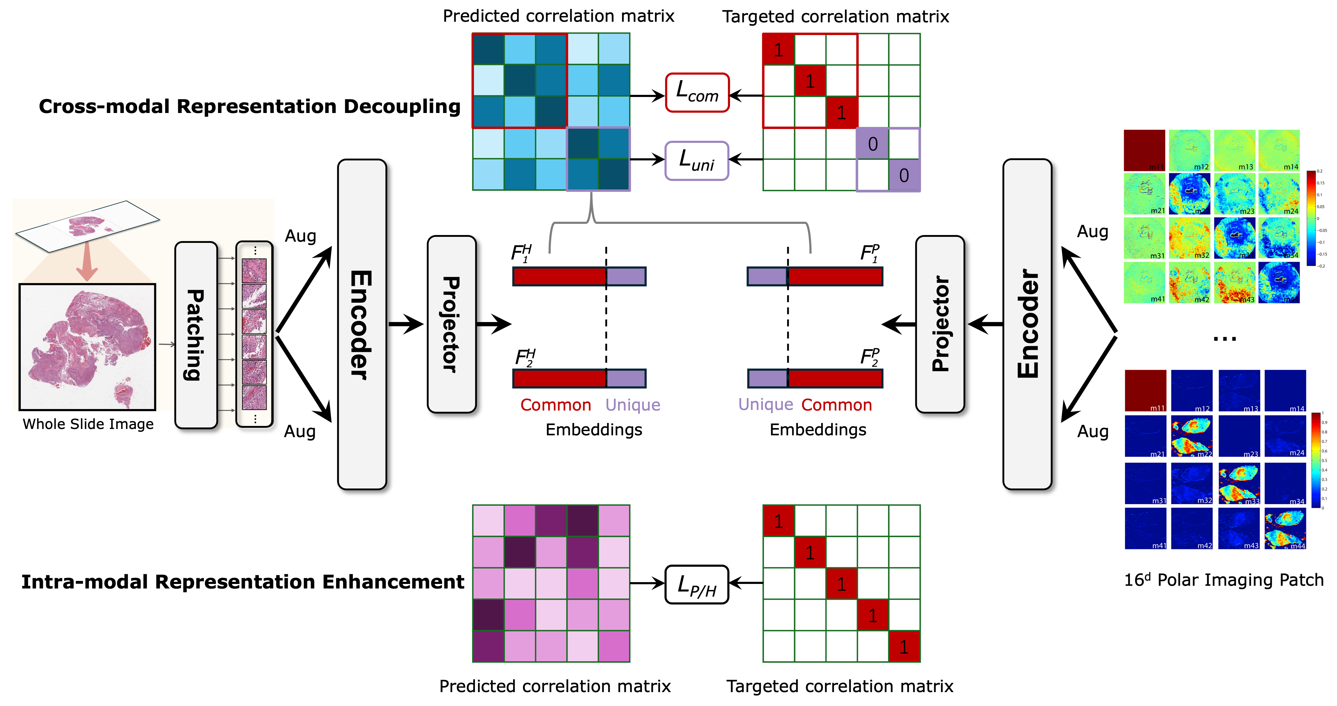
2.1 Cross-correlation Matrix for Feature Dependency Analysis
To measure dependencies between embedding dimensions, we compute cross-correlation matrices. Given two embedding vectors , their correlation matrix is defined as:
| (1) |
where indexes batch samples, and , index embedding dimensions. The resulting square matrix contains values ranging from -1 to 1, quantifying feature correlation across different dimensions. In the cross-modal case, the correlation matrix is calculated between two embeddings from different modalities, such as and in Figure 1.
2.2 Cross-modal Representation Decoupling
To ensure proper feature disentanglement, we partition the embedding space into: (common representations) and (unique representations) with . In Figure 1, the common representations are identical (highlighted in red) and encode modality-invariant features, while the modality-unique representations are decorrelated (highlighted in purple) and capture specialized modality-dependent structures.
To enforce cross-modal common feature alignment, we extract a sub-matrix from the common dimensions of and . The cross-modal redundancy reduction loss is formulated as:
| (2) |
where is a weighting coefficient balancing invariance enforcement (to ensure common features remain aligned across modalities) and decorrelation (to reduce feature redundancy and avoid model collapse).
For modality-unique representations, we extract a separate sub-matrix from and . The objective here is to decorrelate the unique dimensions, encouraging them to retain distinct modality-specific information:
| (3) |
where regulates the balance between inter-modal decorrelation and redundancy reduction. However, simply enforcing decorrelation does not guarantee that the unique embeddings retain meaningful pathology-related features, as they may collapse into random, decorrelated noise. To address this issue, we introduce intra-modal representation enhancement.
2.3 Intra-modal Representation Enhancement
To reinforce feature stability and prevent collapse of decoupled unique dimensions, we introduce intra-modal training that covers all the embedding dimensions, ensuring that the model learns robust within-modality representations. For each modality, a cross-correlation matrix (or ) is generated from the full dimensions of the embedding vectors and (or and ), optimizing:
| (4) |
where regulate the balance between feature invariance and redundancy minimization. This prevents degeneration and ensures intra-modal consistency. Similar implementation for and to obtain .
Overall training objective. The final self-supervised learning objective integrates cross-modal common representation learning, modality-unique decomposition, and intra-modal consistency constraints:
| (5) |
This ensures modality-invariant feature alignment, modality-specific feature decorrelation, and intra-modal representation reinforcement. By incorporating these objectives, PolarHE effectively fuses H&E and polarization imaging, leading to richer pathology representations and superior downstream performance.
3 Experiments
3.1 Polarization System and Paired Dataset Construction
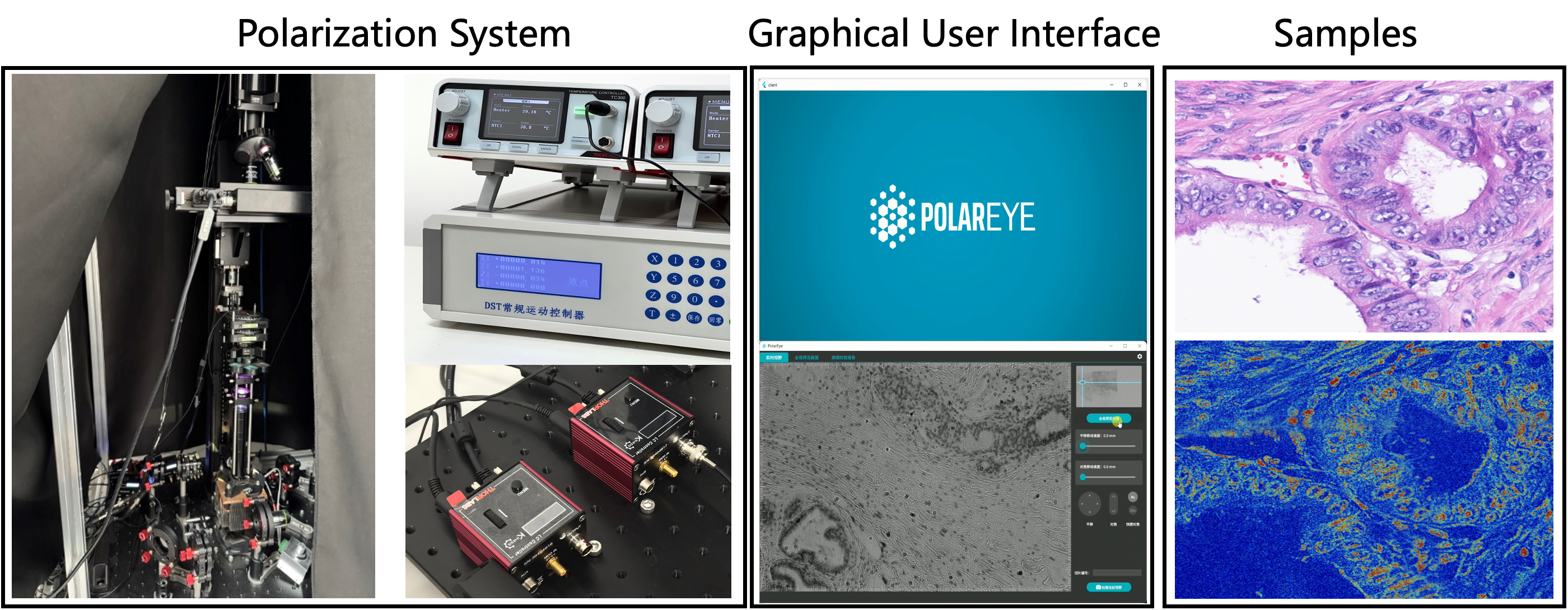
Polarization Imaging System for Pathology. To facilitate multimodal pathology image analysis, we construct a polarization imaging system for data acquisition and processing, as illustrated in Figure 2. This system is designed to capture full-vector polarization imaging, enabling comprehensive tissue characterization beyond conventional H&E staining. We employ a 635nm red light source as the incident light and perform full-field scanning at 4× magnification to ensure uniform tissue coverage across all pathology slides. Each field of view undergoes polarization measurement, generating a 16-dimensional polarization matrix for each pixel . This results in a 16-channel polarization representation, providing rich structural and birefringence information, which is essential for analyzing tissue anisotropy and microstructural organization.
PolarHE Paired Dataset Collection. To establish a precisely aligned multimodal dataset, we store the corresponding H&E-stained whole-slide images (WSIs) alongside the polarization images, preserving their structural and morphological details. To enable direct pixel-wise comparison, we perform image registration between the two modalities, ensuring precise spatial alignment. This results in a dataset of 26 paired PolarHE WSIs, from which we extract 4035 multimodal image patches of size 2304 × 1296. These patches are further cropped into 224 × 224 regions for model input, providing a rich and diverse dataset that enables SSL and downstream pathology analysis. This dataset facilitates the joint learning of common and unique representations across imaging modalities, making it an ideal benchmark for computational pathology.
Implementation. Each branch of PolarHE consists of a dedicated backbone and a three-layer MLP projector, each with an output dimension of 8192. We adopt the UNI ViT-L/16 encoder [3] as the backbone. Training is performed on embedding representations obtained after the projector, where the feature space is partitioned into common and unique components, with 75% allocated to common features. The backbone is subsequently transferred to downstream tasks, while the trade-off parameter () for the loss terms is set to 0.0051 [28].
3.2 Downstream Patch Classification
Datasets. To assess the effectiveness of PolarHE, we conduct patch classification experiments on two benchmark histopathology datasets: Chaoyang Dataset [29] consists of colon tissue slides collected from Chaoyang Hospital. Each patch is 512 × 512 pixels, with the dataset split as follows: Training Set: 1,111 normal, 842 serrated, 1,404 adenocarcinoma, and 664 adenoma patches; Testing Set: 705 normal, 321 serrated, 840 adenocarcinoma, and 273 adenoma patches. MHIST Dataset [22] consists of 2,175 training images and 977 testing images from 328 WSIs. These images are labeled into two classes: Hyperplastic Polyps (HPs) and Sessile Serrated Adenomas (SSAs). From these WSIs, 3,152 diagnostically relevant patches of size 224 × 224 pixels are extracted,representing clinically significant tissue regions.
Patch Classification by Linear Probing. To validate the effectiveness of our pretrained model, we perform linear probing [11] on the Chaoyang and MHIST datasets, demonstrating that PolarHE’s learned representations are highly discriminative, requiring minimal task-specific fine-tuning. Our method, PolarHE, achieves state-of-the-art performance on both datasets. On the Chaoyang Dataset, it achieves an accuracy of 86.70%, an F1 score of 86.42%, and an AUC of 95.96%. On the MHIST Dataset, it achieves an accuracy of 89.06%, an F1 score of 88.97%, and an AUC of 94.10%. These results, presented in Table 1, demonstrate that PolarHE significantly outperforms existing methods, validating the effectiveness of our dual-modality fusion.
| Chaoyang Dataset | MHIST Dataset | ||||||
|---|---|---|---|---|---|---|---|
| Method | Acc | F1 | AUC | Method | Acc | F1 | AUC |
| DINO [1] | 80.2 | - | - | DINO [1] | 83.10 | 81.60 | 89.78 |
| HSA-NRL [29] | 83.40 | 76.54 | 94.51 | BarlowTwins [28] | 81.27 | - | - |
| SSAT [4] | 82.52 | - | - | MoCo-v2 [11] | 85.88 | - | - |
| ViT-DAE [24] | 80.12 | 78.60 | - | Bitfit [27] | 82.40 | 80.86 | 89.60 |
| INCV [2] | 80.34 | 74.11 | 92.63 | SSF [15] | 82.29 | 80.85 | 89.29 |
| 2HSL [7] | 82.90 | - | - | RMSGD [9] | 82.58 | - | - |
| OUSM [25] | 80.53 | 73.70 | 93.69 | TPP [14] | 83.42 | 82.21 | 91.38 |
| SELF [18] | 80.49 | 75.31 | 93.99 | DiRL [12] | 78.20 | - | 87.1 |
| DiRL [12] | 83.60 | - | 95.0 | Virchow [19] | 83.40 | 83.5 | - |
| SH-PEFT [16] | 84.80 | 80.6 | - | CTransPath [20] | 83.75 | - | - |
| UNI [3] | 85.51 | 85.29 | 95.21 | UNI [3] | 85.63 | 85.57 | 91.98 |
| PolarHE (ours) | 86.70 | 86.42 | 95.96 | PolarHE (ours) | 89.06 | 88.97 | 94.10 |
Ablation Study. Table 2 shows removing intra-modal training or modality decoupling reduces accuracy, confirming their importance. Without intra-modal training, performance drops, especially on Chaoyang, highlighting its role in reinforcing modality-specific features. Without modality decoupling, accuracy also declines, showing the need for feature separation. The full model performs best, validating both components. For common representation ratio, the downstream performance increases and decreases smoothly along with the change of the percentage of common dimensions, which indicates the sparsity of the common embedding space.
| Loss Component | Chaoyang | MHIST |
|---|---|---|
| PolarHE (ours) | 86.70 | 89.06 |
| w/o intra | 84.15 | 86.25 |
| w/o decoup. | 85.92 | 87.06 |
| w/o intra&decoup. | 85.51 | 85.63 |
| Common Ratio | Chaoyang | MHIST |
|---|---|---|
| 50% | 84.95 | 87.70 |
| 75% | 86.70 | 89.06 |
| 85% | 86.27 | 88.33 |
3.3 Polarization Property Visualization
Polarization imaging introduces a contrast mechanism for visualizing tissue microstructure and biochemical properties, enabling precise differentiation of pathological features. Unlike H&E staining, where collagen fibers appears pink (indistinguishable from muscle and blood vessels), the fast-axis orientation effectively highlights collagen organization in full-vector polarization imaging, aiding in structural differentiation.
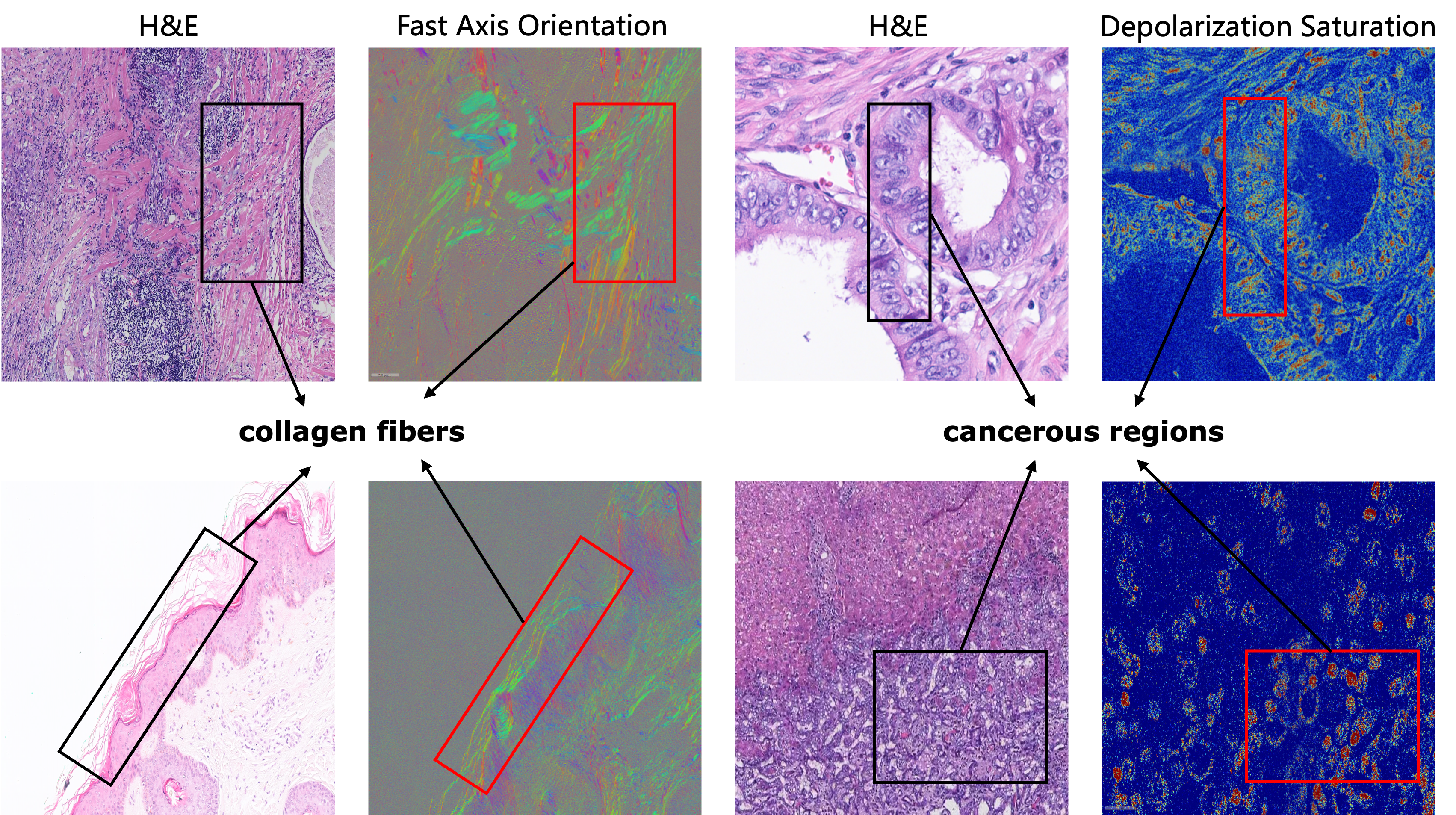
Similarly, endometrioid carcinoma exhibits unique polarization signatures. Compared to normal hepatocytes, cancerous regions show increased DNA replication activity, higher nuclear stiffness, and stronger depolarization signals. The difference in nuclear depolarization saturation provides a potential marker for distinguishing cancerous regions, assessing tumor differentiation, and evaluating malignancy progression. These findings suggest that polarization imaging provides valuable tissue biomarkers, offering enhanced diagnostic capabilities beyond conventional H&E analysis.
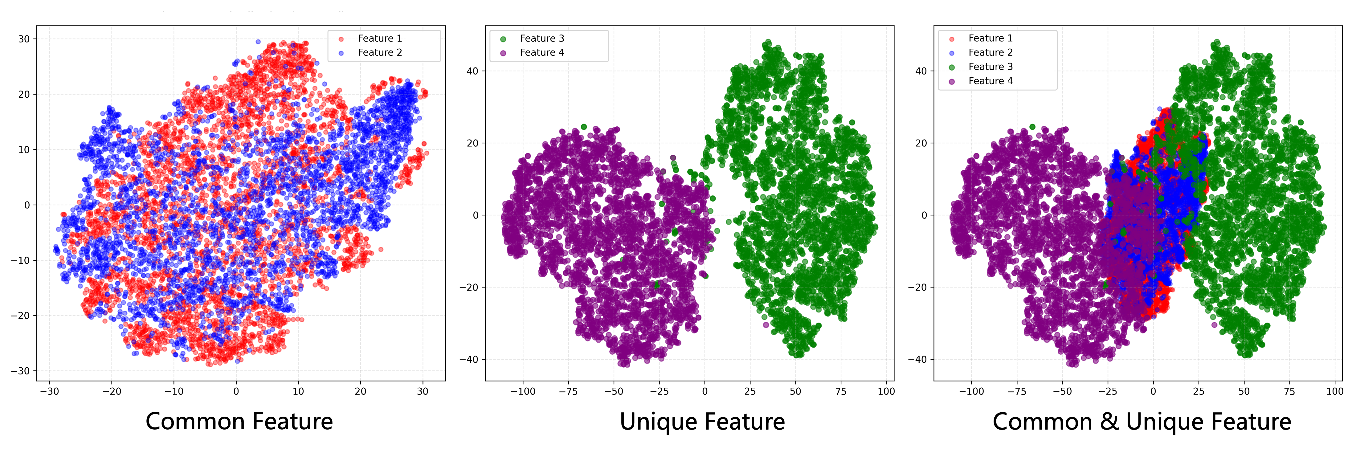
3.4 T-SNE Visualization of Common and Unique Representations
To further investigate the representations learned by PolarHE, we employ t-SNE visualization to analyze the distribution of common and unique features: Shared features between polarization and H&E images exhibit strong alignment in feature space, confirming effective cross-modal feature learning; Unique modality-specific features remain well-separated, demonstrating that our model successfully preserves discriminative modality-dependent information. The distinct separation of common and unique features highlights the effectiveness of PolarHE in learning robust multimodal features, reinforcing the complementary role of polarization imaging in pathology analysis.
4 Conclusion
We propose PolarHE, a dual-modality fusion framework that integrates polarization imaging with H&E staining for enhanced representation learning in pathology. 1By leveraging a feature decomposition strategy, our approach effectively disentangles common and modality-specific features, leading to improved multimodal representation learning. Through comprehensive experiments, we demonstrated that the integration of H&E and polarization representations leads to significantly better performance compared to existing methods. Additionally, t-SNE visualizations confirm that our model successfully aligns shared features while preserving modality-specific distinctions, reinforcing the complementary nature of polarization imaging. These findings underscore the untapped potential of polarization imaging as a powerful auxiliary modality in computational pathology. By enhancing diagnostic accuracy and model robustness, PolarHE marks a promising step toward more interpretable and automated pathology analysis, paving the way for advancements in multimodal deep learning for clinical applications.
References
- [1] Caron, M., Touvron, H., Misra, I., Jégou, H., Mairal, J., Bojanowski, P., Joulin, A.: Emerging properties in self-supervised vision transformers. In: ICCV. pp. 9650–9660 (2021)
- [2] Chen, P., Liao, B.B., Chen, G., Zhang, S.: Understanding and utilizing deep neural networks trained with noisy labels. In: International conference on machine learning. pp. 1062–1070. PMLR (2019)
- [3] Chen, R.J., Ding, T., Lu, M.Y., Williamson, D.F., Jaume, G., Song, A.H., Chen, B., Zhang, A., Shao, D., Shaban, M., et al.: Towards a general-purpose foundation model for computational pathology. Nature Medicine 30(3), 850–862 (2024)
- [4] Das, S., Jain, T., Reilly, D., Balaji, P., Karmakar, S., Marjit, S., Li, X., Das, A., Ryoo, M.S.: Limited data, unlimited potential: A study on vits augmented by masked autoencoders. In: Proceedings of the IEEE/CVF Winter Conference on Applications of Computer Vision. pp. 6878–6888 (2024)
- [5] Dong, Y., Wan, J., Si, L., Meng, Y., Dong, Y., Liu, S., He, H., Ma, H.: Deriving Polarimetry Feature Parameters to Characterize Microstructural Features in Histological Sections of Breast Tissues. IEEE Transactions on Biomedical Engineering 68, 881–892 (2021)
- [6] Dong, Y., Wan, J., Wang, X., Xue, J.H., Zou, J., He, H., Li, P., Hou, A., Ma, H.: A polarization-imaging-based machine learning framework for quantitative pathological diagnosis of cervical precancerous lesions. IEEE Transactions on Medical Imaging 40(12), 3728–3738 (2021)
- [7] Galdran, A., Verjans, J.W., Carneiro, G., González Ballester, M.A.: Multi-head multi-loss model calibration. In: International Conference on Medical Image Computing and Computer-Assisted Intervention. pp. 108–117. Springer (2023)
- [8] He, C., He, H., Chang, J., Chen, B., Ma, H., Booth, M.J.: Polarisation optics for biomedical and clinical applications: a review. Light: Science & Applications 10(1), 194 (2021)
- [9] Hosseini, M.S., Tuli, M., Plataniotis, K.N.: Exploiting explainable metrics for augmented sgd. In: Proceedings of the IEEE/CVF Conference on Computer Vision and Pattern Recognition. pp. 10296–10306 (2022)
- [10] Hou, A., Wang, X., Fan, Y., Miao, W., Dong, Y., Tian, X., Zou, J., Ma, H.: Polarimetry feature parameter deriving from mueller matrix imaging and auto-diagnostic significance to distinguish hsil and cscc. Journal of Innovative Optical Health Sciences 15(01), 2142008 (2022)
- [11] Kang, M., Song, H., Park, S., Yoo, D., Pereira, S.: Benchmarking self-supervised learning on diverse pathology datasets. In: Proceedings of the IEEE/CVF Conference on Computer Vision and Pattern Recognition. pp. 3344–3354 (2023)
- [12] Kapse, S., Das, S., Zhang, J., Gupta, R.R., Saltz, J., Samaras, D., Prasanna, P.: Attention de-sparsification matters: Inducing diversity in digital pathology representation learning. Medical Image Analysis 93, 103070 (2024)
- [13] Koohbanani, N.A., Unnikrishnan, B., Khurram, S.A., Krishnaswamy, P., Rajpoot, N.: Self-path: Self-supervision for classification of pathology images with limited annotations. IEEE Transactions on Medical Imaging 40(10), 2845–2856 (2021)
- [14] Lei, X., Ye, Y., Chen, Z., Shu, M., Xia, Y.: Pre-training everywhere: Parameter-efficient fine-tuning for medical image analysis via target parameter pre-training. arXiv preprint arXiv:2408.15011 (2024)
- [15] Lian, D., Zhou, D., Feng, J., Wang, X.: Scaling & shifting your features: A new baseline for efficient model tuning. Advances in Neural Information Processing Systems 35, 109–123 (2022)
- [16] Liu, M., Xu, L., Liu, S., Zhang, J.: Sparsity-and hybridity-inspired visual parameter-efficient fine-tuning for medical diagnosis. In: International Conference on Medical Image Computing and Computer-Assisted Intervention. pp. 627–637. Springer (2024)
- [17] Nguyen, A.T., Vuong, T.T.L., Kwak, J.T.: Towards a text-based quantitative and explainable histopathology image analysis. In: International Conference on Medical Image Computing and Computer-Assisted Intervention. pp. 514–524. Springer (2024)
- [18] Nguyen, D.T., Mummadi, C.K., Ngo, T.P.N., Nguyen, T.H.P., Beggel, L., Brox, T.: Self: Learning to filter noisy labels with self-ensembling. arXiv preprint arXiv:1910.01842 (2019)
- [19] Vorontsov, E., Bozkurt, A., Casson, A., Shaikovski, G., Zelechowski, M., Liu, S., Severson, K., Zimmermann, E., Hall, J., Tenenholtz, N., et al.: Virchow: A million-slide digital pathology foundation model. arXiv preprint arXiv:2309.07778 (2023)
- [20] Wang, X., Yang, S., Zhang, J., Wang, M., Zhang, J., Huang, J., Yang, W., Han, X.: Transpath: Transformer-based self-supervised learning for histopathological image classification. In: Medical Image Computing and Computer Assisted Intervention–MICCAI 2021: 24th International Conference, Strasbourg, France, September 27–October 1, 2021, Proceedings, Part VIII 24. pp. 186–195. Springer (2021)
- [21] Wang, Y., Albrecht, C.M., Braham, N.A.A., Liu, C., Xiong, Z., Zhu, X.X.: Decoupling common and unique representations for multimodal self-supervised learning. In: European Conference on Computer Vision. pp. 286–303. Springer (2024)
- [22] Wei, J., Suriawinata, A., Ren, B., Liu, X., Lisovsky, M., Vaickus, L., Brown, C., Baker, M., Tomita, N., Torresani, L., et al.: A petri dish for histopathology image analysis. In: Artificial Intelligence in Medicine: 19th International Conference on Artificial Intelligence in Medicine, AIME 2021, Virtual Event, June 15–18, 2021, Proceedings. pp. 11–24. Springer (2021)
- [23] Wu, L., Zhuang, J., Chen, H.: Voco: A simple-yet-effective volume contrastive learning framework for 3d medical image analysis. In: Proceedings of the IEEE/CVF Conference on Computer Vision and Pattern Recognition. pp. 22873–22882 (2024)
- [24] Xu, X., Kapse, S., Gupta, R., Prasanna, P.: Vit-dae: Transformer-driven diffusion autoencoder for histopathology image analysis. In: International Conference on Medical Image Computing and Computer-Assisted Intervention. pp. 66–76. Springer (2023)
- [25] Xue, C., Dou, Q., Shi, X., Chen, H., Heng, P.A.: Robust learning at noisy labeled medical images: Applied to skin lesion classification. In: 2019 IEEE 16th International symposium on biomedical imaging (ISBI 2019). pp. 1280–1283. IEEE (2019)
- [26] Yang, K., Liu, F., Liang, S., Xiang, M., Han, P., Liu, J., Dong, X., Wei, Y., Wang, B., Shimizu, K., et al.: Data-driven polarimetric imaging: a review. Opto-Electronic Science 3(2), 230042–1 (2024)
- [27] Zaken, E.B., Ravfogel, S., Goldberg, Y.: Bitfit: Simple parameter-efficient fine-tuning for transformer-based masked language-models. arXiv preprint arXiv:2106.10199 (2021)
- [28] Zbontar, J., Jing, L., Misra, I., LeCun, Y., Deny, S.: Barlow twins: Self-supervised learning via redundancy reduction. In: International Conference on Machine Learning. pp. 12310–12320. PMLR (2021)
- [29] Zhu, C., Chen, W., Peng, T., Wang, Y., Jin, M.: Hard sample aware noise robust learning for histopathology image classification. IEEE transactions on medical imaging 41(4), 881–894 (2021)