T-cell receptor specificity landscape revealed through de novo peptide design
Abstract
T-cells play a key role in adaptive immunity by mounting specific responses against diverse pathogens. An effective binding between T-cell receptors (TCRs) and pathogen-derived peptides presented on Major Histocompatibility Complexes (MHCs) mediate an immune response. However, predicting these interactions remains challenging due to limited functional data on T-cell reactivities. Here, we introduce a computational approach to predict TCR interactions with peptides presented on MHC class I alleles, and to design novel immunogenic peptides for specified TCR-MHC complexes. Our method leverages HERMES, a structure-based, physics-guided machine learning model trained on the protein universe to predict amino acid preferences based on local structural environments. Despite no direct training on TCR-pMHC data, the implicit physical reasoning in HERMES enables us to make accurate predictions of both TCR-pMHC binding affinities and T-cell activities across diverse viral epitopes and cancer neoantigens, achieving up to 72% correlation with experimental data. Leveraging our TCR recognition model, we develop a computational protocol for de novo design of immunogenic peptides. Through experimental validation in three TCR-MHC systems targeting viral and cancer peptides, we demonstrate that our designs—with up to five substitutions from the native sequence—activate T-cells at success rates of up to 50%. Lastly, we use our generative framework to quantify the diversity of the peptide recognition landscape for various TCR-MHC complexes, offering key insights into T-cell specificity in both humans and mice. Our approach provides a platform for immunogenic peptide and neoantigen design, opening new computational paths for T-cell vaccine development against viruses and cancer.
I Introduction
T-cells play a key role in the adaptive immune system, contributing to immune surveillance and response to pathogens. T-cells recognize pathogen-derived epitopes in the form of short protein fragments (peptides) displayed on specific molecules known as Major Histocompatibility Complexes (MHCs). The binding between a T-cell receptor (TCR) and a peptide-MHC (pMHC) is essential to mediate a protective T-cell response to an infection. To defend the host against a multitude of pathogens, a highly diverse TCR repertoire is generated through a somatic VDJ recombination process, and selected to target infected cells with remarkable sensitivity and specificity.
Predicting the TCR-pMHC binding specificity is an important step in characterizing the efficacy of the immune system in countering different pathogens. Such understanding would aid with disease diagnoses and development of diagnostic tests for early detection of autoimmune diseases [1]. A predictive model for TCR-pMHC specificity can be used to design engineered TCRs that recognize cancer antigens [2], enhancing the effectiveness of adoptive cell transfer and CAR T-cell therapies [3, 4], as well as the design of soluble TCRs and bispecific T-cell engager therapeutics [5]. Moreover, it can enable the design of optimized antigens for vaccines to elicit robust T-cell responses against specific pathogens or tumors [6, 7].
One bottleneck in deciphering the TCR-pMHC code is the lack of well-curated datasets. High-throughput immune repertoire sequencing, combined with comparative analyses of hosts facing similar immune challenges, and the development of functional assays (e.g., pMHC-tetramer staining [8] or MIRA [9]), have enabled the identification of diverse TCR pools with potential reactivity to specific antigens [10], collected in repositories like Immune Epitope Database (IEDB) [11] and VDJdb [12]. However, the current databases are unbalanced, with only a handful of dominant peptides (e.g. derived from SARS-CoV-2, CMV, EBV, Influenza, etc.) and MHC alleles present, each linked to hundreds of TCRs. As a result, statistical inference and machine learning methods trained on such data to predict TCR-pMHC reactivities lack generalizability beyond their training data [13, 14, 15]. On the structural side, only a few hundred TCR-pMHC crystal structures have been experimentally generated to date. This is in part because TCR-pMHC interactions are typically of low affinity and are difficult to stabilize to the level needed for CryoEM or X-ray crystallography [16]. Importantly, this scarcity hinders computational prediction methods like AlphaFold in modeling reliable TCR-pMHC structures [17, 18].
Machine learning models, such as large language models, pre-trained on vast unannotated protein datasets can generate meaningful data representations [19, 20]. Fine-tuning these models on smaller, labeled datasets for specific tasks (e.g., predicting mutation impacts on stability or function) has proven highly effective [21, 22, 23, 24]. Pre-trained sequence-based protein language models have been employed to predict reactivities of antibodies to antigens [23, 24], or TCRs to pMHC complexes [25, 15]. However, lack of generalizability due to sparse and skewed training data has remained a challenge in these models.
Protein-protein interactions in general, and immune-pathogen interaction in particular, rely on the complementarity in the 3D structures and amino acid compositions of the interacting protein pairs. Even though the interacting amino acids can be far apart in a protein sequence, they are close to each other in structure, resulting in local interactions that can determine immune-pathogen recognition. Machine learning models trained on protein structures instead of sequences can learn local structural representations for amino acid statistics, which tend to be more generalizable across different protein families and beyond their training sets [30, 31, 32, 26, 27, 33].
Here, we present a structure-based approach to predict interaction affinities between TCRs and peptides presented on MHC class I, and to design reactive peptides for specific TCR-MHC-I complexes (TCR-MHC for short). Due to the limited availability of structural data for TCRs, we use HERMES– a physics-guided, 3D-equivariant machine learning model trained on the entire protein universe to predict amino acid preferences based on their local structural environments [27, 26]. We demonstrate that HERMES reliably predicts interaction affinities for diverse TCR-pMHC complexes, and the activity of T-cells in response to peptides presented by MHC class I molecules. Moreover, we show that HERMES can be used to design novel peptides binding specific TCR-MHC complexes, achieving up to 50% experimental validation accuracy across diverse TCR-MHC systems. Our peptide design platform can facilitate the computational design of neoantigen libraries for cancer vaccines. By leveraging our design algorithms, we characterize the specificity of a diverse range of TCRs, providing a quantitative measure for the diversity of the peptide-MHC antigens that a TCR can recognize in humans and mice.
II Model
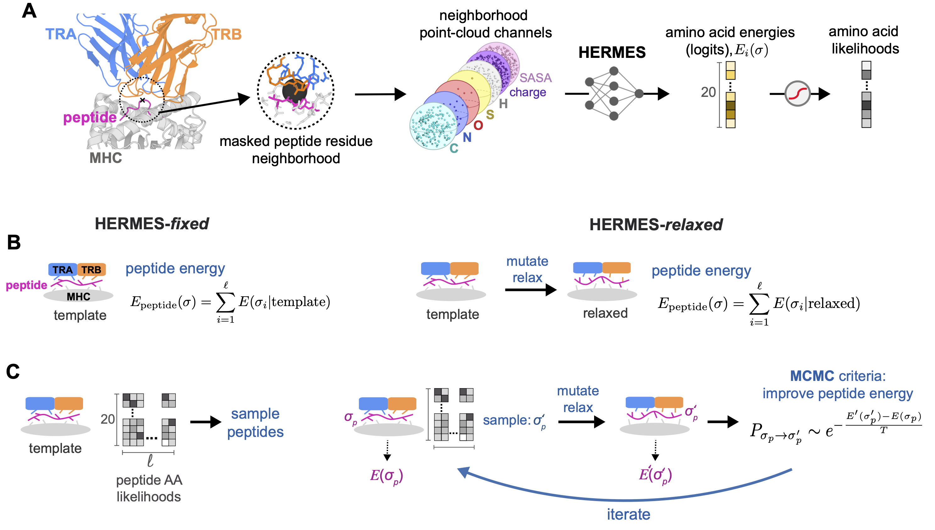
T-cell response is mediated by the interactions between TCRs and pMHC complexes. To model the TCR-pMHC interaction, we use HERMES [26], a 3D rotationally equivariant structure-based neural network model, trained on the protein universe; see Fig. 1A. HERMES predicts the amino acid propensity from its surrounding 3D protein environment, a measure that we have previously shown to serve as a reliable proxy for assessing the effect of mutations on protein stability or binding [27, 26].
For TCR-pMHC complexes, we seek to determine how changes in a peptide sequence impact the binding affinity of a TCR-pMHC complex, and ultimately, the T-cell response to the antigen. To do so, we characterize a score, which we term peptide energy, for a peptide , given its surrounding TCR and MHC structural environment,
| (1) |
We assume that each peptide residue contributes linearly to the total energy by the amount , where is the amino acid at position of the peptide. A residue’s energy contribution is evaluated by HERMES (logits of the network), taking in as input the atomic composition of the surrounding TCR, MHC, and the rest of the peptide (excluding the residue) within a 10 Å of ’s -Carbon; see Fig. 1 and SI for details.
To compute peptide energy, we input to HERMES an experimentally or computationally determined structure of a specified TCR-MHC complex bound to at least one peptide variant. Experimental data on the impact of peptide substitutions on the TCR-pMHC structure is limited, and computational models often fail to capture subtle conformational changes in the structure due to amino acid substitutions in a peptide. Given these constraints, we adopt two approaches to estimate peptide energies across diverse peptide variants for a given TCR-MHC complex:
-
•
HERMES-fixed: In the simplest approach, we choose a TCR-pMHC structure with the same TCR and MHC as our query and a peptide sequence closest to the peptide of interest. This structure is then used as our template in HERMES to compute the peptide energy as described in eq. 1 (Fig. 1B). This method does not alter the underlying structure and assumes that peptide amino acid substitutions do not significantly change the conformation of the TCR-pMHC complex.
-
•
HERMES-relaxed: Since amino acid substitutions can locally reshape a protein complex, we introduce a more involved protocol to account for these structural changes. We begin with the closest available structure for the TCR-pMHC complex. We then mutate the original peptide to the desired state and apply a relaxation procedure using PyRosetta to properly pack the substituted amino acid [28] (SI). During relaxation, side-chain and backbone atoms of the peptide are allowed to move, while only the side-chain atoms of the MHC and TCR chains within a 10 Å radius of the peptide are flexible. We then calculate the peptide energy with HERMES (eq.1), using the relaxed structure as input (Fig. 1B); since PyRosetta’s relaxation procedure is stochastic, we average the peptide energy across 100 realizations of these relaxations.
III Results
Predicting binding affinity of TCR-pMHC complexes
Binding between TCRs and pMHC complexes is necessary to mediate a T-cell response. The binding affinity of a natural TCR-pMHC complex is relatively low, with a dissociation constant in the range of [38, 16, 39], in contrast to nanomolar affinities for antibody-antigen complexes. Engineered TCRs can achieve much higher affinities in a nanomolar [40] to picomolar [41, 42] range.
Surface plasmon resonance (SPR) spectroscopy provides accurate measurements of the dissociation constant () for specific TCR-pMHC systems. In these experiments, one protein–either the TCR or the MHC–is immobilized on a conducting plate, and the other is introduced in solution to bind to it. This binding alters the local refractive index near the plate, affecting the resonance signal. We used a published dataset of SPR-measured affinities for two TCR-MHC complexes binding to an ensemble of peptides, for which crystal structures of the complexes with at least one peptide are available [43, 44]; see Table S1 for details.
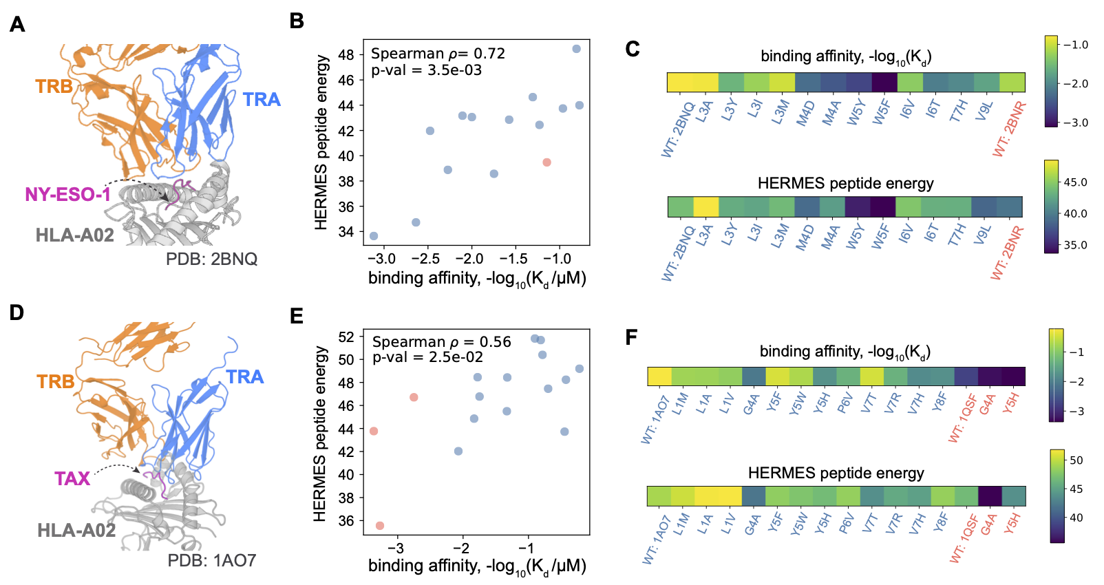
In our first example, we examined TCR 1G4 binding to NY-ESO-1 peptide variants, which is a cancer-testis antigen commonly expressed in many cancers and is targeted by immunotherapies [45]. We used two structural templates, one with the wild-type peptide SLLMWITQC (NY-ESO-1), and the other with a more immunogenic peptide, in which the Cysteine at position 9 is substituted by Valine (C9V) [46, 34] (Fig. 2A). For variants differing by a single amino acid, our predictions with HERMES-relaxed correlated well (72% Spearman correlation) with experimentally measured affinities from refs. [43, 44](Fig. 2B, C).
Next, we tested the A6 TCR binding to the HTLV-1 Tax peptide LLFGYPVYV. We employed two structural templates: one with the wild-type, the other with the mutant peptide with substitution Y8A [35] (Fig. 2D and Table S1). Again, the HERMES-relaxed peptide energies correlated well (56% Spearman ) with the affinities, measured in ref. [44] (Fig. 2E, F).
Overall, HERMES predictions aligned closely with this limited experimental affinity measurements and performed as well as or better than BLOSUM-based scoring [47], ProteinMPNN [30], and TCRdock [17] (Table S2). Notably, unlike genome-averaged methods such as BLOSUM, HERMES is sensitive to the local structural environment, assigning different scores to the same mutation depending on context (Fig. S1). HERMES can predict relative peptide binding affinities for TCR-MHC complexes, provided a suitable structural template is available. Prediction quality suffers in the absence of reliable structural templates, as seen also in other methods such as TCRdock [17] (Table S2). Lastly, HERMES-relaxed peptide energies show better agreement with the affinity measurements than HERMES-fixed (Table S2). However, the small size of this dataset prevents us from drawing broader conclusions about the relative utility of either approach for this problem.
Predicting T-cell response to peptide antigens
Aside from the affinity of TCR-pMHC interactions, other factors play important roles in determining whether and how a T-cell responds. These factors include the phenotypic state of a T-cell (e.g., naive or memory), the reactivity of its TCR to competing self-antigens presented on MHC molecules [48, 49], and the avidity of TCR-pMHC interactions, determined by the clustering of TCRs with co-receptors on the T-cell membrane and the density of pMHC molecules on antigen-presenting cells [50]. T-cell activation can be characterized using a diverse set of functional assays that measure T-cell proliferation, labeled cellular activation markers through flow cytometry, and production of cytokines such as interferon gamma (IFN-). As such, measuring T-cell activity is much easier than measuring the affinity of TCR-pMHC complexes, which requires techniques like SPR. Although our structure-based HERMES model is more attuned to predicting the binding affinity of TCR-pMHC complexes, the ease of data production prompted us to assess the accuracy of our method in predicting T-cell activity.
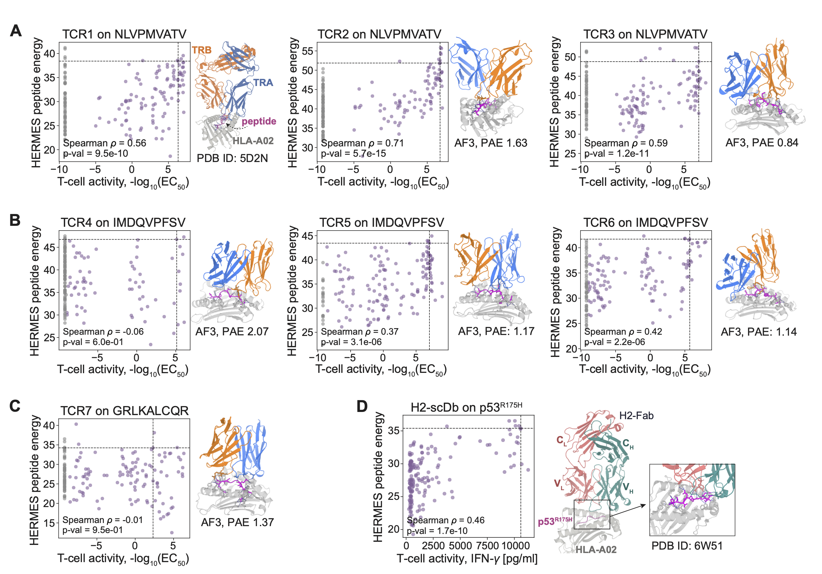
As the first system, we examined T-cell responses to all single-point mutants of a native peptide, as measured in ref. [36] for different TCR-pMHC systems. Specifically, we make predictions for (i) three TCRs (TCRs 1-3) recognizing variants of a highly immunogenic HLA-A*02:01-restricted human cytomegalovirus (CMV) epitope NLVPMVATV (NLV), (ii) three TCRs (TCRs 4-6) recognizing variants of the weaker HLA-A02:01-restricted melanoma gp100 self-antigen IMDQVPFSV, and (iii) a TCR (TCR 7) recognizing variants of weakly immunogenic HLA-B*27:05-restricted pancreatic cancer neo-peptide GRLKALCQR, believed to have elicited an effective immune response in long-term cancer survivors [36]. T-cell activity was quantified by determining the half-maximal effective concentration (EC50) from the dose-response curves of 4-1BB+ CD8+ T-cells across varying peptide concentrations; see Methods on details of inferring EC50.
With the exception of TCR1 (where a TCR-pMHC template with HLA-A*02:01 and one peptide variant was already available), we relied on AlphaFold3 (AF3) [29] to model the remaining TCR-pMHC complexes; see Tables S1, S3 for template information and AF3 sequence inputs for TCRs 2-7. As shown in Figs. 3A, and S2, HERMES-fixed peptide energy predictions (eq. 1) correlate well with experimental EC50, reaching up to 71% Spearman correlation for TCR2 recognizing the CMV peptides. Correlations are lower for TCRs 4-6 targeting variants of the melanoma gp100 self-antigen, with TCR6 achieving 42% Spearman correlation as the best prediction in this class (Fig. 3B). HERMES underperforms for TCR7 responding to the pancreatic cancer neo-peptide GRLKALCQR (Fig. 3C). Still, except for TCR4, which has the largest AF3 Predicted Alignment Error (PAE) of 2.07 at the TCR-pMHC interface (i.e., a lower confidence in its fold), HERMES predictions outperform those of TULIP [15], the state-of-the-art sequence-based language model trained to predict TCR-pMHC interactions (Table S4). The reduced accuracy of HERMES for cancer epitopes compared to the variants of the viral CMV epitope could reflect the impact of antagonism from self-antigens similar to tumor epitopes, hindering T-cell responses [48, 49]—though additional experiments are needed to confirm this.
Next, we examined the therapeutic T-cell responses to all single point mutants of the HLA-A*02:01-restricted p53-derived neoepitope p53R175H, HMTEVVRHC, measured in ref. [5]. This T-cell therapy approach used a designed bi-specific antibody that binds both to the TCR-CD3 complex on T-cells and to the variants of the p53R175H-MHC complex on tumor cells (Table S1). A strong binding to an epitope activates the engaged T-cell, and the extent of such activity was measured by the production of IFN-. The resulting HERMES-fixed predicted peptide energies (eq. 1) show a Spearman with the experimentally measured IFN- production (Figs. 3D, S2).
Our analysis shows that, despite not being explicitly trained for this purpose, HERMES peptide energy effectively predicts T-cell responses to neoepitopes presented by different MHC-I alleles. Moreover, HERMES predictions are not limited to TCR-pMHC interactions but also extend to antibody-pMHC interactions, commonly leveraged in T-cell therapies for their higher achievable affinities. The HERMES predictions on T-cell activities perform as well or better than other computational methods including BLOSUM62 [47], ProteinMPNN [30], and TCRdock [17]; see Table S4 for a detailed benchmark of different approaches, including different HERMES models.
Design of novel peptides reactive to a TCR-MHC complex
Vaccination protocols aim to elicit robust immune responses that induce lasting immune memory in individuals against viral infections and cancer. Peptide-based cancer vaccines represent a promising therapeutic strategy to mount an effective T-cell response, offering the potential to be highly personalized to an individual’s unique tumor antigens and T-cell repertoire [55, 7]. However, the clinical success of cancer vaccines has been limited by factors such as low peptide immunogenicity and insufficient uptake by antigen-presenting cells [56].
Recent approaches have employed deep mutational scanning (DMS) experiments to identify more immunogenic cancer neoantigens [36]. However, these experiments are often limited to scanning single amino acid mutations from the wild-type (native) peptide due to the combinatorial explosion when exploring multiple mutations.
Here, we present a pipeline for designing peptides with enhanced TCR-MHC reactivity by leveraging HERMES’s ability in predicting T-cell response to peptides presented by MHC-I molecules. Our structure-based approach starts with an existing template structure–experimentally or computationally resolved–of a TCR-MHC in complex with at least one peptide variant, which we refer to as the wild-type peptide. We use two methods to sample peptides, with different degrees of complexity; see Fig. 1C for a schematic description of the two pipelines.
In the basic pipeline (HERMES-fixed), we sequentially mask the atoms of each amino acid along the peptide, one at a time. Using the HERMES model, we predict the probability of each amino acid type for a given residue, based on the local structural environment within 10 Å of the masked residue. Repeating this procedure for every amino acid along the peptide yields a position weight matrix (PWM) that represents the probabilities of different amino acids at each peptide position. Peptides are then sampled by drawing from this PWM. It should be noted that HERMES-fixed retains the structural fingerprints of the original peptide, as it does not account for local structural changes induced by amino acid substitutions; see Fig. 1C and SI for more details.
To incorporate structural flexibility and relaxation following amino acid substitutions, we introduce the design pipeline of HERMES-relaxed, using simulated annealing and Markov chain Monte Carlo (MCMC) sampling. We begin with the template TCR-MHC structure and a completely random peptide sequence, which is packed in silico within the structure using PyRosetta’s packing functionality, followed by the Relax protocol [28]. During relaxation, both side-chain and backbone atoms of the peptide are allowed to move, while only the side-chain atoms of the MHC and TCR chains within 10 Å of the peptide are flexible. With this relaxed structure, we apply the HERMES-fixed method to generate a PWM and sample a new peptide, which is then packed and relaxed to form a new structure. We use MCMC to determine whether to accept the newly sampled peptide by comparing its HERMES peptide energy (favorability), evaluated in its relaxed conformation, to that of the original peptide (eq. 1). We then iteratively perform this procedure, incrementally reducing the MCMC temperature at each step, until the results converge; see Figures 1C, and S3, and SI for more details.
The peptides designed by both approaches are then filtered by TCRdock [17] or AlphaFold3 [29], using the Predicted Alignment Error (PAE) between TCR and pMHC interfaces, to assure folding and binding of the resulting TCR-pMHC structures. For each system, we define a specific PAE threshold close to the TCRDock PAE evaluated for the system’s TCR-MHC in complex with the native (wild-type) peptide, which is known a priori to activate T-cells. As a result, the PAE thresholds vary slightly across different systems; see SI for details.
We tested the accuracy of our peptide design pipeline in three systems, with the structural templates taken from (i) the cancer-testis antigen NY-ESO-1 in complex with the TCR 1G4 and HLA-A*02:01, (ii) a peptide derived from the Epstein-Barr virus (EBV) in complex with the TK3 TCR and the HLA-B*35:01, and (iii) the immunotherapeutic target MAGE-A3 in complex with an engineered TCR and HLA-A*01:01. For brevity, we refer to these three systems as NY-ESO, EBV and MAGE, respectively; See Table S5 for information on the specific templates used in each case. It should be noted that as we progressed from NY-ESO to EBV and MAGE, we chose to explore the sequence space farther from the wild-type peptides, leading to more challenging designs.
We validated our designs using Jurkat cells expressing a specific TCR and an endogenous NFAT-eGFP reporter to indicate T-cell activation in response to different peptides. We tested the TCR-MHC specificity in all systems–NY-ESO, MAGE, EBV–under 96 different conditions, including the de novo designed peptides, positive controls (wild-type peptides from the template structures), and an unstimulated “no peptide” control. Peptides were presented on artificial APCs (aAPCs), expressing the specified HLA in each system. For NY-ESO designs, we determined the the percentage of GFP-positive cells through both flow cytometry and fluorescence microscopy. Given the consistency between the two experimental approaches (Fig. S4) we relied on fluorescence microscopy only to measure GFP levels induced by different peptides for MAGE and EBV designs; see SI for experimental details.
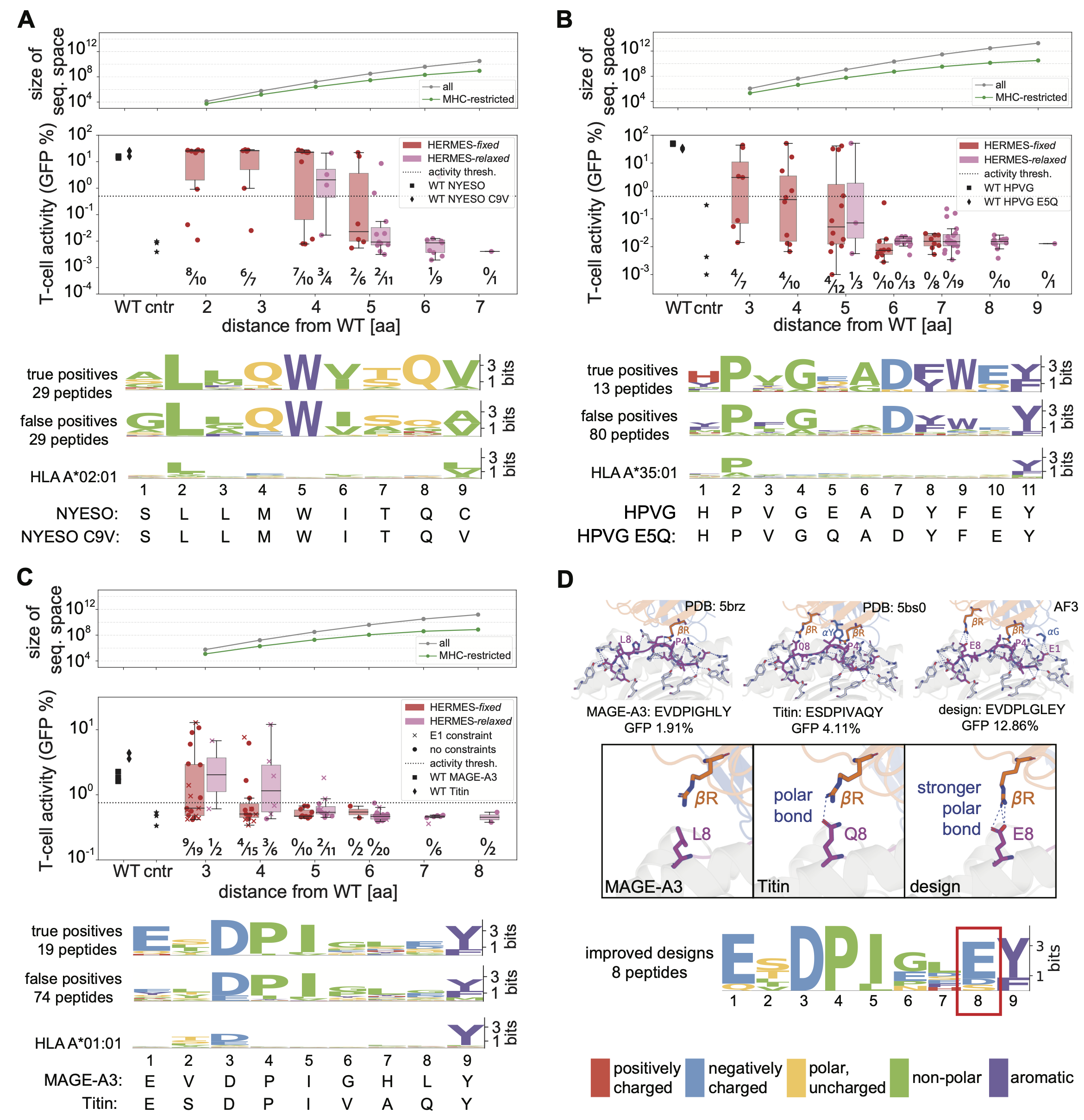
We used two structural templates for the NY-ESO system: one containing the original 9-amino acid peptide SLLMWITQC, and another with the more immunogenic C9V substitution [46, 34]. Our de novo designs differed from the closest wild-type peptide by 2 to 7 amino acids, with a TCRDock PAE . For this system only, we selected 35 negative designs (designed by HERMES, but with PAE ) for experimental testing: only one of these was a false negative, which interestingly, had a desirable AF3 PAE score (as low as the wild-type peptides); see Figs. S5, S6. For the 58 positive designed peptides (PAE ), we achieved an overall design accuracy of 50%, with GFP levels significantly higher than the negative control; see SI for details. The accuracy of predictions decreases as the sequence divergence of the de novo designs from the wild-type peptides increases (Fig. 4A, S6). HERMES-fixed achieved higher design accuracy of 70% within its smaller designed sequence subspace of up to 5 substitutions from the wild-types, whereas HERMES-relaxed explored sequences further from the wild-type peptides (4-7 substitutions) but with a reduced accuracy of 24% (Fig. 4A). The main difference between the true and false positive sequences was at position 8–from strongly preferring glutamine within the true positives, to having similar preferences among glutamine, glycine, arginine, or methionine within the false positives (Fig. 4A).
For the EBV system, we used two structural templates: one with the 11-amino acid peptide HPVGEADYFEY—commonly referred to as HPVG—derived from the viral latent nuclear protein EBNA-1, and the other with an epitope from a wide-spread viral variant, with a glutamine at position 5 [52]. Unlike NY-ESO, the HPVG peptide has a more flexible conformation with a helical turn within the MHC binding pocket. Our de novo designs differed from the closest wild-type peptide by 3 to 9 amino acids, with a TCRDock PAE . Across the 93 de novo designs, we achieved an overall accuracy of 14%, associated with the peptides that induced significant GFP levels in T-cells. The accuracy of predictions decreases with increasing the sequence divergence from the wild-types (Fig. 4B, S7). Notably, we achieved 40% accuracy among the designs within 3-5 amino acid distance of one of the wild-type templates, while none of the designs with larger than 5 amino acid distance were successful. Lastly, we observe a strong preference for glutamic acid at position 10 of the successful designs relative to the false positives (Fig. 4B).
For the MAGE system, we used two structural templates: one with the 9-amino acid MAGE-A3 peptide EVDPIGHLY and another with the Titin-derived self-peptide ESDPIVAQY, which is expressed in cardiomyocytes [53]. The Titin-derived peptide is known to have triggered an off-target response of the affinity-enhanced MAGE-A3-specific engineered TCR that resulted in severe cardiac toxicity and deaths of two patients [57]. Our de novo designs differed from the closest wild-type peptide by 3 to 8 amino acids, with a TCRDock PAE (Fig. 4C). From the resulting 93 de novo designs, 20% activated T-cells with significant levels of GFP expression. Similar to the other systems, the accuracy of predictions decreases with increasing the sequence divergence from the wild-types (Fig. 4C). We achieved 30% accuracy among the designs with 3-5 to amino acid distance, while none of the designs with larger than 5 amino acid distance from the wild-types were successful.
Fig. 4C shows differences in amino acid composition between successful and failed designs. Alanine and glycine scanning experiments have previously suggested that MAGE-A3 peptide variants with an E-DPI- - -Y motif generally activate the T-cells in this system [58]. Consistent with these findings, the fraction of T-cell activating variants in our designs increases as their sequences more closely resemble this motif (Fig. S8).
In a subset of designs using the MAGE-A3 template, we fixed the glutamic acid at position 1 (E1), which is part of the E-DPI—Y pattern. In both MAGE-A3 and Titin E1 is within 4 Å of the MHC and forms strong polar interactions with it (Fig. 4D). Fixing E1 improved the design accuracy to , compared to when this amino acid was not constrained. Even without this constraint in the design protocol, E1 appeared in of successful designs, and in none of the unsuccessful ones (Fig. S8). Notably, among the more distant designs made with HERMES-relaxed, only those containing a fixed E1 were able to activate T-cells (Fig. 4C). This suggests that constraining essential amino acids can enable broader exploration of sequence space at other positions, possibly due to epistatic interactions. However, further investigation is needed to confirm this.
Lastly, several of our designs outperformed the wild-type peptides in activating T-cells: eight surpassed Titin’s GFP levels, and eleven exceeded MAGE-A3’s (Fig. 4C). These improved designs strongly favor glutamic acid (E) at position 8 of the peptide, where MAGE-A3 has leucine (L), and Titin has glutamine (Q) (Fig. 4D). In Titin, the glutamine forms a polar bond with an arginine (R) in the interacting TCR, and it is likely responsible for increasing Titin’s activity relative to MAGE-A3’s. The glutamic acid substitution in our designs further strengthens this polar bond (Fig. 4D), resulting in a stronger T-cell response. For NY-ESO and EBV, the GFP levels induced by the wild-type peptides are relatively high, and many of our designs induced comparable T-cell responses (Fig. 4).
Although no direct experimental comparisons were performed, computational evaluations using TCRdock PAE and presentation score by MHC-I alleles indicate that HERMES designs outperform or match those generated by ProteinMPNN [30], the corresponding MHC position weight matrix [59, 60, 61, 54], or the BLOSUM substitution matrix [47]; see SI and Figs. S9, S10, and S11 for details. A notable exception is in the MAGE system, where ProteinMPNN designs generally have more favorable PAE scores, albeit with a lower proportion of designs passing the antigen presentation filter by the MHC molecule (Fig. S11).
In all the three systems we used the PAE values from TCRdock and AF3 as filtering criteria to select designs for experimental validation (see SI). The PAE values reported by the structure prediction algorithms have been shown to be noisy indicators of protein folding fidelity and design quality [62]. While larger experimental libraries are needed to draw definitive conclusions, it appears that PAEs from TCRdock and AF3 have some complementary predictive strengths (Figs. S5, S12), and therefore, combining their information could lead to a more robust design decisions (SI).
Overall, HERMES demonstrates a potential for designing highly immunogenic peptides up to five amino acid substitutions away from the wild-type–a task that would otherwise require exploring a vast sequence space of roughly to possibilities for a 9-residue to an 11-residue peptide (Fig. 4A-C). This capability makes HERMES a powerful platform for the de novo design of immunogenic peptides and neoantigens, offering a new computational path for the development of peptide-based T-cell vaccines against viruses and cancer.
Structural basis for TCR specificity
TCRs exhibit substantial degeneracy in their recognition, with some autoimmune T-cells experimentally shown to recognize over one million distinct peptides [64]. However, the full extent of TCR degeneracy for typical receptors remains unclear, largely due to the limitations in high-throughput experimental assays for TCR recognition of many peptides presented on different MHCs.
Our computational peptide design framework helps address this limitation, at least for the TCR-MHC complexes with a known structural template. Specifically, for a given TCR-MHC pair, we can us HERMES-fixed model to generate a peptide position weight matrix (PWM), representing the ensemble of peptides presented by the MHC and recognized by the TCR. We use the entropy of this PWM as a proxy for TCR degeneracy.
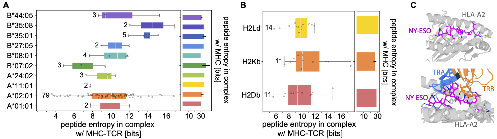
Fig. 5A shows the entropy of these peptide distributions for 105 TCR-MHC pairs across 10 human MHC-I alleles; see Table S6 for details on these structures. The median peptide entropy across these TCR-MHC pairs is approximately 10 bits, indicating that a typical TCR-MHC pair can recognize on the order of peptides. In contrast, examining the degeneracy of MHC-I presentation alone in humans–using the peptide PWMs gathered from the MHC Motif Atlas [54]–yields a median entropy of 31 bits, implying that MHC-I molecules can typically recognize distinct peptides (Fig. 5A). A similar analysis on 36 TCR-MHC pairs spanning three mouse MHC-I alleles shows comparable trends, with a MHC-I degeneracy on the order of peptides (29 bits), and the TCR-MHC degeneracy of peptides (10 bits); see Fig. 5B, and Table S6 for details on these structures. This pronounced reduction in entropy—observed by comparing the distribution of peptides recognized by MHC-I molecules alone to that recognized by TCR-MHC-I complexes—quantifies how TCRs constrain the accessible antigenic shape space, as illustrated by the polar interactions that they form with peptides in Fig. 5C.
Because our analyses focus on interactions involving typical TCR and MHC molecules, our entropy-based degeneracy estimates are smaller than those from previous theoretical studies, which often relied on experimental observations from autoimmune T-cells or limited synthetic peptide libraries [65]. Moreover, the HERMES-fixed model generates PWMs based on peptides whose backbone conformations match those of the wild-type template, likely underestimating the true degeneracy of TCR-MHC complexes. Although HERMES-relaxed can partially address this issue, its designs are only reliable within roughly five amino acids of the native peptides (Fig. 4), preventing a full exploration of the possible peptide poses for a given TCR-MHC complex. Nonetheless, our estimate can serve as informative lower bounds on typical MHC-I and TCR degeneracies.
IV Discussion
In this work, we introduced a structure-based machine learning model to predict TCR-pMHC interactions and to design novel reactive peptides for TCR-MHC complexes. Our approach builds on HERMES, a physically motivated equivariant neural network model that we previously developed [26, 27]. Trained on the protein universe, HERMES predicts amino acid propensities based on their atomic environments in protein structures, and has been shown to implicitly learn biophysical interatomic interactions in proteins [27], enabling it to make highly generalizable predictions. This generalizability is particularly important for TCR-pMHC complexes, where only a modest number of experimentally resolved structures are available, and computational algorithms often struggle to accurately predict new structures [17, 18].
We found that HERMES-predicted peptide energies closely tracked both TCR-pMHC binding affinities and peptide-induced T-cell activities across diverse systems, including variants from a CMV-derived viral epitope [36], the NY-ESO cancer-testis antigen [43, 44], and a melanoma self-antigen [36]. Notably, HERMES also accurately predicted the activity of p53-derived neoepitope variants in the context of a bispecific antibody-mediated T-cell therapy [5]. This result underscores the algorithm’s broad applicability and generalizability: given a reliable structural template close to the system of interest, HERMES can predict peptide-induced T-cell responses, whether through direct TCR interactions or antibody-mediated mechanisms.
Based on the HERMES predictions for TCR-pMHC reactivities, we presented a computational protocol for de novo design of immunogenic peptides that can elicit robust T-cell responses. Starting from the structural template of a TCR-MHC with a native (wild-type) peptide, we developed an MCMC search guided by HERMES peptide energies to explore the vast sequence space of roughly () possibilities for 9-residue (11-residue) peptides, to identify immunogenic candidates. Experimental validation across three systems—with native peptides from NY-ESO and MAGE cancer epitopes, plus a viral epitope from EBV—revealed that our designs reliably activated T-cells, even with up to five substitutions from the native peptide used in the structural template. Notably, several MAGE-derived variants outperformed or matched the native peptide in T-cell activation, demonstrating the method’s potential to enhance peptide potency. These results show the potential of HERMES for designing peptide-based cancer vaccines with neoantigens, providing an avenue for personalized immunotherapy strategies.
Accurately modeling T-cell specificity remains challenging, in part due to the limited functional data on TCR-epitope pairs, and the highly cross-reactive nature of TCR-epitope interactions. A design protocol like the one presented here, coupled with experimental validation, offers a promising path forward for future studies. By iteratively generating candidate peptide libraries, testing them experimentally, and refining the model with each iteration, it should be possible to more efficiently explore the high dimensional antigenic sequence space. This active learning strategy has a potential for revealing the “shape space” of TCR-epitope interactions, with broad applicabilities for early disease diagnosis, and for design of targeted immunotherapies. Moreover, this approach can help mitigate immunotherapy-related toxicity by identifying and excluding potential self-reactivities–beyond single point mutations in the target epitope–such as the off-target Titin reactivity observed in an engineered T-cell therapy against MAGE-A3 that led to a severe cardiac toxicity [57].
Although our findings highlight the promise of structure-based models for immune recognition, a key limitation remains the scarcity of high-quality TCR-pMHC structural templates, despite advances from tools like AlphaFold3 [29] and TCRDock [17]. On the other hand, while TCR sequences are more widely available, sequence-based models often lack generalizability because existing paired TCR-pMHC data are skewed toward just a few peptides [12, 11]. A promising direction for future work lies in developing multi-modal flexible methods that integrate structural and sequence-based models, leveraging both the depth and generalizability offered by structural information and the scalability of sequence data.
Although our study primarily examines MHC class I-restricted peptides, the broad applicability of structure-based modeling suggests that similar methods could be adapted for the more complex TCR-peptide-MHCII interactions, thereby offering new insights into CD4+ T-cell responses. Moreover, incorporating additional immunological factors—such as TCR clustering, co-receptor interactions, cross-reactivity with self-antigens, and functional immune profiling—can further enhance the accuracy of T-cell response predictions and improve the resulting de novo peptide design pipeline.
V Data and code availability
Data and code for all the analyses can be found in the Github repository: https://github.com/StatPhysBio/tcr_antigen_design. Code for the original HERMES [26] model can be found in the Github repository: https://github.com/StatPhysBio/hermes.
Acknowledgment
We thank Mikhail V. Pogorelyy for valuable comments and help with CellCyte. This work has been supported by the National Institutes of Health MIRA awards R35 GM142795 (AN, GV, MNP) and R35 GM141457 (PB), the CAREER award from the National Science Foundation grant 2045054 (AN), the National Institutes of Health R01 award AI136514 (PT and PB), the Royalty Research Fund from the University of Washington no. A153352 (AN, MNP), the Allen School Computer Science & Engineering Research Fellowship from the Paul G. Allen School of Computer Science & Engineering at the University of Washington (GV), the Microsoft Azure award from the eScience institute at the University of Washington (AN, GV, MNP), the TIRTL Bluesky Initiative at St. Jude Children’s Research Hospital (PT), and the ALSAC at St. Jude Children’s Research Hospital (PT and AAM). This work is also supported, in part, through the Departments of Physics and Computer Science and Engineering, and the College of Arts and Sciences at the University of Washington (AN, GV, MNP). This work was performed in part during the 2023 summer workshop “Statistical Physics and Adaptive Immunity” at the Aspen Center for Physics, which is supported by National Science Foundation grant PHY-2210452. This work also benefited from discussions during the 2024 program “Interactions and Co-evolution between Viruses and Immune Systems” at the Kavli Institute for theoretical physics (KITP), which is supported by National Science Foundation grants PHY-2309135, and PHY-2309135, and the Gordon and Betty Moore Foundation Grant No. 2919.02.
Competing Interest Statement
PT is on the Scientific Advisory Board of Immunoscape and Shennon Bio, has received research support and personal fees from Elevate Bio, and consulted for 10X Genomics, Illumina, Pfizer, Cytoagents, Sanofi, Merck, and JNJ.
References
- [1] Attaf M, Huseby E, Sewell AK (2015) T cell receptors as predictors of health and disease Cell. Mol. Immunol. 12:391–399.
- [2] Liu B, et al. (2024) Design of high specificity binders for peptide-MHC-I complexes bioRxiv 2024.11.28.625793.
- [3] Leidner R, et al. (2022) Neoantigen T-cell receptor gene therapy in pancreatic cancer N. Engl. J. Med. 386:2112–2119.
- [4] Mirazee JM, et al. (2024) Enhancing CAR T cell therapy through in silico modeling of hinge sequence length based on epitope location Biophys. J. 123:552a.
- [5] Hsiue EHC, et al. (2021) Targeting a neoantigen derived from a common TP53 mutation Science 371:eabc8697.
- [6] Blass E, Ott PA (2021) Advances in the development of personalized neoantigen-based therapeutic cancer vaccines Nat. Rev. Clin. Oncol. 18:215–229.
- [7] Rojas LA, et al. (2023) Personalized RNA neoantigen vaccines stimulate T cells in pancreatic cancer Nature 618:144–150.
- [8] Zhang SQ, et al. (2018) High-throughput determination of the antigen specificities of T cell receptors in single cells Nat. Biotechnol. 36:1156–1159.
- [9] Nolan S, et al. (2025) A large-scale database of T-cell receptor beta (TCR) sequences and binding associations from natural and synthetic exposure to SARS-CoV-2 Front. Immunol. 16:1488851.
- [10] Dash P, et al. (2017) Quantifiable predictive features define epitope-specific T cell receptor repertoires Nature 547:89–93.
- [11] Vita R, et al. (2019) The Immune Epitope Database (IEDB): 2018 update Nucleic Acids Res. 47:D339–D343.
- [12] Shugay M, et al. (2018) VDJdb: a curated database of T-cell receptor sequences with known antigen specificity Nucleic Acids Res. 46:D419–D427.
- [13] Isacchini G, Walczak AM, Mora T, Nourmohammad A (2021) Deep generative selection models of T and B cell receptor repertoires with soNNia Proc. Natl. Acad. Sci. U. S. A. 118:e2023141118.
- [14] Weber A, Born J, Rodriguez Martínez M (2021) TITAN: T-cell receptor specificity prediction with bimodal attention networks Bioinformatics 37:i237–i244.
- [15] Meynard-Piganeau B, Feinauer C, Weigt M, Walczak AM, Mora T (2024) TULIP: A transformer-based unsupervised language model for interacting peptides and T cell receptors that generalizes to unseen epitopes Proc. Natl. Acad. Sci. U. S. A. 121:e2316401121.
- [16] Piepenbrink KH, Gloor BE, Armstrong KM, Baker BM (2009) Methods for quantifying T cell receptor binding affinities and thermodynamics Methods Enzymol. 466:359–381.
- [17] Bradley P (2023) Structure-based prediction of T cell receptor:peptide-MHC interactions Elife 12:e82813.
- [18] Yin R, et al. (2023) TCRmodel2: high-resolution modeling of T cell receptor recognition using deep learning Nucleic Acids Res. 51:569–576.
- [19] Rives A, et al. (2021) Biological structure and function emerge from scaling unsupervised learning to 250 million protein sequences Proc. Natl. Acad. Sci. U. S. A. 118:e2016239118.
- [20] Lin Z, et al. (2023) Evolutionary-scale prediction of atomic-level protein structure with a language model Science 379:1123–1130.
- [21] Madani A, et al. (2023) Large language models generate functional protein sequences across diverse families Nat. Biotechnol. 41:1099–1106.
- [22] Schmirler R, Heinzinger M, Rost B (2024) Fine-tuning protein language models boosts predictions across diverse tasks Nat. Commun. 15:7407.
- [23] Wang Y, et al. (2024) An explainable language model for antibody specificity prediction using curated influenza hemagglutinin antibodies Immunity 57:2453–2465.e7.
- [24] Hie BL, et al. (2024) Efficient evolution of human antibodies from general protein language models Nat. Biotechnol. 42:275–283.
- [25] Bravi B, et al. (2023) A transfer-learning approach to predict antigen immunogenicity and T-cell receptor specificity Elife 12:e85126.
- [26] Visani GM, et al. (2024) HERMES: Holographic Equivariant neuRal network model for Mutational Effect and Stability prediction bioRxiv 2024.07.09.602403.
- [27] Pun MN, et al. (2024) Learning the shape of protein microenvironments with a holographic convolutional neural network Proc. Natl. Acad. Sci. U. S. A. 121:e2300838121.
- [28] Chaudhury S, Lyskov S, Gray JJ (2010) PyRosetta: a script-based interface for implementing molecular modeling algorithms using Rosetta Bioinformatics 26:689–691.
- [29] Abramson J, et al. (2024) Accurate structure prediction of biomolecular interactions with AlphaFold 3 Nature 630:493–500.
- [30] Dauparas J, et al. (2022) Robust deep learning-based protein sequence design using ProteinMPNN Science 378:49–56.
- [31] Blaabjerg LM, et al. (2023) Rapid protein stability prediction using deep learning representations Elife 12:e82593.
- [32] Michalewicz K, Barahona M, Bravi B (2024) ANTIPASTI: Interpretable prediction of antibody binding affinity exploiting normal modes and deep learning Structure 32:2422–2434.e5.
- [33] Diaz DJ, et al. (2024) Stability Oracle: a structure-based graph-transformer framework for identifying stabilizing mutations Nat. Commun. 15:6170.
- [34] Chen JL, et al. (2005) Structural and kinetic basis for heightened immunogenicity of T cell vaccines J. Exp. Med. 201:1243–1255.
- [35] Garboczi DN, et al. (1996) Structure of the complex between human T-cell receptor, viral peptide and HLA-A2 Nature 384:134–141.
- [36] Łuksza M, et al. (2022) Neoantigen quality predicts immunoediting in survivors of pancreatic cancer Nature 606:389–395.
- [37] Yang X, et al. (2015) Structural basis for clonal diversity of the public T cell response to a dominant human Cytomegalovirus Epitope J. Biol. Chem. 290:29106–29119.
- [38] Davis MM, et al. (1998) Ligand recognition by alpha beta T cell receptors Annu. Rev. Immunol. 16:523–544.
- [39] Zhong S, et al. (2013) T-cell receptor affinity and avidity defines antitumor response and autoimmunity in T-cell immunotherapy Proc. Natl. Acad. Sci. U. S. A. 110:6973–6978.
- [40] Soto CM, et al. (2013) MHC-class I-restricted CD4 T cells: a nanomolar affinity TCR has improved anti-tumor efficacy in vivo compared to the micromolar wild-type TCR Cancer Immunol. Immunother. 62:359–369.
- [41] Li Y, et al. (2005) Directed evolution of human T-cell receptors with picomolar affinities by phage display Nat. Biotechnol. 23:349–354.
- [42] Dunn SM, et al. (2006) Directed evolution of human T cell receptor CDR2 residues by phage display dramatically enhances affinity for cognate peptide-MHC without increasing apparent cross-reactivity Protein Sci. 15:710–721.
- [43] Aleksic M, et al. (2010) Dependence of T cell antigen recognition on T cell receptor-peptide MHC confinement time Immunity 32:163–174.
- [44] Pettmann J, et al. (2021) The discriminatory power of the T cell receptor Elife 10:e67092.
- [45] Esfandiary A, Ghafouri-Fard S (2015) New York esophageal squamous cell carcinoma-1 and cancer immunotherapy Immunotherapy 7:411–439.
- [46] Chen JL, et al. (2000) Identification of NY-ESO-1 peptide analogues capable of improved stimulation of tumor-reactive CTL J. Immunol. 165:948–955.
- [47] Henikoff S, Henikoff JG (1992) Amino acid substitution matrices from protein blocks Proc. Natl. Acad. Sci. U. S. A. 89:10915–10919.
- [48] Altan-Bonnet G, Germain RN (2005) Modeling T cell antigen discrimination based on feedback control of digital ERK responses PLoS Biol. 3:e356.
- [49] Achar SR, et al. (2022) Universal antigen encoding of T cell activation from high-dimensional cytokine dynamics Science 376:880–884.
- [50] Stone JD, Chervin AS, Kranz DM (2009) T-cell receptor binding affinities and kinetics: impact on T-cell activity and specificity Immunology 126:165–176.
- [51] Gras S, et al. (2010) Allelic polymorphism in the T cell receptor and its impact on immune responses J. Exp. Med. 207:1555–1567.
- [52] Liu YC, et al. (2014) A molecular basis for the interplay between T cells, viral mutants, and human leukocyte antigen micropolymorphism J. Biol. Chem. 289:16688–16698.
- [53] Raman MCC, et al. (2016) Direct molecular mimicry enables off-target cardiovascular toxicity by an enhanced affinity TCR designed for cancer immunotherapy Sci. Rep. 6:18851.
- [54] Tadros DM, Eggenschwiler S, Racle J, Gfeller D (2023) The MHC Motif Atlas: a database of MHC binding specificities and ligands Nucleic Acids Res. 51:D428–D437.
- [55] Vormehr M, Türeci, Ö, Sahin U (2019) Harnessing tumor mutations for truly individualized cancer vaccines Annu. Rev. Med. 70:395–407.
- [56] Song K, Pun SH (2024) Design and evaluation of synthetic delivery formulations for peptide-based cancer vaccines BME Front. 5:0038.
- [57] Linette GP, et al. (2013) Cardiovascular toxicity and titin cross-reactivity of affinity-enhanced T cells in myeloma and melanoma Blood 122:863–871.
- [58] Cameron BJ, et al. (2013) Identification of a Titin-derived HLA-A1-presented peptide as a cross-reactive target for engineered MAGE A3-directed T cells Sci. Transl. Med. 5:197ra103.
- [59] Lundegaard C, et al. (2008) NetMHC-3.0: accurate web accessible predictions of human, mouse and monkey MHC class I affinities for peptides of length 8-11 Nucleic Acids Res. 36:W509–12.
- [60] Lundegaard C, Lund O, Nielsen M (2008) Accurate approximation method for prediction of class I MHC affinities for peptides of length 8, 10 and 11 using prediction tools trained on 9mers Bioinformatics 24:1397–1398.
- [61] Andreatta M, Nielsen M (2016) Gapped sequence alignment using artificial neural networks: application to the MHC class I system Bioinformatics 32:511–517.
- [62] Roney JP, Ovchinnikov S (2022) State-of-the-Art Estimation of Protein Model Accuracy Using AlphaFold Phys. Rev. Lett. 129:238101.
- [63] Webb AI, et al. (2004) Functional and structural characteristics of NY-ESO-1-related HLA A2-restricted epitopes and the design of a novel immunogenic analogue J. Biol. Chem. 279:23438–23446.
- [64] Wooldridge L, et al. (2012) A single autoimmune T cell receptor recognizes more than a million different peptides J. Biol. Chem. 287:1168–1177.
- [65] Sewell AK (2012) Why must T cells be cross-reactive? Nat. Rev. Immunol. 12:669–677.