Enhancing Fetal Plane Classification Accuracy with Data Augmentation Using Diffusion Models
Abstract
Ultrasound imaging is widely used in medical diagnosis, especially for fetal health assessment. However, the availability of high-quality annotated ultrasound images is limited, which restricts the training of machine learning models. In this paper, we investigate the use of diffusion models to generate synthetic ultrasound images to improve the performance on fetal plane classification. We train different classifiers first on synthetic images and then fine-tune them with real images. Extensive experimental results demonstrate that incorporating generated images into training pipelines leads to better classification accuracy than training with real images alone. The findings suggest that generating synthetic data using diffusion models can be a valuable tool in overcoming the challenges of data scarcity in ultrasound medical imaging.
Keywords Diffusion Model Data Augmentation Fetal Ultrasound Deep Learning
1 INTRODUCTION
Over the past decade, deep learning has achieved great success in medical image diagnostics, including tasks such as nodule detection Pehrson et al. (2019), image segmentation Wang et al. (2022) and classification Cai et al. (2020). Deep learning models, such as convolutional neural networks (CNNs) LeCun et al. (1989), and residual networks (ResNets) He et al. (2016), trained on large datasets, can learn intricate patterns in medical images, improving diagnostic accuracy and supporting healthcare professionals. However, the successful application of machine learning to medical imaging is heavily dependent on the availability of large, high-quality annotated datasets Alzubaidi et al. (2021). In many fields of medicine, obtaining such datasets can be difficult, and this issue is especially pronounced in ultrasound imaging Huang et al. (2021).
Ultrasound imaging plays a crucial role in prenatal care, providing valuable and non-invasive insights into fetal health and development Brattain et al. (2018). Despite their widespread use, one of the key challenges in developing deep learning models for ultrasound imaging is data scarcity. There are several reasons why obtaining a large dataset of labeled ultrasound images is difficult.
Firstly, due to privacy concerns, hospitals and medical institutions are often unable to release patient data, making it challenging to build large public datasets DuMont Schütte et al. (2021). Furthermore, even if hospitals are willing to share data, obtaining labeled data is costly and time-consuming. Expert knowledge is required to correctly annotate medical images, and for ultrasound, this usually means that radiologists or sonographers must manually label features of interest, such as fetal planes or abnormalities. This process is not only slow but also expensive, which limits the amount of labeled ultrasound data available for training machine learning models.
Given these challenges, there has been growing interest in the use of synthetic data and generative models to supplement real-world data. Recent advances in generative models, such as Generative Adversarial Networks (GANs) Goodfellow et al. (2014) and Diffusion Models Ho et al. (2020), have opened up new possibilities for creating realistic synthetic medical images. These models can learn complex data distributions from real images and generate new synthetic images that resemble the originals. This approach has shown promise in a variety of medical imaging applications. For instance, Frid-Adar, M., et al. (2018) Frid-Adar et al. (2018) demonstrated the use of GANs for generating synthetic liver lesions in CT images. Motamed et al. (2017) Motamed et al. (2021) showed the effectiveness of using GANs to generate synthetic chest X-ray images for training classifiers. Yi et al. (2019) Yi et al. (2019) provided a comprehensive review of GAN-based techniques for generating synthetic medical images. Pan et al. (2022) Pan et al. (2023) demonstrated that diffusion models generate more realistic synthetic CT and MRI images for medical image segmentation than GANs. Hardy et al. (2023) Hardy et al. (2023) showed that generating realistic synthetic liver ultrasound images with a diffusion model can enhance liver disease classification performance. However, the application of generative models to ultrasound imaging still remains less explored, particularly for fetal ultrasound, where data scarcity is a significant challenge.
In this work, we propose the use of diffusion models to generate synthetic fetal ultrasound images and investigate their potential to improve the performance of machine learning classifiers. Unlike GANs, which can suffer from training instability and mode collapse Kodali et al. (2017), diffusion models offer more stable training dynamics and produce high-quality, diverse samples, making them well-suited for this application Dhariwal and Nichol (2021). Specifically, we hypothesize that combining real ultrasound images with generated images can enhance classifier accuracy, especially in cases where annotated real data is limited. By training the model first on synthetic images and then fine-tuning it on real images, we aim to overcome the data scarcity problem in fetal ultrasound imaging. We make the following key contributions:
-
•
We generate in total synthetic ultrasound images of fetal planes using a trained diffusion model. These images, which will be made publicly available, can serve as a valuable resource for future research in medical image classification.
-
•
We conduct experiments with six different classifiers and show that pretraining the classifiers with synthetic images followed by fine-tuning on real images results in improved classification accuracy compared to training only with real images.
-
•
To gain deeper insights into the classifier’s behavior, we analyze the confusion matrices. Our findings reveal that the improvement in classification performance is particularly significant for imbalanced classes with small numbers of images.
-
•
Furthermore, we observe a consistent trend: as the number of generated synthetic images increases, the classification performance after pretraining and fine-tuning consistently improves. These results demonstrate the effectiveness of leveraging synthetic data to enhance the robustness and accuracy of medical image classifiers, especially in scenarios where real-world data is limited or imbalanced.
By addressing the data scarcity issue and demonstrating the potential of synthetic ultrasound images in improving classifier performance, our work opens up new possibilities for the use of generative models in medical image analysis, particularly in fields where obtaining large labeled datasets is challenging.
2 RELATED WORK
The challenge of data scarcity in medical imaging has led researchers to explore a variety of solutions to augment training datasets. These approaches can broadly be categorized into traditional methods, and the more recent deep learning-based methods. This section provides an overview of the key works in each of these categories.
2.1 Traditional Approaches
Traditionally, data scarcity in medical imaging has been addressed through various data augmentation techniques. These methods aim to artificially expand the size of a dataset without requiring additional labeled images. Early data augmentation strategies for medical images included transformations such as rotation, scaling, cropping, flipping, and elastic deformations. These techniques were particularly useful in fields like MRI and CT imaging, where the generation of new images from the same subject could help the model generalize better Goceri (2023). For general imaging, methods such as image cropping and noise addition have been widely used. Simple geometric transformations, such as rotation and scaling have been applied broadly as well. While these methods can be effective in increasing the volume of data, they often fail to generate truly novel variations of the data, which limits their ability to capture the full diversity of possible real-world cases Goceri (2023).
Another traditional approach to tackling data scarcity is transfer learning, which has been used in many medical imaging tasks Sanford et al. (2020). Transfer learning allows models trained on large, publicly available datasets (such as ImageNet) to be adapted to medical datasets with fewer labeled samples. While effective in some contexts, transfer learning still relies heavily on the availability of a reasonable amount of high-quality annotated data, making it less suitable in situations where data is extremely limited or highly specialized, such as in fetal ultrasound imaging.
2.2 Deep Learning-Based Approaches
The rise of deep learning has revolutionized medical image analysis, neural networks becoming the most widely used in medical imaging. However, these methods often require large amounts of labeled data to perform well. This has led to significant interest in using deep learning to generate synthetic medical images as a way of alleviating data scarcity.
In particular, Generative Adversarial Networks (GANs) have been explored as a way to generate synthetic medical images. GANs consist of two neural networks: a generator that creates synthetic data, and a discriminator that attempts to distinguish between real and fake data. GANs have been applied to various imaging modalities, including CT, MRI, and ultrasound. For instance, Frid-Adar et al. (2018) Frid-Adar et al. (2018) used GANs to generate synthetic CT images of liver lesions, which were used to augment a classifier for liver lesion detection. Similarly, Motamed et al. (2017) Motamed et al. (2021) showed the effectiveness of using GANs to generate synthetic chest X-ray images for training classifiers. In ultrasound imaging, GANs have shown promise in generating synthetic images for various purposes. Montero et al. (2021) Montero et al. (2021) applied a GAN-based approach to generate synthetic fetal ultrasound images, focusing on augmenting data for training classifiers on fetal development stages. While GANs have shown considerable success, one challenge with these models is the difficulty in training them, especially in medical imaging, where the distribution of data can be complex and diverse. In addition to GANs, Variational Autoencoders (VAEs) have also been used for generating synthetic medical images. VAEs are another class of generative models that learn a probabilistic mapping between a low-dimensional latent space and high-dimensional image space. Studies such as Dorent et al. (2023) Dorent et al. (2023) have demonstrated the potential of VAEs for generating synthetic ultrasound and MRI images for the purposes of data augmentation.
In recent years, diffusion models have emerged as a powerful new class of generative models, particularly in the field of image synthesis Ho et al. (2020). Diffusion models work by simulating a gradual “noising” process that adds random noise to data in a series of steps, and then by learning how to reverse this process they can generate high-quality samples. One key advantage of diffusion models over GANs and VAEs is their ability to produce high-fidelity images with more stable training dynamics.
The use of diffusion models in medical imaging is a relatively recent development, but promising results have already been reported in various applications. Dhariwal and Nichol (2021) Dhariwal and Nichol (2021) demonstrated the use of diffusion models for generating high-quality images in a variety of domains, including natural images, and showed that they outperform GANs in terms of image quality and diversity. In medical imaging, recent studies have started to explore diffusion models for generating synthetic data. Pan et al. (2022) Pan et al. (2023) applied diffusion models to generate synthetic CT and MRI images for medical image segmentation tasks, showing that diffusion models can generate more realistic images compared to traditional methods like GANs. Similarly, in the field of ultrasound imaging, Hardy et al. (2023) Hardy et al. (2023) used a diffusion model to generate realistic synthetic liver ultrasound images, demonstrating that such generated data could improve the performance of a liver disease classification model. However, the application of diffusion models to fetal ultrasound images remains an underexplored area, and there is a gap in the literature regarding how these models can be applied to enhance classification performance for fetal health assessment.
3 METHODS
In this section, we describe the methodology used to generate synthetic fetal ultrasound images and train a classifier to evaluate their utility in improving classification performance. We employ a classifier-guided diffusion model, which combines the power of generative models and supervised learning to generate class-specific images. Our method involves two main components: the training of the diffusion model and the classification task.
3.1 Classifier-Guided Diffusion Model
We use a classifier-guided diffusion model to generate synthetic ultrasound images Dhariwal and Nichol (2021). In this approach, the diffusion model is conditioned on the classifier’s predictions, allowing it to generate images that belong to specific classes. This method enhances the model’s ability to produce class-specific images that better align with real-world data, improving the quality and relevance of the generated samples. The overall process consists of two interconnected models: the diffusion model and the classifier.
The diffusion model is trained to learn the data distribution of fetal ultrasound images across different classes. A key feature of this approach is that we condition the image generation process on the class labels. Unlike traditional diffusion models that generate images from random noise, the classifier-guided diffusion model is trained such that it can guide the image generation towards specific class categories during the denoising process. During training, the classifier provides the model with class-specific information, which helps the diffusion model generate images that are representative of the corresponding class.
The classifier is trained simultaneously with the diffusion model to predict the class label of a given image. The classifier is a half U-Net (containing only the encoder part) that learns to differentiate between the various classes of fetal ultrasound images. Using a half U-Net allows for efficient feature extraction with fewer parameters, while still capturing essential spatial hierarchies needed for accurate class prediction.
During the training phase, the classifier is used to provide feedback to the diffusion model, ensuring that the generated images match the intended class distribution. The classifier-guided diffusion model allows us to generate high-quality synthetic images that are not only realistic but also tailored to each class of interest, improving the relevance of the synthetic data for training downstream classifiers.
3.2 Ultrasound Image Dataset
To train and evaluate our proposed method, we use the FETAL_PLANES_DB Burgos-Artizzu et al. (2020), a commonly used maternal-fetal ultrasound image dataset. The FETAL_PLANES_DB contains in total images of fetal planes, which are essential in prenatal care for monitoring fetal development. The dataset is divided into six distinct classes, i.e., Abdomen, Brain, Femur, Thorax, the mother’s cervix (widely used for prematurity screening), and a general category to include any other less common image plane. These classes represent different features that are crucial for assessing fetal health.
| Class index | Class name | # images | # training images | # test images |
|---|---|---|---|---|
| 0 | Fetal abdomen | |||
| 1 | Fetal brain | |||
| 2 | Fetal femur | |||
| 3 | Fetal thorax | |||
| 4 | Maternal cervix | |||
| 5 | Other | |||
| Total |
The FETAL_PLANES_DB dataset provides a valuable resource for training models in fetal ultrasound image classification. However, due to the relatively small size of the dataset, we face the common challenge of data scarcity, which makes it difficult to train robust deep learning models. Additionally, as shown in Table 1 there is an imbalance between different classes, which can further hinder model performance. To address these issues, we augment the dataset using the classifier-guided diffusion model.
3.3 Synthetic Image Generation
We generate synthetic images in two rounds. In each round, for each of the six classes, we generate synthetic images, yielding a total of synthetic images. Therefore, in total, we have synthetic images. These images are generated with the help of the diffusion model, which conditions the image generation process on the class label, ensuring that each set of images corresponds to a particular class. The generated images exhibit characteristics that are consistent with the anatomical features of fetal ultrasound images, as guided by the class-specific classifier.
| Class name | # synthetic images | # synthetic images of Round | # synthetic images of Round |
|---|---|---|---|
| Fetal abdomen | |||
| Fetal brain | |||
| Fetal femur | |||
| Fetal thorax | |||
| Maternal cervix | |||
| Other | |||
| Total |
The generated images are then used to augment the real FETAL_PLANES_DB dataset, which contains images. The augmented dataset is used in a downstream classification task. This synthetic data generation process not only alleviates the issue of limited labeled data but also introduces diversity in the training data, helping the model generalize better to unseen data.
3.4 Evaluation of Generated Images: Downstream Classification Tasks
First, we use synthetic images in the first round of generation and conduct a two-stage training process. In the first stage, we pretrain the classifier on the synthetic images generated by the classifier-guided diffusion model. These images are distributed across the six classes, and the classifier learns to distinguish between them. The pretraining process helps the classifier to better understand the data distribution and feature representations specific to each class. In the second stage, the pretrained classifier is fine-tuned using the real images from the FETAL_PLANES_DB dataset. The fine-tuning step ensures that the classifier adjusts to the true distribution of real ultrasound images and improves its generalization ability. This two-stage approach helps overcome the data scarcity problem by leveraging synthetic images to boost performance on a relatively small set of real data. After training, the classifier’s performance is evaluated using standard classification metrics, such as accuracy, and confusion matrix. The results are compared with classifiers trained solely on real images to assess the impact of the synthetic data. Finally, we perform similar procedure as above using the whole synthetic images from the two rounds of generation. A second round of image generation was conducted to investigate whether increasing the number of synthetic images would lead to further improvements in classification performance.
4 EXPERIMENTS
In this section, we describe the details of the experimental setup used to evaluate the proposed method. This includes the dataset acquisition and preparation process, data preprocessing, the architecture of the models used, and the techniques employed for image generation and classification. We also provide details on the evaluation metrics used to assess the quality of the generated images and the classification performance.
4.1 Dataset Acquisition and Preparation
The FETAL_PLANES_DB is a publicly available maternal-fetal ultrasound image dataset †††https://zenodo.org/records/3904280. As shown in Table 1, the FETAL_PLANES_DB dataset was split into an training and test set, resulting in images for training and images for testing. This common split ensures a representative sample of the data is used to train the model while reserving a separate set of images to evaluate its generalization ability on unseen data. The portion (training set) is used to train the model. The model learns patterns and relationships within the data to make accurate predictions on new data. The portion (test set) was used to assess the model’s performance on unseen data. This helped identify how well the model generalized to real-world scenarios beyond the training data. By splitting the data in this way, a balance between training the model effectively could be achieved and ensuring it performed well on data it has not encountered before.
The FETAL_PLANES_DB dataset contains a limited number of images per class, making it challenging to train robust deep learning models directly on the real data. To address this issue, we used the classifier-guided diffusion model to generate synthetic images, thereby augmenting the dataset and enhancing the generalization ability of the model.
4.2 Data Preprocessing
Before training the models, several preprocessing steps on the real images were performed. First, all images were normalized to have pixel values in the range . This ensures that the neural networks can learn efficiently by reducing the risk of vanishing or exploding gradients. Second, the ultrasound images in the dataset were resized to a uniform resolution of pixels. This size was chosen to maintain sufficient detail in the images while reducing computational overhead. Each image in the dataset was assigned one of the six class labels corresponding to different fetal anatomical planes. The synthetic images generated by the diffusion model were also labelled according to their respective class. We also applied traditional data augmentation techniques to real images. Random rotations of the images between and was employed to simulate different scanning angles. Horizontal and vertical flippings were used to account for different fetal orientations in the ultrasound.
4.3 Model Architectures
For this study, a two-part architecture was used: a diffusion model for image generation and five different classifiers for downstream classification tasks. The diffusion model was based on a state-of-the-art architecture for image generation, specifically a classifier-guided diffusion model Dhariwal and Nichol (2021). The model consists of an encoder-decoder architecture with several layers of convolutional and residual blocks. The diffusion process introduces random noise to the images and learns to reverse this process to generate clean, realistic images. During training, the model was conditioned on class labels to ensure that the generated images belong to the correct class. For the downstream classification tasks, we used five different classifiers to assess the performance of the synthetic images generated by the diffusion model. These classifiers represent a range of architectures, from traditional CNNs to modern transformer-based models. The details of five classifiers are as follows.
-
•
ResNet50 He et al. (2016): A deep convolutional neural network with residual connections that enable it to learn deep representations while avoiding the vanishing gradient problem.
-
•
DenseNet169 Huang et al. (2017): A variant of CNNs where each layer is connected to every other layer in a dense fashion, improving feature reuse and gradient flow.
-
•
ViT_b_32 (Vision Transformer) Dosovitskiy (2020): A transformer-based model that splits images into patches and uses self-attention mechanisms to learn global image representations. The “b_32” refers to the base version of ViT with input patch size of .
-
•
Swin_t (Swin Transformer) Liu et al. (2021): A hierarchical transformer model designed for vision tasks. It uses shifted windowing mechanisms for efficient computation and local-global feature learning.
-
•
MedMamba Yue and Li (2024): A medical-specific model that combines transformers and CNNs tailored for medical image analysis. MedMamba uses medical image priors to improve its performance on healthcare datasets.
The goal is to compare how well each of these models performs when trained on real images, synthetic images, and augmented data using the proposed method.
4.4 Diffusion Model Training and Image Generation
During training, we used real images from the FETAL_PLANES_DB dataset to learn the image distribution, as shown in Table 1. The diffusion model was trained to generate images that match the characteristics of the real ultrasound images in each class. Following Dhariwal and Nichol (2021) Dhariwal and Nichol (2021), we used mean-squared error loss between the true noise and the predicted noise to train the diffusion model, and used cross-entropy loss to train a half U-Net to provide classifier-guided information to the diffusion model. After training, the model can generate synthetic images by conditioning on the class label. As shown in Table 2, in each round, for each of the six classes, we generated synthetic images, resulting in a total of synthetic images.
5 Results
In this section, we present the experimental results obtained from our proposed method. We evaluated both the qualitative and quantitative performance of the generated images and the classifiers trained on real, synthetic, and augmented data.
5.1 Visualization of Synthetic Images
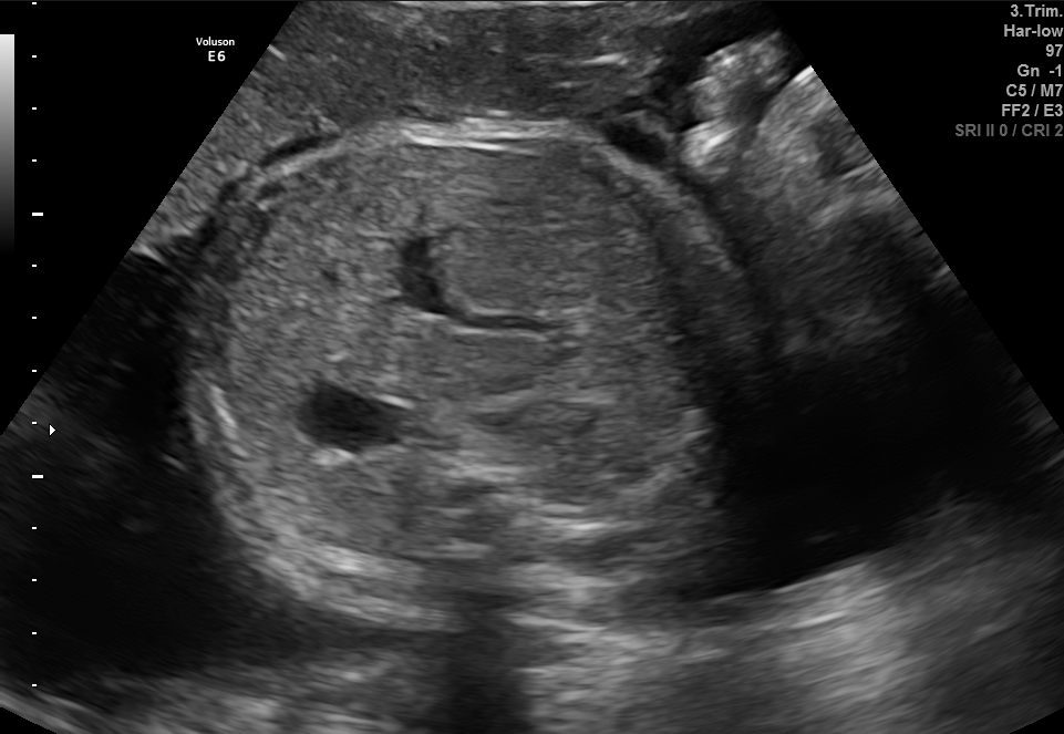
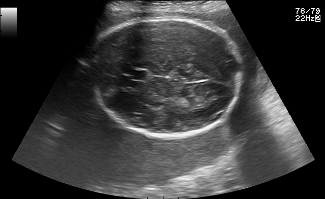
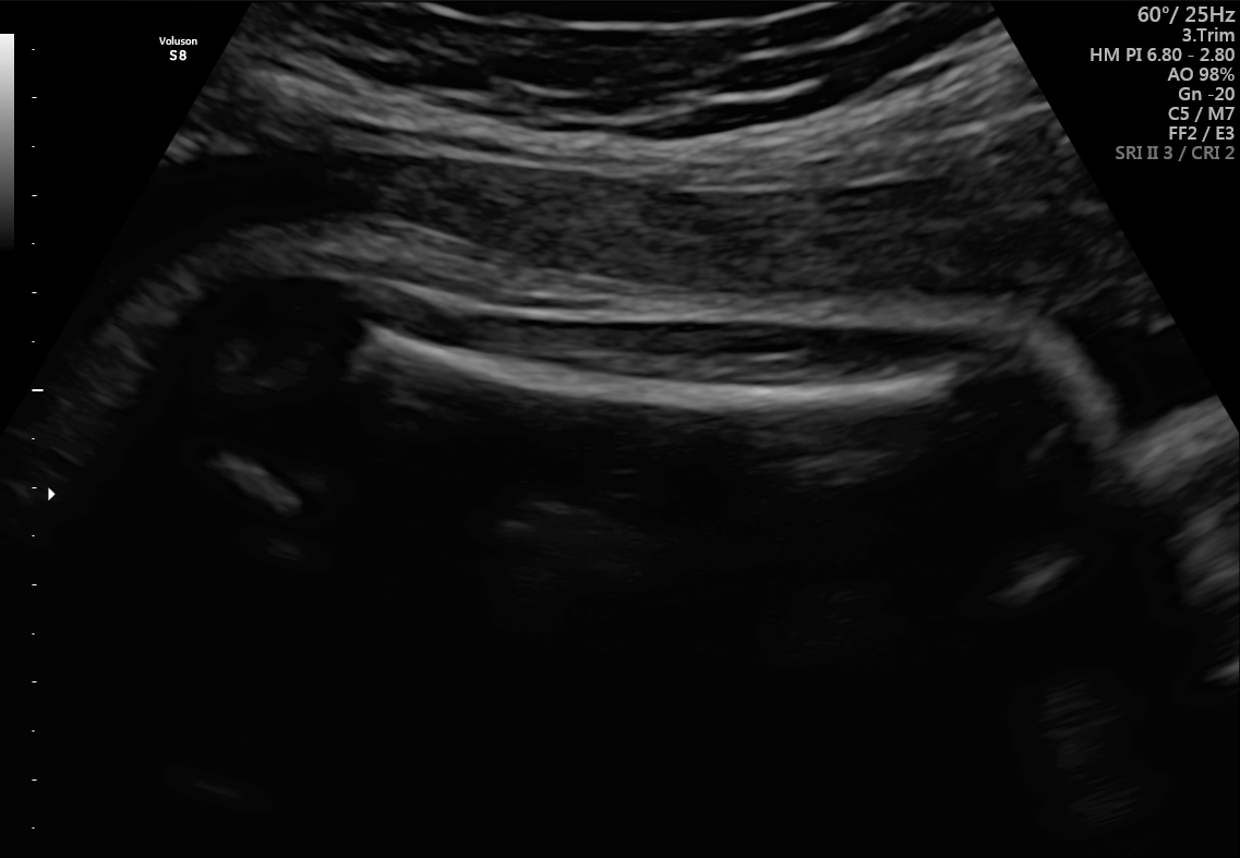
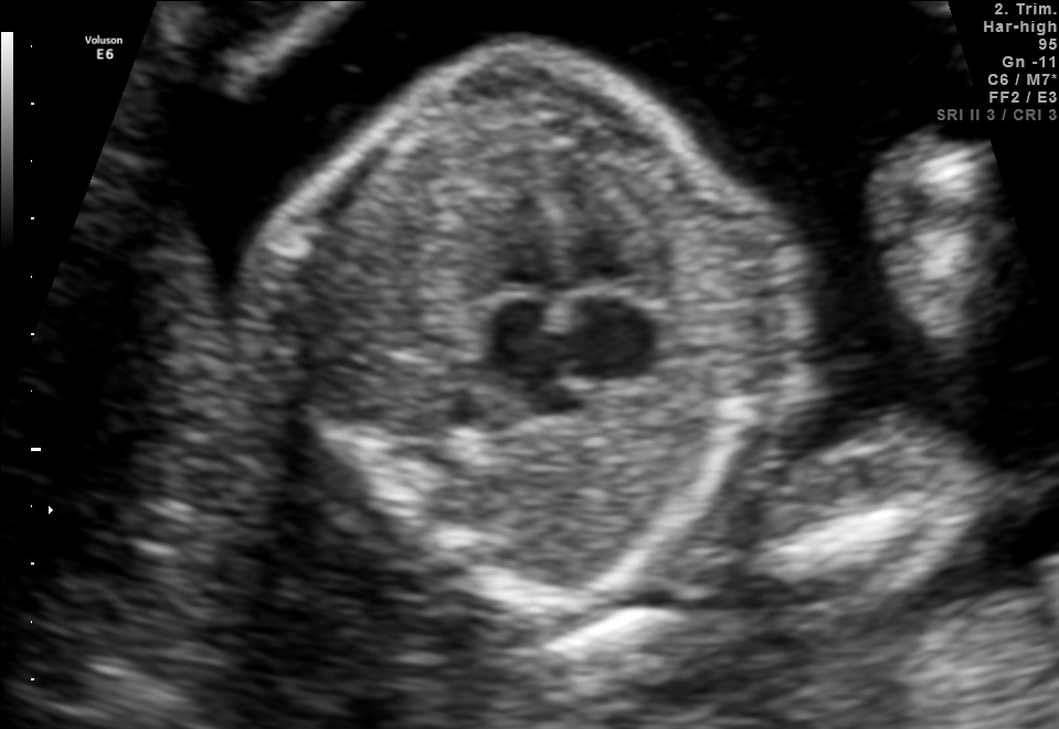
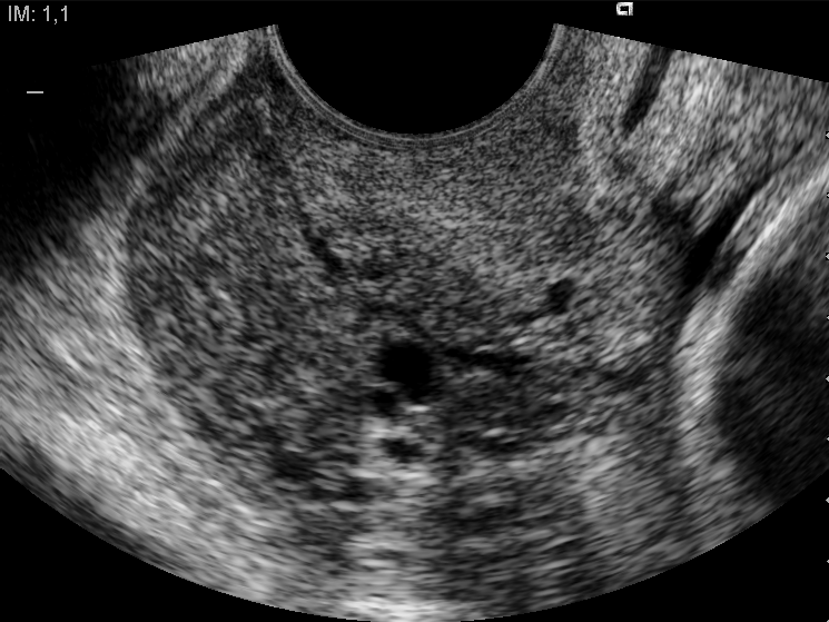
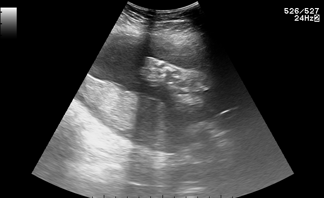
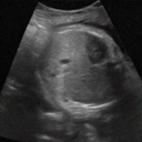
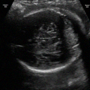
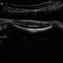
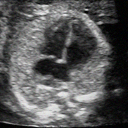
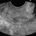
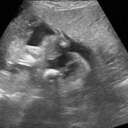
The generated images were visually assessed by examining a sample of synthetic images from each of the six classes. The synthetic images were compared with the real images from the FETAL_PLANES_DB dataset in Figure 1 to evaluate their realism and similarity. Example images from the diffusion model are shown in Figure 2, where we observe that the synthetic images closely resemble the real images in terms of texture, structure, and class-specific features. This addition emphasizes the visual quality of the generated images, which was a key aspect of evaluating the success of diffusion models in this context.
5.2 Downstream Classification Results
5.2.1 Classification Accuracy
The baseline for comparison is Real Data Only, which means training classifiers only on real images from the FETAL_PLANES_DB dataset ( images, see Table 1). Synthetic Data Only means classifiers trained solely on synthetic images generated by the classifier-guided diffusion model. Our method is Pretraining + Fine-Tuning, which means classifiers pretrained on synthetic images and fine-tuned on real images, as proposed and described in Section 3.4.
| Training accuracy | Test accuracy | |
|---|---|---|
| Real Data Only ( real images) | ||
| Synthetic Data Only ( synthetic images) | ||
| Pretraining ( synthetic images) + Fine-Tuning ( real images) | ||
| Mixed Data ( synthetic images + real images) | ||
| Synthetic Data Only ( synthetic images) | ||
| Pretraining ( synthetic images) + Fine-Tuning ( real images) |
Table 3 presents the results of training and test accuracy, using ViT_b_32 (Vision Transformer) Dosovitskiy (2020) as the base classifier model, and Table 4 shows the same results for Swin_t (Swin Transformer) Liu et al. (2021), as mentioned in Section 4.3. Results of using other models, i.e., ResNet50 He et al. (2016), DenseNet169 Huang et al. (2017), MedMamba Yue and Li (2024), can be found in the appendix due to limited space.
For training the ViT_b_32 classifier, the Adam optimizer Kingma (2014) was used with learning rate and cross-entropy loss function. Each configuration was trained for epochs with batch size , and saved the best training and test accuracy during the training procedure. For Pretraining + Fine-Tuning, after pretraining, the checkpoint was fine-tuned on real data saved from pretraining with synthetic data, with the same setup for the optimizer and loss function.
The generated images significantly contribute to improving the classification performance. Classifiers trained solely on synthetic data (“Synthetic Data Only”) achieve a respectable level of accuracy. However, the “Pretraining + Fine-Tuning” approach, where classifiers were pretrained on synthetic images and then fine-tuned on real data, demonstrates a substantial improvement in both training and test accuracy compared to the baseline of training solely on real data (“Real Data Only”). This highlights the effectiveness of using synthetic data generated by the diffusion model to enhance the robustness and generalization capabilities of the classification models.
| Training accuracy | Test accuracy | |
|---|---|---|
| Real Data Only ( real images) | ||
| Synthetic Data Only ( synthetic images) | ||
| Pretraining ( synthetic images) + Fine-Tuning ( real images) | ||
| Mixed Data ( synthetic images + real images) | ||
| Synthetic Data Only ( synthetic images) | ||
| Pretraining ( synthetic images) + Fine-Tuning ( real images) |
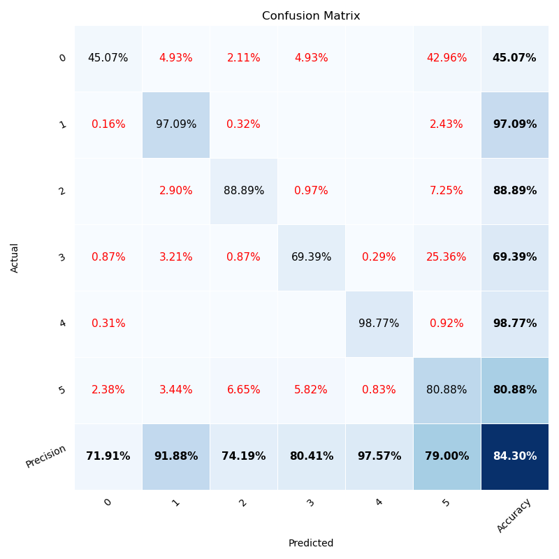
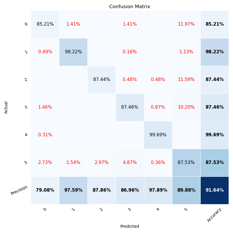
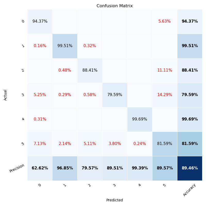
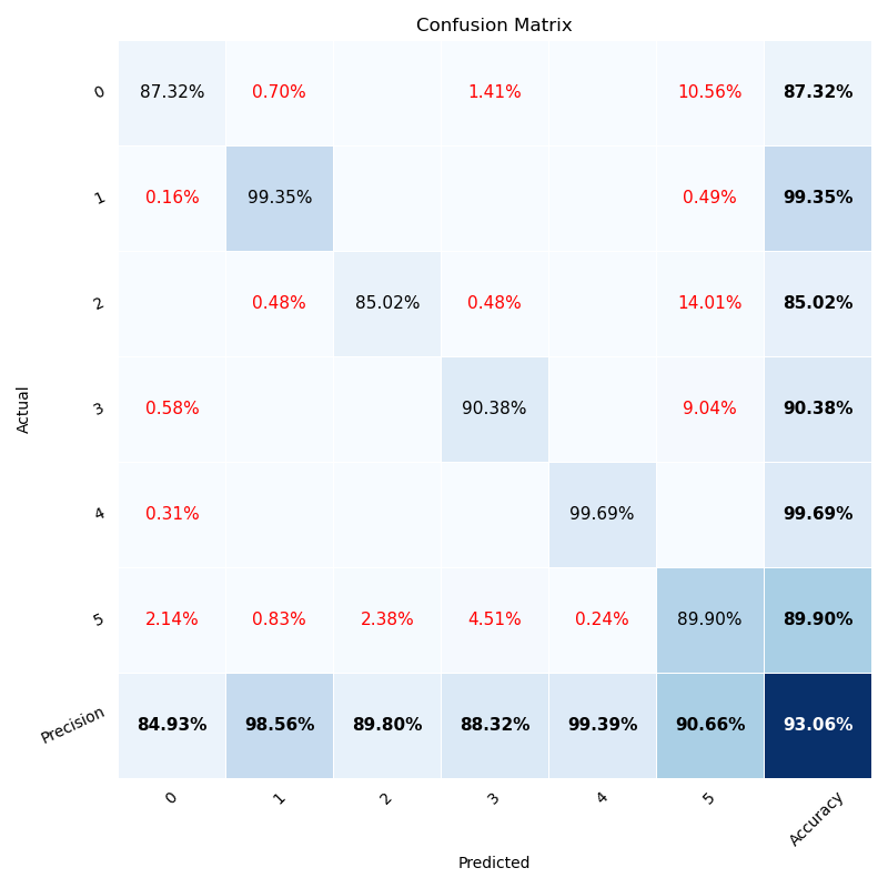
5.2.2 Confusion Matrix
To gain deeper insights into the classification performance beyond overall accuracy, we analyzed the confusion matrices for each method. The confusion matrix provides a detailed breakdown of the classifier’s performance by revealing the number of true positives, true negatives, false positives, and false negatives for each class. This analysis allows us to identify specific areas where the classifier excels and where it struggles, such as class-specific biases or difficulties in distinguishing between certain classes. By examining the confusion matrices, we can pinpoint areas for improvement and refine the model accordingly.


According to Figure 3(a), training classifiers exclusively on real-world datasets often encounters difficulties when dealing with classes containing a limited number of images. This data scarcity leads to low test accuracy, particularly for underrepresented classes. For example, for Fetal abdomen and Fetal Femur classes, where the number of training images were and (Table 1), the test accuracies are only and , respectively. By initially pretraining the classifier on a large dataset of synthetically generated data, its performance was significantly enhanced. The proposed two-stage process effectively addresses the issue of imbalanced classification, which is a direct consequence of data scarcity. Pretraining on synthetic data provides a more robust foundation, enabling the classifier to better recognize patterns and improve accuracy, especially for those classes with limited real-world examples. For the same two classes, i.e., Fetal abdomen and Fetal Femur classes, the test accuracy was significantly improved, achieving and , respectively. Similar conclusions regarding the performance of Swin_t can be drawn from the confusion matrices presented in Figure 4.


6 CONCLUSION AND FUTURE WORK
In this study, we proposed a novel approach to address the challenge of data scarcity in medical image analysis, specifically for fetal ultrasound images. By leveraging a classifier-guided diffusion model, we generated synthetic ultrasound images from the publicly available FETAL_PLANES_DB dataset. These synthetic images were used to pretrain classifiers, which were then fine-tuned using real images to improve performance.
We evaluated our approach using six different classifiers: ResNet50, DenseNet169, ViT_b_32, Swin_t, and MedMamba. The experimental results demonstrated that our method of combining synthetic data with real data for transfer learning significantly improved the classification accuracy when compared to training with real data alone. The experimental results on downstream classification tasks showed that the synthetic images generated by our diffusion model were highly realistic and similar to real fetal ultrasound images. Our contributions can be summarized as follows:
-
•
We generated a large-scale dataset of synthetic fetal ultrasound images, which will be made publicly available to help advance research in medical image analysis.
-
•
We demonstrated that using synthetic images for pretraining and real images for fine-tuning can improve classifier performance compared to training with real images only.
-
•
We provided a comparative study using multiple classifiers and found that our approach works well across a diverse set of models, including deep CNNs and transformer-based architectures.
This work highlights the potential of generative models in augmenting medical datasets and enhancing classifier performance in scenarios with limited data. However, there are several directions for future work that could further improve and expand the impact of our approach:
-
•
Improved Generative Models: While the classifier-guided diffusion model demonstrated promising results, further advancements in generative modeling could be explored. For instance, the use of contrastive learning could be explored to generate more discriminative features in the synthetic images.
-
•
Multimodal Imaging: While our study focuses on ultrasound images, extending this method to other types of medical imaging, such as MRI or CT scans, could offer broader applications in clinical practice. In particular, combining synthetic data from multiple modalities may help create more robust models that can generalize across different imaging techniques.
-
•
Incorporating Expert Knowledge: One potential improvement is to involve domain experts, such as radiologists and sonographers, in the image generation process. This could be done by using semi-supervised learning or active learning approaches, where radiologists label or provide feedback on the generated images. Such feedback could help the model generate even more realistic and clinically relevant images.
-
•
Longitudinal and Temporal Data: A key challenge in prenatal ultrasound imaging is capturing changes over time. In future work, we could explore temporal models that take into account the dynamic nature of fetal development. This would enable the generation of synthetic images that reflect the evolution of fetal structures over time, which is important for predictive models in maternal-fetal medicine.
In summary, this research demonstrates the value of combining synthetic data generation with real data for training medical image classifiers, providing a viable solution to data scarcity in ultrasound imaging. The proposed method holds promise for improving model performance in healthcare applications, and future research could build on this work to make synthetic data generation an integral part of medical image analysis, ultimately enhancing the quality and accessibility of healthcare.
References
- Pehrson et al. [2019] Lea Marie Pehrson, Michael Bachmann Nielsen, and Carsten Ammitzbøl Lauridsen. Automatic pulmonary nodule detection applying deep learning or machine learning algorithms to the lidc-idri database: a systematic review. Diagnostics, 9(1):29, 2019.
- Wang et al. [2022] Risheng Wang, Tao Lei, Ruixia Cui, Bingtao Zhang, Hongying Meng, and Asoke K Nandi. Medical image segmentation using deep learning: A survey. IET image processing, 16(5):1243–1267, 2022.
- Cai et al. [2020] Lei Cai, Jingyang Gao, and Di Zhao. A review of the application of deep learning in medical image classification and segmentation. Annals of translational medicine, 8(11), 2020.
- LeCun et al. [1989] Yann LeCun, Bernhard Boser, John Denker, Donnie Henderson, Richard Howard, Wayne Hubbard, and Lawrence Jackel. Handwritten digit recognition with a back-propagation network. Advances in neural information processing systems, 2, 1989.
- He et al. [2016] Kaiming He, Xiangyu Zhang, Shaoqing Ren, and Jian Sun. Deep residual learning for image recognition. Proceedings of the IEEE conference on computer vision and pattern recognition, pages 770–778, 2016.
- Alzubaidi et al. [2021] Laith Alzubaidi, Muthana Al-Amidie, Ahmed Al-Asadi, Amjad J Humaidi, Omran Al-Shamma, Mohammed A Fadhel, Jinglan Zhang, Jesus Santamaría, and Ye Duan. Novel transfer learning approach for medical imaging with limited labeled data. Cancers, 13(7):1590, 2021.
- Huang et al. [2021] Qinghua Huang, Zhaoji Miao, Shichong Zhou, Cai Chang, and Xuelong Li. Dense prediction and local fusion of superpixels: A framework for breast anatomy segmentation in ultrasound image with scarce data. IEEE Transactions on Instrumentation and Measurement, 70:1–8, 2021.
- Brattain et al. [2018] Laura J Brattain, Brian A Telfer, Manish Dhyani, Joseph R Grajo, and Anthony E Samir. Machine learning for medical ultrasound: status, methods, and future opportunities. Abdominal radiology, 43:786–799, 2018.
- DuMont Schütte et al. [2021] August DuMont Schütte, Jürgen Hetzel, Sergios Gatidis, Tobias Hepp, Benedikt Dietz, Stefan Bauer, and Patrick Schwab. Overcoming barriers to data sharing with medical image generation: a comprehensive evaluation. NPJ digital medicine, 4(1):141, 2021.
- Goodfellow et al. [2014] Ian Goodfellow, Jean Pouget-Abadie, Mehdi Mirza, Bing Xu, David Warde-Farley, Sherjil Ozair, Aaron Courville, and Yoshua Bengio. Generative adversarial nets. Advances in neural information processing systems, 27, 2014.
- Ho et al. [2020] Jonathan Ho, Ajay Jain, and Pieter Abbeel. Denoising diffusion probabilistic models. Advances in neural information processing systems, 33:6840–6851, 2020.
- Frid-Adar et al. [2018] Maayan Frid-Adar, Idit Diamant, Eyal Klang, Michal Amitai, Jacob Goldberger, and Hayit Greenspan. Gan-based synthetic medical image augmentation for increased cnn performance in liver lesion classification. Neurocomputing, 321:321–331, 2018.
- Motamed et al. [2021] Saman Motamed, Patrik Rogalla, and Farzad Khalvati. Data augmentation using generative adversarial networks (gans) for gan-based detection of pneumonia and covid-19 in chest x-ray images. Informatics in medicine unlocked, 27:100779, 2021.
- Yi et al. [2019] Xin Yi, Ekta Walia, and Paul Babyn. Generative adversarial network in medical imaging: A review. Medical image analysis, 58:101552, 2019.
- Pan et al. [2023] Shaoyan Pan, Tonghe Wang, Richard LJ Qiu, Marian Axente, Chih-Wei Chang, Junbo Peng, Ashish B Patel, Joseph Shelton, Sagar A Patel, Justin Roper, et al. 2d medical image synthesis using transformer-based denoising diffusion probabilistic model. Physics in Medicine & Biology, 68(10):105004, 2023.
- Hardy et al. [2023] Romain Hardy, Joe Klepich, Ryan Mitchell, Steve Hall, Jericho Villareal, and Cornelia Ilin. Improving nonalcoholic fatty liver disease classification performance with latent diffusion models. Scientific Reports, 13(1):21619, 2023.
- Kodali et al. [2017] Naveen Kodali, Jacob Abernethy, James Hays, and Zsolt Kira. On convergence and stability of gans. arXiv preprint arXiv:1705.07215, 2017.
- Dhariwal and Nichol [2021] Prafulla Dhariwal and Alexander Nichol. Diffusion models beat gans on image synthesis. Advances in neural information processing systems, 34:8780–8794, 2021.
- Goceri [2023] Evgin Goceri. Medical image data augmentation: techniques, comparisons and interpretations. Artificial Intelligence Review, 56(11):12561–12605, 2023.
- Sanford et al. [2020] Thomas H Sanford, Ling Zhang, Stephanie A Harmon, Jonathan Sackett, Dong Yang, Holger Roth, Ziyue Xu, Deepak Kesani, Sherif Mehralivand, Ronaldo H Baroni, et al. Data augmentation and transfer learning to improve generalizability of an automated prostate segmentation model. American Journal of Roentgenology, 215(6):1403–1410, 2020.
- Montero et al. [2021] Alberto Montero, Elisenda Bonet-Carne, and Xavier Paolo Burgos-Artizzu. Generative adversarial networks to improve fetal brain fine-grained plane classification. Sensors, 21(23):7975, 2021.
- Dorent et al. [2023] Reuben Dorent, Nazim Haouchine, Fryderyk Kogl, Samuel Joutard, Parikshit Juvekar, Erickson Torio, Alexandra J Golby, Sebastien Ourselin, Sarah Frisken, Tom Vercauteren, et al. Unified brain mr-ultrasound synthesis using multi-modal hierarchical representations. In International conference on medical image computing and computer-assisted intervention, pages 448–458. Springer, 2023.
- Burgos-Artizzu et al. [2020] Xavier P Burgos-Artizzu, David Coronado-Gutiérrez, Brenda Valenzuela-Alcaraz, Elisenda Bonet-Carne, Elisenda Eixarch, Fatima Crispi, and Eduard Gratacós. Evaluation of deep convolutional neural networks for automatic classification of common maternal fetal ultrasound planes. Scientific Reports, 10(1):10200, 2020.
- Huang et al. [2017] Gao Huang, Zhuang Liu, Laurens Van Der Maaten, and Kilian Q Weinberger. Densely connected convolutional networks. Proceedings of the IEEE conference on computer vision and pattern recognition, pages 4700–4708, 2017.
- Dosovitskiy [2020] Alexey Dosovitskiy. An image is worth 16x16 words: Transformers for image recognition at scale. arXiv preprint arXiv:2010.11929, 2020.
- Liu et al. [2021] Ze Liu, Yutong Lin, Yue Cao, Han Hu, Yixuan Wei, Zheng Zhang, Stephen Lin, and Baining Guo. Swin transformer: Hierarchical vision transformer using shifted windows. Proceedings of the IEEE/CVF international conference on computer vision, pages 10012–10022, 2021.
- Yue and Li [2024] Yubiao Yue and Zhenzhang Li. Medmamba: Vision mamba for medical image classification. arXiv preprint arXiv:2403.03849, 2024.
- Kingma [2014] Diederik P Kingma. Adam: A method for stochastic optimization. arXiv preprint arXiv:1412.6980, 2014.
Appendix A More Experimental Results
Results of training and test accuracy of using other models, i.e., ResNet50 He et al. [2016], DenseNet169 Huang et al. [2017], MedMamba Yue and Li [2024], can be found in Table 5, Table 6, and Table 7, respectively.
| Training accuracy | Test accuracy | |
|---|---|---|
| Real Data Only ( real images) | ||
| Synthetic Data Only ( synthetic images) | ||
| Pretraining ( synthetic images) + Fine-Tuning ( real images) | ||
| Synthetic Data Only ( synthetic images) | ||
| Pretraining ( synthetic images) + Fine-Tuning ( real images) |