The R-Vessel-X Project111The research leading to these results was funded by the French Agence Nationale de la Recherche (Grant Agreement ANR-18-CE45-0018).
Abstract
1) Objectives: This technical report presents a synthetic summary and the principal outcomes of the project R-Vessel-X (“Robust vascular network extraction and understanding within hepatic biomedical images”) funded by the French Agence Nationale de la Recherche, and developed between 2019 and 2023. 2) Material and methods: We used datasets and tools publicly available such as IRCAD, Bullitt or VascuSynth to obtain real or synthetic angiographic images. The main contributions lie in the field of 3D angiographic image analysis: filtering, segmentation, modeling and simulation, with a specific focus on the liver. 3) Results: We paid a particular attention to open-source software diffusion of the developed methods, by means of 3D Slicer plugins for the liver anatomy segmentation (SlicerRVXLiverSegmentation) and vesselness filtering (SlicerRVXVesselnessFilters), and an online demo for the generation of synthetic and realistic vessels in 2D and 3D (OpenCCO). 4) Conclusion: The R-Vessel-X project provided extensive research outcomes, covering various topics related to 3D angiographic image analysis, such as filtering, segmentation, modeling and simulation. We also developed open-source and free softwares so that the research communities in biomedical engineering can use these results in their future research.
keywords:
Image analysis , Angiographic images , Vessels , Hepatic vasculature , Open-source software1 Introduction
1.1 Context
Observing, analysing and/or modeling human vascular networks is a key-step for various medical applications, including e.g. diagnosis, surgery, drug delivery. Medical image acquisition, such as X-ray Computed Tomography (CT) or Magnetic Resonance Imaging (MRI) plays a central role in this context.
Despite numerous developments in this field, computer extraction and modeling of vascular networks from 3-dimensional (3D) medical images is still a challenge, due to their high complexity and their partial visibility within most current medical acquisition systems, whatever the organ considered (brain, liver, heart, retina, etc.). Moreover, the reproducibility of such approaches is rarely addressed and experiments are often conducted on private datasets.
1.2 Related Works
After 30 years of research, vascular structure analysis from 3D images, that mainly relies on vessel segmentation, remains a complex issue, especially in the case of 3D images [39, 44]. Most efforts of 3D vessel segmentation were geared towards brain and heart vessels. Nonetheless, the liver has also been under investigation, leading to various contributions. An exhaustive state of the art is beyond the scope of this article. The interested reader can refer to two recent surveys proposed by [10] and [54] and the herein references.
In the context of liver vessel segmentation, most methods were designed to process X-ray Computed Tomography (CT) images, that constitute the state of the art for liver imaging. During the period of the project (from 2019 to 2023), except few methods relying on classical image processing techniques (e.g. graph cuts [18]), a majority of the proposed approaches relied on deep learning paradigms: convolutional neural networks [30, 50], generative adversarial networks and autoencoders [7], [53] and multiscale paradigms [14], graph neural networks [40], deep supervision [20] or transformers [52, 51].
Although remaining less frequent, Magnetic Resonance Imaging (MRI) is becoming a relevant alternative to CT for liver image analysis [38]. A survey of artificial intelligence methods for MRI liver analysis was recently proposed by [22]. During the last years, various strategies were considered. [16] relied on a classical clustering (K-means) approach. [41] used convolutional neural networks to process MRI T2⋆ relaxometry. [23, 17] took advantage of dynamic MR images to carry out classification-based analysis of structures including vessels.
1.3 Objectives
The purpose of the R-Vessel-X (“Robust vascular network extraction and understanding within hepatic biomedical images”) project was to develop novel solutions to extract vascular networks within 3D medical (CT or MR) images, with a specific focus on the liver.
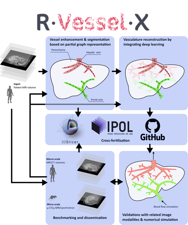
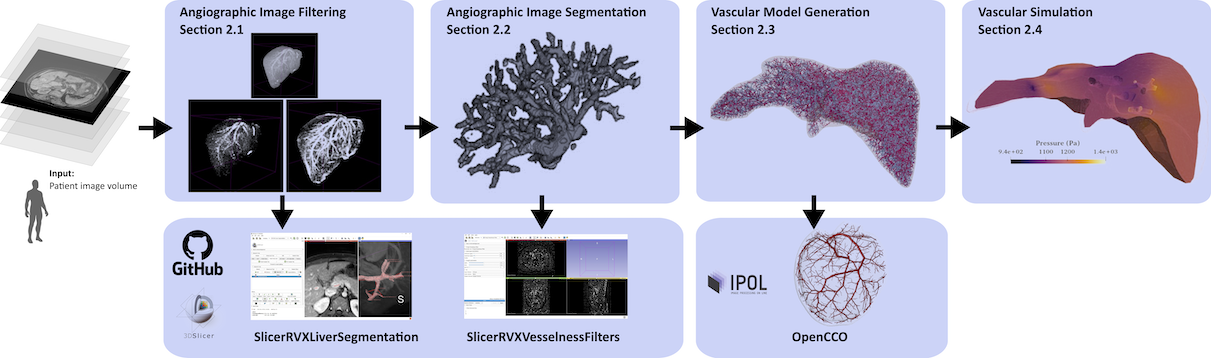
The scientific part of the project was organized as five interlinked work packages (WPs), visually summarized in Fig. 1. The principal contributions of these WPs, together with pointers on related publications that provide more insight on the developed methods and tools, are given in the next section. The project outcomes were dedicated to various biomedical engineering topics related to 3D angiographic image analysis: filtering, segmentation, modeling and simulation, as depicted in Fig. 2.
|
|
|
|
|---|---|---|
| (a) | (b) | (c) |
2 Contributions
2.1 Angiographic Image Filtering
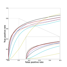
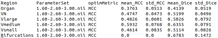
Filtering with the purpose of denoising and/or enhancing the vascular signal vs. the remaining patterns has been one of the first research topics in angiographic image analysis. Over the years, a wide range of filters were proposed, from derivative-based [13] to non-linear approaches [43]. Despite a large choice of operators, filtering remains a complex task. Indeed, the efficiency of a filter depends on the type of acquisition and on the vascular structure being imaged. Generally the proposed filters are also governed by many parameters that require ad hoc setting. It follows that choosing the right filter with the right parameters is tricky, a fortiori in absence of a general benchmarking framework. Building such a framework has been the purpose of the PhD thesis carried out by Jonas Lamy [31]. Its purposes were twofold: (1) designing a generic protocol that allows to run and fairly compare filtering operators with controlled parameters, and (2) providing reliable versions of the most popular angiographic filters. This thesis finally led to revisiting seven gold standard filters, embedded in a C++ library. They were compared on three public datasets: IRCAD (liver CT images), Bullitt (brain MR images) and the synthetic tool VascuSynth. Their performances were investigated in various areas: in the whole organ, around the vascular network and at the bifurcations, together with a multiscale approach guided by the diameters of the vessels. Fig. 4 compares the behavior of the seven selected filters in a bifurcation area. Two versions (based on different parameter optimisation strategies) were proposed, first in [34], then in [33]. An open source version222https://github.com/JonasLamy/LiverVesselness of the code and a demonstrator333https://kerautret.github.io/LiverVesselnessIPOLDemo/ were made available in [32], in order to allow any interested user to assess the performance of his/her own filters on any angiographic data. Note that using the online demonstration platform, users can freely test the filters on their own volume they can upload to the server. Fig. 3 illustrates a typical example of experiment generated from the proposed filtering benchmark that can be customized with various metrics (mean/std MCC or Dice), and on different region domains (organ, vessel neighborhood). Different representations can be generated (histogram (a), ROC curve (b), and raw data results). An example of results from experiments carried out on 8 methods is given in Tab. 1 for different regions of the vascular network.
Section 2.5 also presents an open-source software proposing all those filters into a single 3D Slicer plug-in.
2.2 Angiographic Image Segmentation
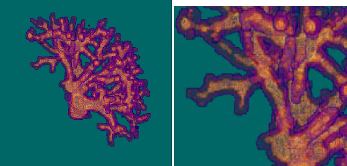
The segmentation of vessels in medical images has been covered by various methodologies over the last decades. Many recent ones rely on deep learning models derived from the U-Net [46]. The work conducted by Abir Affane [1] during her PhD was first to adapt and compare 3D models to liver vessel segmentation, namely 3D U-Net, Dense U-Net, MultiRes U-Net on the IRCAD dataset [2]. In addition, different image structures were tested as inputs of the neural networks, with complete volumes, slabs (groups of slices) and patches. It followed from this first study that the Dense U-Net approach, trained on image volumes, constitutes the best compromise in terms of performance vs. number of parameters. In a second time, a novel approach was designed, by leveraging the developments of the two PhD theses. Indeed, as exposed in Table 2, thanks to vesselness filters, it was possible to train deep learning models from vascular patterns provided by these operators, alternatively to native angiographic images [3]. By comparing different filters, it was observed that the vesselness filter proposed by [25], used as a pre-processing step on CT images from IRCAD with a Dense U-Net model, was an accurate solution for hepatic vascular segmentation, as illustrated in Fig. 5. By applying a few post-processing operators, we were able to obtain faithful segmentations, with a satisfactory preservation of bifurcations and small vessels.
| Preprocessing | U-Net | Dense U-Net | MultiRes U-Net |
|---|---|---|---|
| Without | 0.586 | 0.671 | 0.694 |
| Jerman filter | 0.712 | 0.856 | 0.835 |
| RORPO filter | 0.236 | 0.271 | 0.334 |
| Sato filter | 0.512 | 0.848 | 0.591 |
| Zhang filter | 0.201 | 0.311 | 0.212 |
A major issue in medical image segmentation by deep learning models is the annotation of volumes by clinical experts. We present later in Section 2.5 a 3D Slicer plug-in dedicated to this task for liver anatomy from CT or MR images.
2.3 Vascular Model Generation
Despite being composed of simple tubular elements, vascular networks are highly complex structures. This complexity derives from their branching topology, their tortuous geometry and their highly multiscale organization, that goes from macroscopic vessels to microscopic ones. It follows that angiographic imaging devices are not able to capture the complete information related to vascular networks. Due to these limitations, some research efforts were dedicated to model the vessels, based on anatomical, physiological and biomechanical hypotheses. This led to numerical approaches for vascular model generation. In particular, the CCO (Constrained Constructive Optimization) scheme was proposed by [48], first in 2 dimensions and then by [27] in 3 dimensions. The CCO algorithm incrementally builds a binary tree by adding at each iteration a new terminal point. Beyond a simple topological process, the strength of CCO lies in the spatial embedding of this tree, that defines a position for each branching point and a size for each segment based on physiologically-compliant rules guiding a spatially constrained optimization process.
 |
 |
| (a) | (b) |
 |
 |
| (a) | (b) |
This seminal work led to the further development of variants, including the popular VascuSynth [19] tool and non-open implementations of CCO, e.g. by [24]. Motivated by research reproducibility with open software dissemination, and convinced by the relevance of CCO as a way to relevantly complete the information missed by angiographic images, we revisited the CCO algorithm and we proposed an open-source implementation together with a demonstrator444https://opencco-team.github.io/OpenCCO-IPOL_Demo described by [28]. Fig. 6 illustrates a result obtained from the online demonstration where the user defines a restricted domain defined on the heart wall. Revisiting this algorithm led us to investigate some tricky parts of the optimization process, in particular the non-linear equation system governing the progressive construction of the vascular tree, initially solved as proposed by [26]. These efforts opened the way to new efficient optimization strategies, such as proposed by [9] and paving the way towards improved vascular tree modeling processes.
In section 2.4, we expose how we employed this software in order to model the liver vasculature for the simulation of hepatic vascular functioning.
2.4 Vascular Simulation
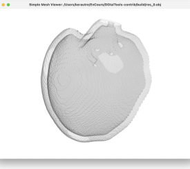
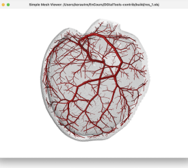
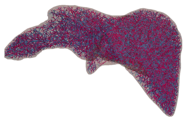
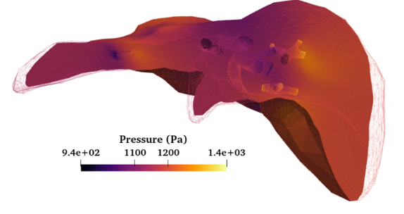
We finally addressed the challenge of numerical simulation of liver perfusion, by taking into account vessels from medical images. This is a very hard task, due to the multi-scale high complexity of the liver functioning, from hepatic lobules to fine then larger vessels, and to the partial representation of liver anatomy acquired from medical images [37]. During the post-doc fellowship carried out by Mohamed A. Chetoui, and to represent a large volume of the liver vasculature with the finest vessels, we used the aforementioned OpenCCO tool, as illustrated in Fig. 7 (a). We generated this vascular network starting from the larger branches of hepatic and portal veins obtained from medical images. The liver volume boundary was also extracted from such images. Then, we developed a novel computing method to obtain physiological parameters (pressure, flow, permeability) of hepatic perfusion [8]. We opted for a multi-compartment approach [45], and by coupling Darcy law and computational fluid dynamics, in order to calculate these parameters inside the liver parenchyma and vessels. An example of result obtained with our system is depicted in Fig. 7 (b), where we show the pressure calculated within the liver volume. We tested our approach on 1D and 3D configurations to validate the correct behavior of the simulation regarding a in-silico realistic liver physiology model. Moreover, we developed this complete pipeline into a single Python framework by using the FreeFEM library [21].
2.5 Software developments
During the project, we developed and disseminated two official open-source 3D Slicer [29] plug-ins.
SlicerRVXVesselnessFilters555https://github.com/R-Vessel-X/SlicerRVXVesselnessFilters gathers the aforementioned vesselness filters (Section 2.1) into a single framework, which facilitates their use for biomedical and medical image processing in 3D Slicer. Any user can set the parameters related to each algorithm, and interactively visualize the result.
SlicerRVXLiverSegmentation666https://github.com/R-Vessel-X/SlicerRVXLiverSegmentation aims at interactively aiding clinical experts and biomedical engineers in liver anatomy segmentation from medical images (e.g. CT, MRI), comprising the parenchyma, hepatic and portal vessels and potential tumors. We shew in [35] that this tool enables a faster annotation compared to commercial solutions, mostly for MRI data. It can thus be applied in medical analysis contexts, as we did e.g. for registration [11]. Moreover, this is the very first time that such plug-in incorporates deep models based on MONAI [6] for automatic liver volume segmentation.
3 Concluding Remarks and Perspectives
The R-Vessel-X project provided extensive research outcomes, covering various topics related to 3D angiographic image analysis, such as filtering, segmentation, modeling and simulation. We also developed softwares so that researchers, medical doctors and engineers can use essential vascular tools in their daily practice. The SlicerRVXLiverSegmentation plug-in is used in the radiology department at CHU (University Hospital) of Clermont-Ferrand, which enables clinical research in cardio-vascular pathologies. As perspective, the combination of these independent products would be beneficial for the community. Our goal is to propose new public and open-source plug-ins or softwares still related to angiographic imaging. Furthermore, we aim to continue our investigations on biomedical pre-clinical imaging [4], and draw relations with human-scale vascular systems. This project also enabled new on-going researches, for example in deep vessel segmentation for the reconnection of vascular structures by an hybrid approach [12] or the combination of vesselness filters in deep U-Net models [15].
Compliance with Ethical Standards
During the R-Vessel-X project, we used datasets and tools publicly available such as IRCAD [49], Bullitt [5] or VascuSynth [19] to obtain angiographic images. Also, each referenced article produced during the project met the requirements for ethical standards to get published. As a matter of reproducibility and availability of our developments, we created permanent links for our softwares SlicerRVXLiverSegmentation [35], SlicerRVXVesselnessFilters [33] and OpenCCO [28].
Acknowledgement
The research leading to these results was funded by the French Agence Nationale de la Recherche (Grant Agreement ANR-18-CE45-0018), which is gratefully acknowledged by the authors.
References
- Affane [2022] Affane, A., 2022. Robust liver vessel segmentation in medical images using 3-D deep learning approaches. Ph.D. thesis. University of Clermont Auvergne, Clermont-Ferrand, France.
- Affane et al. [2021] Affane, A., Kucharski, A., Chapuis, P., Freydier, S., Lebre, M.A., Vacavant, A., Fabijańska, A., 2021. Segmentation of liver anatomy by combining 3-D U-Net approaches. Journal of Applied Sciences 11, 4895.
- Affane et al. [2022] Affane, A., Lamy, J., Lebre, M.A., Vacavant, A., 2022. Robust deep 3-D architectures based on vascular patterns for liver vessel segmentation. Informatics in Medicine Unlocked 34, 101111.
- Alvarez et al. [2023] Alvarez, M., de Pina, D.R., Rositi, H., Vacavant, A., 2023. Comparison of unet and dc-unet models for an efficient segmentation and visualization of rodent hepatic vascular network from x-ray phase contrast imaging, in: ISBI, Procs., IEEE. pp. 1–5.
- Bullitt et al. [2005] Bullitt, E., Zeng, D., Gerig, G., Aylward, S., Joshi, S., Smith, J.K., Lin, W., Ewend, M.G., 2005. Vessel tortuosity and brain tumor malignancy: A blinded study. Academic Radiology 12, 1232–1240.
- Cardoso et al. [2022] Cardoso, M.J., Li, W., Brown, R., Ma, N., Kerfoot, E., Wang, Y., Murray, B., Myronenko, A., Zhao, C., Yang, D., Nath, V., He, Y., Xu, Z., Hatamizadeh, A., Zhu, W., Liu, Y., Zheng, M., Tang, Y., Yang, I., Zephyr, M., Hashemian, B., Alle, S., Darestani, M.Z., Budd, C., Modat, M., Vercauteren, T., Wang, G., Li, Y., Hu, Y., Fu, Y., Gorman, B., Johnson, H.J., Genereaux, B.W., Erdal, B.S., Gupta, V., Diaz-Pinto, A., Dourson, A., Maier-Hein, L., Jaeger, P.F., Baumgartner, M., Kalpathy-Cramer, J., Flores, M., Kirby, J.S., Cooper, L.A.D., Roth, H.R., Xu, D., Bericat, D., Floca, R., Zhou, S.K., Shuaib, H., Farahani, K., Maier-Hein, K.H., Aylward, S.R., Dogra, P., Ourselin, S., Feng, A., 2022. MONAI: an open-source framework for deep learning in healthcare. CoRR abs/2211.02701.
- Cheema et al. [2021] Cheema, M.N., Nazir, A., Yang, P., Sheng, B., Li, P., Li, H., Wei, X., Qin, J., Kim, J., Feng, D.D., 2021. Modified GAN-CAED to minimize risk of unintentional liver major vessels cutting by controlled segmentation using CTA/SPET-CT. IEEE Transactions on Industrial Informatics 17, 7991–8002.
- Chetoui et al. [2023] Chetoui, M., Morvan, M., Beaurepaire, P., Vacavant, A., 2023. A new open-source workflow for multiscale modeling of hepatic perfusion, in: Congress of the European Society of Biomechanics, Maastricht, The Netherlands.
- de Chou et al. [2024] de Chou, R.S., Srir, M.A., Najman, L., Passat, N., Talbot, H., Vignon-Clementel, I.E., 2024. Convex optimization for binary tree-based transport networks, in: DGMM, Procs., pp. 217–228.
- Ciecholewski and Kassjanski [2021] Ciecholewski, M., Kassjanski, M., 2021. Computational methods for liver vessel segmentation in medical imaging: A review. Sensors 21, 2027.
- Debroux et al. [2020] Debroux, N., Lienemann, G., Magnin, B., Guyader, C.L., Vacavant, A., 2020. A time-dependent joint segmentation and registration model: Application to longitudinal registration of hepatic DCE-MRI sequences, in: IPTA, Procs., IEEE. pp. 1–6.
- Esteves et al. [2024] Esteves, S.C., Vacavant, A., Merveille, O., 2024. A plug-and-play framework for curvilinear structure segmentation based on a learned reconnecting regularization. Neurocomputing 599, 128055.
- Frangi et al. [1998] Frangi, A.F., Niessen, W.J., Vincken, K.L., Viergever, M.A., 1998. Muliscale vessel enhancement filtering, in: MICCAI, Procs., pp. 130–137.
- Gao et al. [2023] Gao, Z., Zong, Q., Wang, Y., Yan, Y., Wang, Y., Zhu, N., Zhang, J., Wang, Y., Zhao, L., 2023. Laplacian salience-gated feature pyramid network for accurate liver vessel segmentation. IEEE Transactions on Medical Imaging 42, 3059–3068.
- Garret et al. [2024] Garret, G., Vacavant, A., Frindel, C., 2024. Deep vessel segmentation based on a new combination of vesselness filters, in: ISBI, Procs., IEEE.
- Goceri et al. [2017] Goceri, E., Shah, Z., Gurcan, M., 2017. Vessel segmentation from abdominal magnetic resonance images: Adaptive and reconstructive approach. International Journal for Numerical Methods in Biomedical Engineering 33, e2811.
- Guo et al. [2022] Guo, Q., Song, H., Fan, J., Ai, D., Gao, Y., Yu, X., Yang, J., 2022. Portal vein and hepatic vein segmentation in multi-phase MR images using flow-guided change detection. IEEE Transactions on Image Processing 31, 2503–2517.
- Guo et al. [2020] Guo, X., Xiao, R., Zhang, T., Chen, C., Wang, J., Wang, Z., 2020. A novel method to model hepatic vascular network using vessel segmentation, thinning, and completion. Medical & Biological Engineering & Computing 58, 709–724.
- Hamarneh and Jassi [2010] Hamarneh, G., Jassi, P., 2010. Vascusynth: Simulating vascular trees for generating volumetric image data with ground-truth segmentation and tree analysis. Computerized Medical Imaging and Graphics 34, 605–616.
- Hao et al. [2022] Hao, W., Zhang, J., Su, J., Song, Y., Liu, Z., Liu, Y., Qiu, C., Han, K., 2022. HPM-Net: Hierarchical progressive multiscale network for liver vessel segmentation in CT images. Computer Methods and Programs in Biomedicine 224, 107003.
- Hecht [2012] Hecht, F., 2012. New development in FreeFem++. Journal of numerical mathematics 20, 251–265. URL: https://freefem.org/.
- Hill et al. [2021] Hill, C.E., Biasiolli, L., Robson, M.D., Grau, V., Pavlides, M., 2021. Emerging artificial intelligence applications in liver magnetic resonance imaging. World Journal of Gastroenterology 27, 6825–6843.
- Ivashchenko et al. [2020] Ivashchenko, O.V., Rijkhorst, E.J., ter Beek, L.C., Hoetjes, N.J., Pouw, B., Nijkamp, J., Kuhlmann, K.F., Ruers, T.J., 2020. A workflow for automated segmentation of the liver surface, hepatic vasculature and biliary tree anatomy from multiphase mr images. Magnetic Resonance Imaging 68, 53–65.
- Jaquet et al. [2019] Jaquet, C., Najman, L., Talbot, H., Grady, L.J., Schaap, M., Spain, B., Kim, H.J., Vignon-Clementel, I.E., Taylor, C.A., 2019. Generation of patient-specific cardiac vascular networks: A hybrid image-based and synthetic geometric model. IEEE Transactions on Biomedical Engineering 66, 946–955.
- Jerman et al. [2016] Jerman, T., Pernus, F., Likar, B., Spiclin, Z., 2016. Enhancement of vascular structures in 3D and 2D angiographic images. IEEE Transactions on Medical Imaging 35, 2107–2118.
- Kamiya and Togawa [1972] Kamiya, A., Togawa, T., 1972. Optimal branching structure of the vascular tree. The Bulletin of Mathematical Biophysics 34, 431–438.
- Karch et al. [1999] Karch, R., Neumann, F., Neumann, M., Schreiner, W., 1999. A three-dimensional model for arterial tree representation, generated by constrained constructive optimization. Computers in Biology and Medicine 29, 19–38.
- Kerautret et al. [2023] Kerautret, B., Ngo, P., Passat, N., Talbot, H., Jaquet, C., 2023. OpenCCO: An implementation of Constrained Constructive Optimization for generating 2D and 3D vascular trees. Image Processing On Line 13, 258–279.
- Kikinis et al. [2014] Kikinis, R., Pieper, S.D., Vosburgh, K.G., 2014. 3D Slicer: A Platform for Subject-Specific Image Analysis, Visualization, and Clinical Support. Springer New York, New York, NY. pp. 277–289.
- Kitrungrotsakul et al. [2019] Kitrungrotsakul, T., Han, X., Iwamoto, Y., Lin, L., Foruzan, A.H., Xiong, W., Chen, Y., 2019. Vesselnet: A deep convolutional neural network with multi pathways for robust hepatic vessel segmentation. Computerized Medical Imaging and Graphics 75, 74–83.
- Lamy [2023] Lamy, J., 2023. Study of vesselness filters applied to 3D medical images. Ph.D. thesis. Lumière University Lyon 2, France.
- Lamy et al. [2021] Lamy, J., Kerautret, B., Merveille, O., Passat, N., 2021. Structure, concept and result reproducibility of the benchmark on vesselness filters, in: RRPR, Procs., pp. 121–135.
- Lamy et al. [2022a] Lamy, J., Merveille, O., Kerautret, B., Passat, N., 2022a. A benchmark framework for multiregion analysis of vesselness filters. IEEE Transactions on Medical Imaging 41, 3649–3662.
- Lamy et al. [2020] Lamy, J., Merveille, O., Kerautret, B., Passat, N., Vacavant, A., 2020. Vesselness filters: A survey with benchmarks applied to liver imaging, in: ICPR, Procs., pp. 3528–3535.
- Lamy et al. [2022b] Lamy, J., Pelletier, T., Lienemann, G., Magnin, B., Kerautret, B., Passat, N., Finet, J., Vacavant, A., 2022b. The 3D Slicer RVXLiverSegmentation plug-in for interactive liver anatomy reconstruction from medical images. Journal of Open Source Software 7, 3920.
- Law and Chung [2008] Law, M.W., Chung, A., 2008. Three dimensional curvilinear structure detection using optimally oriented flux, in: ECCV, Procs., Springer. pp. 368–382.
- Lebre et al. [2017] Lebre, M., Arrouk, K., Van, A.V., Leborgne, A., Grand-Brochier, M., Beaurepaire, P., Vacavant, A., Magnin, B., Abergel, A., Chabrot, P., 2017. Medical image processing and numerical simulation for digital hepatic parenchymal blood flow, in: SASHIMI@MICCAI, Procs., Springer. pp. 99–108.
- Lebre et al. [2019] Lebre, M., Vacavant, A., Grand-Brochier, M., Rositi, H., Abergel, A., Chabrot, P., Magnin, B., 2019. Automatic segmentation methods for liver and hepatic vessels from CT and MRI volumes, applied to the Couinaud scheme. Computers in Biology and Medicine 110, 42–51.
- Lesage et al. [2009] Lesage, D., Angelini, E.D., Bloch, I., Funka-Lea, G., 2009. A review of 3D vessel lumen segmentation techniques: Models, features and extraction schemes. Medical Image Analysis 13, 819–845.
- Li et al. [2022] Li, R., Huang, Y., Chen, H., Liu, X., Yu, Y., Qian, D., Wang, L., 2022. 3D graph-connectivity constrained network for hepatic vessel segmentation. IEEE Journal of Biomedical and Health Informatics 26, 1251–1262.
- Liu et al. [2020] Liu, M.Z., Vanguri, R., Mutasa, S., Ha, R., Liu, Y., Button, T., Jambawalikar, S., 2020. Channel width optimized neural networks for liver and vessel segmentation in liver iron quantification. Computers in Biology and Medicine 122, 103798.
- Meijering et al. [2004] Meijering, E., Jacob, M., Sarria, J.C., Steiner, P., Hirling, H., Unser, e.M., 2004. Design and validation of a tool for neurite tracing and analysis in fluorescence microscopy images. Journal of Cytometry Part A 58, 167–176.
- Merveille et al. [2018] Merveille, O., Talbot, H., Najman, L., Passat, N., 2018. Curvilinear structure analysis by ranking the orientation responses of path operators. IEEE Transactions on Pattern Analysis and Machine Intelligence 40, 304–317.
- Moccia et al. [2018] Moccia, S., De Momi, E., El Hadji, S., S. Mattos, L., 2018. Blood vessel segmentation algorithms - Review of methods, datasets and evaluation metrics. Computer Methods and Programs in Biomedicine 158, 71–91.
- Rohan et al. [2016] Rohan, E., Lukes, V., Jonásová, A., 2016. Modeling of the contrast-enhanced perfusion test in liver based on the multi-compartment flow in porous media. CoRR abs/1605.09162. arXiv:1605.09162.
- Ronneberger et al. [2015] Ronneberger, O., Fischer, P., Brox, T., 2015. U-Net: Convolutional networks for biomedical image segmentation, in: MICCAI, Procs., pp. 234--241.
- Sato et al. [1998] Sato, Y., Nakajima, S., Shiraga, N., Atsumi, H., Yoshida, S., Koller, T., Gerig, G., Kikinis, R., 1998. Three-dimensional multi-scale line filter for segmentation and visualization of curvilinear structures in medical images. Medical Image Analysis 2, 143--168.
- Schreiner and Buxbaum [1993] Schreiner, W., Buxbaum, P., 1993. Computer-optimization of vascular trees. IEEE Transactions on Biomedical Engineering 40, 482--491.
- Soler et al. [2010] Soler, L., Hostettler, A., Agnus, V., Charnoz, A., Fasquel, J., Moreau, J., Osswald, A., Bouhadjar, M., Marescaux, J., 2010. 3D image reconstruction for comparison of algorithm database: A patient specific anatomical and medical image database. Technical Report. IRCAD. Strasbourg, France.
- Su et al. [2021] Su, J., Liu, Z., Zhang, J., Sheng, V.S., Song, Y., Zhu, Y., Liu, Y., 2021. DV-Net: Accurate liver vessel segmentation via dense connection model with D-BCE loss function. Knowledge-Based Systems 232, 107471.
- Wang et al. [2023] Wang, X., Zhang, X., Wang, G., Zhang, Y., Shi, X., Dai, H., Liu, M., Wang, Z., Meng, X., 2023. TransFusionNet: Semantic and spatial features fusion framework for liver tumor and vessel segmentation under JetsonTX2. IEEE Journal of Biomedical and Health Informatics 27, 1173--1184.
- Wu et al. [2023] Wu, M., Qian, Y., Liao, X., Wang, Q., Heng, P., 2023. Hepatic vessel segmentation based on 3D swin-transformer with inductive biased multi-head self-attention. BMC Medical Imaging 23, 91.
- Yan et al. [2021] Yan, Q., Wang, B., Zhang, W., Luo, C., Xu, W., Xu, Z., Zhang, Y., Shi, Q., Zhang, L., You, Z., 2021. Attention-guided deep neural network with multi-scale feature fusion for liver vessel segmentation. IEEE Journal of Biomedical and Health Informatics 25, 2629--2642.
- Zhang et al. [2022] Zhang, J., Wu, F., Chang, W., Kong, D., 2022. Techniques and algorithms for hepatic vessel skeletonization in medical images: A survey. Entropy 24, 465.
- Zhang et al. [2018] Zhang, R., Zhou, Z., Wu, W., Lin, C.C., Tsui, P.H., Wu, S., 2018. An improved fuzzy connectedness method for automatic three-dimensional liver vessel segmentation in CT images. Journal of Healthcare Engineering 2018.