These authors contributed equally as senior authors
[1,2]Steffen Rulands
\equalcontThese authors contributed equally as senior authors
$These authors contributed equally as first authors
1]Max Planck Institute for the Physics of Complex Systems, Noethnitzer Str. 38, 01187 Dresden, Germany 2]Ludwigs-Maximilians-Universität München, Arnold Sommerfeld Center for Theoretical Physics, Theresienstr. 37, 80333 München, Germany 3] Charité - Universitätsmedizin Berlin, Center for Stroke Research Berlin, Charitéplatz 1, 10117 Berlin, Germany 4] Charité - Universitätsmedizin Berlin, Department of Neurology with Experimental Neurology, Charitéplatz 1, 10117 Berlin, Germany 5] Berlin Institute of Health at Charité - Universitätsmedizin Berlin, Core Unit pluripotent Stem Cells and Organoids, Charitéplatz 1, 10117 Berlin, Germany 6] Radcliffe Department of Medicine, University of Oxford, Oxford, UK
Universal quasi-particle kinetics control the cell death decision
Abstract
Understanding how fluctuations propagate across spatial scales is central to our understanding of inanimate matter from turbulence to critical phenomena. In contrast to physical systems, biological systems are organized into a hierarchy of processes on a discrete set of spatial scales: they are compartmentalized. Here, we show that dynamic compartmentalization of stochastic systems leads to emergent, quasi-particle-like kinetics which are used by cells to perform key biological functions. Specifically, we derive a general theory that predicts the emergence of a single degree of freedom irrespective of system specifics. We obtain equations of motion and response characterising its unique kinetic properties. We experimentally demonstrate the biological relevance of quasi-particle kinetics in the decision of cells to commit suicide (apoptosis). Using fluorescent microscopy, we show that the response of cells to apoptotic stimuli exhibits quasi-particle like kinetics which establish a low-pass filter for cellular stress signals. By highlighting that cells manipulate how noise and signals propagate across spatial scales, our work reveals a new mechanism of cell fate decision-making.
keywords:
Compartmentalization, Stochastic Resetting, Apoptosis, Mitochondrial DynamicsUnderstanding how fluctuations propagate across spatial scales is the foundation of theories of inanimate matter. For example, in turbulence velocity fluctuations induced on the macroscopic scale propagate to the microscopic scale and dissipate there [1, 2, 3]. In critical phenomena, fluctuations propagate self-similarly from the microscopic to the macroscopic scale, leading to divergences in thermodynamic response functions [4, 5, 6]. Understanding these phenomena required theoretical methodologies in the form of renormalization group theory [7, 8, 9] or coupled modes theory [10, 11, 12], which allow for calculating the emergent effect of all intermediary scales on physical properties of interest.
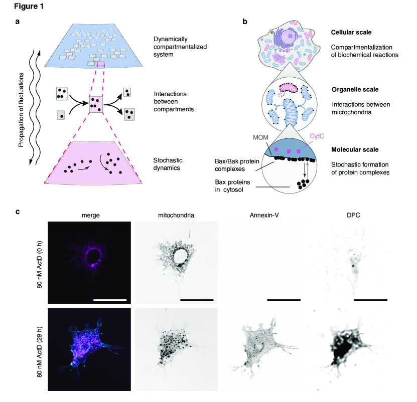
In contrast to inanimate matter, biological systems are organized into a discrete hierarchy of non-equilibrium processes on vastly different spatial scales [13, 14, 15, 16]. Molecular processes are often embedded into subcellular structures termed organelles and cells interact to form complex organs that constitute organisms. Biological systems have explicit mechanisms that propagate fluctuations and signals across scales. As an example, the decision of cells to commit suicide (i.e. apoptosis) is mediated by complexes of apoptosis-regulating proteins such as Bax [17] (Fig. 1b). Bax is recruited from the cytosol to the outer membrane of subcellular compartmens termed mitochondria (mitochondrial outer membrane, MOM) at a rate that depends on the cell’s stress level [18, 19]. In the MOM, these proteins undergo stochastic dynamics that lead to the formation of pores. These pores release the protein cytochrome C (CytC) from mitochondria, thereby irreversibly executing apoptosis [20, 18]. The mitochondria themselves are highly dynamic organelles that undergo rapid fusion and fission [21], which leads to concentration changes of protein complexes on their membranes. This gives rise to a two-way propagation of fluctuations and signals between the molecular and the organelle scale (Fig. 1b). Figure 1c shows fluorescent microscopy images of the morphological changes of mitochondria and early apoptotic features of a cell subject to an apoptotic stimulus. Beyond the fundamental biological mechanisms, understanding the regulation of apoptosis is fundamental to understanding development of organisms [17], and the development of disease [22].
Here, we show that compartmentalized systems exhibit an emergent degree of freedom whose kinetic properties are used by cells to regulate cell decisions. We study a paradigmatic class of compartmentalized stochastic systems, in which stochastic processes are embedded into interacting compartments. We derive the emergent degrees of freedom that arise from the propagation of fluctuations across the hierarchy of scales and experimentally demonstrate that cells make use of this to regulate apoptosis. Specifically, we develop a general theory of the propagation of noise and signals in a general class of dynamically compartmentized systems. We identify an emergent quasi-particle-like degree of freedom describing the kinetics of molecular pathways on the cellular scale. By deriving effective equations of motion, we show that this quasi-particle differs qualitatively in the response dynamics predicted by the biochemistry of the signaling pathway alone. We demonstrate the functional relevance of the quasi-particle in the context of apoptosis, where we show the emergence of a kinetic low-pass filter that allows cells to distinguish slow, biologically relevant changes that require the cell to commit suicide from fast, irrelevant fluctuations. We validate our findings experimentally by quantifying cell death in cell cultures subject to varying duration and strengths of apoptotic stimuli.
Results
Emergent quasi-particles in compartmentalized systems
To derive an effective theory of compartmentalized systems, we define a paradigmatic, yet general, theoretical framework for compartmentalized stochastic processes. We consider a system divided into compartments that are defined by their respective sizes . Each compartment hosts a stochastic process represented by the intrinsic variable within each compartment. In analogy to biochemical systems, we refer to as a concentration. can be any stochastic process with multiplicative Gaussian white noise and arbitrary noise amplitude. In this case, the time evolution of follows a Langevin equation of the form with a deterministic ”force” , noise amplitude and noise . Most biochemical systems fall into this category of stochastic processes. Compartments themselves undergo compartment dynamics like compartment growth, compartment fission, or compartment fusion. For homeostatic conditions, among all compartment processes, compartment fusion, and fission are the dominant contributions to fluctuations in the concentrations (Supplemental Theory 2.3). The state of the system is then defined by the combination of concentrations and compartment sizes for each compartment, .
Compartment dynamics affect the size of each compartment, but they also lead to fluxes in the concentrations as particles are exchanged between compartments. This leads to a nontrivial feedback between processes on the compartment scale and processes on the microscopic scale - fluctuations and signals propagate across scales.
In physics, one is usually interested in macroscopic observables, like pressure or conductivity. We therefore usually integrate out microscopic degrees of freedom. In biology, the molecular scale is important and target point of many experimental measurements and medical therapies. We therefore aim to derive an effective theory on the smallest scale. To this end, we integrate out probability fluxes between the microscopic and the compartment scale to derive an effective description of the time evolution of the probability that a randomly chosen compartment has a concentration at a given time ,
| (1) |
Here, the first term on the right-hand side describes the deterministic drift of the probability density function. This drift happens with a velocity that is proportional to the deterministic force and additional feedback from the compartment scales. This feedback is given by an additional effective interaction potential which describes an attractive force to the mean in the concentration space. The second, diffusive term on the right-hand side describes the dispersive spreading of due to noise.
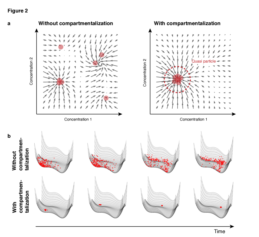
Equation (1) therefore describes the balance between the dispersion of the probability density function due to the diffusion term and its contractions due to the effective interactions between compartments. As a result, one intuitively expects the probability density function to obtain a fixed shape with constant variance. In plasma physics, Eq. (1) is known as the Vlasov-McKean equation, which admits solutions where the probability density function localizes (see also [14] and the Supplemental Theory for a derivation). Analogous to localization phenomena observed in condensed matter physics [23, 24, 25, 26], we refer to this localized mode as a quasi-particle. (see the Supplemental Theory 2.5. for a detailed discussion of the use of this term).
Therefore, the time evolution of the stochastic multi-scale system is effectively described by a single degree of freedom, which is the position of the quasi-particle in concentration space. We therefore next derive equations of motion for the quasi-particle. The kinetics of the quasi-particle is effectively described in terms of its “position” as the geometric median of the distribution of compartment concentrations, , and its “internal deformation” as the non-parametric skew, . In the limit of weak forces and weak noise to the highest order in the derivatives of , the equations of motion for the quasi-particle read , and (Supplemental Theory 2.6.). is a parameter that is proportional to and for times much longer than the time scales much longer than it quantifies the width of the distribution . Therefore, the deterministic motion in concentration space is complemented by an effective drift proportional to the non-parametric skew . This drift takes a value that exponentially approaches a steady state given by the second derivative of the force . For time scales much longer than the time scale of the compartment dynamics the equation of motion of the quasi-particle simplifies to
| (2) |
With this equation of motion, we have reduced the dynamics of a high-dimensional multi-scale system to the time evolution of a single degree of freedom, . The simplicity of the equation of motion allows graphical integration and thus offers an intuitive approach for predicting the emergent consequences of dynamic compartmentalization. Moreover, despite its simplicity, the equation of motion quantitatively agrees with results from numerical simulations of the full stochastic dynamics (Supplemental Theory Fig. S11).
One can gain intuition about the kinetics of the quasi-particle and the unusual appearance of the second derivative of the force by formal analogy to a simple mechanical system of three equal masses coupled by overdamped springs with spring constant (Supplemental Theory Figure Fig. S4). In this system, the position of the central mass is equivalent to and its deflection from the center of mass reflects . The force acting on the central mass is a combination of forces from the left and right masses resembling the discrete second derivative of the force field equivalent to the third derivative of the potential. Notably, with Eq. (2), the time evolution of the stochastic multi-scale system has been reduced to an ordinary differential equation that can be solved graphically or by using standard tools from dynamical systems theory.
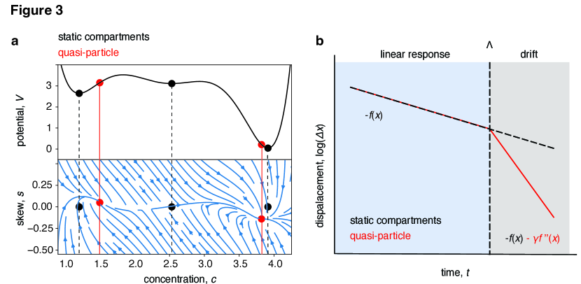
The dynamics of the quasi-particle defined by Eq. (2) exhibit a qualitatively different behavior compared to non-compartmentalized systems. First, the stationary states of the quasi-particle position do not necessarily coincide with the fixed points of (Fig. 3a). In general, since the quasi-particle distorts the force on length scales smaller than its variance and deepens it on much longer length scales, the number of metastable states is reduced compared to the number of fixed points of (Supplemental Theory 2.8.). In equilibrium biochemical systems this implies that steady states do not coincide with the minima of the Gibbs free energy defined by the chemical reactions.
Secondly, dynamically compartmentalized systems respond differently to perturbations. Specifically, the linearized response kinetics of the quasi-particle to perturbations in the force exhibit two temporal regimes. On short time scales, the response kinetics is dominated by a fast relaxation of to its steady state value. On time scales much longer than , the dynamics are governed by nonlinear dependencies in force (Fig. 3c). This behavior can be understood intuitively in the spring system analogy, where perturbations of the position of the central mass on time scales much smaller than the time scale associated with the spring constant (retardation time, ) lead to a deflection independent of the positions of the other masses akin to a point mass in a potential (Supplemental Theory Figure Fig. S4). Perturbations on time scales much slower than the retardation time are propagated between the masses and lead to a joint response depending on non-local properties of the potential.
Taken together, dynamically compartmentalized systems show qualitatively altered kinetics compared to non-compartmentalized systems. This holds for systems in which compartment dynamics happen on timescales faster than macroscopic changes in concentration. This raises the question of whether this behavior is used by cells to perform biological functions.
Quasi-particle kinetics in apoptosis
To test whether the emergent quasi-particle kinetics is used by cells to perform a biological function we now test these predictions in the context of a paradigmatic cell fate decision, that of apoptosis. Apoptosis is executed by the permeabilization of the MOM leading to release of the protein CytC from the mitochondrial intermembrane space, which triggers the disintegration of the cell [20, 18]. The release of CytC is facilitated by protein complexes of the apoptosis executing proteins Bax, which is recruited to the MOM, and Bak, which is resident in the MOM. Both proteins undergo stochastic kinetics that can lead to the formation of pores in the MOM [18, 19]. Notably, mitochondria are highly dynamic organelles that undergo continuous cycles of fusion and fission, which lead to the redistribution of Bax complexes among mitochondria. Therefore, apoptosis is an example of a dynamically compartmentalized system. Mitochondria undergo fusion and fission on a time scale of seconds to minutes [27, 28]. This is comparable to the time scale of Bax concentration changes on the MOM to lead to MOM permeabilization (MOMP) [27, 29]. Because of this, the kinetic parameters of apoptosis regulation fall into a regime where the quasi-particle kinetics, Eq. (2), is applicable.
By using the known biochemistry of Bax complex formation [18, 19, 30] we can derive the force term of Eq. (2) and the kinetic parameters of mitochondrial dynamics [31, 32, 27] fix the value of . This force term takes the form of a bistable system [33, 34, 35, 36] with one stable state at a low Bax concentration and a further stable state at a high Bax concentration, separated by unstable steady state positioned at an intermediary concentration (Fig. 4a, Supplemental Theory 3.2.).
To investigate whether apoptotic decision making involves quasi-particle dynamics, we performed two sets of experiments in which we compared cell cultures with dynamic mitochondria and cell cultures in which mitochondrial fission was inhibited. In the first experiment, we aimed to test the static properties of the quasi-particle to find out whether a quasi particle forms by the localization of Bax concentrations in concentration space, as illustrated in Fig. 2 b. In the context of the translocation dynamics of Bax to the mitochondrial membrane, we predict reduced variability of Bax concentration across different mitochondria inside the same cell, when cells are subjected to apoptotic stimuli.
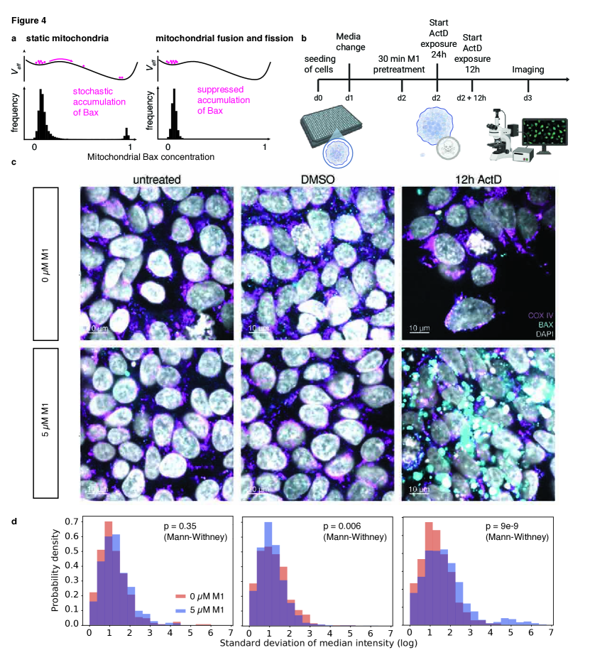
To test this, we quantified Bax localization on mitochondria in fluorescent microscopy experiments. Human induced pluripotent stem cell (hiSPC) cultures were treated with the Bax-acitvating apoptotic stimulus (actinomycin D, Act D) for 12 or 24 hours, with the mitochondrial intermembrane space and Bax fluorescently labeled (see Experimental Methods). Although this method cannot fully label the outer mitochondrial membrane, CoxIV-labeled pixels were used to robustly identify a subset of mitochondrial regions. In turn, the CoxIV co-localized median Bax intensity provides a robust measure of membrane-localized Bax, and the CoxIV area-weighted standard deviation over co-localized median Bax concentrations within the same cell serves as a robust approximation of Bax concentration variability. To assess the effect of multi-scale feedback, we proceed analogously with a second cell culture that was treated before the experiment with the functional mitochondrial fission inhibitor hydrazone M1 [37], which effectively diminishes quasi-particle dynamics.
We observed both for ActD untreated and 12h treated cells that the variability of membrane-bound Bax was significantly increased (two-sided Mann-Whitney U-test p < 0.05) when cells were pretreated with M1 Fig. 4c, d. Specifically, we found that the M1-treated cells comprise a sub-population with strongly increased Bax variability inside a single cell which we did not observe in cells not treated with M1. These cells correspond to cells with Bax puncta only on a subset of CoxIV label mitochondrial regions. The absence of this sub-population in cells not treated with M1 indicates that mitochondrial dynamics is necessary to localize subcellular mitochondrial Bax concentrations in the concentration phase space, a key characteristic of the quasi-particle predicted by Eq. (1).
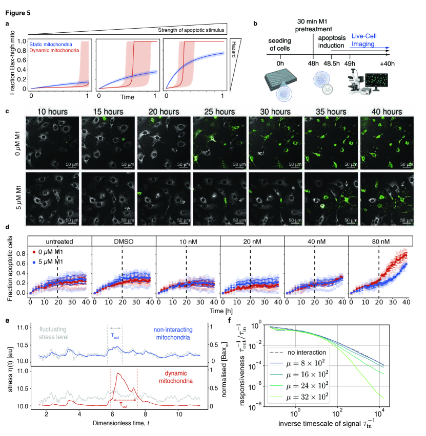
In the second set of experiments we next tested the kinetic properties of the quasi-particle. To this end we investigated the response of the Bax system to perturbations in the form of a constant stimulus which biologically describes the concentration of stress mediators in the cytosol and the ensuing recruitment rate of Bax proteins to the MOM. The effect of such a stimulus is a tilt in the effective bistable potential governing mitochondrial Bax concentrations (Supplemental Theory Figure Fig. S6). Equation (2) then predicts a sigmoidal response of mitochondrial Bax concentrations to the stimulus (Supplemental Fig. S9, S10, S11, and Supplemental Theory 3.3.). This means that the response to the stimulus is suppressed on short time scales and enhanced on long time scales compared to the case of static mitochondria. The characteristic time that separates both temporal regimes of the response kinetics decreases with increasing stimulus strength (Fig. 5a).
To test whether such kinetics emerges in cells we subjected cells to apoptotic stimuli of varying strength by administering ActD at different concentrations with or without mitochondrial fission inhibiting M1 pretreatment. We subsequently performed live imaging to monitor the fraction of apoptotic cells labelled by Annexin V (Figure 5b). We found that for a high concentration of ActD, cells without M1 pretreatment showed a stronger response to the apoptotic stimulus compared to cells pretreated with M1. Further, for low concentrations of ActD, cells not preptreated responded significantly weaker compared to M1 treated cells at early timepoints (); We performed a Wilcoxon signed-rank test comparing the two M1 treatment groups while we aggregated the data over all ActD treatment groups and over all early timepoints within the time window which yielded . This suggests that the accumulation dynamics of active apoptotic effects (i.e. the Bax accumulation dynamics) in response to an apoptotic stimuli follows a sigmoidal response dynamics in time as predicted by the kinetic equation of the quasi particle, Eq. (2).
The suppression of the response to stimuli on short time scale with the simultaneous enhancement of the response on long time scales resembles the behaviour of a low-pass filter used in electronics to suppress noise. To investigate whether the Bax system may establish a kinetic low pass filter of metabolite fluctuations we studied its response to a time-correlated stochastic signal . Figure 5e shows exemplary trajectories of a stochastic signal and ensuing Bax (protein) concentrations obtained from full stochastic simulations for physiological parameters (Methods). While in a hypothetical system with static mitochondria, the Bax concentration follows the stochastic signal, for a system with mitochondrial dynamics fast fluctuations in the stochastic signal are suppressed while slow trends tend to be followed and amplified. More general, the exponent characterizing the suppression of fast fluctuations increases with the rate of mitochondrial fusion and fission (Fig. 5f).
Discussion
Biological systems are inherently dynamic, with processes occurring across multiple spatial and temporal scales. Compartmentalization is a characteristic feature of many biological systems which underlies fundamental subcellular processes involved in cellular signaling and decision-making. We demonstrated that interactions between compartments gives rise to emergent quasi-particle kinetics characterized by unique kinetic properties: a suppressed response to extrinsic cues on short timescales and a facilitated, sigmoidal response on longer, persistent timescales.
Although the chemical reaction dynamics within dynamic compartments and the dynamics of the compartments themselves occur on vastly different spatial scales, their timescales can become intertwined. Specifically, macroscopic changes driven by stochastic fluctuations in the chemical reactions enclosed within these compartments often occur on the same timescale as the dynamics of the compartments. A relevant example for this is the stochastic accumulation of Bax proteins on the mitochondrial outer membrane [38] and mitochondrial fusion and fission cycles [27, 31, 32]. This coupling between the two processes creates a kinetic regime, where the emergent behavior cannot be fully understood by considering either spatial scale in isolation.
The biological relevance of the regulation of apoptosis and mitochondrial dynamics is ubiquitous. The canonical relevance lies in morphogenesis of tissues and organs in development as well as removal of superfluous or damaged cells [17]. Furthermore, erroneous activation or inhibition of apoptosis can contribute to the development of disease [22], including cancer [22], ischemic heart disease [22], and stroke [39, 40], among others. This makes apoptosis a desirable target for therapeutic intervention in medicine [41, 42, 43, 44].
The execution of apoptosis is regulated by complex molecular mechanisms [18, 20, 19, 30, 45]. Next to Bax, the structurally and functionally closely related protein Bak, which resides in the MOM, contributes to execution of apoptosis by pore formation [18, 20], and homomeric or heteromeric pores seem to follow different kinetics leading to MOMP [46]. Furthermore, complex protein interactions among the Bcl2 family members in the cytosol or the MOM regulate the events leading to execution of apoptosis [18, 20, 19, 30, 45]. However, the model proposed herein fully accounts for the regulation leading to the accumulation of Bax (or Bak) particles in the MOM and considers the particle concentration of these pro-apoptotic effectors in the MOM as prerequisite for pore formation leading to MOMP.
Beyond their classical function in providing energy for the cell, mitochondria are the integrators for various signaling pathways in cells [21]. Dynamic mitochondria are known to enable cells to cope with various states of stress [21, 47], and it is established that mitochondrial shape dynamically changes when cells die [21]. In the present study, we emphasize the interaction between dynamic mitochondria and Bax proteins, which collaboratively play a pivotal role in helping the cell determine which death-promoting stimuli are significant enough to execute apoptosis. This introduces a novel concept in biology, highlighting how cellular organelles and proteins dynamically interact to fine-tune life-or-death decisions within the cell.
We derived these results under general conditions: that of white Gaussian noise and the mixing of the time scale of the microscopic and compartment dynamics. We therefore expect that the properties of quasi-particle kinetics may also be relevant in the context of other organelle-associated cell decisions, such as the regulation of protein synthesis mediated by the translocation of mTORC1 to lysosomes [48, 49, 50], the maturational dynamics of endosomes [51, 52, 53], or the regulation of oxidative phosphorylation in mitochondria [54, 55, 56]. Sigmoidal responses are ubiquitous in biological systems and in important building block of their function [57, 58, 59]. In molecular systems, they usually emerge from cooperative binding of multiple proteins. Dynamic compartmentalization allows to generate a sigmoidal response with a Hill tuneable coefficient that depends on kinetic parameters of compartment fusion and fission.
Methods
Cell Culture
The human induced pluripotent stem cell (hiPSC) line BIHi250-A (https://hpscreg.eu/cell-line/BIHi250-A) was cultivated in Essential 8 medium prepared according to the original recipe [60] and grown on Geltrex (Thermo, A1413302) coated tissue culture plates. Media change was performed daily for six days with one double-feed on day 7. Cells were passaged when confluency reached approximately 80%. Mouse embryonic fibroblasts (MEFs) immortalized by limited dilution were grown in tissue culture flasks and cultivated in DMEM/F-12 (Thermo, 11320033) supplemented with 5% FBS (PAN Biotech, P30-3030M) and passaged when a confluency of approximately 80% was reached. All cell cultures were kept at 37°C and 5% CO2.
M1 Mitochondrial Fission Inhibitor and Actinomycin D Treatment
For mitochondrial fission inhibition with M1, MEFs were seeded into wells of a 384-well Greiner µClear Imaging plate (Greiner, 781091) at a density of 24,000 cells/cm2. 24 hours after seeding, media was changed. For M1 pre-treatment, M1 (Merck, SML0629-25MG) stock solution (50 mM) was diluted in PBS without calcium and magnesium (PBS-/-, Thermo, 14200075) and sonicated (40 kHz, 5 minutes, room temperature) and then added to respective wells at a final concentration of 5 µM. After 30 minutes of M1 pre-treatment, actinomycin D (ActD, Selleckchem, S8964) was added at concentrations from 0 to 80 nM. Immediately after addition of ActD, tetramethylrhodamine-ethylester (TMRE, Thermo, T669) was added to each well at a final concentration of 50 nM and Annexin-V-FITC (ImmunoTools, 31490013X2) according to the manufacturer’s instructions.
BAX Immunofluorescence
For BAX localization, hiPSCs were detached using TrypLETM Select (Thermo, 12563011) and seeded as single cells into Geltrex-coated 384-well Greiner µClear plates at a density of 24,000 cells/cm² in E8 medium containing 5 µM Y-27632 Rho-associated, coiled-coil containing protein kinase (ROCK) inhibitor (ROCKi, Selleckchem, S1049). After 24 hours, ROCKi was removed by replacing the medium. Following an additional 24-hour culture period, hiPSCs were treated with 12.5 nM ActD for 12 or 24 hours. A subset of cells was pre-treated with the mitochondrial fission inhibitor M1 for 30 minutes prior to ActD incubation as described above. At the experimental endpoint, cells were fixed with 4% paraformaldehyde (PFA, Carl Roth, 30525-89-4) for 15 minutes at 37°C and washed twice with PBS-/- at room temperature (RT). Blocking and permeabilization were performed using a blocking buffer consisting of 5% normal donkey serum (NDS, Milipore, S30-100ML) and 0.3% saponin (Sigma, 47036) in PBS-/- for 45 minutes at RT. Primary antibodies (COX IV (4D11-B3-E8) mouse mAB, Cell Signaling Technology, 11967; Bax (D2E11) rabbit mAb, Cell Signaling Technology, 5023) were diluted in blocking buffer at 1:500 and 1:200, respectively, and incubated with cells for 1 hour at RT. After three 5-minute washes with PBS-/- at RT, secondary antibodies (donkey anti-mouse (HCA)-Alexa 647, Thermo, A31571; donkey anti-rabbit (HCA)-Alexa 488, Thermo, A32790) were diluted 1:500 in blocking buffer and incubated for 2 hours at RT, protected from light. Following three additional 5-minute washes at RT, the final wash included 10 µM Hoechst 33342 (Thermo, H1399). Cells were protected from light until imaging.
Imaging
Live-Cell Imaging
Live-cell imaging was performed on a spinning-disk confocal microscope (Revvity Opera Phenix) with a 40x water immersion objective (NA=1.1), operated by Harmony 5.2 (Revvity) software. Digital phase contrast (DPC) was acquired at 50% laser power and 700 ms exposure time and created with an upper plane at 5 µm, a lower plane at -3 µm, a filter of 1.0, and speckle scale of 10 µm. TMRE was excited with a 561nm laser with a power of 100%, 80 ms exposure time, and at a height of 0 µm. Annexin-V-FITC was imaged using a 488 nm laser, 50% laser power, 40 ms exposure time, and imaging height of 0 µm. Sequential channel order was set to acquire the DPC first, followed by TMRE and then Annexin-V-FITC. Two separate sequences for consecutive image acquisition were set. Sequence 1 consisted of 6 timepoints with intervals of 2 hours. Sequence 2 comprised 30 timepoints with 1-hour intervals. This resulted in 2-hour intervals throughout the first 10 hours of the experiment followed by 30 hours with images taken hourly. For every well, 12 out of 81 fields were selected in which images were taken.
BAX Immunofluorescence Imaging
Imaging was performed on a spinning-disk confocal microscope (Revvity Opera Phenix) with a 63x water immersion objective (NA=1.15), operated by Harmony 5.2. Hoechst was imaged with a 375 nm laser with 40% laser power and 40 ms exposure time, COX IV was imaged with a 647 nm laser, 80% laser power for 80 ms, and BAX was imaged with a 488 nm laser at 80% laser power and 80 ms exposure time. All channels were acquired at an imaging height of 2 µm. Lasers for Hoechst and COX IV were excited together followed by a second exposure with excitation at 488 nm. 12 out of 225 fields were imaged per well.
Image Processing
Image processing was performed with SignalsImageArtist 1.3 (Revvity). Figures with microscopy images were prepared with OMERO.figure v4.4.3 on OMERO plus (Glencoe) .
Live-Cell Imaging
All images were subjected to a basic flatfield correction and brightfield correction. Images were pre-processed with filter-combinations to eliminate background noise. TMRE images were pre-processed with a sliding parabola (curvature 1), followed by Gaussian smoothing (width 2 pixels). The structures of interest (mitochondria) were identified by segmentation in an image calculated by subtracting the image pre-processed with sliding parabola and Gaussian smoothing from an image preprocessed with sliding parabola alone. Annexin-V-FITC images were also pre-processed with a sliding parabola (curvature 0.1, Gaussian smoothing 3 pixels width), but no further image calculation. Cells were identified using the Find Cells Building block (Method C) on the unfiltered TMRE channel as an estimate for the cell area. Morphology and intensity properties of identified cells were calculated and used to only select objects with a cell roundness above 0.3. Furthermore, objects touching the image border were excluded from analysis. Identification of mitochondria was achieved by Find Image Region based on the pre-processed / calculated TMRE images and restricted to the population of previously identified cells. Objects were identified based on an absolute threshold with the lowest intensity 6. Morphology and intensity properties for mitochondrial objects were calculated. Objects were eliminated when the area was 16 px2, when the object length was < 4 px, and when the intensity was 270. To identify Annexin-V-FITC positive vesicles (AV-vesicles), the Find Image Region building block was utilized based on the pre-processed Annexin-V-FITC channel. Within the region of previously identified cells, AV-vesicles were identified by an absolute threshold with the lowest intensity 50. Morphological and intensity properties of AV-vesicles were calculated. All calculated morphology and intensity properties were exported for secondary analysis. Raw data were annotated with treatment group and timepoint information based on well identifiers. Cells with an Annexin-V vesicle area > 0 were classified as apoptotic, while those with a vesicle area of 0 were classified as non-apoptotic. Statistical analysis was performed in python 3.7.13. For each treatment group and timepoint, the mean apoptotic cell count and its corresponding standard error were calculated using the agg function in pandas 1.3.5. For each treatment group and replica, the fraction of apoptotic cells was normalized by subtracting the fraction of apoptotic cells at the timepoint of first observation (). The normalized fraction of apoptotic cells was plotted over time for each treatment group using visualization libraries seaborn 0.12.2 and matplotlib 3.2.2. Additionally, the unweighted mean apoptotic cell count and its standard error were computed across all replicates for each treatment group and timepoint. These aggregated metrics were visualized along with the individual timepoint analysis to enable direct comparison. A Wilcoxon signed-rank test was performed to compare the aggregated data from all ActD treatments combined against the two M1 treatment within the time window .
Immunofluorescence Imaging
Brightfield correction and advanced flatfield correction were applied to all images. Channels were individually pre-processed with combinations of different filters to eliminate background and enhance signal of interest. For BAX signal, images were filtered with a sliding parabola at a curvature of 1 before performing a Gaussian smoothing with a width of 3 px. The image pre-processed with sliding parabola and Gaussian smoothing from an image pre-processed with sliding parabola alone to further eliminate background. The COX IV channel was filtered in the same way with a sliding parabola curvature of 10. Hoechst was pre-processed similarly, with a sliding parabola curvature of 0.1 and a Gaussian smooth width of 2 px. Nuclei were identified by Find Nuclei building block (Method B). After calculation of morphology and intensity properties, only those nuclei were accepted for analysis that showed a mean intensity > 36, a nucleus area > 43 µm2, and a nuclear roundness above 0.7. Cells were identified by a selected region around the nucleus. This region was created with an outer border of -5 µm. COX IV signal was used as an estimation of mitochondrial location within the cell and segmented by the building block Find Image Region based on the pre-processed COX IV channel. The segmentation of the COX IV image region was limited to the region of interest previously defined as cells. Based on calculated morphology and intensity properties, COX IV objects were included in the analysis when the object area was > 0.4µm2 and the intensity > 22. BAX particles were identified with Find Image Region based on the pre-processed BAX channel. BAX particle segmentation was limited to regions identified as COX IV positive. Raw data were annotated with treatment group and time point information based on well identifiers. Statistical analysis was performed using python 3.7.13. For each cell, the median Bax intensity within its COX IV-positive regions was calculated using pandas 1.3.5. Additionally, the standard deviation of Bax intensity within each COX IV-positive region was calculated, weighted by the area of the region. This weighted standard deviation was aggregated at the cell level and stored as a cell attribute. The heavily skewed distribution of the standard deviation of the median Bax intensity per cell was visualized as normalized density plots of the natural logarithm of the standard deviation, using matplotlib 3.2.2. For each ActD treatment, a two-sided Mann-Whitney U-test was performed against the M1 treatment on the standard deviation of the median Bax intensity.
Simulations
To study the multi-scale dynamics of compartmentalized stochastic reaction systems, we used standard SSA (stochastic simulation algorithm) to sample random trajectories of the system dynamics in the compartment and multiplexed the compartment dynamics by stochastically informing the results from the SSA in different compartment with each other. All simulations were implemented in python 3.7.13.
The chemical reactions within each compartment were simulated in parallel with the stochastic evolution of the compartments, with updates occurring at discrete intervals. This separation of timescales allowed for efficient parallelization of the simulation, significantly reducing execution time while maintaining accuracy. Memory management was optimized by discretizing compartment sizes and pre-allocating storage for concentration vectors and reaction matrices.
Acknowledgements
We thank F. Jülicher for the helpful feedback, the members of the BIH at Charité Research IT for maintaining the SIMA and OMERO platforms, and all members of the involved groups for critical discussions. This work was supported by the Core Unit pluripotent Stem Cells and Organoids (CUSCO) of the Berlin Institute of Health (BIH) at Charité – Universitätsmedizin Berlin. This project has received funding from the European Research Council (ERC, grant agreement no. 950349, S.R.) and under grant agreement 825161 (P.M. and H.S.) under the European Union’s Horizon 2020 research and innovation program, and in part by the Einstein Foundation Berlin (EJF-2020-602, EVF-2021-619, EVF-2021-619-2, EVF-BUA-2022-694 to P.M.; EC3R to P.M. and H.S.), the Else Kröner-Fresenius Stiftung (2019_A34, P.M.), the Volkswagen Stiftung (9A866, P.M.), and the Stiftung Charité (StC-VF-2024-59, P.M.). P.M. is Einstein Junior Fellow funded by the Einstein Foundation Berlin.
Code availability
Simulation routines are described in the Supplemental Theory in Section 4. Code snippets are available from the corresponding author upon reasonable request.
Competing interests
The authors declare no competing interests.
References
- [1] Andrei Nikolaevich Kolmogorov, V. Levin, Julian Charles Roland Hunt, Owen Martin Phillips, and David Williams. Dissipation of energy in the locally isotropic turbulence. Proceedings of the Royal Society of London. Series A: Mathematical and Physical Sciences, 434(1890):15–17, January 1997. Publisher: Royal Society.
- [2] Masaki Sano and Keiichi Tamai. A universal transition to turbulence in channel flow. Nature Phys, 12(3):249–253, March 2016. Publisher: Nature Publishing Group.
- [3] Takuma Yamada, Sanae-I. Itoh, Takashi Maruta, Naohiro Kasuya, Yoshihiko Nagashima, Shunjiro Shinohara, Kenichiro Terasaka, Masatoshi Yagi, Shigeru Inagaki, Yoshinobu Kawai, Akihide Fujisawa, and Kimitaka Itoh. Anatomy of plasma turbulence. Nature Phys, 4(9):721–725, September 2008. Publisher: Nature Publishing Group.
- [4] Nigel Goldenfeld. Lectures On Phase Transitions And The Renormalization Group. CRC Press, Boca Raton, June 2019.
- [5] Dapeng Bi, J. H. Lopez, J. M. Schwarz, and M. Lisa Manning. A density-independent rigidity transition in biological tissues. Nature Phys, 11(12):1074–1079, December 2015. Publisher: Nature Publishing Group.
- [6] William Bialek, Andrea Cavagna, Irene Giardina, Thierry Mora, Edmondo Silvestri, Massimiliano Viale, and Aleksandra M. Walczak. Statistical mechanics for natural flocks of birds. Proceedings of the National Academy of Sciences, 109(13):4786–4791, March 2012. Publisher: Proceedings of the National Academy of Sciences.
- [7] J. M. Yeomans. Statistical Mechanics of Phase Transitions. Clarendon Press, Oxford England : New York, 1st edition edition, June 1992.
- [8] Andrea Cavagna, Luca Di Carlo, Irene Giardina, Tomás S. Grigera, Stefania Melillo, Leonardo Parisi, Giulia Pisegna, and Mattia Scandolo. Natural swarms in 3.99 dimensions. Nat. Phys., 19(7):1043–1049, July 2023. Publisher: Nature Publishing Group.
- [9] Fernando Caballero, Cesare Nardini, and Michael E. Cates. From bulk to microphase separation in scalar active matter: a perturbative renormalization group analysis. J. Stat. Mech., 2018(12):123208, December 2018. Publisher: IOP Publishing and SISSA.
- [10] Thomas Christopoulos, Odysseas Tsilipakos, Nikolaos Grivas, and Emmanouil E. Kriezis. Coupled-mode-theory framework for nonlinear resonators comprising graphene. Phys. Rev. E, 94(6):062219, December 2016. Publisher: American Physical Society.
- [11] Liesbeth M. C. Janssen. Mode-Coupling Theory of the Glass Transition: A Primer. Front. Phys., 6, October 2018. Publisher: Frontiers.
- [12] Simone Zanotto, Francesco P. Mezzapesa, Federica Bianco, Giorgio Biasiol, Lorenzo Baldacci, Miriam Serena Vitiello, Lucia Sorba, Raffaele Colombelli, and Alessandro Tredicucci. Perfect energy-feeding into strongly coupled systems and interferometric control of polariton absorption. Nature Phys, 10(11):830–834, November 2014. Publisher: Nature Publishing Group.
- [13] Solenn Patalano, Adolfo Alsina, Carlos Gregorio-Rodriguez, Martin Bachman, Stephanie Dreier, Irene Hernando-Herraez, Paulin Nana, Shankar Balasubramanian, Seirian Sumner, Wolf Reik, and Steffen Rulands. Self-organization of plasticity and specialization in a primitively social insect. Cell Systems, 13(9):768–779.e4, September 2022.
- [14] Felix J. Meigel and Steffen Rulands. Controlling noise with self-organized resetting, September 2024. arXiv:2312.09307 [cond-mat].
- [15] Lorenzo Duso and Christoph Zechner. Stochastic reaction networks in dynamic compartment populations. Proceedings of the National Academy of Sciences, 117(37):22674–22683, September 2020. Publisher: Proceedings of the National Academy of Sciences.
- [16] David F. Anderson and Aidan S. Howells. Stochastic Reaction Networks Within Interacting Compartments. Bull Math Biol, 85(10):87, August 2023.
- [17] Kim Newton, Andreas Strasser, Nobuhiko Kayagaki, and Vishva M. Dixit. Cell death. Cell, 187(2):235–256, January 2024. Publisher: Elsevier.
- [18] Justin Kale, Elizabeth J. Osterlund, and David W. Andrews. BCL-2 family proteins: changing partners in the dance towards death. Cell Death Differ, 25(1):65–80, January 2018. Publisher: Nature Publishing Group.
- [19] Jonathan F. Lovell, Lieven P. Billen, Scott Bindner, Aisha Shamas-Din, Cecile Fradin, Brian Leber, and David W. Andrews. Membrane Binding by tBid Initiates an Ordered Series of Events Culminating in Membrane Permeabilization by Bax. Cell, 135(6):1074–1084, December 2008. Publisher: Elsevier.
- [20] Peter E. Czabotar and Ana J. Garcia-Saez. Mechanisms of BCL-2 family proteins in mitochondrial apoptosis. Nat Rev Mol Cell Biol, 24(10):732–748, October 2023. Publisher: Nature Publishing Group.
- [21] Luis-Carlos Tábara, Mayuko Segawa, and Julien Prudent. Molecular mechanisms of mitochondrial dynamics. Nat Rev Mol Cell Biol, pages 1–24, October 2024. Publisher: Nature Publishing Group.
- [22] Ilio Vitale, Federico Pietrocola, Emma Guilbaud, Stuart A. Aaronson, John M. Abrams, Dieter Adam, Massimiliano Agostini, Patrizia Agostinis, Emad S. Alnemri, Lucia Altucci, Ivano Amelio, David W. Andrews, Rami I. Aqeilan, Eli Arama, Eric H. Baehrecke, Siddharth Balachandran, Daniele Bano, Nickolai A. Barlev, Jiri Bartek, Nicolas G. Bazan, Christoph Becker, Francesca Bernassola, Mathieu J. M. Bertrand, Marco E. Bianchi, Mikhail V. Blagosklonny, J. Magarian Blander, Giovanni Blandino, Klas Blomgren, Christoph Borner, Carl D. Bortner, Pierluigi Bove, Patricia Boya, Catherine Brenner, Petr Broz, Thomas Brunner, Rune Busk Damgaard, George A. Calin, Michelangelo Campanella, Eleonora Candi, Michele Carbone, Didac Carmona-Gutierrez, Francesco Cecconi, Francis K.-M. Chan, Guo-Qiang Chen, Quan Chen, Youhai H. Chen, Emily H. Cheng, Jerry E. Chipuk, John A. Cidlowski, Aaron Ciechanover, Gennaro Ciliberto, Marcus Conrad, Juan R. Cubillos-Ruiz, Peter E. Czabotar, Vincenzo D’Angiolella, Mads Daugaard, Ted M. Dawson, Valina L. Dawson, Ruggero De Maria, Bart De Strooper, Klaus-Michael Debatin, Ralph J. Deberardinis, Alexei Degterev, Giannino Del Sal, Mohanish Deshmukh, Francesco Di Virgilio, Marc Diederich, Scott J. Dixon, Brian D. Dynlacht, Wafik S. El-Deiry, John W. Elrod, Kurt Engeland, Gian Maria Fimia, Claudia Galassi, Carlo Ganini, Ana J. Garcia-Saez, Abhishek D. Garg, Carmen Garrido, Evripidis Gavathiotis, Motti Gerlic, Sourav Ghosh, Douglas R. Green, Lloyd A. Greene, Hinrich Gronemeyer, Georg Häcker, György Hajnóczky, J. Marie Hardwick, Ygal Haupt, Sudan He, David M. Heery, Michael O. Hengartner, Claudio Hetz, David A. Hildeman, Hidenori Ichijo, Satoshi Inoue, Marja Jäättelä, Ana Janic, Bertrand Joseph, Philipp J. Jost, Thirumala-Devi Kanneganti, Michael Karin, Hamid Kashkar, Thomas Kaufmann, Gemma L. Kelly, Oliver Kepp, Adi Kimchi, Richard N. Kitsis, Daniel J. Klionsky, Ruth Kluck, Dmitri V. Krysko, Dagmar Kulms, Sharad Kumar, Sergio Lavandero, Inna N. Lavrik, John J. Lemasters, Gianmaria Liccardi, Andreas Linkermann, Stuart A. Lipton, Richard A. Lockshin, Carlos López-Otín, Tom Luedde, Marion MacFarlane, Frank Madeo, Walter Malorni, Gwenola Manic, Roberto Mantovani, Saverio Marchi, Jean-Christophe Marine, Seamus J. Martin, Jean-Claude Martinou, Pier G. Mastroberardino, Jan Paul Medema, Patrick Mehlen, Pascal Meier, Gerry Melino, Sonia Melino, Edward A. Miao, Ute M. Moll, Cristina Muñoz-Pinedo, Daniel J. Murphy, Maria Victoria Niklison-Chirou, Flavia Novelli, Gabriel Núñez, Andrew Oberst, Dimitry Ofengeim, Joseph T. Opferman, Moshe Oren, Michele Pagano, Theocharis Panaretakis, Manolis Pasparakis, Josef M. Penninger, Francesca Pentimalli, David M. Pereira, Shazib Pervaiz, Marcus E. Peter, Paolo Pinton, Giovanni Porta, Jochen H. M. Prehn, Hamsa Puthalakath, Gabriel A. Rabinovich, Krishnaraj Rajalingam, Kodi S. Ravichandran, Markus Rehm, Jean-Ehrland Ricci, Rosario Rizzuto, Nirmal Robinson, Cecilia M. P. Rodrigues, Barak Rotblat, Carla V. Rothlin, David C. Rubinsztein, Thomas Rudel, Alessandro Rufini, Kevin M. Ryan, Kristopher A. Sarosiek, Akira Sawa, Emre Sayan, Kate Schroder, Luca Scorrano, Federico Sesti, Feng Shao, Yufang Shi, Giuseppe S. Sica, John Silke, Hans-Uwe Simon, Antonella Sistigu, Anastasis Stephanou, Brent R. Stockwell, Flavie Strapazzon, Andreas Strasser, Liming Sun, Erwei Sun, Qiang Sun, Gyorgy Szabadkai, Stephen W. G. Tait, Daolin Tang, Nektarios Tavernarakis, Carol M. Troy, Boris Turk, Nicoletta Urbano, Peter Vandenabeele, Tom Vanden Berghe, Matthew G. Vander Heiden, Jacqueline L. Vanderluit, Alexei Verkhratsky, Andreas Villunger, Silvia von Karstedt, Anne K. Voss, Karen H. Vousden, Domagoj Vucic, Daniela Vuri, Erwin F. Wagner, Henning Walczak, David Wallach, Ruoning Wang, Ying Wang, Achim Weber, Will Wood, Takahiro Yamazaki, Huang-Tian Yang, Zahra Zakeri, Joanna E. Zawacka-Pankau, Lin Zhang, Haibing Zhang, Boris Zhivotovsky, Wenzhao Zhou, Mauro Piacentini, Guido Kroemer, and Lorenzo Galluzzi. Apoptotic cell death in disease—Current understanding of the NCCD 2023. Cell Death Differ, 30(5):1097–1154, May 2023. Publisher: Nature Publishing Group.
- [23] P. W. Anderson. Absence of Diffusion in Certain Random Lattices. Phys. Rev., 109(5):1492–1505, March 1958. Publisher: American Physical Society.
- [24] Philip W. Anderson. Basic Notions Of Condensed Matter Physics. CRC Press, March 2018. Google-Books-ID: 9HhQDwAAQBAJ.
- [25] Paul Romatschke and Michael Strickland. Collective modes of an anisotropic quark-gluon plasma. Phys. Rev. D, 68(3):036004, August 2003. Publisher: American Physical Society.
- [26] Shamik Gupta and Stefano Ruffo. The world of long-range interactions: A bird’s eye view. Int. J. Mod. Phys. A, 32(09):1741018, March 2017. Publisher: World Scientific Publishing Co.
- [27] Patrick D. Bhola, Alexa L. Mattheyses, and Sanford M. Simon. Spatial and Temporal Dynamics of Mitochondrial Membrane Permeability Waves during Apoptosis. Biophysical Journal, 97(8):2222–2231, October 2009.
- [28] Sarah B. Berman, Ying-bei Chen, Bing Qi, J. Michael McCaffery, Edmund B. Rucker, III, Sandra Goebbels, Klaus-Armin Nave, Beth A. Arnold, Elizabeth A. Jonas, Fernando J. Pineda, and J. Marie Hardwick. Bcl-xL increases mitochondrial fission, fusion, and biomass in neurons. Journal of Cell Biology, 184(5):707–719, March 2009.
- [29] M. Rehm, H. J. Huber, C. T. Hellwig, S. Anguissola, H. Dussmann, and J. H. M. Prehn. Dynamics of outer mitochondrial membrane permeabilization during apoptosis. Cell Death Differ, 16(4):613–623, April 2009. Publisher: Nature Publishing Group.
- [30] Lieven P. Billen, Candis L. Kokoski, Jonathan F. Lovell, Brian Leber, and David W. Andrews. Bcl-XL Inhibits Membrane Permeabilization by Competing with Bax. PLOS Biology, 6(6):e147, June 2008. Publisher: Public Library of Science.
- [31] Michal Cagalinec, Dzhamilja Safiulina, Mailis Liiv, Joanna Liiv, Vinay Choubey, Przemyslaw Wareski, Vladimir Veksler, and Allen Kaasik. Principles of the mitochondrial fusion and fission cycle in neurons. J Cell Sci, 126(Pt 10):2187–2197, May 2013.
- [32] Verónica Eisner, Guy Lenaers, and György Hajnóczky. Mitochondrial fusion is frequent in skeletal muscle and supports excitation–contraction coupling. Journal of Cell Biology, 205(2):179–195, April 2014.
- [33] Jun Cui, Chun Chen, Haizhu Lu, Tingzhe Sun, and Pingping Shen. Two Independent Positive Feedbacks and Bistability in the Bcl-2 Apoptotic Switch. PLOS ONE, 3(1):e1469, January 2008. Publisher: Public Library of Science.
- [34] Chun Chen, Jun Cui, Haizhu Lu, Rui Wang, Shuai Zhang, and Pingping Shen. Modeling of the Role of a Bax-Activation Switch in the Mitochondrial Apoptosis Decision. Biophysical Journal, 92(12):4304–4315, June 2007.
- [35] Der-Fen Suen, Kristi L. Norris, and Richard J. Youle. Mitochondrial dynamics and apoptosis. Genes Dev., 22(12):1577–1590, June 2008.
- [36] Sabrina L. Spencer and Peter K. Sorger. Measuring and Modeling Apoptosis in Single Cells. Cell, 144(6):926–939, March 2011.
- [37] Danling Wang, Jianing Wang, Ghislain M. C. Bonamy, Shelly Meeusen, Richard G. Brusch, Carolina Turk, Pengyu Yang, and Peter G. Schultz. A Small Molecule Promotes Mitochondrial Fusion in Mammalian Cells. Angewandte Chemie International Edition, 51(37):9302–9305, 2012. _eprint: https://onlinelibrary.wiley.com/doi/pdf/10.1002/anie.201204589.
- [38] Kai Cao, Joel S. Riley, Rosalie Heilig, Alfredo E. Montes-Gómez, Esmee Vringer, Kevin Berthenet, Catherine Cloix, Yassmin Elmasry, David G. Spiller, Gabriel Ichim, Kirsteen J. Campbell, Andrew P. Gilmore, and Stephen W. G. Tait. Mitochondrial dynamics regulate genome stability via control of caspase-dependent DNA damage. Developmental Cell, 57(10):1211–1225.e6, May 2022.
- [39] Jakob Walther, Elena Marie Kirsch, Lina Hellwig, Sarah S. Schmerbeck, Paul M. Holloway, Alastair M. Buchan, and Philipp Mergenthaler. Reinventing the Penumbra — the Emerging Clockwork of a Multi-modal Mechanistic Paradigm. Transl. Stroke Res., 14(5):643–666, October 2023.
- [40] Philipp Mergenthaler, Joyce S. Balami, Ain A. Neuhaus, Amin Mottahedin, Gregory W. Albers, Peter M. Rothwell, Jeffrey L. Saver, Martin E. Young, and Alastair M. Buchan. Stroke in the Time of Circadian Medicine. Circ Res, 134(6):770–790, March 2024.
- [41] Xin Niu, Hetal Brahmbhatt, Philipp Mergenthaler, Zhi Zhang, Jing Sang, Michael Daude, Fabian G. R. Ehlert, Wibke E. Diederich, Eve Wong, Weijia Zhu, Justin Pogmore, Jyoti P. Nandy, Maragani Satyanarayana, Ravi K. Jimmidi, Prabhat Arya, Brian Leber, Jialing Lin, Carsten Culmsee, Jing Yi, and David W. Andrews. A Small-Molecule Inhibitor of Bax and Bak Oligomerization Prevents Genotoxic Cell Death and Promotes Neuroprotection. Cell Chemical Biology, 24(4):493–506.e5, April 2017. Publisher: Elsevier.
- [42] Kaiming Li, Mark F van Delft, and Grant Dewson. Too much death can kill you: inhibiting intrinsic apoptosis to treat disease. The EMBO Journal, 40(14):e107341, July 2021. Publisher: John Wiley & Sons, Ltd.
- [43] Justin P. Pogmore, David Uehling, and David W. Andrews. Pharmacological Targeting of Executioner Proteins: Controlling Life and Death. J. Med. Chem., 64(9):5276–5290, May 2021. Publisher: American Chemical Society.
- [44] Sarah T. Diepstraten, Mary Ann Anderson, Peter E. Czabotar, Guillaume Lessene, Andreas Strasser, and Gemma L. Kelly. The manipulation of apoptosis for cancer therapy using BH3-mimetic drugs. Nat Rev Cancer, 22(1):45–64, January 2022. Publisher: Nature Publishing Group.
- [45] Christian Bogner, Justin Kale, Justin Pogmore, Xiaoke Chi, Aisha Shamas-Din, Cécile Fradin, Brian Leber, and David W. Andrews. Allosteric Regulation of BH3 Proteins in Bcl-xL Complexes Enables Switch-like Activation of Bax. Molecular Cell, 77(4):901–912.e9, February 2020.
- [46] Katia Cosentino, Vanessa Hertlein, Andreas Jenner, Timo Dellmann, Milos Gojkovic, Aida Peña-Blanco, Shashank Dadsena, Noel Wajngarten, John S. H. Danial, Jervis Vermal Thevathasan, Markus Mund, Jonas Ries, and Ana J. Garcia-Saez. The interplay between BAX and BAK tunes apoptotic pore growth to control mitochondrial-DNA-mediated inflammation. Molecular Cell, 82(5):933–949.e9, March 2022.
- [47] Rubén Quintana-Cabrera and Luca Scorrano. Determinants and outcomes of mitochondrial dynamics. Molecular Cell, 83(6):857–876, March 2023. Publisher: Elsevier.
- [48] Suchithra Menon, Christian C. Dibble, George Talbott, Gerta Hoxhaj, Alexander J. Valvezan, Hidenori Takahashi, Lewis C. Cantley, and Brendan D. Manning. Spatial Control of the TSC Complex Integrates Insulin and Nutrient Regulation of mTORC1 at the Lysosome. Cell, 156(4):771–785, February 2014.
- [49] Brittany Angarola and Shawn M. Ferguson. Coordination of Rheb lysosomal membrane interactions with mTORC1 activation. F1000Res, 9:F1000 Faculty Rev–450, May 2020.
- [50] Bernadette Carroll, Dorothea Maetzel, Oliver DK Maddocks, Gisela Otten, Matthew Ratcliff, Graham R Smith, Elaine A Dunlop, Joao F Passos, Owen R Davies, Rudolf Jaenisch, Andrew R Tee, Sovan Sarkar, and Viktor I Korolchuk. Control of TSC2-Rheb signaling axis by arginine regulates mTORC1 activity. eLife, 5:e11058, January 2016. Publisher: eLife Sciences Publications, Ltd.
- [51] Willem Stoorvogel, Ger J. Strous, Hans J. Geuze, Viola Oorschot, and Alan L. Schwartzt. Late endosomes derive from early endosomes by maturation. Cell, 65(3):417–427, May 1991.
- [52] Lionel Foret, Jonathan E. Dawson, Roberto Villasenor, Claudio Collinet, Andreas Deutsch, Lutz Brusch, Marino Zerial, Yannis Kalaidzidis, and Frank Juelicher. A General Theoretical Framework to Infer Endosomal Network Dynamics from Quantitative Image Analysis. Current Biology, 22(15):1381–1390, August 2012.
- [53] Angela Wandinger-Ness and Marino Zerial. Rab Proteins and the Compartmentalization of the Endosomal System. Cold Spring Harb Perspect Biol, 6(11):a022616, November 2014. Company: Cold Spring Harbor Laboratory Press Distributor: Cold Spring Harbor Laboratory Press Institution: Cold Spring Harbor Laboratory Press Label: Cold Spring Harbor Laboratory Press Publisher: Cold Spring Harbor Lab.
- [54] Vitaly A. Selivanov, Tatyana V. Votyakova, Jennifer A. Zeak, Massimo Trucco, Josep Roca, and Marta Cascante. Bistability of Mitochondrial Respiration Underlies Paradoxical Reactive Oxygen Species Generation Induced by Anoxia. PLoS Comput Biol, 5(12):e1000619, December 2009.
- [55] Vitaly A. Selivanov, Marta Cascante, Mark Friedman, Mark F. Schumaker, Massimo Trucco, and Tatyana V. Votyakova. Multistationary and Oscillatory Modes of Free Radicals Generation by the Mitochondrial Respiratory Chain Revealed by a Bifurcation Analysis. PLOS Computational Biology, 8(9):e1002700, September 2012. Publisher: Public Library of Science.
- [56] Sunil Nath. The thermodynamic efficiency of ATP synthesis in oxidative phosphorylation. Biophysical Chemistry, 219:69–74, December 2016.
- [57] Boris N. Kholodenko. Cell-signalling dynamics in time and space. Nat Rev Mol Cell Biol, 7(3):165–176, March 2006. Publisher: Nature Publishing Group.
- [58] Michael Hinczewski and D. Thirumalai. Cellular Signaling Networks Function as Generalized Wiener-Kolmogorov Filters to Suppress Noise. Phys. Rev. X, 4(4):041017, October 2014. Publisher: American Physical Society.
- [59] Qiang Zhang, Sudin Bhattacharya, and Melvin E. Andersen. Ultrasensitive response motifs: basic amplifiers in molecular signalling networks. Open Biology, 3(4):130031, April 2013. Publisher: Royal Society.
- [60] Guokai Chen, Daniel R. Gulbranson, Zhonggang Hou, Jennifer M. Bolin, Victor Ruotti, Mitchell D. Probasco, Kimberly Smuga-Otto, Sara E. Howden, Nicole R. Diol, Nicholas E. Propson, Ryan Wagner, Garrett O. Lee, Jessica Antosiewicz-Bourget, Joyce M. C. Teng, and James A. Thomson. Chemically defined conditions for human iPSC derivation and culture. Nat Methods, 8(5):424–429, May 2011. Publisher: Nature Publishing Group.
See pages - of Supplement_for_QP.pdf