[1,2]\fnmLorenzo \surMercolli
[1]\orgdivDepartment of Nuclear Medicine, \orgnameInselspital, Bern University Hostpital, University of Bern, \orgaddress\cityBern, \countrySwitzerland
2]\orgdivARTORG Center for Biomedical Engineering Research, \orgnameUniversity of Bern, \orgaddress\cityBern, \countrySwitzerland
3]\orgnameSiemens Medical Solutions USA, Inc., \orgaddress\cityKnoxville TN, \countryUSA
4]\orgdivFaculty of Physics, Astronomy and Applied Computer Science, \orgnameJagiellonian University, \orgaddress\cityKrakow, \countryPoland
5]\orgdivCentre for Theranostics, \orgnameJagiellonian University, \orgaddress\cityKrakow, \countryPoland
6]\orgnameSiemens Healthineers International AG, \orgaddress\cityZürich, \countrySwitzerland
Positronium Lifetime Imaging with the Biograph Vision Quadra using 124I
Abstract
Purpose: Measuring the ortho-positronium (oPs) lifetime in human tissue bears the potential of adding clinically relevant information about the tissue microenvironment to conventional positron emission tomography (PET). Through phantom measurements, we investigate the voxel-wise measurement of oPs lifetime using a commercial long-axial field-of-view (LAFOV) PET scanner.
Methods: We prepared four samples with mixtures of Amberlite XAD4, a porous polymeric adsorbent, and water and added between 1.12 MBq and 1.44 MBq of 124I. The samples were scanned in two different setups: once with a couple of centimeters between each sample (15 minutes scan time) and once with all samples taped together (40 minutes scan time). For each scan, we determine the oPs lifetime for the full samples and at the voxel level. The voxel sizes under consideration are 10.03 mm3, 7.13 mm3 and 4.03 mm3.
Results: Amberlite XAD4 allows the preparation of samples with distinct oPs lifetime. Using a Bayesian fitting procedure, the oPs lifetimes in the whole samples are 2.52±0.03 ns, 2.37±0.03 ns, 2.27±0.04 ns and 1.82±0.02 ns, respectively. The voxel-wise oPs lifetime fits showed that even with 4.03 mm3 voxels the samples are clearly distinguishable and a central voxels have good count statistics. However, the situation with the samples close together remains challenging with respect to the spatial distinction of regions with different oPs lifetimes.
Conclusion: Our study shows that positronium lifetime imaging on a commercial LAFOV PET/CT should be feasible under clinical conditions using 124I.
keywords:
Positronium lifetime imaging, Long axial field-of-view PET/CT, 124IIntroduction
Ortho-positronium (oPs), the spin 1 state of an electron-positron bound state, has a significantly longer lifetime in vacuum than the spin 0 state, which is called para-positronium (pPs). The lifetime of pPs is too short in order to interact significantly with the environment [1]. However, oPs has a lifetime of about in vacuum and it can therefore undergo different interactions with surrounding atoms and molecules (see e.g. Refs. [2, 3, 1, 4]). In particular, the oPs’ positron can annihilate with an environmental electron, and thereby, the oPs lifetime can be significantly shortened. This so-called pick-off process makes the oPs lifetime dependent on the atomic and molecular structure of the surrounding material. oPs lifetime is also shortened by a spin exchange process depending on the concentration of oxygen molecules [5, 1, 6]. In vacuum oPs decays into three photons, while in matter due to the pick-off and conversion processes, it may annihilate also into two photons. In principle, both decays can be used for measuring the lifetime of oPs properties. However, it was shown that oPs lifetime imaging based on the two-photon annihilation is 300 times more efficient than oPs imaging based on annihilation into three photons [7, 7, 8].
There is a significant interest in the medical domain for oPs lifetime measurements (see e.g. Refs. [9, 3, 7, 1, 4, 10]), mainly driven by the possibility to measure oxygenation levels in human tissue, [5, 11, 12, 6, 13, 14], to asses tissue pathology in vivo [15, 16, 17, 18, 19] and to sense pH level and electrolytes within the tissue [20, 21, 22, 13, 23]. Recently, the first in vivo positronium images [18] and the first in-vivo measurements of oPs lifetime with clinical positron emission tomography (PET) system [18, 24] were demonstrated. The oPs lifetime has the potential to add diagnostic information, which is currently unavailable or requires additional interventions, such as e.g. biopsy or additional use of hypoxia tracers.
In Refs. [25, 24], we showed that it is possible to do oPs lifetime measurements with a commercial long axial field-of-view (LAFOV) PET scanner [26, 27]. However, in Ref. [24] we also showed that the collection of sufficient count statistics with a PET scanner is a major challenge. The voxel-wise determination of oPs lifetime, what is usually called oPs lifetime imaging, has been shown to be feasible only with long-lived radionuclides, long scan times, large voxel sizes or simplifying the fit models [18, 15, 28, 29, 30, 31]. Usually, a combination of multiple of these methods is required.
In this report, we show that oPs lifetime imaging can be achieved using a commercial PET/CT scanner under conditions typically encountered in clinical practice with respect to isotope, activity concentration, scan time, and voxel size. As highlighted in Refs. [25, 24, 32], \isotope[124]I possesses favorable characteristics for oPs lifetime imaging [32], it is also well suited for oPs imaging with the Biograph Vision Quadra (Siemens Healthineers, USA) [25, 24] and is routinely used in some departments due to the favorable imaging characteristics compared to conventional \isotope[131]I imaging. In contrast to other thyroid-directed PET tracers, like [\isotope[18]F]tetrafluoroborate, \isotope[124]I PET has usually higher uptake and also enables delayed imaging which can be used for dosimetry applications [33, 34, 35]. Using phantom measurements, we identify the conditions under which oPs lifetime imaging is viable and bring to the fore the remaining challenges.
1 Materials and methods
In order to asses the capabilities of Quadra with respect to oPs lifetime imaging, we filled four chemistry tubes with different mixtures of Amberlite XAD4 (Sigma-Aldrich, Co., St. Louis MO, USA) and demineralized water. As shown in Ref. [36], XAD4 allows to vary the oPs lifetime with a simple experimental setup. A relatively low activity of [\isotope[124]I]NaI was added to each tube. Tab. 1 summarizes the details of the sample preparation and Fig. 1 shows the experimental setup. The first tube contained XAD4 that was air-dried for 24 hours. T2 cointained the wet XAD4 (as it is delivered), while for T2 we added of gelatine to wet XAD4. In Tab. 1 is the weight of the wet XAD4 and the gelatine together. To all tubes, about [\isotope[124]I]NaI solution was added.
| Sample | XAD4 | ||||
|---|---|---|---|---|---|
| T1 | Dry | 1.12 | 5.0 | 3.25 | 0.67 |
| T2 | Wet | 1.44 | 5.5 | 3.56 | 0.67 |
| T3 | Gelatine | 1.14 | 4.5 | 3.49 | 0.79 |
| T4 | Deminalized water | 1.26 | 5.0 | 5.0 | 1.0 |
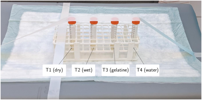
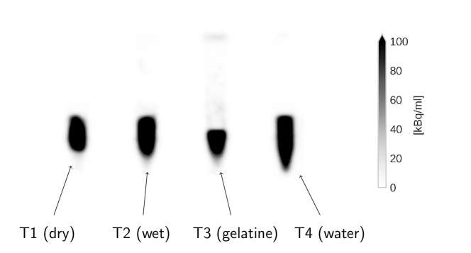
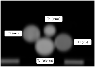
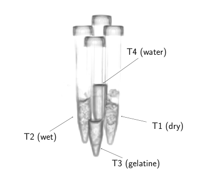
The samples were measured once with a large distance between them, as shown in Fig. 1, and once taped together. In Fig. 2 we show the maximum intensity projection (MIP) of the coincidence PET image of the setup with separated tubes (5 minutes scan time). The voxel size is . The experimental setup with the tubes taped together is depicted Fig. 3 with the top view of a CT slice and a 3D rendering from the CT. In the CT images, the voxel size is .
In addition, we measured the samples 15 minutes (separated) and 40 minutes (taped together) in singles mode. As described in Ref. [25], singles mode in Quadra records all single-crystal interactions into a list mode file. A prototype software was used to sort all three-photon events (), i.e. events with two photons in the annihilation window and one photon in the prompt energy window of . \isotope[124]I has the convenient property of having a prompt photon with an energy of , which Quadra’s detector can fully resolve, and a relatively high branching ratio of [37, 38]. The spatial location of a is determined through the time-of-flight (TOF) information of the two annihilation photons, i.e. there is no image reconstruction along the line of Refs. [39, 40, 41, 42]. The histoimages of are binned into three different voxel sizes of , and , respectively. We consider the voxel size to be on the verge of clinical usefulness. It is also the same order of magnitude as the maximum positron range in water for \isotope[124]I (continuous slowing down approximation range for a positron in water is according to NIST PSTAR). On the other end, we chose the smallest voxel size such that it would really push oPs imaging on Quadra to its limits. According to Refs. [43, 44], is about the spatial resolution that is achievable in coincidence PET imaging with \isotope[124]I. Finally, sits in the middle and could be thought of as similar to the spatial resolution of a SPECT/CT system. As a comparison, we also perform a fit that encompasses all in a single tube.
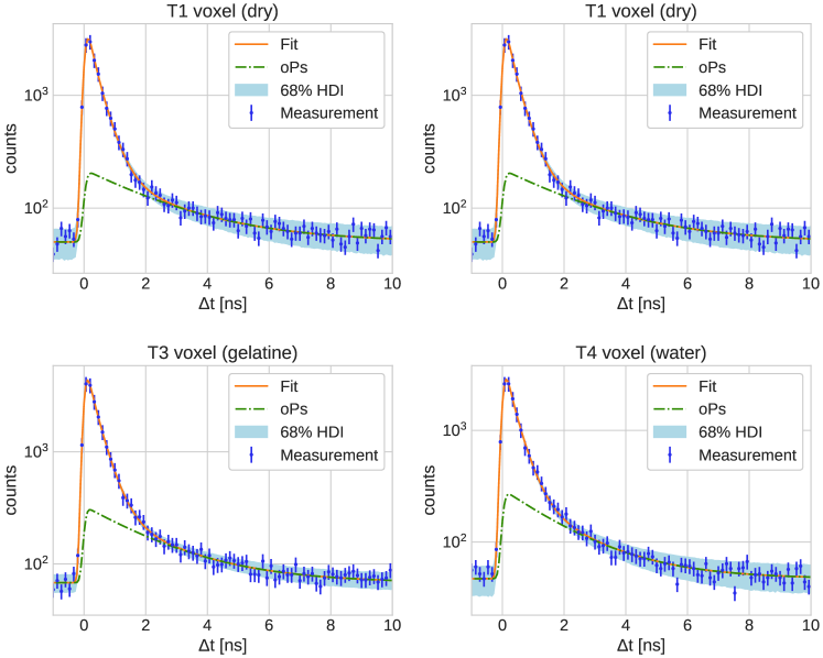
The time difference distributions (TDD) are the binned time differences between the annihilation and prompt photons in each voxel (time bin width is , i.e. slightly above Quadras time resolution). We use the Bayesian fitting procedure discussed in Refs. [25, 24] to determine the oPs lifetime from a measured TDD. The fitting model is the same as in Refs. [25, 24], i.e. a Gaussian function convoluted with three lifetime components for pPs, direct annihilation and oPs. In contrast to Ref. [24], we fix the pPs lifetime and direct annihilation together with the background count number. The background is fixed as the mean value of time differences that are smaller than . We fit the following priors with a Gaussian likelihood to the voxel-wise TDD
| (1) | ||||
where is the background value and are the bin values of the TDD. In the oPs lifetime images, we only selected voxels that have a relative error in the background region of less than . We fit time differences in the range from to .
The posterior distribution of is bell-shaped with hardly any skewness. We therefore report the uncertainty of with a standard deviation, which is estimated with the common point estimate. The uncertainty of the branching ratios is given in terms of the highest density intervals (HDI) of the posterior distribution since a point estimate of the standard deviation would not make sense for a Dirichlet variable.
2 Results
The top four rows of Tab. 2 report the single tube fits, i.e. when all measured time differences in a tube are collected in one TDD (no spatial binning of the data). Clearly, the different humidity levels of the XAD4 powder lead to significantly distinct oPs lifetimes. Furthermore, Tab. 2 includes also the results from fitting a TDD from a single voxel. The voxel is chosen in the central region of each tube. This allows us to get a good intuition about the count statistics for the smallest voxel size. Fig. 4 shows the corresponding single-voxel TDD together with the fit prediction for the oPs lifetime component.
| Sample | |||||||
|---|---|---|---|---|---|---|---|
| T1 (dry) | 2.52±0.03 | 0.073 | [0.071, 0.074] | 0.716 | [0.714, 0.718] | 0.211 | [0.21, 0.213] |
| T2 (wet) | 2.37±0.03 | 0.081 | [0.079, 0.082] | 0.702 | [0.7, 0.704] | 0.217 | [0.216, 0.219] |
| T3 (gelatine) | 2.27±0.04 | 0.073 | [0.071, 0.075] | 0.705 | [0.702, 0.708] | 0.222 | [0.22, 0.223] |
| T4 (water) | 1.82±0.02 | 0.088 | [0.087, 0.09] | 0.644 | [0.641, 0.646] | 0.268 | [0.267, 0.269] |
| T1 voxel | 2.56±0.23 | 0.09 | [0.081, 0.099] | 0.702 | [0.687, 0.716] | 0.208 | [0.2, 0.216] |
| T2 voxel | 2.37±0.23 | 0.073 | [0.062, 0.085] | 0.69 | [0.671, 0.708] | 0.237 | [0.227, 0.247] |
| T3 voxel | 2.3±0.12 | 0.044 | [0.039, 0.05] | 0.745 | [0.736, 0.754] | 0.21 | [0.205, 0.215] |
| T4 voxel | 2.0±0.14 | 0.062 | [0.052, 0.072] | 0.683 | [0.666, 0.7] | 0.255 | [0.246, 0.264] |
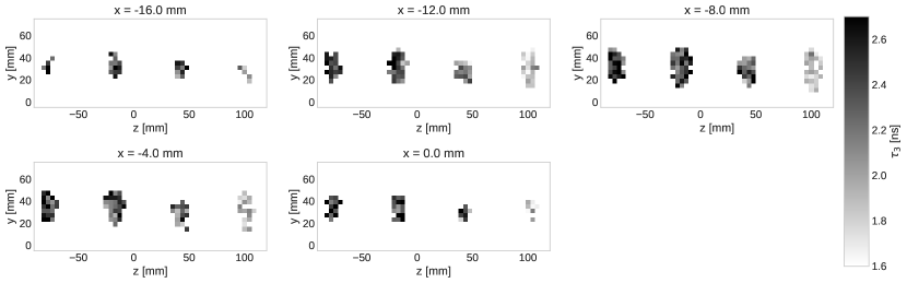
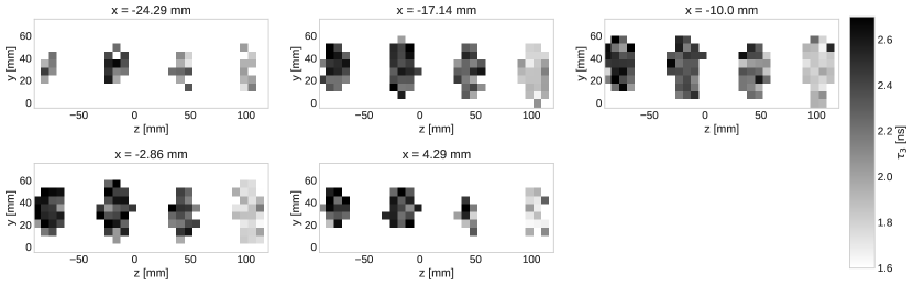
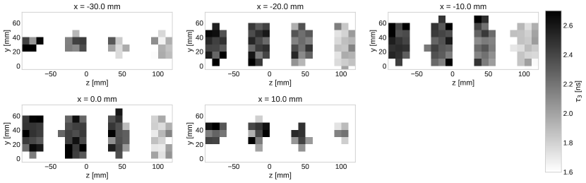
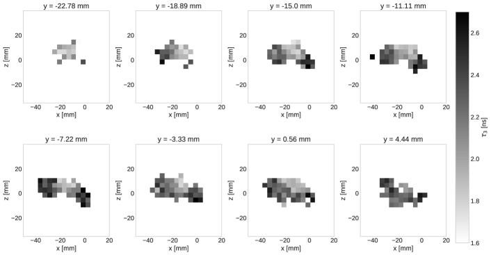
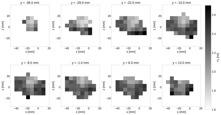
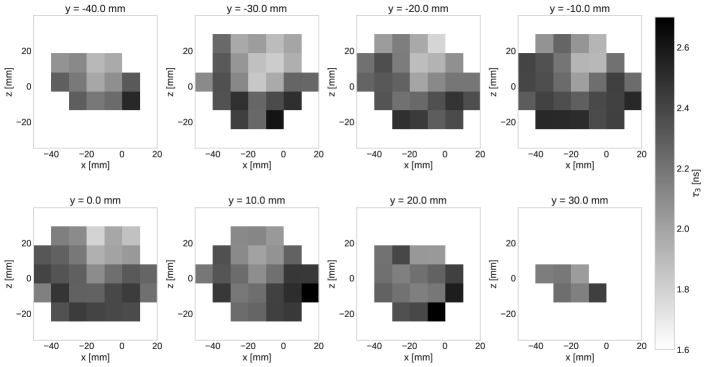
In Figs. 5 and 6 we show 2D slices of the oPs lifetime images for the two scans with separated and taped-together tubes. The two top rows show slices for the voxel size, while the middle and bottom rows are for the and voxel sizes, respectively. For best visualization, the slices of the separated tubes in Fig. 5 are shown in the plane, analogously to a coronal PET MIP in Fig. 2. For the tubes close together, we chose the plane as in the CT slice in Fig. 3. No post-processing, such as smoothing or any filtering, was applied to the oPs lifetime images.
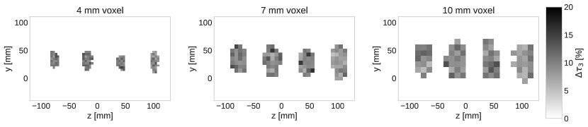
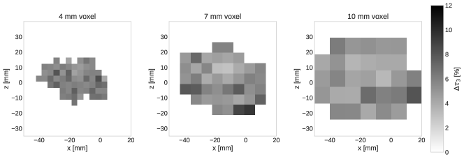
Finally, we show the MIP of the standard deviation for for the three voxel sizes and the two experimental setups in Fig. 7. The color bar in these figures shows the relative uncertainty of for each voxel.
3 Discussion
First, we would like to highlight the low statistical uncertainty of the oPs lifetime in the single tube fit in Tab. 2. Within a few ml, an activity concentration low as and a scan time of 15 minutes, the marginalized posterior distribution of has a relative standard deviation of less than using a commercial LAFOV PET/CT and Ref. [25]’s methodology. Apart from the higher prompt photon branching ratio of \isotope[124]I, the main improvement compared \isotope[82]Rb and \isotope[68]Ga (see Refs. [25, 24]) is Quadra’s capability to resolve \isotope[124]I’s photopeak and thereby increasing the peak signal-to-background ratio (pSBR). In addition, the smaller decay constant \isotope[124]I allows for a longer scan time and larger time-integrated counts. This high statistical precision allows to distinguish the oPs lifetime in the four samples, confirming Ref. [36] in that XAD4 is well suited for performance evaluations and intercomparisons of PET/CT scanners for oPs lifetime imaging.
In the literature, oPs lifetime imaging has shown to be feasible only with \isotope[22]Na and very long scan times and/or large voxel sizes. E.g. Ref. [15] applied a spatial binning of to their tissue sample data. Likewise, Ref. [18] seems to have a voxel size of multiple cm. It remains unclear, how much viable clinical information such voxel sizes may contain. With \isotope[82]Rb and \isotope[68]Ga, i.e. radionuclides that are used in clinical routine, the count statistics and in particular the pSBR are not sufficient for oPs lifetime imaging, given our methodology. This is why in Refs. [25, 24] we refrained from performing voxel-wise oPs lifetime fits.
Considering Fig. 5, it is clear that even with the the oPs lifetime in the four tubes are distinguishable. From left to right, decreases according to the sample filling. Homogeneity of , i.e. of the gray scale, within a single tube clearly gets worse with decreasing voxel size, which simply reflects the decreasing count statistics within a voxel (see also Fig. 7). Likely, the localization of with TOF introduces some uncertainties and we might see events that stem from the tube walls and possibly even the air. The error of increases towards the tube walls, but as Fig. 7 shows, the relative error still remains around . Relaxing the background error condition for fitting voxels could possibly provide a shape of the oPs lifetime images that is more consistent with the real shape of the tubes.
With the second experimental setup, i.e. with the tubes tied together, we wanted to create a very challenging situation for oPs lifetime imaging. Indeed, the top view slices in Fig. 6 do not allow for a clear spatial distinction of the four tubes. With respect to the oPs lifetimes, the shorter of the water tube can be best distinguished from the other tubes in the slices. Also in the smallest voxel size, the light gray area is distinguishable, but in our opinion the limit of oPs lifetime imaging with Quadra is reached at this point. The longer scan of this setup (40 min vs. 15 min) is noticeable in the lower error, as shown in Fig. 7. However, the limiting factor for this experiment is likely the localization of the with TOF. It should be noted that also in the coincidence PET image (reconstructed with OSEM, TOF, matrix size, 4 iterations and 5 subsets, Gauss filter) the tubes are not very well spatially distinguishable. The tube shape is better visible than in Fig. 6, but partial volume effects blur the small gaps between the tubes. The recently proposed reconstruction methods from Refs. [39, 42] suggest that voxel sizes smaller than should be feasible. It would be interesting to see whether the algorithm of Ref. [42] could lead to a substantial improvement on Fig. 6.
In view of clinical applications, a smoothing (or more sophisticated post-processing) of the oPs lifetime images would certainly improve the diagnostic value of the image. It should be mentioned, that the activity concentration in the four samples (see Tab. 1) is still somewhat higher than one could expect in a thyroid cancer patient. E.g. Ref. [45] reports an activity concentration between and in differentiated thyroid cancer metastases, which could be a prime target for oPs lifetime imaging due to hypoxia dependence of the tumor differentiation [46].
4 Conclusions
This brief report demonstrates that oPs lifetime imaging, achieved as a 3D image with as voxel values, is feasible using a commercial PET/CT scanner under clinically viable conditions with respect to the isotope, activity concentration, scan time and voxel sizes. The Quadra scanner, in combination with our data analysis methodology, is able to capture oPs lifetimes with notable precision, even at voxel sizes as small as . These results affirm that the Quadra can yield distinct, voxel-wise lifetime measurements across various sample compositions, enabling diagnostic-level imaging using \isotope[124]I-based compounds. Future work could focus on advanced reconstruction algorithms and smoothing techniques, potentially enhancing both the diagnostic utility and spatial resolution of oPs lifetime images, especially in challenging setups with closely positioned samples.
Declarations
Funding
This research is partially supported by the grant no. 216944 under the Weave/Lead Agency program of the Swiss National Science Foundation and the National Science Centre of Poland through grant OPUS24+LAP No. 2022/47/I/NZ7/03112 and 2021/42/A/ST2/00423.
Competing interests
WMS and MC are full-time employees of Siemens Medical Solutions USA, Inc. HS is a part-time employee of Siemens Healthineers International AG.
PM is an inventor on a patent related to this work. Patent nos.: (Poland) PL 227658, (Europe) EP 3039453, and (United States) US 9,851,456], filed (Poland) 30 August 2013, (Europe) 29 August 2014, and (United States) 29 August 2014; published (Poland) 23 January 2018, (Europe) 29 April 2020, and (United States) 26 December 2017. AR has received research support and speaker honoraria from Siemens. KS received research grants from Novartis and Siemens and conference sponsorships from United Imaging, Siemens, and Subtle Medical not related to the submitted work. All other authors have no conflict of interests to report. RS has received research/travel support from Boehringer Ingelheim Fund and Else Kröner-Fresenius-Stiftung, as well as travel support and lecture fees from Novartis and Boston Scientific, outside the submitted work.
Ethics approval
Not applicable.
Data availability
Evaluated data are available in the Zenodo repository https://doi.org/10.5281/zenodo.13443797.
Consent to participate
Not applicable.
References
- \bibcommenthead
- Bass et al. [2023] Bass, S.D., Mariazzi, S., Moskal, P., Stępień, E.Ł.: Colloquium: Positronium physics and biomedical applications. Rev. Mod. Phys. 95, 021002 (2023) https://doi.org/10.1103/RevModPhys.95.021002
- Vértes et al. [2010] Vértes, A., Nagy, S., Klencsár, Z., Lovas, R.G., Rösch, F.: Handbook of Nuclear Chemistry: Vol. 1: Basics of Nuclear Science. Handbook of Nuclear Chemistry. Springer New York, USA, ??? (2010). https://doi.org/10.1007/978-1-4419-0720-2
- Moskal et al. [2019] Moskal, P., Jasińska, B., Stępień, E.Ł., Bass, S.D.: Positronium in medicine and biology. Nature Reviews Physics 1(9), 527–529 (2019) https://doi.org/10.1038/s42254-019-0078-7
- Hourlier et al. [2024] Hourlier, A., Boisson, F., Brasse, D.: Experimental Uses of Positronium and Potential for Biological Applications. IEEE Transactions on Radiation and Plasma Medical Sciences 8(6), 581–594 (2024) https://doi.org/10.1109/TRPMS.2024.3407981
- Shibuya et al. [2020] Shibuya, K., Saito, H., Nishikido, F., Takahashi, M., Yamaya, T.: Oxygen sensing ability of positronium atom for tumor hypoxia imaging. Communications Physics (173) (2020) https://doi.org/10.1038/s42005-020-00440-z
- Moskal and Stępieǹ [2021] Moskal, P., Stępieǹ, E.Ł.: Positronium as a biomarker of hypoxia. Bio-Algorithms and Med-Systems 17(4), 311–319 (2021) https://doi.org/10.1515/bams-2021-0189
- Moskal and Stępień [2020] Moskal, P., Stępień, E.Ł.: Prospects and Clinical Perspectives of Total-Body PET Imaging Using Plastic Scintillators. PET Clinics 15(4), 439–452 (2020) https://doi.org/10.1016/j.cpet.2020.06.009
- Moskal and Stępień [2022] Moskal, P., Stępień, E.Ł.: Perspectives on translation of positronium imaging into clinics. Frontiers in Physics 10 (2022) https://doi.org/10.3389/fphy.2022.969806
- Moskal et al. [2019] Moskal, P., Kisielewska, D., Curceanu, C., Czerwiński, E., Dulski, K., Gajos, A., Gorgol, M., Hiesmayr, B., Jasińska, B., Kacprzak, K., Kapłon, Ł., Korcyl, G., Kowalski, P., Krzemień, W., Kozik, T., Kubicz, E., Mohammed, M., Niedźwiecki, S., Pałka, M., Pawlik-Niedźwiecka, M., Raczyński, L., Raj, J., Sharma, S., Shivani, Shopa, R.Y., Silarski, M., Skurzok, M., Stępień, E., Wiślicki, W., Zgardzińska, B.: Feasibility study of the positronium imaging with the J-PET tomograph. Physics in Medicine & Biology 64, 055017 (2019) https://doi.org/10.1088/1361-6560/aafe20
- Tashima and Yamaya [2024] Tashima, H., Yamaya, T.: Three-Gamma Imaging in Nuclear Medicine: A Review. IEEE Transactions on Radiation and Plasma Medical Sciences 8(8), 853–866 (2024) https://doi.org/10.1109/trpms.2024.3470836
- Stepanov et al. [2020] Stepanov, P.S., Selim, F.A., Stepanov, S.V., Bokov, A.V., Ilyukhina, O.V., Duplâtre, G., Byakov, V.M.: Interaction of positronium with dissolved oxygen in liquids. Physical chemistry chemical physics: PCCP 22, 5123–5131 (2020) https://doi.org/10.1039/c9cp06105c
- Stepanov et al. [2021] Stepanov, S.V., Byakov, V.M., Stepanov, P.S.: Positronium in biosystems and medicine: A new approach to tumor diagnostics based on correlation between oxygenation of tissues and lifetime of the positronium atom. Physics of Wave Phenomena 29 (2021) https://doi.org/10.3103/S1541308X21020138
- Takyu et al. [2024a] Takyu, S., Matsumoto, K.-i., Hirade, T., Nishikido, F., Akamatsu, G., Tashima, H., Takahashi, M., Yamaya, T.: Quantification of radicals in aqueous solution by positronium lifetime: an experiment using a clinical PET scanner. Japanese Journal of Applied Physics 63(8), 086003 (2024) https://doi.org/10.35848/1347-4065/ad679a
- Takyu et al. [2024b] Takyu, S., Nishikido, F., Tashima, H., Akamatsu, G., Matsumoto, K.-i., Takahashi, M., Yamaya, T.: Positronium lifetime measurement using a clinical pet system for tumor hypoxia identification. Nuclear Instruments and Methods in Physics Research Section A: Accelerators, Spectrometers, Detectors and Associated Equipment 1065, 169514 (2024) https://doi.org/10.1016/j.nima.2024.169514
- Moskal et al. [2021] Moskal, P., Dulski, K., Chug, N., Curceanu, C., Czerwiński, E., Dadgar, M., Gajewski, J., Gajos, A., Grudzień, G., Hiesmayr, B.C., Kacprzak, K., Kapł on, Ł., Karimi, H., Klimaszewski, K., Korcyl, G., Kowalski, P., Kozik, T., Krawczyk, N., Krzemień, W., Kubicz, E., Małczak, P., Niedźwiecki, S., Pawlik-Niedźwiecka, M., Pędziwiatr, M., Raczyński, L., Raj, J., Ruciński, A., Sharma, S., Shivani, Shopa, R.Y., Silarski, M., Skurzok, M., Stępień, E.Ł., Szczepanek, M., Tayefi, F., Wiślicki, W.: Positronium imaging with the novel multiphoton PET scanner. Science Advances 7, 4394 (2021) https://doi.org/10.1126/sciadv.abh4394
- Karimi et al. [2023] Karimi, H., Moskal, P., Żak, A., Stępień, E.Ł.: 3D melanoma spheroid model for the development of positronium biomarkers. Scientific Reports 13(1), 7648 (2023) https://doi.org/10.1038/s41598-023-34571-4
- Moskal et al. [2023] Moskal, P., Kubicz, E., Grudzień, G., Czerwiński, E., Dulski, K., Leszczyński, B., Niedźwiecki, S., Stȩpień, E.Ł.: Developing a novel positronium biomarker for cardiac myxoma imaging. EJNMMI Physics 10, 22 (2023) https://doi.org/10.1186/s40658-023-00543-w
- Moskal et al. [2024] Moskal, P., Baran, J., Bass, S., Choiński, J., Chug, N., Curceanu, C., Czerwiński, E., Dadgar, M., Das, M., Dulski, K., Eliyan, K.V., Fronczewska, K., Gajos, A., Kacprzak, K., Kajetanowicz, M., Kaplanoglu, T., Kapłon, Ł., Klimaszewski, K., Kobylecka, M., Korcyl, G., Kozik, T., Krzemień, W., Kubat, K., Kumar, D., Kunikowska, J., Ms̨czewska, J., Migdał, W., Moskal, G., Mryka, W., Niedźwiecki, S., Parzych, S., Rio, E.P., Raczyński, L., Sharma, S., Shivani, S., Shopa, R.Y., Silarski, M., Skurzok, M., Tayefi, F., Ardebili, K.T., Tanty, P., Wiślicki, W., Królicki, L., Stępień, E.Ł.: Positronium image of the human brain in vivo. Science Advances 10(37), 2840 (2024) https://doi.org/10.1126/sciadv.adp2840
- Avachat et al. [2024] Avachat, A.V., Mahmoud, K.H., Leja, A.G., Xu, J.J., Anastasio, M.A., Sivaguru, M., Di Fulvio, A.: Ortho-positronium lifetime for soft-tissue classification. Scientific Reports 14(1), 21155 (2024) https://doi.org/10.1038/s41598-024-71695-7
- Shimazoe and Uenomachi [2022] Shimazoe, K., Uenomachi, M.: Multi-molecule imaging and inter-molecular imaging in nuclear medicine. Bio-Algorithms and Med-Systems 18(1), 127–134 (2022) https://doi.org/10.2478/bioal-2022-0081
- Shimazoe et al. [2022] Shimazoe, K., Uenomachi, M., Takahashi, H.: Imaging and sensing of pH and chemical state with nuclear-spin-correlated cascade gamma rays via radioactive tracer. Communications Physics 5(1), 24 (2022) https://doi.org/10.1038/s42005-022-00801-w
- Zaleski et al. [2023] Zaleski, R., Kotowicz, O., Górska, A., Zaleski, K., Zgardzińska, B.: Investigation of the ability to detect electrolyte disorder using PET with positron annihilation lifetime spectroscopy. The Journal of Physical Chemistry B 127(46), 9887–9890 (2023) https://doi.org/10.1021/acs.jpcb.3c04208
- Shimazoe et al. [2024] Shimazoe, K., Donghwan, K., Mineo, T., Sato, T., Ohta, S., Tatsumi, T., Sugiyama, A., Yamatsugu, K., Nomura, S., Terabayashi, R., et al.: pH dependence of perturbed angular correlation in DOTA chelated 111 In measured with ring-shape gamma-ray detectors. Interactions 245(1), 22 (2024) https://doi.org/10.1007/s10751-024-01864-7
- Mercolli et al. [2024] Mercolli, L., Steinberger, W.M., Sari, H., Afshar-Oromieh, A., Caobelli, F., Conti, M., Felgosa Cardoso, Â.R., Mingels, C., Moskal, P., Pyka, T., Rathod, N., Schepers, R., Seifert, R., Shi, K., Stępień, E.Ł., Viscione, M., Rominger, A.O.: In vivo positronium lifetime measurements with a long axial field-of-view pet/ct. medRxiv (2024) https://doi.org/10.1101/2024.10.19.24315509 https://www.medrxiv.org/content/early/2024/10/22/2024.10.19.24315509.full.pdf
- Steinberger et al. [2024] Steinberger, W.M., Mercolli, L., Breuer, J., Sari, H., Parzych, S., Niedzwiecki, S., Lapkiewicz, G., Moskal, P., Stępieǹ, E.Ł., Rominger, A., Shi, K., Conti, M.: Positronium lifetime validation measurements using a long-axial field-of-view positron emission tomography scanner. EJNMMI Physics 11(76) (2024) https://doi.org/10.1186/s40658-024-00678-4
- Prenosil et al. [2022] Prenosil, G.A., Sari, H., Fürstner, M., Afshar-Oromieh, A., Shi, K., Rominger, A., Hentschel, M.: Performance characteristics of the Biograph Vision Quadra PET/CT system with a long axial field of view using the NEMA NU 2-2018 standard. Journal of nuclear medicine 63, 476–484 (2022) https://doi.org/10.2967/jnumed.121.261972
- Spencer et al. [2021] Spencer, B.A., Berg, E., Schmall, J.P., Omidvari, N., Leung, E.K., Abdelhafez, Y.G., Tang, S., Deng, Z., Dong, Y., Lv, Y., Bao, J., Liu, W., Li, H., Jones, T., Badawi, R.D., Cherry, S.R.: Performance evaluation of the uEXPLORER total-body PET/CT scanner based on NEMA NU 2-2018 with additional tests to characterize PET scanners with a long axial field of view. Journal of nuclear medicine 62, 861–870 (2021) https://doi.org/10.2967/jnumed.120.250597
- Chen et al. [2024] Chen, Z., Kao, C.-M., Huang, H.-H., An, L.: Enhanced positronium lifetime imaging through two-component reconstruction in time-of-flight positron emission tomography. Frontiers in Physics 12 (2024) https://doi.org/10.3389/fphy.2024.1429344
- Moskal et al. [2020] Moskal, P., Kisielewska, D., Shopa, R.Y., Bura, Z., Chhokar, J., Curceanu, C., Czerwiński, E., Dadgar, M., Dulski, K., Gajewski, J., Gajos, A., Gorgol, M., Del Grande, R., Hiesmayr, B.C., Jasińska, B., Kacprzak, K., Kamińska, A., Kapłon, Ł., Karimi, H., Korcyl, G., Kowalski, P., Krawczyk, N., Krzemień, W., Kozik, T., Kubicz, E., Małczak, P., Mohammed, M., Niedźwiecki, S., Pałka, M., Pawlik-Niedźwiecka, M., Pędziwiatr, M., Raczyński, L., Raj, J., Ruciński, A., Sharma, S., Shivani, S., Silarski, M., Skurzok, M., Stępień, E.Ł., Vandenberghe, S., Wiślicki, W., Zgardzińska, B.: Performance assessment of the 2 gamma positronium imaging with the total-body PET scanners. EJNMMI Physics 7(1) (2020) https://doi.org/10.1186/s40658-020-00307-w
- Moskal [2019] Moskal, P.: Positronium Imaging. In: 2019 IEEE Nuclear Science Symposium and Medical Imaging Conference (NSS/MIC), pp. 1–3 (2019). https://doi.org/10.1109/NSS/MIC42101.2019.9059856
- Shopa and Dulski [2023] Shopa, R.Y., Dulski, K.: Positronium imaging in J-PET with an iterative activity reconstruction and a multi-stage fitting algorithm. Bio-Algorithms and Med-Systems 19(1), 54–63 (2023)
- Takyu et al. [2023] Takyu, S., Ikeda, H., Wakizaka, H., Nishikido, F., Matsumoto, K.-i., Tashima, H., Suzuki, H., Funaki, Y., Watabe, H., Takahashi, M., Yamaya, T.: Positron annihilation lifetime measurement with TOF-PET detectors: feasibility of Iodine-124 use. Applied Physics Express 16(11), 116001 (2023) https://doi.org/10.35848/1882-0786/ad047c
- Plyku et al. [2022] Plyku, D., Hobbs, R.F., Wu, D., Garcia, C., Sgouros, G., Van Nostrand, D.: I-124 PET/CT image-based dosimetry in patients with differentiated thyroid cancer treated with I-131: correlation of patient-specific lesional dosimetry to treatment response. Annals of Nuclear Medicine 36(3), 213–223 (2022) https://doi.org/10.1007/s12149-021-01655-y
- Dittmann et al. [2020] Dittmann, M., Gonzalez Carvalho, J.M., Rahbar, K., Schäfers, M., Claesener, M., Riemann, B., Seifert, R.: Incremental diagnostic value of [18F]tetrafluoroborate PET-CT compared to [131I]iodine scintigraphy in recurrent differentiated thyroid cancer. European Journal of Nuclear Medicine and Molecular Imaging 47(11), 2639–2646 (2020) https://doi.org/10.1007/s00259-020-04727-9
- Ventura et al. [2023] Ventura, D., Dittmann, M., Büther, F., Schäfers, M.a.: Diagnostic Performance of [18F]TFB PET/CT Compared with Therapeutic Activity [131I]Iodine SPECT/CT and [18F]FDG PET/CT in Recurrent Differentiated Thyroid Carcinoma. Journal of Nuclear Medicine 65(2), 192–198 (2023) https://doi.org/10.2967/jnumed.123.266513
- Łapkiewicz et al. [2022] Łapkiewicz, G., Niedźwiecki, S., Moskal, P.: Developing a phantom for the positronium imaging evaluation. Acta Physica Polonica. B, Proceedings Supplement 15(4) (2022) https://doi.org/10.5506/APhysPolBSupp.15.4-A4
- Katakura and Wu [2008] Katakura, J.-i., Wu, Z.D.: Nuclear Data Sheets for A=124. Nuclear Data Sheets 109(7), 1655–1877 (2008) https://doi.org/10.1016/j.nds.2008.06.001
- Das et al. [2023] Das, M., Mryka, W., Beyene, E.Y., Parzych, S., Sharma, S., Stępień, E.Ł., Moskal, P.: Estimating the efficiency and purityfor detecting annihilation and promptphotons for positronium imagingwith j-pet using toy monte carlosimulation. Bio-Algorithms and Med-Systems 19(1), 87–95 (2023) https://doi.org/10.5604/01.3001.0054.1938
- Qi and Huang [2022] Qi, J., Huang, B.: Positronium lifetime image reconstruction for TOF PET. IEEE transactions on medical imaging 41, 2848–2855 (2022) https://doi.org/10.1109/TMI.2022.3174561
- Shibuya et al. [2022] Shibuya, K., Saito, H., Tashima, H., Yamaya, T.: Using inverse laplace transform in positronium lifetime imaging. Physics in Medicine & Biology 67(2), 025009 (2022) https://doi.org/10.1088/1361-6560/ac499b
- Chen et al. [2023] Chen, Z., An, L., Kao, C.-M., Huang, H.-H.: The properties of the positronium lifetime image reconstruction based on maximum likelihood estimation. Bio-Algorithms and Med-Systems 19(1), 1–8 (2023) https://doi.org/10.5604/01.3001.0054.1807
- Huang et al. [2024] Huang, B., Li, T., Arino-Estrada, G., Dulski, K., Shopa, R.Y., Moskal, P., Stepien, E., Qi, J.: SPLIT: Statistical Positronium Lifetime Image Reconstruction via Time-Thresholding. IEEE transactions on medical imaging 43, 2148–2158 (2024) https://doi.org/10.1109/TMI.2024.3357659
- Kertész et al. [2022] Kertész, H., Conti, M., Panin, V., Cabello, J., Bharkhada, D., Beyer, T., Papp, L., Jentzen, W., Cal-Gonzalez, J., Herraiz, J.L., López-Montes, A., Rausch, I.: Positron range in combination with point-spread-function correction: an evaluation of different implementations for [124I]-PET imaging. EJNMMI Physics 9(1) (2022) https://doi.org/10.1186/s40658-022-00482-y
- Kersting et al. [2023] Kersting, D., Moraitis, A., Sraieb, M., Zarrad, F., Umutlu, L., Rischpler, C., Fendler, W.P., Herrmann, K., Weber, M., Conti, M., Fragoso Costa, P., Jentzen, W.: Quantification performance of silicon photomultiplier-based PET for small 18F-, 68Ga- and 124I-avid lesions in the context of radionuclide therapy planning. Physica Medica 114, 103149 (2023) https://doi.org/10.1016/j.ejmp.2023.103149
- Jentzen et al. [2008] Jentzen, W., Freudenberg, L., Eising, E.G., Sonnenschein, W., Knust, J., Bockisch, A.: Optimized 124I PET Dosimetry Protocol for Radioiodine Therapy of Differentiated Thyroid Cancer. Journal of Nuclear Medicine 49(6), 1017–1023 (2008) https://doi.org/10.2967/jnumed.107.047159
- Ma et al. [2022] Ma, B., Wen, S., Luo, Y., Zhang, T., Yang, Y., Shen, C., Zhang, Y., Ji, Q., Qu, N., Wang, Y.: Targeting Tumor Hypoxia Inhibits Aggressive Phenotype of Dedifferentiated Thyroid Cancer. The Journal of Clinical Endocrinology & Metabolism 108(2), 368–384 (2022) https://doi.org/10.1210/clinem/dgac548