Coupled Eikonal problems to model cardiac reentries
in Purkinje network and myocardium
Abstract
We propose a novel partitioned scheme based on Eikonal equations to model the coupled propagation of the electrical signal in the His-Purkinje system and in the myocardium for cardiac electrophysiology. This scheme allows, for the first time in Eikonal-based modeling, to capture all possible signal reentries between the Purkinje network and the cardiac muscle that may occur under pathological conditions. As part of the proposed scheme, we introduce a new pseudo-time method for the Eikonal-diffusion problem in the myocardium, to correctly enforce electrical stimuli coming from the Purkinje network. We test our approach by performing numerical simulations of cardiac electrophysiology in a real biventricular geometry, under both pathological and therapeutic conditions, to demonstrate its flexibility, robustness, and accuracy.
keywords:
Cardiac electrophysiology, Purkinje network, Electrical reentries, Eikonal models, Pseudo-time method, Bundle branch block.PMJs short = PMJs, long = Purkinje-muscle junctions \DeclareAcronymCCS short = CCS, long = cardiac conduction system \DeclareAcronymCRT short = CRT, long = cardiac resynchronization therapy \DeclareAcronymECGs short = ECGs, long = electrocardiograms \DeclareAcronymFMM short = FMM, long = Fast Marching method \DeclareAcronymAV short = AV, long = atrioventricular \DeclareAcronymBDF short = BDF, long = backward differentiation formula \DeclareAcronymCT short = CT, long = computed tomography
1 Introduction
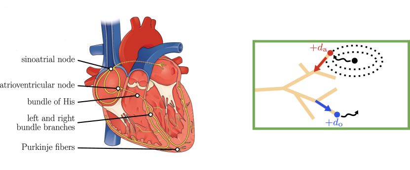
Heart contraction is triggered by an electrical action potential propagating throughout the cardiac muscle. This process is regulated by the \acCCS, schematized in Figure 1(left). The Purkinje network plays a pivotal role in driving the action potential [1, 2, 3, 4]. It is a dense network of conductive fibers, lying on the endocardium, and electrically connected to the surrounding tissue at its terminal points, called Purkinje-muscle junctions (PMJs). Propagation is referred to as orthodromic when excitation travels from the Purkinje network to the muscle, or antidromic when the opposite happens, as depicted in Figure 1(right). Antidromic propagation is typically absent in healthy hearts; however, it becomes relevant under diseased conditions [5, 6, 7], where reentries111Cardiological literature generally refers to reentry as an abnormal pathway for heart signals to travel, forming a reentrant circuit, regardless of whether it involves the His-Purkinje system [8]. In this work, we focus exclusively on reentrant circuits involving the Purkinje network. With a slight misuse of terminology, we refer to such circuits simply as reentries. in the network may occur. Therefore, incorporating both orthodromic and antidromic propagation mechanisms in computational models of cardiac electrophysiology is crucial for accurately reproducing the physical, possibly pathological, processes.
Existing cardiac computational models often lack an explicit representation of the Purkinje network, and usually surrogate it through realistic stimulation protocols based on sparse endocardial sources [9, 10, 11, 12, 13, 14, 15, 16], on space-dependent conduction velocities [17, 18, 19, 20], or on the introduction of a fast endocardial layer [21, 22, 23, 24, 25, 26, 27, 28, 29, 30, 31]. Other studies include an explicit representation of the His-Purkinje system, either rule-based [7, 32, 33, 34, 35, 36, 37, 38, 39, 40, 41, 42, 43, 44, 45, 46] or patient-specific [6, 47, 48, 49]. However, only a limited number of works have developed models that account for antidromic propagation, thus allowing a bidirectionally-coupled Purkinje-myocardium model.
The main issue in modeling this interplay is capturing all the possible reentries of the electrical signal between the network and the muscle. Reentrant processes have been modeled using the Monodomain equation both for network and for muscular propagation [7, 33, 36, 50, 51], or using Bidomain models [40, 42]. The work in [6] proposed an algorithm for Purkinje-muscle coupling using an Eikonal-Eikonal model [52], which accounts for antidromic propagation arising from possible muscular sources. However, this algorithm makes the simplifying assumption that the signal propagating from the network to the muscle cannot reenter the network, as would occur, for instance, in the presence of bundle branch blocks [33, 36, 50, 51]. Recently, a reaction-Eikonal model [23], that incorporates the Purkinje network through a monolithic approach [43, 45], yet without a detailed discussion of antidromic reentries, has been used to replicate clinical outcomes, such as for \acCRT, His bundle pacing, and \acECGs. In conclusion, to the best of our knowledge, no studies have been conducted using Eikonal models to address the modeling of bidirectional propagation with a discussion on reentries. Models of this kind would be useful, particularly thanks to the computational efficiency of solvers for Eikonal equations.
In this work, we present a new Eikonal-Eikonal partitioned scheme for Purkinje-muscle electrophysiology simulations, supporting both orthodromic and antidromic propagation, with a particular focus on reentries. We rely on the Eikonal model for the network and on the Eikonal-diffusion model for the muscle. The main motivations underlying our choices lie in: (i) the modularity offered by partitioned schemes, compared to the monolithic approach, allowing to solve the two problems independently using distinct solvers; and (ii) the computational advantage offered by the Eikonal models, which significantly reduce computational costs compared to the Monodomain and Bidomain ones.
We remark that Monodomain and Bidomain models have the advantage of inherently handling bidirectional propagation and PMJ delays [7], while an Eikonal-Eikonal model necessitates specific algorithms to explicitly address these interactions. In this regard, our novel algorithm improves the versatility and generality of the one proposed in [6], enabling reentries of the electrical signal between the network and the muscle. Moreover, our algorithm requires the development of a new pseudo-time method for the Eikonal-diffusion problem, as classic solvers would not be accurate in this coupling context.
We showcase the accuracy and the efficiency of the proposed computational framework through numerical experiments in realistic geometries and under pathological or therapeutic conditions, such as bundle branch blocks and \acCRT.
This paper is organized as follows. Section 2 outlines the mathematical and numerical models employed in this work. Section 3 details the general parameters and computational setup. Section 4 presents the results of numerical simulations in various pathological scenarios. Finally, Section 5 provides a summary of the conclusions.
2 Mathematical and numerical models
In this section, we introduce the mathematical models used to describe the propagation of electrical signals within the cardiac conduction system. In the subsequent sections we examine in detail the following modeling aspects:
-
1.
the model for the activation of the His-Purkinje system through Eikonal equation;
-
2.
the model for the activation of the myocardium using the Eikonal-diffusion equation;
-
3.
the Purkinje-myocardium coupling to manage reentries between the two domains, resulting from orthodromic and antidromic propagation, employing a novel partitioned scheme for the bidirectional coupling.
2.1 Electrical activation of the Purkinje
2.1.1 Continuous problem
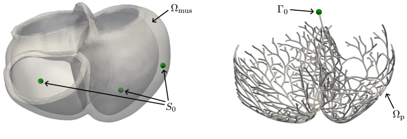
We use the Eikonal equation [6, 7] to describe the propagation of electrical signals within the Purkinje fibers, represented as a one-dimensional domain (see Figure 2(right)):
| (1) |
where represents the unknown activation times of the network, is the conduction velocity, and is the local curvilinear coordinate along the network. is the set of points generating the front. In healthy scenarios, coincides with the atrioventricular node (AV node), whereas in general it may be composed by PMJs supporting antidromic propagation.
Equation (1) provides the activation times at any point of the Purkinje network, including the \acPMJs. Conduction blocks are modeled as disconnections in the network.
2.1.2 Numerical discretization: Fast Marching method
We solve the 1D Eikonal problem using the \acFMM, proposed for the first time in [53] to solve the pure Eikonal equation and already applied to the context of cardiac electrophysiology [7, 54, 55]. In the 1D setting, the \acFMM reduces to the Dijkstra’s shortest path algorithm [56].
Notably, this algorithm automatically updates the activation time for those nodes in the network where a condition is prescribed (i.e. nodes belonging to ), but a signal coming from another stimulation point arrives earlier. In other words, if a stimulus is applied to a point where the solution to (1) would result in an activation earlier than the stimulus itself, then that stimulus is disregarded by the algorithm.
2.2 Electrical activation of the myocardium
2.2.1 Continuous problem
The propagation of the electrical signal in the myocardium , see Figure 2(left), is modeled by the Eikonal-diffusion equation [13, 52]. This model is used to reconstruct the macroscopic propagation of action potential excitation wavefronts during the depolarization phase, allowing to compute the activation time as a spatial function within the myocardium.
We define the activation time as the instant at which the depolarization wavefront reaches the point . The Eikonal-diffusion model reads:
| (2) |
Here, is the outward directed unit vector normal to the boundary of the domain , while is a portion of the physical boundary where the activation time is prescribed. Under normal propagation, coincides with the \acPMJs responsible for orthodromic propagation, because the signal traveling along the Purkinje network enters the muscle through these junctions. In cases of dysfunctions, additional muscular sources could be present, such as ectopic stimulation sites [6]. In (2), is the anisotropic tensor accounting for the orientation of muscular myofibers, and it is related to the conductivity tensor through the membrane capacitance and the surface-to-volume ratio :
| (3) |
where are the conductivities along the fibers (longitudinal) , the sheets (transversal) , and the cross-fibers (normal) directions, respectively. Accurately modeling myocardial fiber arrangement is essential to reproduce the anisotropic propagation occurring in the muscle [14, 57]. We prescribed muscular fibers, employing the strategy proposed by [14, 58], through the algorithm by Bayer et al. [57], which is a Laplace-Dirichlet rule-based method. Finally, the parameter , uniform over the domain , is the velocity of the action potential depolarization planar wavefront along the fiber direction in an infinite cable, under the assumption of a unit surface-to-volume ratio, membrane capacitance, and conductivity [59, 60] and normalized with respect to the diffusion parameter.
The first term in the first equation of (2) is the classical Eikonal term, describing the propagation of a front in an anisotropic domain (where the anisotropy is captured by ). The second term is a generalized Laplacian representing anisotropic diffusion, establishing a connection between the speed of the wavefront and its curvature. Propagation is faster when the wavefront is concave, and slower when it is convex [2, 60].
Problem (2) is very convenient from a computational standpoint, if compared to the Monodomain and the Bidomain models. Firstly, it requires solving a single steady PDE, albeit non-linear, as opposed to the Mondomain and Bidomain problems, which are time-dependent and must be coupled to ODE systems for ionic modeling [2, 52]. More importantly, the activation time, unlike the transmembrane potential, typically does not feature sharp fronts or strong gradients. Therefore, the Eikonal model does not demand for a fine spatial resolution [61, 62, 63, 64], enabling the simulation of the activation of large volumes of cardiac tissue with relatively low computational costs.
2.2.2 Numerical discretization: a novel pseudo-time method
The numerical solution of (2) requires a discretization with a proper treatment of the non-linearity. Classical non-linear solvers, such as Newton’s method, typically have convergence issues in this context, due to the difficulty of finding an appropriate initial guess to problem (2) [52]. To circumvent this issue, one can numerically solve (2) by recovering the steady-state solution of the following parabolic pseudo-time problem [52, 65]:
| (4) |
This is known as pseudo-time method. In (5), we propose a different version of the pseudo-time method, introduced to properly handle the Purkinje-muscle coupled propagation, since standard versions would not be accurate in the context of our new coupling algorithm, as detailed in what follows.
Starting from Equation (4), the time discretization employing a fully implicit \acBDF of order (say BDF; ), along with proper boundary and initial conditions, reads:
| (5) |
where is the numerical solution, , while and are the terms associated to the BDF time discretization222 is a constant coefficient, while refers to the value of the function at the current, or possibly earlier, time steps, or even a combination of them. For instance, for BDF1, and , while for BDF2, and , and so on. [66, 67]. The novelty of our approach lies in the location where Dirichlet-type conditions are imposed. Specifically, the collection of all stimulation points , is replaced now by , containing only the subset of related to active stimuli. At time , active stimuli are identified by:
| (6) |
Dirichlet-type conditions are imposed only on the stimulation points that the algorithm identifies as active, whereas inactive stimuli are disregarded. Therefore, we underline that the solution of (5) is deliberately not consistent with the continuous problem (2), since the steady-state solution only satisfies the condition on a subset of . Thus, (5) can be seen both as an alternative approach to Eikonal modeling of cardiac activation and as a solving methodology. A rigorous derivation of the pseudo-time-continuous counterpart to (5) is not addressed in this paper.
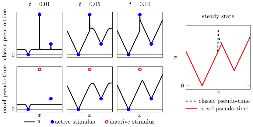
Figure 3 helps clarifying the rationale behind the development of our novel solver. The figure compares the evolution of the solutions obtained by the classic and novel pseudo-time methods for the same set of stimuli. The novel algorithm correctly identifies one stimulus as being inactive, whereas the classic algorithm would lead to a non-physical activation delay in correspondence of that stimulus. The novel pseudo-time formulation is essential for obtaining physically meaningful solutions during the coupling with the Purkinje network.
2.3 Purkinje-muscle fully coupled problem
To address the coupling between the problems (1) and (2), we introduce the following functionals, which map the stimuli to the corresponding activation times both in the myocardium and in the Purkinje network:
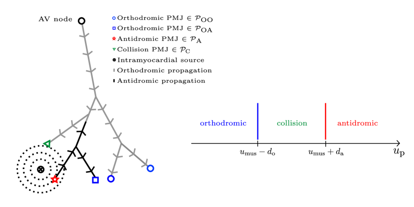
Our starting point is the coupling strategy presented in [6]. The interface conditions of the coupled problem are related to the continuity of the activation times and at the PMJs. The latter are classified into:
-
1.
antidromic \acPMJs, which are activated by the front coming from the muscle, and delay the signal by before transmitting it to the Purkinje network.
-
2.
orthodromic \acPMJs, which are activated by the front coming from the AV node or emerging in the network due to antidromic propagation, and delay the signal by before transmitting it to the myocardium.
-
3.
collision \acPMJs333This is a new category, introduced in this work, to speed up and correctly classify PMJs in Algorithm 1., which arise when the muscular and the network wavefronts collide at the PMJ itself, so they are neither orthodromic nor antidromic. This is possible due to the presence of a delay at the \acPMJs.
Figure 4 schematically illustrates the different types of \acPMJs resulting from orthodromic and antidromic propagation.
We propose hereafter Algorithm 1, designed to overcome the limitations of [6]. The latter made the assumption that the antidromic activation of the \acPMJs is induced exclusively by the muscular sources. This implies that the muscular pathways that activate the network could only be generated by the muscular sources. In other words, with the algorithm of [6], a signal originating from the AV node and entering the muscle through a PMJ cannot reenter the network. Conversely, our approach is able to account for potential occurrence of reentries. Algorithm 1 allows for an excitation front to originate at the AV node, enter the muscle through a PMJ and subsequently reenter the Purkinje network at a different PMJ. This is achieved by incorporating subiterations in the algorithm, thus repeatedly solving the two Eikonal problems and exchanging information at the \acPMJs until the maximum number of iterations is reached, and it is made possible by the use of the new pseudo-time method described in Section 2.2.2. We fix the number of iterations to , although this can be straightforwardly improved through a suitable stopping criterion.
The \acPMJs classification, showed in Figure 4, is updated through the function classify_pmjs, illustrated in Algorithm 2.
3 Simulations setting
In this section, we briefly introduce the general setting of the numerical simulations that will be presented in Section 4. Section 3.1 describes the computational domains considered for the 3D ventricular geometry and the 1D Purkinje network, while Section 3.2 summarizes the common parameter settings for all the numerical simulations. Finally, Section 3.3 is dedicated to the computational environment adopted to implement the algorithms and to perform the simulations.
3.1 Computational domains
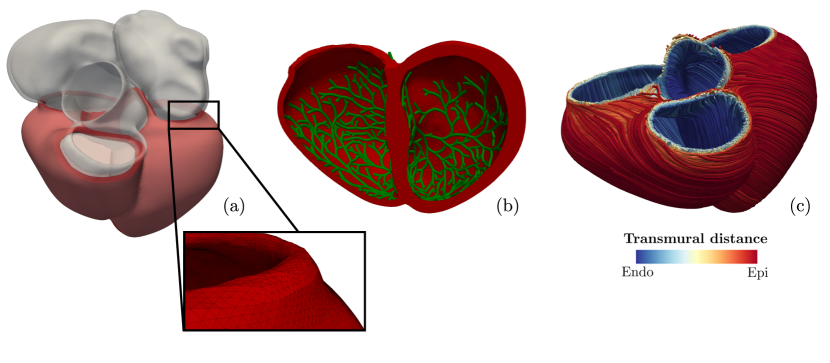
The 3D cardiac computational domain, depicted in Figure 5, is a patient-specific human biventricular geometry, reconstructed from a \acCT scan of a patient. This geometry is obtained from a publicly available cohort of four-chamber hearts presented in [62]. The mesh was segmented from a \acCT scan of the heart of an 83-years-old male who was recruited for a \acCRT upgrade following heart failure. Since this is not a healthy heart, it exhibits certain non-physiological features such as muscular hypertrophy and myocardial thickening. Moreover, endocardial structures, such as papillary muscles, are not included in the segmentation.
The extraction of the ventricular portion and the volumetric mesh generation (with an average edge length of ) was performed using vmtk444http://www.vmtk.org/ and the methods described in [69]. The main characteristics of the obtained mesh are summarized in Table 1.
Incorporating a patient-specific His-Purkinje system represents a distinct challenge, since currently no imaging technique is able to reconstruct a patient-specific network for human hearts. We used the open-source Python library fractal-tree555https://github.com/fsahli/fractal-tree to generate the 1D Purkinje network [41]. This approach employs a semi-automatic algorithm based on a fractal law, designed to generate a network standing on an endocardial surface discretized by triangles, mimicking the tree structure of the Purkinje itself. An example of generated network is reported in Figure 5(b). The absence of endocardial structures in the considered 3D geometries prevents an accurate reconstruction of the network, allowing only to capture its principal features. Some significant elements, particularly the moderator band in the right ventricle, have been omitted.
3.2 Simulation parameters
| element shape | tetrahedra |
|---|---|
| #elements | 5’268’810 |
| #nodes(DoFs) | 927’097 |
| aspect ratio | 1.57 0.53 |
| scaled jacobian | 0.63 0.14 |
| average cell diameter | |
| min cell diameter | |
| max cell diameter |
| Description | Parameter | Reference |
|---|---|---|
| Stimulation sites and shape | ||
| Conduction block sites | ||
| Conductivity values | [14] | |
| Depolarization velocity of the myocardium | [14] | |
| Conduction velocity of the Purkinje | [71, 7] | |
| Orthodromic PMJ delay | [7] | |
| Antidromic PMJ delay | [7] |
Table 2 reports the main physical parameters useful for solving the Eikonal model. Following [14], we select the conductivity values in the myocardium to obtain the conduction velocities of in the fiber direction, in the sheet direction and in the normal direction [14, 71]. We selected the conduction velocity in the Purkinje fibers to be [27, 71, 48]. At the \acPMJs, the signal experiences a delay of for orthodromic propagation and a shorter delay of for antidromic propagation [7].
Finally, pointwise stimuli in the muscle are applied at the \acPMJs, while spherical stimuli of radius are imposed in correspondence of possible ectopic or artifical stimuli. The placement of muscular sources and possible conduction blocks is evaluated case-by-case and passed as input through their point coordinates.
3.3 Simulation environment
The numerical framework presented in Section 2 has been implemented in lifex [72, 73, 74]666https://lifex.gitlab.io, a high-performance C++ finite element library for cardiovascular modeling, based on deal.II [75, 76, 77].
Simulations have been performed using the HPC facilities of the Mathematics Department at Politecnico di Milano, running on one node with 20 cores 4x Xeon E5-2640 v4 (2.4GHz) with 384GB of RAM. Running iterations777The choice , in Algorithm 1, is done to balance reliable results with reduced computational time. of Algorithm 2 takes less than 2 .
4 Numerical results
This section is dedicated to present numerical results from electrophysiology simulations using the model framework introduced in Section 2 and based on the setting presented in Section 3. Computational simulations offer a valuable tool for medical decision-making. This ability to simulate different scenarios with relative computational ease, without intervening on the patient, represents, in perspective, a notable application [2, 3, 23, 45, 78]. We consider four test (T) scenarios, illustrating both healthy and pathological propagation:
-
1.
T-H: healthy propagation. The atrioventricular node represents the unique stimulation point for ventricular activation and only orthodromic propagation is present [4].
- 2.
- 3.
- 4.
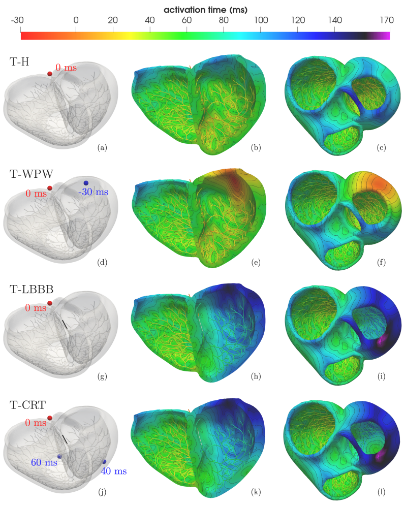
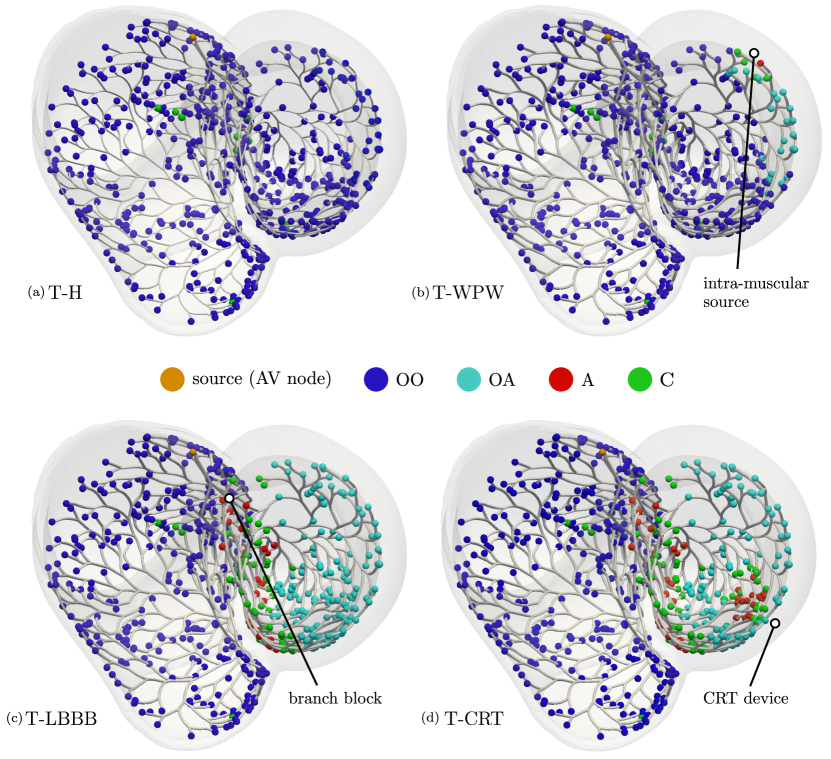
Figures 6 and 7 compare the activation times and the \acPMJs classification of the four test cases, respectively. The two figures are completed by Tables 3 and 4, which collect synthetical indices related to the activation maps and to the \acPMJs classification.
| TAT | EAT | ||
|---|---|---|---|
| T-H | 140.7 | 27.6 | |
| T-WPW | 120.5 | -30.0 | |
| T-LBBB | 174.0 | 31.1 | |
| T-CRT | 173.4 | 31.1 |
| T-H | T-WPW | T-LBBB | T-CRT | |
|---|---|---|---|---|
| OO | 522 | 490 | 305 | 305 |
| OA | 0 | 27 | 142 | 108 |
| A | 0 | 1 | 23 | 39 |
| C | 9 | 13 | 61 | 79 |
4.1 T-H: healthy propagation
We present a simulation characterized by healthy propagation, which will be used as a baseline for comparisons with the pathological scenarios. Figure 6(b,c) shows the resulting activation map. Only orthodromic propagation is present, following His-Purkinje activation, which takes approximately , in alignment with the values recorded in-vivo [27]. The sub-endocardial layer is the first portion of the muscle to activate, with the signal progressively spreading throughout the myocardium and transmurally towards the epicardium. The propagation follows an apico-basal pattern, favored by the scarce presence of Purkinje fibers near the ventricular bases. The total ventricular activation times are reported to be around in healthy patients [48, 80]. The latest activation time, recorded in Table 4, is slightly elevated, likely due to the inclusion of the valvular plane in the geometry and to muscle thickening caused by the patient’s hypertrophy.
4.2 T-WPW: Wolff-Parkinson-White syndrome
The Wolff-Parkinson-White (WPW) syndrome is a pathology characterized by the presence of an accessory pathway between the left atrium and the left ventricle, named bundle of Kent [81, 82, 83]. Since we only consider a ventricular geometry, we follow the modeling approach proposed in [6], which surrogates the effect of WPW syndrome on the ventricles by introducing an intramuscular source at the termination of the Kent bundle. This induces a pre-excitation of the left ventricle.
The simulation pacing setup is shown in Figure 6(d). A muscular source was placed in the posterolateral portion, near the basal plane, of the left ventricular myocardium, consistently with in-vivo observations of the location of the Kent bundle [6, 84]. Here, a spherical stimulus with radius was applied at time to replicate the pre-excitation of the muscle, considering that the activation of the AV node starts later at , which is used as reference time across all the scenarios.
Figure 6(e,f) shows the the resulting activation map. A front propagating through the muscle is clearly observed before the signal starts to propagate through the network, causing the pre-excitation of the upper posterolateral portion of the left ventricle. At the orthodromic \acPMJs, focal activation is still visible. Away from the muscular source, all the \acPMJs are orthodromic, while near it antidromic or collision \acPMJs arise, as we can see in Figure 7(b).
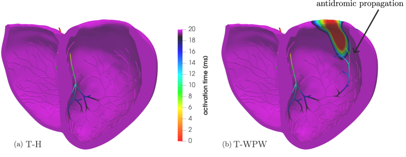
Figure 8 reports a comparison between T-H and T-WPW, highlighting the effects of antidromic propagation. We observe that, in the presence of only orthodromic activation, in Figure 8(a), the signal reaches only the three branches diverging from the main bundle of the Purkinje network. Meanwhile, thanks to the pre-excitation of the muscle starting at , the signal enters the network antidromically in Figure 8(b). Here, two wavefronts are distinguishable in opposite directions. Close to the ectopic stimulation point, all the types of \acPMJs are present: antidromic, collision and orthodromic activated by antidromic propagation (OA-labelled \acPMJs).
4.3 T-LBBB: complete left bundle branch block
The second pathological scenario considered is a complete left bundle branch block (LBBB). The latter consists in the interruption of the electric signal propagation in a segment of the left branch, resulting in an alteration of ventricular synchrony [5, 85].
The atrioventricular node was stimulated as in the healthy case at time (see Section 4.1). A conduction block is located along a segment of the left bundle branch, shown in black in Figure 6(g).
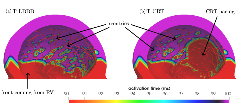
The total activation time is , as reported in Table 3 and shown in Figure 6(h,i). The signal originates from the atrioventricular node and travels through the His bundle. At the conduction block, the signal path in the left branch is interrupted, continuing toward the ventricle solely through the right branch. In Figure 6(h), we can notice how the right ventricle undergoes normal orthodromic activation, while the signal in the left ventricle arrives later. The wavefront reaches the left ventricle through the interventricular septum, see Figure 6(h), coherently with what is observed in patients with complete branch blocks. Consequently, impulses enter in the left branch of network antidromically. Figure 7(c) shows that the conduction block determines the presence of antidromic \acPMJs near the interventricular septum. Moreover, no orthodromic \acPMJs activated by the signal coming from the AV node (OO-labelled \acPMJs) are present in the left ventricle. In contrast, we see many OA-labelled \acPMJs, which result from a signal that has entered the network antidromically from the muscle and, due to the higher conduction velocity in the Purkinje fibers, is able to reemerge orthodromically from a different PMJ. A high number of OA \acPMJs is confirmed also by Table 4.
Figure 9(a) displays the activation map for the time frame , highlighting the reentry propagation from the muscle to the network. The higher transmission velocity in the network causes the signal to re-emerge again in the muscle. In Figure 9(a), a primary front is coming from the right ventricle, overcome by the stimuli arriving from the terminations of the left network. We emphasize that our new proposed Algorithm 1 made it possible to numerically observe this reentry effect. In contrast, the methodologies outlined in [6] would not be able to capture such reentries, providing a less biophysically accurate solution.
4.4 T-CRT: cardiac resynchronization therapy
The last presented scenario combines the two sources of antidromic propagation, both the conduction block and the muscular source, into a single simulation, showcasing the versatility of the algorithm for Purkinje-muscle coupling. This test case aims to simulate the effects of \acfCRT in presence of a complete left bundle branch block. \acCRT is a treatment that involves biventricular pacing to help restoring the normal rhythm and timing of the heartbeat [45, 78, 79]. This involves implanting electrodes in the heart to produce artificial stimuli that compensate for the propagation imbalance caused by a particular pathology. Typically, one lead is placed at the apex of the right ventricle, and another on the free wall midway up the left ventricle [1, 79, 86, 87]. The effect of artificial pacing is modeled by defining two appropriate epicardial stimuli [45, 78].
Figure 6(j) resumes the initial setup of the simulation. A stimulus at time is imposed at the AV node, with a complete conduction block located as in T-LBBB (see Section 4.3). Two additional spherical stimuli are imposed in order to mimic the two leads of \acCRT. Activation at these points occurs at time and , consisting of spherical stimuli with radius . Notice that this is an arbitrary choice, as is their location, which usually corresponds to the termination of two cardiac veins. The correct positioning and timing may vary a lot from patient to patient and are evaluated in clinical practice on individual specifics.
The biventricular activation map is depicted in Figure 6(k,l). The CRT lead in the right ventricle has, on purpose, a higher activation time than the one ensured by orthodromic propagation, in order to verify that it produces no effect. Indeed, the novel pseudo-time method, proposed in Section 2.2.2, correctly operates in this situation. Conversely, the lead in the left ventricle produces a stimulus that impacts the solution, as shown in Figure 6(k,l). This is even more visible in Figure 9(b), where the artificial pacing is evident, along with the signal entering the network via antidromic \acPMJs. Several wavefronts are present in the muscle: one main front, which comes from the interventricular septum, and one produced by the \acCRT lead, but also many pointwise stimuli at \acPMJs due to reentries. The right ventricle activates normally, while the left ventricle shows alterations compared to the healthy case. The latter are partially compensated by the artificial pacing, ensuring better activation of the lateral wall of the left ventricle. Figure 9 also provides a comparison between T-LBBB (a) and T-CRT (b): the anterolateral portion of the left ventricle is activated thanks to \acCRT. In T-CRT, in addition to the antidromic and collision \acPMJs in correspondence to the interventricular septum, further \acPMJs of these types are present near the \acCRT device, as depicted in Figure 7(d). Moreover, many orthodromic \acPMJs activated due to antidromic propagation (OA type) are visible in the free wall of the left ventricle, consistently with the reentry effect.
5 Conclusions
In this paper, we studied the interaction of the electrical excitation in the cardiac muscle and the Purkinje network using Eikonal equations, focusing on modeling electrical reentries. We proposed a novel Eikonal-diffusion pseudo-time method capable of correctly imposing active stimuli in the muscle while discarding inactive ones. Furthermore, we introduced a novel Purkinje-muscle coupling partitioned scheme (Algorithm 1) that effectively manages possible reentries between the two domains by iteratively computing activation times and exchanging information at the interface, represented by the \acPMJs. Building on and extending the work presented in [6], we addressed its limitations, achieving improved versatility and accuracy.
While this work primarily focused on methodological and modeling aspects, we also performed numerical simulations in realistic pathological and therapeutic scenarios. The results underscore the critical role of the Purkinje network and highlight the importance of accurately capturing electrical reentries to better reproduce the pathophysiological processes. The numerical simulations showcased the accuracy, flexibility and robustness of our algorithm, successfully handling arbitrary combinations of Purkinje network, stimulation setups and conduction blocks.
A promising direction for future work is to rigorously define the continuous formulation of the pseudo-time-discrete problem (5), which is not consistent with (2) and implicitly introduces a novel notion of solution, whose properties warrant further investigation. Additionally, our numerical experiments relied on Purkinje networks reconstructed through rule-based algorithms, rather than patient-specific. However, thanks to the modularity of the proposed scheme, the latter can be included straightforwardly. Finally, investigating pathological scenarios from an electro-mechanical perspective may provide deeper insights into their effects on cardiac function.
Acknowledgements
M.B., R.P. and L.D. received support from the project PRIN2022, MUR, Italy, 2023–2025 202232A8AN “Computational modeling of the heart: from efficient numerical solvers to cardiac digital twins”. R.P. has also received support from the INdAM GNCS project CUP E53C23001670001 “Mathematical models and numerical methods for the construction of cardiac digital twins”. C.V. has been partially supported by: i) the European Union-Next Generation EU, Mission 4, Component 1, CUP: D53D23018770001, under the research project MUR PRIN22-PNRR n.P20223KSS2, “Machine learning for fluid structure interaction in cardiovascular problems: efficient solutions, model reduction, inverse problems”, ii) the Italian Ministry of Health within the PNC PROGETTO HUB LIFE SCIENCE - DIAGNOSTICA AVANZATA (HLS-DA) “INNOVA”, PNCE3-2022-23683266–CUP: D43C22004930001, within the “Piano Nazionale Complementare Ecosistema Innovativo della Salute” - Codice univoco investimento: PNCE3-2022-23683266; iii) the Italian research project MUR PRIN22 n.2022L3JC5T “Predicting the outcome of endovascular repair for thoracic aortic aneurysms: analysis of fluid dynamic modeling in different anatomical settings and clinical validation”; iv) Italian Ministry of Health within the project “CAL.HUB.RIA” - CALABRIA HUB PER RICERCA INNOVATIVA ED AVANZATA. Code: T4-AN-09, CUP: F63C22000530001. M.B., R.P., L.D. and C.V. acknowledge their membership to INdAM group GNCS - Gruppo Nazionale per il Calcolo Scientifico (National Group for Scientific Computing, Italy). M.B. and R.P. acknowledge the INdAM GNCS project CUP E53C23001670001. M.B., R.P. and L.D. acknowledge the “Dipartimento di Eccellenza 2023-2027”, MUR, Italy, Dipartimento di Matematica, Politecnico di Milano.
Declaration of competing interest
All the authors declare that they have no known competing financial interests or personal relationships that could have appeared to influence the work reported in this paper.
CRediT authorship contribution statement
S. Brunati: Conceptualization, Methodology, Software, Simulation, Data curation, Formal analysis, Investigation, Visualization, Writing – original draft, Writing – review & editing. M. Bucelli: Conceptualization, Methodology, Software, Formal analysis, Investigation, Visualization, Writing – original draft, Writing – review & editing. R. Piersanti: Conceptualization, Methodology, Formal analysis, Investigation, Visualization, Writing – original draft, Writing – review & editing. L. Dede’: Supervision, Funding acquisition, Project administration, Writing – review & editing. C. Vergara: Conceptualization, Supervision, Funding acquisition, Project administration, Writing – original draft, Writing – review & editing.
References
- [1] P. A. Iaizzo, Handbook of cardiac anatomy, physiology, and devices, Springer Science & Business Media, 2010.
- [2] A. Quarteroni, A. Manzoni, C. Vergara, The cardiovascular system: mathematical modelling, numerical algorithms and clinical applications, Acta Numerica 26 (2017) 365–590.
- [3] A. Quarteroni, L. Dede’, A. Manzoni, C. Vergara, Mathematical modelling of the human cardiovascular system: data, numerical approximation, clinical applications, Vol. 33, Cambridge University Press, 2019.
- [4] M. A. Bagni, D. Brambilla, L. Catacuzzeno, M. Catalano, M. Contini, R. Crescenzo, M. D’Amelio, C. Faggio, M. P. Gallo, E. Grasselli, R. Gulino, G. Leanza, R. Maccarone, A. Michelucci, D. A. Ragozzino, I. Sirangelo, C. Veronesi, Fisiologia, Edises, 2023.
- [5] M. Romanò, Testo-atlante di elettrocardiografia pratica: Approccio clinico ragionato all’elettrocardiogramma, Springer Science & Business Media, 2010.
- [6] S. Palamara, C. Vergara, D. Catanzariti, E. Faggiano, C. Pangrazzi, M. Centonze, F. Nobile, M. Maines, A. Quarteroni, Computational generation of the Purkinje network driven by clinical measurements: the case of pathological propagations, International Journal for Numerical Methods in Biomedical Engineering 30 (12) (2014) 1558–1577.
- [7] C. Vergara, M. Lange, S. Palamara, T. Lassila, A. F. Frangi, A. Quarteroni, A coupled 3D-1D numerical Monodomain solver for cardiac electrical activation in the myocardium with detailed Purkinje network, Journal of Computational Physics 308 (2016) 218–238.
- [8] D. Fleischmann, T. Pop, J. De Bakker, Reentry mechanism within the His-Purkinje system in man during extrasystolic stimulation of the right ventricle, in: Cardiac Pacing: Diagnostic and Therapeutic Tools, Springer, 1976, pp. 194–204.
- [9] E. J. Vigmond, M. Hughes, G. Plank, L. J. Leon, Computational tools for modeling electrical activity in cardiac tissue, Journal of Electrocardiology 36 (2003) 69–74.
- [10] D. Nickerson, N. Smith, P. Hunter, New developments in a strongly coupled cardiac electromechanical model, EP Europace 7 (s2) (2005) S118–S127.
- [11] D. Keller, R. Kalayciyan, O. Dössel, G. Seemann, Fast creation of endocardial stimulation profiles for the realistic simulation of body surface ECGs, in: World Congress on Medical Physics and Biomedical Engineering, September 7-12, 2009, Munich, Germany: Vol. 25/4 Image Processing, Biosignal Processing, Modelling and Simulation, Biomechanics, Springer, 2010, pp. 145–148.
- [12] M. J. Bishop, G. Plank, R. A. Burton, J. E. Schneider, D. J. Gavaghan, V. Grau, P. Kohl, Development of an anatomically detailed mri-derived rabbit ventricular model and assessment of its impact on simulations of electrophysiological function, American Journal of Physiology-Heart and Circulatory Physiology 298 (2) (2010) H699–H718.
- [13] A. Quarteroni, T. Lassila, S. Rossi, R. Ruiz-Baier, Integrated heart—coupling multiscale and multiphysics models for the simulation of the cardiac function, Computer Methods in Applied Mechanics and Engineering 314 (2017) 345–407.
- [14] R. Piersanti, P. C. Africa, M. Fedele, C. Vergara, L. Dede’, A. F. Corno, A. Quarteroni, Modeling cardiac muscle fibers in ventricular and atrial electrophysiology simulations, Computer Methods in Applied Mechanics and Engineering 373 (2021) 113468.
- [15] F. Regazzoni, M. Salvador, P. C. Africa, M. Fedele, L. Dede’, A. Quarteroni, A cardiac electromechanical model coupled with a lumped-parameter model for closed-loop blood circulation, Journal of Computational Physics 457 (2022) 111083.
- [16] P. C. Africa, M. Salvador, P. Gervasio, L. Dede’, A. Quarteroni, A matrix–free high–order solver for the numerical solution of cardiac electrophysiology, Journal of Computational Physics 478 (2023) 111984.
- [17] T. P. Usyk, I. J. LeGrice, A. D. McCulloch, Computational model of three-dimensional cardiac electromechanics, Computing and Visualization in Science 4 (2002) 249–257.
- [18] S. Göktepe, E. Kuhl, Electromechanics of the heart: a unified approach to the strongly coupled excitation–contraction problem, Computational Mechanics 45 (2010) 227–243.
- [19] N. A. Trayanova, Whole-heart modeling: applications to cardiac electrophysiology and electromechanics, Circulation Research 108 (1) (2011) 113–128.
- [20] M. Sermesant, R. Chabiniok, P. Chinchapatnam, T. Mansi, F. Billet, P. Moireau, J.-M. Peyrat, K. Wong, J. Relan, K. Rhode, M. Ginks, P. Lambiase, H. Delingette, M. Sorine, C. A. Rinaldi, D. Chapelle, R. Razavi, N. Ayache, Patient-specific electromechanical models of the heart for the prediction of pacing acute effects in CRT: a preliminary clinical validation, Medical Image Analysis 16 (1) (2012) 201–215.
- [21] M. Kotikanyadanam, S. Göktepe, E. Kuhl, Computational modeling of electrocardiograms: A finite element approach toward cardiac excitation, International Journal for Numerical Methods in Biomedical Engineering 26 (5) (2010) 524–533.
- [22] B. Baillargeon, N. Rebelo, D. D. Fox, R. L. Taylor, E. Kuhl, The Living Heart project: a robust and integrative simulator for human heart function, European Journal of Mechanics-A/Solids 48 (2014) 38–47.
- [23] A. Neic, F. O. Campos, A. J. Prassl, S. A. Niederer, M. J. Bishop, E. J. Vigmond, G. Plank, Efficient computation of electrograms and ECGs in human whole heart simulations using a reaction-Eikonal model, Journal of Computational Physics 346 (2017) 191–211.
- [24] A. W. Lee, U. C. Nguyen, O. Razeghi, J. Gould, B. S. Sidhu, B. Sieniewicz, J. Behar, M. Mafi-Rad, G. Plank, F. W. Prinzen, C. A. Rinaldi, K. Vernooy, S. A. Niederer, A rule-based method for predicting the electrical activation of the heart with cardiac resynchronization therapy from non-invasive clinical data, Medical Image Analysis 57 (2019) 197–213.
- [25] K. Gillette, M. A. Gsell, A. J. Prassl, E. Karabelas, U. Reiter, G. Reiter, T. Grandits, C. Payer, D. Štern, M. Urschler, J. D. Bayer, C. M. Augustin, A. Neic, T. Pock, E. J. Vigmond, G. Plank, A framework for the generation of digital twins of cardiac electrophysiology from clinical 12-leads ECGs, Medical Image Analysis 71 (2021) 102080.
- [26] K. Gillette, M. A. Gsell, M. Strocchi, T. Grandits, A. Neic, M. Manninger, D. Scherr, C. H. Roney, A. J. Prassl, C. M. Augustin, E. J. Vigmond, G. Plank, A personalized real-time virtual model of whole heart electrophysiology, Frontiers in Physiology 13 (2022) 907190.
- [27] G. Del Corso, R. Verzicco, F. Viola, A fast computational model for the electrophysiology of the whole human heart, Journal of Computational Physics 457 (2022) 111084.
- [28] M. Fedele, R. Piersanti, F. Regazzoni, M. Salvador, P. C. Africa, M. Bucelli, A. Zingaro, L. Dede’, A. Quarteroni, A comprehensive and biophysically detailed computational model of the whole human heart electromechanics, Computer Methods in Applied Mechanics and Engineering 410 (2023) 115983.
- [29] R. Piersanti, F. Regazzoni, M. Salvador, A. F. Corno, L. Dede’, C. Vergara, A. Quarteroni, 3D-0D closed-loop model for the simulation of cardiac biventricular electromechanics, Computer Methods in Applied Mechanics and Engineering 391 (2022) 114607.
- [30] M. Bucelli, A. Zingaro, P. C. Africa, I. Fumagalli, L. Dede’, A. Quarteroni, A mathematical model that integrates cardiac electrophysiology, mechanics, and fluid dynamics: Application to the human left heart, International Journal for Numerical Methods in Biomedical Engineering 39 (3) (2023) e3678.
- [31] E. Zappon, M. Salvador, R. Piersanti, F. Regazzoni, L. Dede’, A. Quarteroni, An integrated heart–torso electromechanical model for the simulation of electrophysiological outputs accounting for myocardial deformation, Computer Methods in Applied Mechanics and Engineering 427 (2024) 117077.
- [32] S. Abboud, O. Berenfeld, D. Sadeh, Simulation of high-resolution QRS complex using a ventricular model with a fractal conduction system. Effects of ischemia on high-frequency QRS potentials., Circulation Research 68 (6) (1991) 1751–1760.
- [33] O. Berenfeld, J. Jalife, Purkinje-muscle reentry as a mechanism of polymorphic ventricular arrhythmias in a 3-dimensional model of the ventricles, Circulation Research 82 (10) (1998) 1063–1077.
- [34] E. J. Vigmond, C. Clements, Construction of a computer model to investigate sawtooth effects in the Purkinje system, IEEE transactions on biomedical engineering 54 (3) (2007) 389–399.
- [35] T. Ijiri, T. Ashihara, T. Yamaguchi, K. Takayama, T. Igarashi, T. Shimada, T. Namba, R. Haraguchi, K. Nakazawa, A procedural method for modeling the Purkinje fibers of the heart, The Journal of Physiological Sciences 58 (7) (2008) 481–486.
- [36] K. Ten Tusscher, A. V. Panfilov, Modelling of the ventricular conduction system, Progress in Biophysics and Molecular Biology 96 (1-3) (2008) 152–170.
- [37] D. Romero, R. Sebastian, B. H. Bijnens, V. Zimmerman, P. M. Boyle, E. J. Vigmond, A. F. Frangi, Effects of the Purkinje system and cardiac geometry on biventricular pacing: a model study, Annals of Biomedical Engineering 38 (2010) 1388–1398.
- [38] R. Sebastian, V. Zimmerman, D. Romero, D. Sanchez-Quintana, A. F. Frangi, Characterization and modeling of the peripheral cardiac conduction system, IEEE Transactions on Medical Imaging 32 (1) (2012) 45–55.
- [39] R. M. Bordas, K. Gillow, D. Gavaghan, B. Rodríguez, D. Kay, A Bidomain model of the ventricular specialized conduction system of the heart, SIAM Journal on Applied Mathematics 72 (5) (2012) 1618–1643.
- [40] E. Behradfar, A. Nygren, E. J. Vigmond, The role of Purkinje-myocardial coupling during ventricular arrhythmia: a modeling study, PloS one 9 (2) (2014) e88000.
- [41] F. S. Costabal, D. E. Hurtado, E. Kuhl, Generating Purkinje networks in the human heart, Journal of Biomechanics 49 (12) (2016) 2455–2465.
- [42] M. Landajuela, C. Vergara, A. Gerbi, L. Dede’, L. Formaggia, A. Quarteroni, Numerical approximation of the electromechanical coupling in the left ventricle with inclusion of the Purkinje network, International Journal for Numerical Methods in Biomedical Engineering 34 (7) (2018) e2984.
- [43] M. Strocchi, A. W. Lee, A. Neic, J. Bouyssier, K. Gillette, G. Plank, M. K. Elliott, J. Gould, J. M. Behar, B. Sidhu, V. Mehta, M. J. Bishop, E. J. Vigmond, C. A. Rinaldi, S. A. Niederer, His-bundle and left bundle pacing with optimized atrioventricular delay achieve superior electrical synchrony over endocardial and epicardial pacing in left bundle branch block patients, Heart Rhythm 17 (11) (2020) 1922–1929.
- [44] M. Peirlinck, F. S. Costabal, J. Yao, J. Guccione, S. Tripathy, Y. Wang, D. Ozturk, P. Segars, T. Morrison, S. Levine, E. Kuhl, Precision medicine in human heart modeling: perspectives, challenges, and opportunities, Biomechanics and Modeling in Mechanobiology 20 (2021) 803–831.
- [45] M. Strocchi, K. Gillette, A. Neic, M. K. Elliott, N. Wijesuriya, V. Mehta, E. J. Vigmond, G. Plank, C. A. Rinaldi, S. A. Niederer, Comparison between conduction system pacing and cardiac resynchronization therapy in right bundle branch block patients, Frontiers in Physiology 13 (2022) 1011566.
- [46] S. Reza, B. Kovarovic, D. Bluestein, Assessing post-tavr cardiac conduction abnormalities risk using an electromechanically coupled beating heart, Biomechanics and Modeling in Mechanobiology (2024) 1–17.
- [47] O. Camara, A. Pashaei, R. Sebastian, A. F. Frangi, Personalization of fast conduction Purkinje system in Eikonal-based electrophysiological models with optical mapping data, in: Statistical Atlases and Computational Models of the Heart: First International Workshop, STACOM 2010, and Cardiac Electrophysiological Simulation Challenge, CESC 2010, Held in Conjunction with MICCAI 2010, Beijing, China, September 20, 2010. Proceedings 1, Springer, 2010, pp. 281–290.
- [48] S. Palamara, C. Vergara, E. Faggiano, F. Nobile, An effective algorithm for the generation of patient-specific Purkinje networks in computational electrocardiology, Journal of Computational Physics 283 (2015) 495–517.
- [49] J. Camps, L. A. Berg, Z. J. Wang, R. Sebastian, L. L. Riebel, R. Doste, X. Zhou, R. Sachetto, J. Coleman, B. Lawson, V. Grau, K. Burrage, A. Bueno-Orovio, R. Weber dos Santos, B. Rodriguez, Digital twinning of the human ventricular activation sequence to clinical 12-lead ECGs and magnetic resonance imaging using realistic Purkinje networks for in silico clinical trials, Medical Image Analysis 94 (2024) 103108.
- [50] E. J. Vigmond, B. D. Stuyvers, Modeling our understanding of the His-Purkinje system, Progress in Biophysics and Molecular Biology 120 (1-3) (2016) 179–188.
- [51] E. J. Vigmond, J. Bouyssier, J. Bayer, M. Haïssaguerre, H. Ashikaga, On the nature of delays allowing anatomical re-entry involving the Purkinje network: a simulation study, EP Europace 23 (Supplement_1) (2021) i71–i79.
- [52] P. Colli Franzone, L. F. Pavarino, S. Scacchi, Mathematical cardiac electrophysiology, Vol. 13, Springer, 2014.
- [53] J. A. Sethian, Fast marching methods, SIAM review 41 (2) (1999) 199–235.
- [54] E. Pernod, M. Sermesant, E. Konukoglu, J. Relan, H. Delingette, N. Ayache, A multi-front Eikonal model of cardiac electrophysiology for interactive simulation of radio-frequency ablation, Computers & Graphics 35 (2) (2011) 431–440.
- [55] T. Grandits, K. Gillette, A. Neic, J. Bayer, E. Vigmond, T. Pock, G. Plank, An inverse Eikonal method for identifying ventricular activation sequences from epicardial activation maps, Journal of Computational Physics 419 (2020) 109700.
- [56] E. W. Dijkstra, A note on two problems in connexion with graphs, in: Edsger Wybe Dijkstra: His Life, Work, and Legacy, Springer, 2022, pp. 287–290.
- [57] J. D. Bayer, R. C. Blake, G. Plank, N. A. Trayanova, A novel rule-based algorithm for assigning myocardial fiber orientation to computational heart models, Annals of biomedical engineering 40 (2012) 2243–2254.
- [58] P. C. Africa, R. Piersanti, M. Fedele, L. Dede’, A. Quarteroni, lifex-fiber: an open tool for myofibers generation in cardiac computational models, BMC Bioinformatics 24 (1) (2023) 143.
- [59] P. Colli Franzone, L. Guerri, Spreading of excitation in 3D models of the anisotropic cardiac tissue. i. Validation of the Eikonal model, Mathematical Biosciences 113 (2) (1993) 145–209.
- [60] P. Colli Franzone, L. Guerri, M. Pennacchio, B. Taccardi, Spread of excitation in 3D models of the anisotropic cardiac tissue. ii. Effects of fiber architecture and ventricular geometry, Mathematical Biosciences 147 (2) (1998) 131–171.
- [61] P. Colli Franzone, L. F. Pavarino, S. Scacchi, Reduced macroscopic models: The Monodomain and Eikonal models, Mathematical Cardiac Electrophysiology (2014) 123–148.
- [62] M. Strocchi, C. M. Augustin, M. A. Gsell, E. Karabelas, A. Neic, K. Gillette, O. Razeghi, A. J. Prassl, E. J. Vigmond, J. M. Behar, J. Gould, B. Sidhu, C. A. Rinaldi, M. J. Bishop, G. Plank, S. A. Niederer, A publicly available virtual cohort of four-chamber heart meshes for cardiac electro-mechanics simulations, PloS one 15 (6) (2020) e0235145.
- [63] S. Stella, F. Regazzoni, C. Vergara, L. Dede’, A. Quarteroni, A fast cardiac electromechanics model coupling the Eikonal and the nonlinear mechanics equations, Mathematical Models and Methods in Applied Sciences 32 (08) (2022) 1531–1556.
- [64] L. Gander, R. Krause, M. Weiser, F. Sahli Costabal, S. Pezzuto, On the accuracy of Eikonal approximations in cardiac electrophysiology in the presence of fibrosis, in: International Conference on Functional Imaging and Modeling of the Heart, Springer, 2023, pp. 137–146.
- [65] A. Quarteroni, L. Formaggia, A. Veneziani, Complex systems in biomedicine, Springer, 2006.
- [66] A. Quarteroni, R. Sacco, F. Saleri, Numerical mathematics, Vol. 37, Springer Science & Business Media, 2010.
- [67] D. Forti, L. Dedè, Semi-implicit bdf time discretization of the navier–stokes equations with vms-les modeling in a high performance computing framework, Computers & Fluids 117 (2015) 168–182.
- [68] R. Piersanti, R. Bradley, S. Y. Alid, A. Quarteroni, L. Dede, N. A. Trayanova, Defining myocardial fiber bundle architecture in atrial digital twins, arXiv preprint arXiv:2410.11601 (2024).
- [69] M. Fedele, A. Quarteroni, Polygonal surface processing and mesh generation tools for the numerical simulation of the cardiac function, International Journal for Numerical Methods in Biomedical Engineering 37 (4) (2021) e3435.
- [70] C. Stimpson, C. Ernst, P. Knupp, P. Pébay, D. Thompson, The Verdict library reference manual, Sandia National Laboratories Technical Report 9 (6) (2007) 8.
- [71] I. Cavero, H. Holzgrefe, Internodal conduction pathways: revisiting a century-long debate on their existence, morphology, and location in the context of 2023 best science, Advances in Physiology Education 47 (4) (2023) 838–850.
- [72] P. C. Africa, lifex: A flexible, high performance library for the numerical solution of complex finite element problems, SoftwareX 20 (2022) 101252.
- [73] P. C. Africa, R. Piersanti, F. Regazzoni, M. Bucelli, M. Salvador, M. Fedele, S. Pagani, L. Dede’, A. Quarteroni, lifex-ep: a robust and efficient software for cardiac electrophysiology simulations, BMC Bioinformatics 24 (1) (2023) 389.
- [74] M. Bucelli, The lifex library version 2.0, arXiv preprint arXiv:2411.19624 (2024).
- [75] W. Bangerth, R. Hartmann, G. Kanschat, deal.II—a general-purpose object-oriented finite element library, ACM Transactions on Mathematical Software (TOMS) 33 (4) (2007) 24–es.
- [76] D. Arndt, W. Bangerth, D. Davydov, T. Heister, L. Heltai, M. Kronbichler, M. Maier, J.-P. Pelteret, B. Turcksin, D. Wells, The deal.II finite element library: Design, features, and insights, Computers & Mathematics with Applications 81 (2021) 407–422.
- [77] D. Arndt, W. Bangerth, M. Feder, M. Fehling, R. Gassmöller, T. Heister, L. Heltai, M. Kronbichler, M. Maier, P. Munch, J.-P. Pelteret, S. Sticko, B. Turcksin, D. Wells, The deal.II library, version 9.4, Journal of Numerical Mathematics 30 (3) (2022) 231–246.
- [78] A. W. Lee, C. M. Costa, M. Strocchi, C. A. Rinaldi, S. A. Niederer, Computational modeling for cardiac resynchronization therapy, Journal of Cardiovascular Translational Research 11 (2018) 92–108.
- [79] F. Leyva, S. Nisam, A. Auricchio, 20 years of cardiac resynchronization therapy, Journal of the American College of Cardiology 64 (10) (2014) 1047–1058.
- [80] D. Durrer, R. T. Van Dam, G. Freud, M. J. Janse, F. L. Meijler, R. C. Arzbaecher, Total excitation of the isolated human heart, Circulation 41 (6) (1970) 899–912.
- [81] W. C. Sealy, J. J. Gallagher, E. L. Pritchett, The surgical anatomy of Kent bundles based on electrophysiological mapping and surgical exploration, The Journal of Thoracic and Cardiovascular Surgery 76 (6) (1978) 804–815.
- [82] S. M. Al-Khatib, E. L. Pritchett, Clinical features of Wolff-Parkinson-White syndrome, American Heart Journal 138 (3) (1999) 403–413.
- [83] S. Mittal, Atrioventricular accessory path: (the Kent bundle), in: Insights into Electrocardiograms with MCQs, Springer, 2023, pp. 97–110.
- [84] B. F. Waller, C. M. Orr, J. D. Slack, C. A. Pinkerton, J. V. Tassel, T. Peters, B. Waller, Anatomy, histology, and pathology of coronary arteries: A review relevant to new interventional and imaging techniques-part iv, Clinical Cardiology 15 (9) (1992) 675–687.
- [85] J. H. McAnulty, S. H. Rahimtoola, Bundle branch block, Progress in Cardiovascular Diseases 26 (4) (1984) 333–354.
- [86] C. Butter, A. Auricchio, C. Stellbrink, M. Schlegl, E. Fleck, W. Hörsch, E. Huvelle, J. Ding, A. Kramer, P. T. in Congestive Heart Failure II Study Group, W. Hörsch, E. Huvelle, J. Ding, A. Kramer, Should stimulation site be tailored in the individual heart failure patient?, The American Journal of Cardiology 86 (9) (2000) K144–K151.
- [87] P. T. Mortensen, P. Sogaard, H. Mansour, J. Ponsonaille, D. Gras, A. Lazarus, W. Reiser, C. Alonso, C. M. Linde, M. Lunati, B. Kramm, E. M. Harrison, Sequential biventricular pacing: evaluation of safety and efficacy, Pacing and Clinical Electrophysiology 27 (3) (2004) 339–345.