BOston Neonatal Brain Injury Data for Hypoxic Ischemic Encephalopathy (BONBID-HIE): II. 2-year Neurocognitive Outcome and NICU Outcome
Abstract
Hypoxic Ischemic Encephalopathy (HIE) affects approximately 1-5/1000 newborns globally and leads to adverse neurocognitive outcomes in 30% to 50% of cases by two years of age. Despite therapeutic advances with Therapeutic Hypothermia (TH), prognosis remains challenging, highlighting the need for improved biomarkers. This paper introduces the second release of the Boston Neonatal Brain Injury Dataset for Hypoxic-Ischemic Encephalopathy (BONBID-HIE), an open-source, comprehensive MRI and clinical dataset featuring 237 patients, including NICU outcomes and 2-year neurocognitive outcomes from Massachusetts General Hospital and Boston Children’s Hospital.
Background & Summary
HIE is a clinical syndrome due to a lack of blood flow and oxygen to the brain. It affects around 1-5/1000 newborns globally (Graham et al., 2008; Lee et al., 2013). Despite advancements in Therapeutic Hypothermia (TH), the prognosis for many infants remains challenging, with 35%–50% suffering adverse neurocognitive outcomes by 2 years of age (Shankaran et al., 2005; Azzopardi et al., 2009; Gluckman et al., 2005). Therefore, 63 of the 130 ongoing HIE-related trials worldwide are testing whether new therapies (Laptook et al., 2017; Shankaran et al., 2017; Liu et al., 2006; Potter et al., 2018; Nuñez-Ramiro et al., 2019; Liang et al., 2019; Cotten et al., 2014) can supplement TH and further reduce adverse outcomes. However, therapeutic innovation is slow and inconclusive, for 1) before therapy, patients at high risk of developing adverse outcomes cannot be identified; 2) after therapy, outcomes cannot be measured until age 2 years (Laptook et al., 2021). Both issues point to a lack of a neonatal biomarker that can predict adverse 2-year outcomes. To facilitate the development of biomarkers in HIE study, we present BOston Neonatal Brain Injury Dataset for Hypoxic Ischemic Encephalopathy (BONBID-HIE), an open-source, comprehensive, and representative MRI and clinical dataset for HIE. This paper introduces the second part of the BONBID-HIE data. This release contains raw and derived diffusion parameter maps, as well as NICU outcome and 2-year outcome with 237 patients. Our data was from Massachusetts General Hospital (MGH) and Boston Children’s Hospital (BCH). It includes MRIs from different scanners (Siemens 3T and GE 1.5T), different MRI protocols, and from patients of different races/ethnicities and ages (0-14 days postnatal age). Part I of our data release (Bao et al., 2023) focuses on lesion detection, while Part II (this paper) is focus on clinical, treatment, and neurologic outcome data for further developing prognostic biomarkers.
Methods
Overview
Retrospective Data Collection
Data was retrospectively collected from MGH and BCH. Inclusion criteria were: (1) term-born (at physician discretion) (2) clinical diagnosis of HIE; (3) no comorbidities such as hydrocephalus or congenital syndromes; and (4) high-quality MRI acquired in Day 0-14 after birth. Exclusion criteria were: (1) excessive motion artifacts or missing images; or (2) primary perinatal stroke, focal artery ischemic stroke, or hemorrhage.
Clinical and demographic data were extracted from electronic health records (EHRs). MRIs were conducted using either a GE 1.5T Signa scanner or Siemens 3T TrioTim or PrismaFit scanners. Apparent diffusion coefficient (ADC) maps were generated by the scanners themselves, using Syngo software for Siemens scanners (Forman et al., 2016) and the Advantage Windows Workstation for GE scanners (Provenzale et al., 2007; Dudink et al., 2007). For further MRI details, please refer to our Part I paper (Bao et al., 2023).
Data Records
The dataset is available on Zenodo (https://zenodo.org/records/13690270). All data has been made publicly available under the CC-BY-NC-ND license (https://creativecommons.org/licenses/by-nc-nd/4.0/deed.en).
Dataset Characteristics
| A. Demographics and Clinical Characteristics | ||
| Maternal Information | ||
| Maternal age at delivery (years) | N=236 | |
| Race | White (117), Black or African American (13), Hispanic or Latino (18), Multi Race (5), Unknown (69), Other (15) | N=237 |
| Delivery | C-section (143), Vaginal (94) | N=237 |
| Antepartum hemorrhage | Yes (43), No (194) | N=237 |
| Thyroid dysfunction | Yes (12), No (225) | N=237 |
| Pre-eclampsia | Yes (10), No (227) | N=237 |
| Fetal decels | Yes (153), No (84) | N=237 |
| Shoulder dystocia | Yes (11), No (226) | N=237 |
| Chorioamnionitis | Yes (26), No (206) | N=232 |
| Emergency c-section | Yes (127), No (104) | N=231 |
| Neonatal Information | ||
| Age at scan (days) | N=232 | |
| Gestational age at birth (weeks) | N=237 | |
| Birth weight (g) | N=144 | |
| Infant head circumference (cm) | N=165 | |
| Sex | Male (137), Female (100) | N=237 |
| 1-minute APGAR scores | N=237 | |
| 5-minute APGAR scores | N=235 | |
| 10-minute APGAR scores | N=207 | |
| Lowest pH value in umbilical cord | N=225 | |
| Therapeutic hypothermia before MRI? | Yes (171), No (66) | N=237 |
| Endotracheal tube (ETT) in NICU | Yes (168), No (59) | N=227 |
| Total parenteral nutrition (TPN) in NICU | Yes (209), No (27) | N=236 |
| Seizures NICU | Yes (115), No (122) | N=237 |
| Length of stay in NICU (days) | N=237 | |
| B. NICU Outcome | Deceased (33), Survived (204) | N=237 |
| C. 2-year Neurocognitive Outcome | Adverse (103), Normal (104) | N=207 |
| Developmental Delay | Yes (61), No (173) | N=173 |
| Cerebraipalsy | Yes (26), No (147) | N=173 |
| Visual impairment | Yes (12), No (159) | N=171 |
| Hear impairment | Yes (14), No (157) | N=171 |
| D. BSID-III Scores | ||
| age at test (months) | N=64 | |
| Raw score - cognition | N=21 | |
| Composite score - cognition | N=61 | |
| Percentile - cognition | N=60 | |
| Scaled score - cognition | N=19 | |
| Raw score - language total | n/a | |
| Raw score - language (receptive communication) | N=21 | |
| Raw score - language (expressive communication) | N=21 | |
| Composite score - language | N=46 | |
| Percentile - language | N=47 | |
| Raw score - motor total | n/a | |
| Raw score - motor (fine motor) | N=21 | |
| Raw score - motor (gross motor) | N=20 | |
| Scaled score - motor (fine motor) | N=27 | |
| Scaled score - motor (gross motor) | N=19 | |
| Composite score (motor) | N=50 | |
| Percentile - motor | N=50 |
Clinical Information
Table 1A lists the demographics and clinical characteristics of mothers and neonates. Maternal information includes demographics (age at delivery, race), birth mode (C-section or vaginal), and complications during pregnancy and delivery. Neonatal information includes demographics (age at MRI scan, gestational age at birth, birth weight, head circumference, sex), birth conditions (1/5/10-minute APGAR scores, lowest pH value in umbilical cord), treatment (hypothermia or not), and complications in the neonatal intensive care unit (NICU), including seizure (yes/no), length of stay (in days), the use of endotracheal tube (ETT, yes/no), and the administration of total parenteral nutrition (TPN, yes/no). In each row, we also listed the number of patients who had such information available.
MRI Information
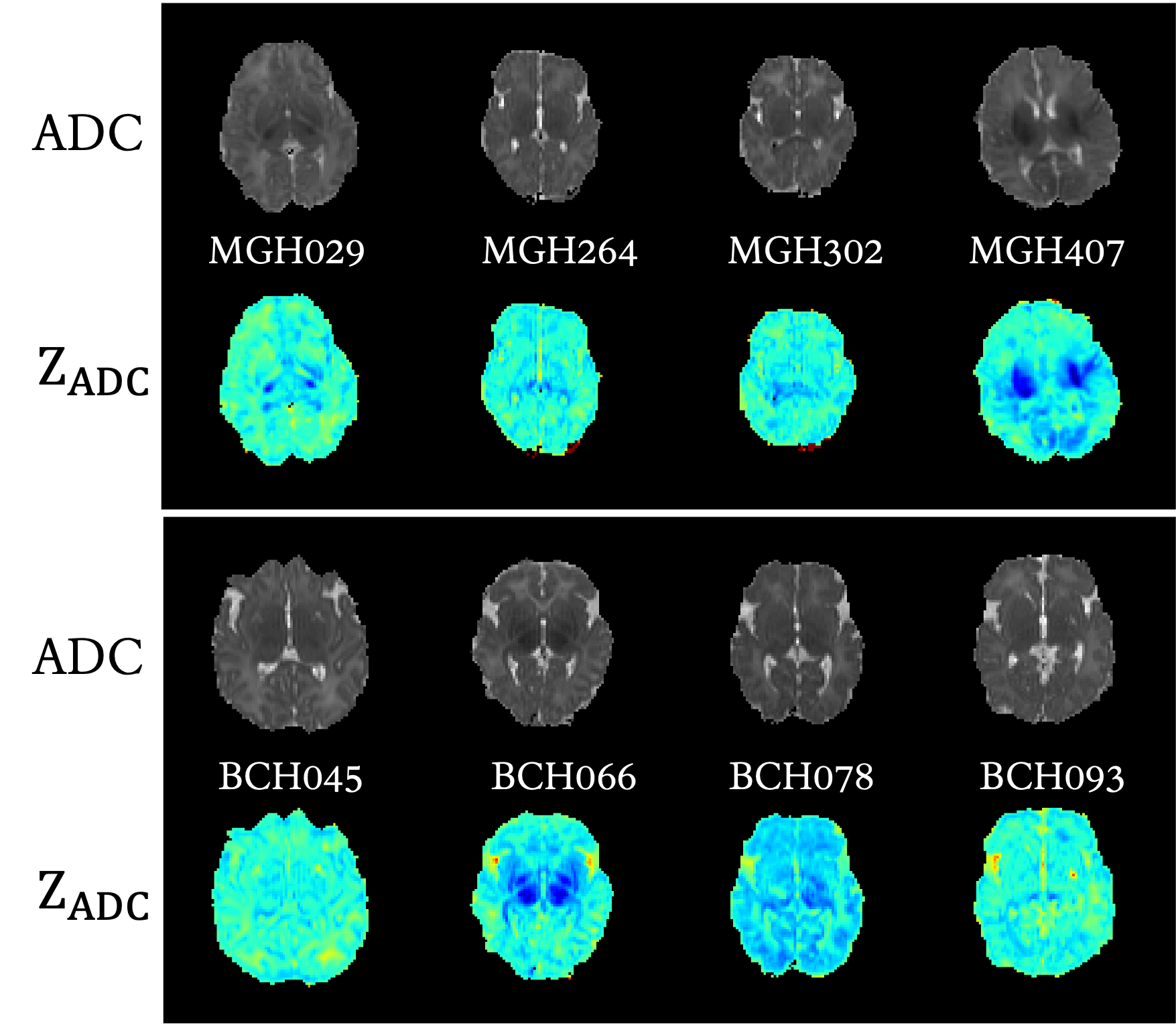
We provide ADC and for HIE outcome predictions, such information has been verified crucial for HIE outcome predictions. Figure 1 shows the ADC map, map from MGH and BCH. For details of ADC and , please refer our Part.I paper (Bao et al., 2023).
| NICU Outcome with MRIs | 2-year Outcome with MRIs | ||||||
| Site | Total | Survived | Deceased | Site | Total | Normal | Adverse |
| BCH | 72 | 65 | 7 | BCH | 72 | 30 | 42 |
| MGH | 107 | 94 | 13 | MGH | 84 | 53 | 31 |
Due to not all outcomes being collected with MRIs, we list the distribution of MRIs with NICU outcomes and 2-year outcomes in Table 2.
NICU Outcome
Table 1B lists the NICU outcome which represents the patient status at NICU discharge.
2-year Neurocognitive Outcome
Table 1C lists characteristics of 2-year neurocognitive outcomes. Outcome is defined as normal versus adverse, according to clinical criteria and NRN recommendations (Laptook et al., 2017; Shankaran et al., 2017). An adverse outcome is defined if the Bayley (version III) cognitive score in any of the Bayley-III domains, GMFCS level between 2 to 5, or blindness/hearing impairment. Otherwise, the patient had normal outcomes. Table 1C lists characteristics of Bayle version III scores (BSID-III scores).
Data structure and file formats
The data is organized in the format shown in Figure 2. BONBID-HIE provides, per patient: (i) 1ADC_ss: skull stripped ADC map; (ii) 2Z_ADC: map; (iii) 3LABEL: expert lesion annotations; and (iv) clinical data: clinical variables as written in Table 1A. (v) NICU outcome: status when discharged at NICU. (vi): 2-year outcome: 2-year neurocognitive outcome, noted that patients deceased before 2-year appointment are marked as adverse outcome.

Acknowledgements
This work was supported, in part, by NIH R03HD104891, R21NS121735, and R61NS126792.
Competing interests
None.
References
- Azzopardi et al. (2009) Denis V Azzopardi, Brenda Strohm, A David Edwards, Leigh Dyet, Henry L Halliday, Edmund Juszczak, Olga Kapellou, Malcolm Levene, Neil Marlow, Emma Porter, et al. Moderate hypothermia to treat perinatal asphyxial encephalopathy. New England Journal of Medicine, 361(14):1349–1358, 2009.
- Bao et al. (2023) Rina Bao, Ya’nan Song, Sara V Bates, Rebecca J Weiss, Anna N Foster, Camilo Jaimes Cobos, Susan Sotardi, Yue Zhang, Randy L Gollub, P Ellen Grant, et al. Boston neonatal brain injury dataset for hypoxic ischemic encephalopathy (bonbid-hie): Part i. mri and manual lesion annotation. bioRxiv, 2023.
- Cotten et al. (2014) C Michael Cotten, Amy P Murtha, Ronald N Goldberg, Chad A Grotegut, P Brian Smith, Ricki F Goldstein, Kimberley A Fisher, Kathryn E Gustafson, Barbara Waters-Pick, Geeta K Swamy, et al. Feasibility of autologous cord blood cells for infants with hypoxic-ischemic encephalopathy. The Journal of Pediatrics, 164(5):973–979, 2014.
- Dudink et al. (2007) Jeroen Dudink, Maarten Lequin, Carola van Pul, Jan Buijs, Nikk Conneman, Johannes van Goudoever, and Paul Govaert. Fractional anisotropy in white matter tracts of very-low-birth-weight infants. Pediatric Radiology, 37:1216–1223, 2007.
- Forman et al. (2016) Christoph Forman, Jens Wetzl, Carmel Hayes, and Michaela Schmidt. Compressed sensing: a paradigm shift in mri. MAGNETOM Flash, 66:9–13, 2016.
- Gluckman et al. (2005) Peter D Gluckman, John S Wyatt, Denis Azzopardi, Roberta Ballard, A David Edwards, Donna M Ferriero, Richard A Polin, Charlene M Robertson, Marianne Thoresen, Andrew Whitelaw, et al. Selective head cooling with mild systemic hypothermia after neonatal encephalopathy: multicentre randomised trial. The Lancet, 365(9460):663–670, 2005.
- Graham et al. (2008) Ernest M Graham, Kristy A Ruis, Adam L Hartman, Frances J Northington, and Harold E Fox. A systematic review of the role of intrapartum hypoxia-ischemia in the causation of neonatal encephalopathy. American Journal of Obstetrics and Gynecology, 199(6):587–595, 2008.
- Laptook et al. (2017) Abbot R Laptook, Seetha Shankaran, Jon E Tyson, Breda Munoz, Edward F Bell, Ronald N Goldberg, Nehal A Parikh, Namasivayam Ambalavanan, Claudia Pedroza, Athina Pappas, et al. Effect of therapeutic hypothermia initiated after 6 hours of age on death or disability among newborns with hypoxic-ischemic encephalopathy: a randomized clinical trial. JAMA, 318(16):1550–1560, 2017.
- Laptook et al. (2021) Abbot R Laptook, Seetha Shankaran, Patrick Barnes, Nancy Rollins, Barbara T Do, Nehal A Parikh, Shannon Hamrick, Susan R Hintz, Jon E Tyson, Edward F Bell, et al. Limitations of conventional magnetic resonance imaging as a predictor of death or disability following neonatal hypoxic–ischemic encephalopathy in the late hypothermia trial. The Journal of Pediatrics, 230:106–111, 2021.
- Lee et al. (2013) Anne CC Lee, Naoko Kozuki, Hannah Blencowe, Theo Vos, Adil Bahalim, Gary L Darmstadt, Susan Niermeyer, Matthew Ellis, Nicola J Robertson, Simon Cousens, et al. Intrapartum-related neonatal encephalopathy incidence and impairment at regional and global levels for 2010 with trends from 1990. Pediatric Research, 74(1):50–72, 2013.
- Liang et al. (2019) Shi-Peng Liang, Qian Chen, Yi-Bing Cheng, Ying-Ying Xue, and Hai-Jun Wang. Comparative effects of monosialoganglioside versus citicoline on apoptotic factor, neurological function and oxidative stress in newborns with hypoxic-ischemic encephalopathy. Coll Physicians Surg Pak, 29(4):324–327, 2019.
- Liu et al. (2006) Zulian Liu, Tengbin Xiong, and Catherine Meads. Clinical effectiveness of treatment with hyperbaric oxygen for neonatal hypoxic-ischaemic encephalopathy: systematic review of chinese literature. British Medical Journal, 333(7564):374, 2006.
- Nuñez-Ramiro et al. (2019) Antonio Nuñez-Ramiro, Isabel Benavente-Fernández, Eva Valverde, Malaika Cordeiro, Dorotea Blanco, Hector Boix, Fernando Cabañas, Mercedes Chaffanel, Belén Fernández-Colomer, Jose Ramón Fernández-Lorenzo, et al. Topiramate plus cooling for hypoxic-ischemic encephalopathy: a randomized, controlled, multicenter, double-blinded trial. Neonatology, 116(1):76–84, 2019.
- Potter et al. (2018) Molly Potter, Ted Rosenkrantz, and R Holly Fitch. Behavioral and neuroanatomical outcomes in a rat model of preterm hypoxic-ischemic brain injury: effects of caffeine and hypothermia. International Journal of Developmental Neuroscience, 70:46–55, 2018.
- Provenzale et al. (2007) James M Provenzale, Luxia Liang, David DeLong, and Leonard E White. Diffusion tensor imaging assessment of brain white matter maturation during the first postnatal year. American Journal of Roentgenology, 189(2):476–486, 2007.
- Shankaran et al. (2005) Seetha Shankaran, Abbot R Laptook, Richard A Ehrenkranz, Jon E Tyson, Scott A McDonald, Edward F Donovan, Avroy A Fanaroff, W Kenneth Poole, Linda L Wright, Rosemary D Higgins, et al. Whole-body hypothermia for neonates with hypoxic–ischemic encephalopathy. New England Journal of Medicine, 353(15):1574–1584, 2005.
- Shankaran et al. (2017) Seetha Shankaran, Abbot R Laptook, Athina Pappas, Scott A McDonald, Abhik Das, Jon E Tyson, Brenda B Poindexter, Kurt Schibler, Edward F Bell, Roy J Heyne, et al. Effect of depth and duration of cooling on death or disability at age 18 months among neonates with hypoxic-ischemic encephalopathy: a randomized clinical trial. JAMA, 318(1):57–67, 2017.