MedDiff-FM: A Diffusion-based Foundation Model for Versatile Medical Image Applications
Abstract
Diffusion models have achieved significant success in both the natural image and medical image domains, encompassing a wide range of applications. Previous investigations in medical images have often been constrained to specific anatomical regions, particular applications, and limited datasets, resulting in isolated diffusion models. This paper introduces a diffusion-based foundation model to address a diverse range of medical image tasks, namely MedDiff-FM. MedDiff-FM leverages 3D CT images from multiple publicly available datasets, covering anatomical regions from head to abdomen, to pre-train a diffusion foundation model, and explores the capabilities of the diffusion foundation model across a variety of application scenarios. The diffusion foundation model handles multi-level image processing both at the image-level and patch-level, and utilizes position embedding to establish multi-level spatial relationships as well as anatomical structures and region classes to control certain anatomical regions. MedDiff-FM manages several downstream tasks seamlessly, including image denoising, anomaly detection, and image synthesis. MedDiff-FM is also capable of performing lesion generation and lesion inpainting by rapidly fine-tuning the diffusion foundation model using ControlNet with task-specific conditions. Experimental results demonstrate the effectiveness of MedDiff-FM in addressing diverse downstream medical image tasks.
Keywords:
Diffusion Model Foundation Model Image Denoising Anomaly Detection Image Synthesis.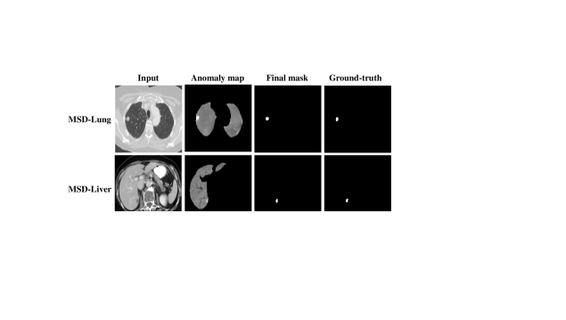
1 Introduction
Denoising diffusion probabilistic models (DDPMs) [19] have gained widespread applications in both natural image and medical image domains in recent times. DDPMs are capable of generating images of high-quality and diversity as well as maintaining training stability. Latent diffusion models (LDMs) [44] project images from pixel space to latent space and operate diffusion process in the latent space, which is more computationally efficient. Besides, stable diffusion [44] is known as a powerful pre-trained text-to-image latent diffusion model that can generate photo-realistic images given text prompts within seconds. In addition to text-to-image synthesis, there are other kinds of conditional image synthesis such as semantic-to-image synthesis [57, 53], and layout-to-image synthesis [71]. Incorporating diffusion models to synthesize high-quality long videos [3, 63, 5] are also flourishing.
Furthermore, utilizing diffusion models for medical image synthesis is meaningful because of the scarcity of data for certain diseases and the differences in patient populations and demographics. Therefore, synthesizing high-quality medical images using diffusion models is a promising method for augmenting medical datasets while preserving privacy. There are several studies for generating 2D and 3D medical images. ArSDM [11] and NASDM [51] synthesize 2D medical images using semantic masks conditioned semantic diffusion models (SDMs) [57]. Pinaya et al. [42], MedSyn [66], and GuideGen [10] are designed for generating 3D CT or MRI images based on textual condition, semantic condition, or a combination of both.
The significant success achieved by DDPM in image synthesis has demonstrated its potential capabilities to the world. Therefore, diffusion models have been explored in other tasks such as image super-resolution [48, 67], and image editing [38, 25, 4], and have shown their amazing abilities. Utilizing pre-trained text-to-image diffusion models for downstream tasks [70, 28] is also a promising approach. For medical images, diffusion models can also be applied to image denoising [65, 12], anomaly detection [62, 64, 2], and other related tasks.
However, previous methods mainly focus on specific medical image tasks, particular anatomical regions, and limited datasets. Moreover, these trained diffusion models lack generalization ability and are relatively isolated from each other. Therefore, this paper aims to introduce a pre-trained diffusion foundation model that covers different anatomical regions, leverages large-scale datasets, and addresses a variety of medical image tasks.
In this paper, we propose MedDiff-FM, a diffusion-based foundation model to satisfy these demands. MedDiff-FM deals with multiple anatomical regions, including head, neck, chest, and abdomen, utilizes publicly available medical image datasets from different institutions, and handles a diverse range of downstream applications. The diffusion foundation model accommodates multi-level medical images, accepting both image-level and patch-level inputs. To construct the relationships between multi-level inputs, we draw insights from Patch Diffusion [58], a patch-level diffusion model that adopts patch coordinate conditioning. We not only adapt the coordinate position conditioning from 2D to 3D for medical images but also advance multi-level relationships. The proposed position embedding for 3D CT images constructs multi-level spatial relationships between image-level and patch-level medical images. Moreover, MedDiff-FM leverages anatomical structures and region classes to better control and generate higher-quality medical images.
MedDiff-FM deals with a diverse range of downstream tasks without fine-tuning, including image denoising, anomaly detection, and image synthesis. Through fine-tuning with ControlNet, MedDiff-FM is also able to perform lesion generation and lesion inpainting under task-specific conditions. During inference, when processing entire CT volumes using the patch-level diffusion model, MedDiff-FM employs a patch-based sliding window sampling strategy with overlapping windows and smoothed noise estimates [41] to mitigate boundary artifacts. Experimental results indicate that MedDiff-FM is effective in addressing a variety of downstream medical image tasks.
The contributions of this work are summarized as follows:
-
•
We propose MedDiff-FM, a diffusion-based foundation model that leverages 3D CT images from diverse datasets and multiple anatomical regions, to pre-train a diffusion foundation model for handling a wide range of medical image tasks.
-
•
MedDiff-FM deals with medical images flexibly, achieving multi-level image processing (i.e., image-level and patch-level), and leverages position embedding to build spatial relationships between image-level and patch-level 3D CT images, along with anatomical structures and region classes to condition certain anatomical regions.
-
•
MedDiff-FM provides seamless applications for several downstream tasks, including image denoising, anomaly detection, and image synthesis.
-
•
Through rapid fine-tuning of the diffusion foundation model via ControlNet, MedDiff-FM demonstrates effective lesion generation and lesion inpainting under task-specific conditions.
2 Related Work
2.1 Patch-based Diffusion Models
Recently, denoising diffusion probabilistic models [19] have demonstrated superior generative capabilities due to sample quality and diversity, while maintaining training stability. However, in the context of 3D medical images, where voxel dimensions are often large, the patch sizes that diffusion models can process are limited. While image-level processing captures global information in medical images, it also requires patch-level processing to capture local details.
Patch Diffusion [58] is a patch-level diffusion model training method, which adopts patch coordinate conditioning and patch size scheduling to balance global encoding effectiveness and training efficiency. To deal with multi-level medical images, and construct the relationship between image-level and patch-level inputs, MedDiff-FM not only utilizes the coordinate position conditioning from Patch Diffusion but also advances multi-level representations, and extends the coordinate position conditioning from 2D to 3D, in order to establish the multi-level relationships of medical images.
Özdenizci et al. [41] design a patch-based diffusion approach that processes arbitrary sized images during inference, and utilizes smoothed noise estimates across overlapping patches. Therefore, to deal with entire CT volumes with the patch-level diffusion model, MedDiff-FM leverages patch-based sliding window sampling strategy with overlapping windows and smoothed noise estimates to eliminate artificial boundaries.
2.2 Diffusion Applications in Natural Images
In the natural image domain, stable diffusion (SD) [44] has emerged as a powerful pre-trained text-to-image generation model. Stable diffusion is a latent diffusion model [44] which performs diffusion process in the low-dimensional latent space instead of the high-dimensional pixel space. ControlNet [68] generalizes the pre-trained text-to-image diffusion models to more diverse conditions, such as canny edges, human poses, and depth maps. Besides text-to-image diffusion models, text-to-video diffusion models [3, 63, 5] have also witnessed significant development, such as Sora [5].
In addition to unconditional and conditional image generation tasks, diffusion models have also been applied to other tasks [8]. For example, image super-resolution [48, 67], image editing [38, 25, 4], and image-to-image translation [47, 56]. Moreover, Zhao et al. [70] and Kondapaneni et al. [28] adapt pre-trained text-to-image diffusion models to diverse downstream tasks, and show the capabilities of pre-trained diffusion models.
2.3 Diffusion Applications for Medical Images
Beyond the natural image domain, diffusion models are also flourishing in the medical image domain. Most of the diffusion models for medical images are 2D models related to 2D medical images, such as X-rays, CT slices, and MRI slices. Zhuang et al. [72] generate 2D abdominal CT images using mask and edge conditions. ArSDM [11] generates colonoscopy polyp images with polyp masks. NASDM [51] synthesizes nuclei pathology images conditioned on nuclei masks.
However, 2D diffusion models concentrate on the intra-slice information and omit the inter-slice information of 3D medical images such as CT and MRI. Pinaya et al. [42] synthesize 3D brain MRIs with covariable conditions such as brain structure volumes. Medical Diffusion [27] generates unconditional 3D CT and MRI images. MedGen3D [16] is introduced for 3D thoracic CT and brain MRI synthesis with their aligned segmentation masks. MedSyn [66] is proposed for generating high-fidelity 3D chest CT images with textual guidance. GuideGen [10] is a text-guided diffusion model for paired abdominal CT scan and anatomical structure generation.
Diffusion models for medical images have diverse applications under different medical image scenarios [26], beyond medical image synthesis. For example, DDM2 [65] adopts a self-supervised method for diffusion MRI denoising. CoreDiff [12] introduces a contextual error-modulated generalized diffusion model for low-dose CT denoising. Wolleb et al. [62] utilize a class conditional diffusion model for the anomaly detection of brain tumors and pleural effusion. AnoDDPM [64] and AutoDDPM [2] are both designed for the anomaly detection of brain tumors from 2D MRI images. Jimenez-Perez et al. [22] attempt to pre-train a diffusion model on chest X-rays for reconstruction and segmentation tasks.
3 Methodology
In this section, we first provide an overview of MedDiff-FM, then introduce the multi-resolution integrated medical diffusion foundation model and the position embedding for 3D CT images, and finally discuss the application of MedDiff-FM to downstream tasks.
3.1 Overview of MedDiff-FM
Fig. 1 illustrates the overview of MedDiff-FM. MedDiff-FM is trained on a diverse range of publicly available 3D CT datasets from different institutions. These 3D CT datasets cover different anatomical regions, including the head, neck, chest, and abdomen, encompassing a variety of anatomical structures and eliminating the limitations of focusing on a single anatomical region. The pre-training of MedDiff-FM on different anatomical regions and diverse medical image datasets enables it to be applied to downstream tasks across multiple anatomical regions, demonstrating strong generalization capabilities. MedDiff-FM can be directly applied to downstream tasks, including image denoising, anomaly detection, and image synthesis, without the need for fine-tuning, which significantly conserves resources and enhances convenience. Additionally, by fine-tuning MedDiff-FM, lesion generation and lesion inpainting can be achieved by incorporating task-specific conditions using ControlNet.
3.2 Multi-resolution Integrated Medical Diffusion Foundation Model
As depicted in Fig. 2, the diffusion foundation model accepts multi-level medical image inputs, specifically image-level inputs and patch-level inputs. Given a 3D medical image , we randomly apply one of three operations: resizing the image to the patch size, randomly cropping the image to twice the patch size and then resizing it to the patch size, or randomly cropping the image to the patch size. The first operation yields image-level inputs, whereas the latter two yield patch-level inputs. The patch size used in this paper is 128 × 128 × 128. The multi-level input follows the data distribution . For diffusion timesteps, it produces a sequence of noisy images , where follows a standard Gaussian distribution. In the denoising process, the denoising U-Net [45] progressively transforms random noises into images. The denoising U-Net takes as inputs the noisy image , the current timestep , the position embedding, and additional conditions, and outputs the estimated noise .
During the pre-training of the diffusion foundation model, we leverage two conditioning signals, the region condition and the anatomy condition , to achieve multi-perspective control over the denoising process. The region condition utilizes anatomical region classes that indicate whether the anatomical region belongs to the head and neck (HaN), chest, or abdomen. The anatomy condition leverages anatomical structure masks derived from TotalSegmentator [61] and thresholding methods to impose control over anatomical structures. Since the diffusion foundation model takes multi-level medical image inputs, we aim to establish explicit spatial relationships between image-level and patch-level inputs. We adopt position encoding to obtain position embedding of the X, Y, and Z coordinates, which we will discuss further in Section 3.3.
The overall training objective of MedDiff-FM is formulated as:
| (1) |
When it is necessary to fine-tune the diffusion foundation model, we utilize ControlNet to incorporate task-specific conditions. ControlNet leverages the weights of the neural network blocks, creating a locked copy to preserve the knowledge of the pre-trained diffusion model and a trainable copy to adapt to additional conditions. These copies are connected through zero convolution modules, which gradually adjust the parameters starting from zero. A target encoder is employed to extract features from the target condition , enabling more effective injection of auxiliary conditions into the diffusion foundation model.
The objective function for fine-tuning MedDiff-FM is formulated as:
| (2) |
3.3 3D CT Image Position Embedding
The voxel dimensions in 3D medical images are often large; however, the image sizes that diffusion models can process are limited. The image-level processing may lead to the neglect of local details, while the patch-level processing may lose holistic perception. Therefore, we propose the diffusion foundation model that is designed to handle both image-level and patch-level inputs simultaneously, aiming to integrate multi-level medical image information.
To construct relationships between image-level and patch-level inputs, we draw insights from Patch Diffusion [58], a patch-level diffusion model that utilizes patch coordinate conditioning. Instead of solely using patch coordinates to represent the relative position of the patch to the original image, we construct multi-level position relationships, as illustrated in Fig. 3. The multi-level position relationships establish explicit connections between the global and local information in medical images. In addition, we adapt the 2D coordinate position conditioning to 3D for medical images. The coordinate positions are pixel-level and normalized to , where denotes the upper left back corner and denotes the bottom right front corner. The three position coordinate channels , , and represent the X, Y, and Z coordinate positions, respectively. Furthermore, we employ position encoding function [39] to better encode positional information. The process is formulated as follows, where denotes the maximum frequency.
| (3) | |||
| (4) | |||
| (5) |
During inference, leveraging the established multi-level position relationships, MedDiff-FM utilizes a patch-based sliding window sampling strategy to deal with entire CT volume processing or generation using the patch-level diffusion model. The multi-level position relationships establish spatial relations between each patch and the whole volume. An example of patch-level whole volume synthesis is shown in Fig. 4. The diffusion model generates patch-level images, which are then combined to form the entire CT volume. To mitigate artificial boundaries between overlapping windows, we employ the smoothed noise estimates [41] across overlapping patches. At each denoising timestep , the mean estimated noise based sampling updates are applied to overlapping pixels across patches.
3.4 Downstream Tasks without Fine-tuning
MedDiff-FM can be used to accomplish multiple downstream tasks, including image synthesis, image denoising, and anomaly detection, without the need for fine-tuning. As shown in Fig. 4, MedDiff-FM synthesizes whole CT volumes of flexible sizes based on patch-level sampling. The image synthesis is beneficial for augmenting medical image datasets and enhancing data diversity.
3.4.1 Image Denoising.
CT is a prevalent imaging modality in clinical diagnosis, but the associated radiation exposure poses potential health risks. Low-dose CT (LDCT) effectively reduces the radiation dose but often suffers from significant noise and artifacts, which can impair diagnostic accuracy. Consequently, the image denoising techniques are essential for enhancing the quality of LDCT images. We leverage the denoising capabilities of MedDiff-FM to progressively reduce noise in LDCT images over multiple time steps, thus improving image quality.
3.4.2 Anomaly Detection.
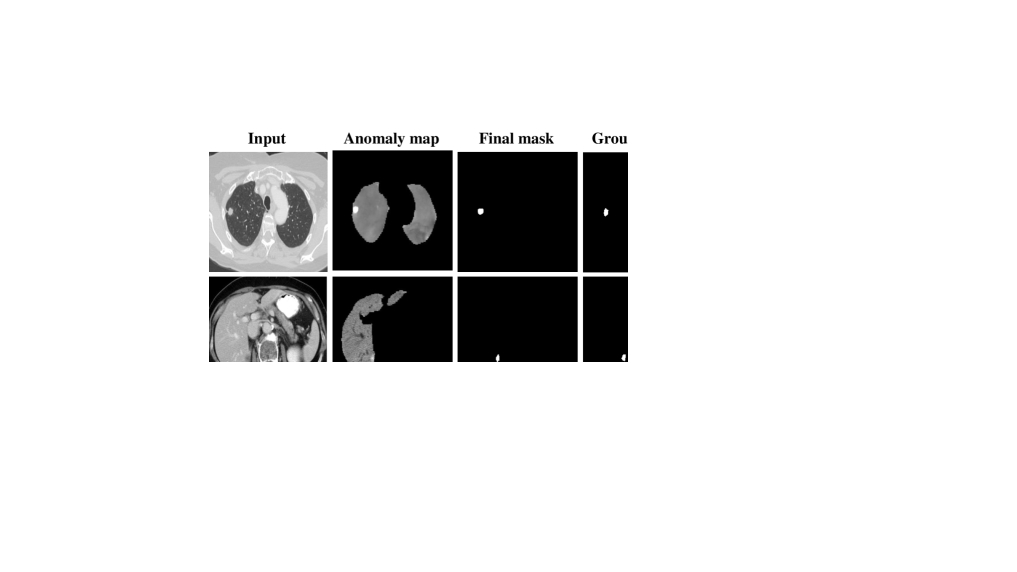
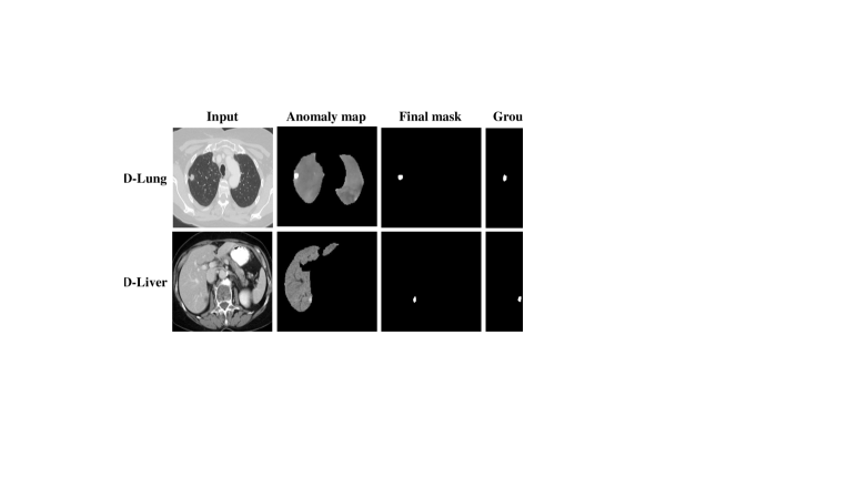
Anomaly detection aims at identifying irregularities or abnormalities in medical images, aiding in diagnosis and treatment. To accomplish anomaly detection of given anatomical regions, we utilize anatomical structure masks to focus on specific anatomical regions while masking other regions. Therefore, given the masked unhealthy image , we add noise at a fixed time step to obtain the noisy unhealthy image . We then directly predict the original image, yielding the reconstructed healthy image . The anomaly map is calculated as . We apply a threshold to binarize the anomaly map, resulting in a binary mask .
3.5 Downstream Tasks with Fine-tuning
3.5.1 Lesion Generation.
By rapidly fine-tuning MedDiff-FM using lesion patches, we can effectively achieve the lesion generation task, despite having limited lesion data. We employ lesion masks as the target condition to enable the controlled generation of lesion images. The generated lesion images can contribute to various tasks such as lesion segmentation.
3.5.2 Lesion Inpainting.
We employ the lesion generation model to perform the lesion inpainting task rather than repeatedly fine-tuning MedDiff-FM. Since the lesion generation model is already capable of generating lesions, lesion inpainting can utilize this capability straightforwardly. Lesion inpainting can better leverage the information from the original CT image rather than relying entirely on image generation. We adopt the image inpainting method proposed in RePaint [33], which combines the known region from the original image with the inpainted region from the model output at each step.
| Dataset Name | # Cases |
| Head and Neck | 362 |
| StructSeg [50] | 50 |
| INSTANCE2022 [31, 32] | 130 |
| HaN-Seg [43] | 42 |
| SegRap2023 [34] | 140 |
| Chest | 1,040 |
| SegTHOR [29] | 40 |
| CT-RATE [15] | 1,000 |
| Abdomen | 1,732 |
| AbdomenCT-1K [36] | 1,062 |
| AMOS22 [21] | 500 |
| BTCV [30] | 30 |
| CHAOS [23, 24] | 20 |
| WORD [35] | 120 |
| Whole Body | 2,242 |
| TotalSegmentator [61] | 1,228 |
| AutoPET [13] | 1,014 |
| Total | 5,376 |
4 Experiments and Results
In this section, we first describe the experimental setup, including datasets, implementation details, and evaluation metrics. Next, we evaluate the downstream tasks, which can be divided into two categories: those that do not require fine-tuning of MedDiff-FM and those that require fine-tuning of MedDiff-FM. The first category includes image synthesis, image denoising, and anomaly detection, while the second category includes lesion generation and lesion inpainting.
4.1 Experimental Schemes
4.1.1 Datasets.
MedDiff-FM datasets. We collect several publicly available medical image datasets, covering diverse anatomical regions and structures, for developing MedDiff-FM. The medical image datasets and their corresponding number of cases used in our experiments are shown in Table 1. We gather a total of 5,376 CT volumes, consisting of 362 head and neck volumes, 1,040 chest volumes, 1,732 abdomen volumes, and 2,242 whole body volumes. To balance between different anatomical regions, we only adopt 1,000 chest cases of the CT-RATE [15] dataset. These datasets are randomly divided into 90% for MedDiff-FM training and 5% for validation, with the remaining 5% reserved for evaluating MedDiff-FM on the downstream image synthesis task.
| Task | Dataset | # Cases |
| Image Denoising | Mayo 2016 [37] | 10 |
| Anomaly Detection | MSD-Lung [52, 1] | 63 |
| MSD-Liver [52, 1] | 131 | |
| Lesion Generation & | MSD-Lung [52, 1] | 63 |
| Lesion Inpainting | MED-LN [46] | 90 |
| MSD-Liver [52, 1] | 131 | |
| ABD-LN [46] | 86 |
Task-specific datasets. To further validate MedDiff-FM on other downstream tasks, we adopt the datasets listed in Table 2. The Mayo 2016 dataset [37], also known as 2016 NIH-AAPM-Mayo Clinic Low-Dose CT Grand Challenge, contains 1 mm full-dose and quarter-dose CT images from 10 patients. Covering both chest and abdomen regions, the Mayo 2016 dataset is used to evaluate the image denoising capabilities of MedDiff-FM. MSD-Lung and MSD-Liver, Task06 and Task03 of Medical Segmentation Decathlon (MSD) [52, 1], are used for the evaluation of anomaly detection. Additionally, we adopt four datasets to evaluate lesion generation and lesion inpainting tasks, of which MED-LN and ABD-LN are acquired from [46], containing mediastinal and abdominal lymph nodes respectively. The four datasets are each randomly divided, with 80% used for fine-tuning MedDiff-FM, specifically for ControlNet training and validation, and the remaining 20% for testing.
| Anatomical Region | Method | MS-SSIM | LPIPS | FID | MMD | Dice |
| HaN | DDPM | 0.7596 | 0.0201 | 0.2757 | 0.0394 | 0.80 |
| MedDiff-FM | 0.7821 | 0.0132 | 0.1269 | 0.0265 | 0.89 | |
| Chest | DDPM | 0.7657 | 0.0261 | 0.2675 | 0.0656 | 0.73 |
| MedDiff-FM | 0.7942 | 0.0263 | 0.1853 | 0.0545 | 0.75 | |
| Abdomen | DDPM | 0.6194 | 0.0339 | 0.2903 | 0.0573 | 0.86 |
| MedDiff-FM | 0.6412 | 0.0238 | 0.2775 | 0.0459 | 0.90 | |
| Average | DDPM | 0.7168 | 0.0265 | 0.1430 | 0.0541 | 0.80 |
| MedDiff-FM | 0.7411 | 0.0211 | 0.1021 | 0.0422 | 0.84 |
4.1.2 Implementation Details.
We implement all the methods using PyTorch and carry out all the experiments using NVIDIA GeForce RTX 3090 GPUs. To obtain the anatomical structures used in MedDiff-FM, we leverage the automated whole body medical image segmentation tool, TotalSegmentator v2 [61], to segment the CT images. Incorporating the 117 classes segmented by TotalSegmentator, we further derive the body class using a thresholding method, resulting in 118 classes in total. For data preprocessing, we first resample the image voxel spacing to 1.0 mm × 1.0 mm × 1.0 mm. Next, for whole body CT images, we split the head and neck, chest, and abdomen regions based on the segmentation results from TotalSegmentator and crop the images corresponding to these anatomical regions. For different anatomical regions, we apply varying window widths and levels for window truncation. For the head and neck region, the window level is set to 50 and the window width to 400; for the chest region, the window level is -500 and the window width is 1800; and for the abdomen region, the window level is 60 and the window width is 360. Subsequently, the data ranges are normalized to . The diffusion timesteps is 1,000 with cosine noise schedule. The patch size used in our experiment is 128 × 128 × 128. The model channels for MedDiff-FM and ControlNet are 32 and the number of residual blocks is 1, with the spatial transformer operating at a spatial resolution of 16 × 16 × 16. For training, we utilize loss and Adam optimizer, with a learning rate of , a batch size of 1, and 4 gradient accumulation steps. The MedDiff-FM is trained for around 150 epochs, and the ControlNet is trained for around 10k steps.
4.1.3 Evaluation Metrics.
We utilize metrics widely used to assess synthesis quality and diversity, including Multi-Scale Structural Similarity Index Measure (MS-SSIM) [60], Learned Perceptual Image Patch Similarity (LPIPS) [69], Fréchet Inception Distance (FID) [18], and Maximum Mean Discrepancy (MMD) [14]. The feature extractor is a 3D pre-trained ResNet [17] from MedicalNet [7].
To further measure the consistency between the generated images and the given anatomical structure conditions for image synthesis tasks. We extract the segmented anatomical structures of the generated images using TotalSegmentator [61], and calculate the Dice coefficient (Dice) between these structures and the given anatomical conditions for major organs. For the head and neck region, this includes the brain and skull; for the chest region, the lung, heart, and aorta; and for the abdomen region, the spleen, kidney, gallbladder, liver, stomach, pancreas, small bowel, duodenum, and colon.
To evaluate the distribution similarity between the generated lesion images and the real lesion images for lesion generation and lesion inpainting tasks, we train segmentation models on real lesion images with nnU-Net [20]. Then we utilize the trained segmentation models to segment the generated lesion images, and calculate the Dice between the segmentation results and ground-truths. We compare the Dice coefficients for real lesion images and generated lesion images to assess their distribution similarity.
For the image denoising task, we use structural similarity index measure (SSIM) [59] and peak signal-to-noise ratio (PSNR) to validate image denoising performance, following Noise2Sim [40].
For the anomaly detection task, we adopt commonly used Area Under the Receiver Operating Characteristic Curve (AUC), specificity (SPE), sensitivity (SEN) and accuracy (ACC) to assess the anomaly detection results, as demonstrated in [55].
4.2 Effectiveness of Position Embedding
To demonstrate the effectiveness of position embedding, we utilize MedDiff-FM to accomplish the patch-level whole CT volume synthesis task. Based on the patch-level sampling strategy, MedDiff-FM can synthesize whole CT volumes of flexible sizes.
The patch-level whole volume synthesis results are listed in Table 3. MedDiff-FM significantly outperforms DDPM across all anatomical regions and various generation metrics, demonstrating remarkable generative capabilities. Additionally, the higher Dice score of MedDiff-FM demonstrates better consistency between the synthetic images and the generation conditions. The visualization results are shown in Fig. 5. The CT volumes generated by MedDiff-FM demonstrate overall consistency, with richer and clearer details compared to DDPM. Whether at the global image-level or the local patch-level, the generated image quality is superior.
In summary, these quantitative and qualitative results indicate that position embedding successfully constructs multi-level spatial relationships between each local patch and the whole CT volume.
| Method | SSIM ↑ | PSNR ↑ | |
| LDCT | 0.8434 | 23.44 | |
| Supervised Method | RED-CNN [6] | 0.9030 | 28.58 |
| MAP-NN [49] | 0.9013 | 28.28 | |
| Unsupervised Method | BM3D [9] | 0.8830 | 27.28 |
| Noise2Sim [40] | 0.9045 | 28.38 | |
| MedDiff-FM | 0.9123 | 28.10 |
| Dataset | Method | AUC ↑ | SPE ↑ | SEN ↑ | ACC ↑ |
| MSD-Lung | MedDiff-FM (900) | 0.9785 | 0.9909 | 0.5665 | 0.9906 |
| MedDiff-FM (950) | 0.9816 | 0.9778 | 0.7208 | 0.9776 | |
| Fully Supervised | 0.9876 | 0.9996 | 0.7856 | 0.9995 | |
| MSD-Liver | MedDiff-FM (800) | 0.9924 | 0.9954 | 0.5141 | 0.9945 |
| MedDiff-FM (900) | 0.9937 | 0.9956 | 0.6229 | 0.9948 | |
| Fully Supervised | 0.9988 | 0.9996 | 0.7964 | 0.9993 |
4.3 Evaluation of Downstream Tasks without Fine-tuning
To validate the effectiveness of directly leveraging the pre-trained MedDiff-FM model in addressing downstream tasks, we evaluate it on the image denoising and anomaly detection tasks.
4.3.1 Image Denoising.
We evaluate the image denoising capabilities of MedDiff-FM on the Mayo 2016 dataset using the official 25% dose CT images. We utilize MedDiff-FM for denoising the LDCT images with 50 steps. The quantitative results are illustrated in Table 4. The outcomes of RED-CNN [6], MAP-NN [49], and BM3D [9] are adopted from Noise2Sim [40]. Results in the table indicate that MedDiff-FM demonstrates strong denoising capabilities. MedDiff-FM obviously outperforms other methods in terms of SSIM, while its PSNR performance is comparable. MedDiff-FM can be seamlessly utilized for image denoising, eliminating the need for additional fine-tuning specific to the denoising task and thus reducing the consumption of spatiotemporal resources. As displayed in Fig. 6, MedDiff-FM significantly enhances image quality, exhibiting considerable denoising capabilities.
| Lesion generation | Lesion inpainting | ||||||
| Dataset | Method | MS-SSIM | LPIPS | FID | MMD | Dice | Dice |
| MSD-Lung | Real | - | - | - | - | 0.74 | 0.74 |
| From scratch | 0.6606 | 0.0135 | 0.2059 | 0.1018 | 0.16 | 0.42 | |
| MedDiff-FM (fine-tune) | 0.6731 | 0.0136 | 0.2332 | 0.0998 | 0.28 | 0.77 | |
| MSD-Liver | Real | - | - | - | - | 0.71 | 0.71 |
| From scratch | 0.5991 | 0.0151 | 0.1304 | 0.0679 | 0.40 | 0.71 | |
| MedDiff-FM (fine-tune) | 0.6121 | 0.0138 | 0.1690 | 0.0727 | 0.48 | 0.71 | |
| MED-LN | Real | - | - | - | - | 0.28 | 0.28 |
| From scratch | 0.7946 | 0.0059 | 0.3057 | 0.0538 | 0.01 | 0.30 | |
| MedDiff-FM (fine-tune) | 0.7982 | 0.0074 | 0.1195 | 0.0183 | 0.22 | 0.34 | |
| ABD-LN | Real | - | - | - | - | 0.51 | 0.51 |
| From scratch | 0.5267 | 0.0304 | 0.1765 | 0.0570 | 0.32 | 0.51 | |
| MedDiff-FM (fine-tune) | 0.5802 | 0.0211 | 0.2425 | 0.0594 | 0.50 | 0.53 | |
4.3.2 Anomaly Detection.
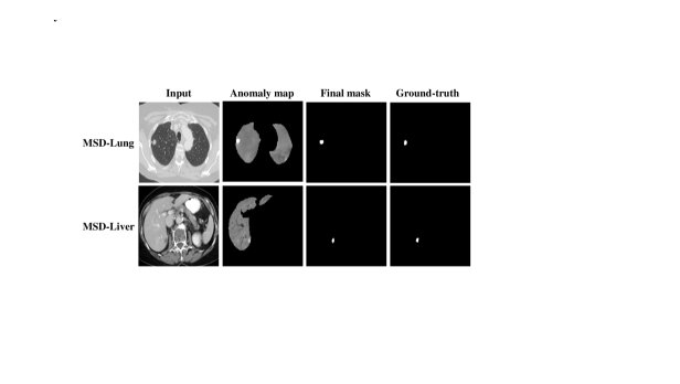
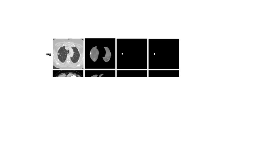
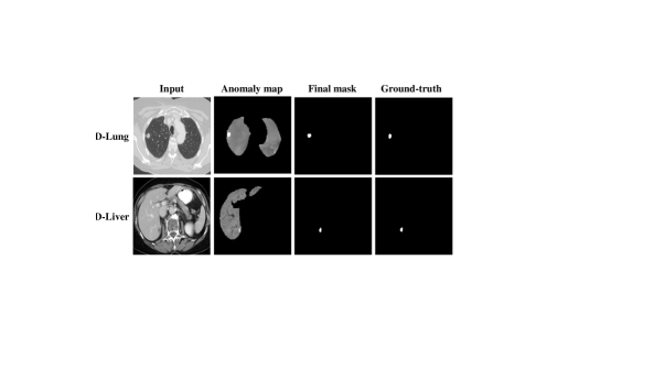
We utilize MedDiff-FM to accomplish the anomaly detection task on the MSD-Lung and MSD-Liver datasets. Since we employ anatomical structure masks to focus on specific anatomical regions, we exclude lesions that are not located within the liver or lung regions segmented by TotalSegmentator [61]. We provide the results of fully supervised learning using nnU-Net [20] on the test set for reference. Table 5 demonstrates the anomaly detection results. The number in parentheses represents the diffusion steps. MedDiff-FM employs Gaussian noise, and when adding noise to unhealthy CT images, small time steps struggle to disrupt tumor structures [64]. The results indicate that utilizing MedDiff-FM for anomaly detection in CT images can achieve excellent performance. Fig. 7 visualizes the anomaly detection results, where MedDiff-FM successfully detects the lung and liver tumors.
4.4 Evaluation of Downstream Tasks with Fine-tuning
To further demonstrate the capabilities of MedDiff-FM, we fine-tune it for lesion-related tasks, including lesion generation and lesion inpainting. We fine-tune MedDiff-FM on four datasets: MSD-Lung, MSD-Liver, MED-LN, and ABD-LN.

4.4.1 Lesion Generation.
The lesion generation results are demonstrated in Table 6. Those generative metrics assess the quality of the generated images from a holistic perspective. However, for lesion generation, it is crucial to concentrate more on the quality of the lesions themselves. Utilizing segmentation models trained on real lesion images to detect synthetic lesion images and calculate the Dice score provides a more accurate assessment of synthetic lesion quality. The results indicate that the fine-tuned MedDiff-FM significantly outperforms the model trained from scratch in terms of lesion generation. The visualization of lesion generation samples is shown in Fig. 8.
4.4.2 Lesion Inpainting.
The lesion inpainting results are presented in the last column of Table 6. Since image inpainting does not change the holistic structure of the original image, it is unnecessary to use generative metrics to evaluate the quality of the entire image. Instead, we just utilize the Dice score to evaluate the effectiveness of MedDiff-FM in lesion inpainting. Additionally, the qualitative results are depicted in Fig. 9.
5 Discussion
The proposed MedDiff-FM covers multiple anatomical regions and handles various downstream tasks, demonstrating robust generalization capabilities. The architecture of the diffusion foundation model is flexible, supporting multi-level image processing. However, there are some limitations in this work. First, we utilize anatomical structures to control local details so as to generate higher-quality medical images, which limits flexibility. Future work could explore more flexible, textual conditions. Second, while MedDiff-FM is specifically designed for CT images, it highlights the potential for diffusion-based foundation models to be extended to other medical imaging modalities. Moreover, for the whole image generation, the patch-based sliding window inference strategy, in conjunction with the progressive denoising process, imposes substantial computational burdens. To address this, future work could explore methods like consistency models [54] to accelerate the sampling process.
6 Conclusion
In conclusion, this paper presents MedDiff-FM, a diffusion-based foundation model that deals with a wide range of medical image tasks. MedDiff-FM utilizes 3D CT images from diverse publicly available datasets and focuses on multiple anatomical regions, overcoming the limitations of previous works that were constrained by specific anatomical regions and particular tasks. MedDiff-FM is capable of handling multi-level image processing with position embedding to build multi-level spatial relationships, and using anatomical structures and regions as conditions. The pre-trained diffusion foundation model can seamlessly perform tasks such as image denoising, anomaly detection, and image synthesis. Furthermore, MedDiff-FM deals with lesion generation and lesion inpainting through rapid fine-tuning via ControlNet with task-specific conditions. Experimental results highlight the effectiveness of MedDiff-FM, making it a valuable tool for various medical image applications.
References
- [1] Antonelli, M., Reinke, A., Bakas, S., Farahani, K., Kopp-Schneider, A., Landman, B.A., Litjens, G., Menze, B., Ronneberger, O., Summers, R.M., et al.: The medical segmentation decathlon. Nature communications 13(1), 4128 (2022)
- [2] Bercea, C.I., Neumayr, M., Rueckert, D., Schnabel, J.A.: Mask, stitch, and re-sample: Enhancing robustness and generalizability in anomaly detection through automatic diffusion models. arXiv preprint arXiv:2305.19643 (2023)
- [3] Blattmann, A., Rombach, R., Ling, H., Dockhorn, T., Kim, S.W., Fidler, S., Kreis, K.: Align your latents: High-resolution video synthesis with latent diffusion models. In: Proceedings of the IEEE/CVF Conference on Computer Vision and Pattern Recognition. pp. 22563–22575 (2023)
- [4] Brooks, T., Holynski, A., Efros, A.A.: Instructpix2pix: Learning to follow image editing instructions. In: Proceedings of the IEEE/CVF Conference on Computer Vision and Pattern Recognition. pp. 18392–18402 (2023)
- [5] Brooks, T., Peebles, B., Holmes, C., DePue, W., Guo, Y., Jing, L., Schnurr, D., Taylor, J., Luhman, T., Luhman, E., Ng, C., Wang, R., Ramesh, A.: Video generation models as world simulators (2024), https://openai.com/research/video-generation-models-as-world-simulators
- [6] Chen, H., Zhang, Y., Kalra, M.K., Lin, F., Chen, Y., Liao, P., Zhou, J., Wang, G.: Low-dose ct with a residual encoder-decoder convolutional neural network. IEEE transactions on medical imaging 36(12), 2524–2535 (2017)
- [7] Chen, S., Ma, K., Zheng, Y.: Med3d: Transfer learning for 3d medical image analysis. arXiv preprint arXiv:1904.00625 (2019)
- [8] Croitoru, F.A., Hondru, V., Ionescu, R.T., Shah, M.: Diffusion models in vision: A survey. IEEE Transactions on Pattern Analysis and Machine Intelligence 45(9), 10850–10869 (2023)
- [9] Dabov, K., Foi, A., Katkovnik, V., Egiazarian, K.: Image denoising by sparse 3-d transform-domain collaborative filtering. IEEE Transactions on image processing 16(8), 2080–2095 (2007)
- [10] Dai, L., Zhang, R., Huang, Z., Zhang, X.: Guidegen: A text-guided framework for joint ct volume and anatomical structure generation. arXiv preprint arXiv:2403.07247 (2024)
- [11] Du, Y., Jiang, Y., Tan, S., Wu, X., Dou, Q., Li, Z., Li, G., Wan, X.: Arsdm: colonoscopy images synthesis with adaptive refinement semantic diffusion models. In: International conference on medical image computing and computer-assisted intervention. pp. 339–349. Springer (2023)
- [12] Gao, Q., Li, Z., Zhang, J., Zhang, Y., Shan, H.: Corediff: Contextual error-modulated generalized diffusion model for low-dose ct denoising and generalization. IEEE Transactions on Medical Imaging (2023)
- [13] Gatidis, S., Hepp, T., Früh, M., La Fougère, C., Nikolaou, K., Pfannenberg, C., Schölkopf, B., Küstner, T., Cyran, C., Rubin, D.: A whole-body fdg-pet/ct dataset with manually annotated tumor lesions. Scientific Data 9(1), 601 (2022)
- [14] Gretton, A., Borgwardt, K.M., Rasch, M.J., Schölkopf, B., Smola, A.: A kernel two-sample test. The Journal of Machine Learning Research 13(1), 723–773 (2012)
- [15] Hamamci, I.E., Er, S., Almas, F., Simsek, A.G., Esirgun, S.N., Dogan, I., Dasdelen, M.F., Wittmann, B., Simsar, E., Simsar, M., et al.: A foundation model utilizing chest ct volumes and radiology reports for supervised-level zero-shot detection of abnormalities. arXiv preprint arXiv:2403.17834 (2024)
- [16] Han, K., Xiong, Y., You, C., Khosravi, P., Sun, S., Yan, X., Duncan, J.S., Xie, X.: Medgen3d: A deep generative framework for paired 3d image and mask generation. In: International Conference on Medical Image Computing and Computer-Assisted Intervention. pp. 759–769. Springer (2023)
- [17] He, K., Zhang, X., Ren, S., Sun, J.: Deep residual learning for image recognition. In: Proceedings of the IEEE conference on computer vision and pattern recognition. pp. 770–778 (2016)
- [18] Heusel, M., Ramsauer, H., Unterthiner, T., Nessler, B., Hochreiter, S.: Gans trained by a two time-scale update rule converge to a local nash equilibrium. Advances in neural information processing systems 30 (2017)
- [19] Ho, J., Jain, A., Abbeel, P.: Denoising diffusion probabilistic models. Advances in neural information processing systems 33, 6840–6851 (2020)
- [20] Isensee, F., Jaeger, P.F., Kohl, S.A., Petersen, J., Maier-Hein, K.H.: nnu-net: a self-configuring method for deep learning-based biomedical image segmentation. Nature methods 18(2), 203–211 (2021)
- [21] Ji, Y., Bai, H., Ge, C., Yang, J., Zhu, Y., Zhang, R., Li, Z., Zhanng, L., Ma, W., Wan, X., et al.: Amos: A large-scale abdominal multi-organ benchmark for versatile medical image segmentation. Advances in neural information processing systems 35, 36722–36732 (2022)
- [22] Jimenez-Perez, G., Osorio, P., Cersovsky, J., Montalt-Tordera, J., Hooge, J., Vogler, S., Mohammadi, S.: Dino-diffusion. scaling medical diffusion via self-supervised pre-training. arXiv preprint arXiv:2407.11594 (2024)
- [23] Kavur, A.E., Gezer, N.S., Barış, M., Aslan, S., Conze, P.H., Groza, V., Pham, D.D., Chatterjee, S., Ernst, P., Özkan, S., Baydar, B., Lachinov, D., Han, S., Pauli, J., Isensee, F., Perkonigg, M., Sathish, R., Rajan, R., Sheet, D., Dovletov, G., Speck, O., Nürnberger, A., Maier-Hein, K.H., Bozdağı Akar, G., Ünal, G., Dicle, O., Selver, M.A.: CHAOS Challenge - combined (CT-MR) healthy abdominal organ segmentation. Medical Image Analysis 69, 101950 (Apr 2021). https://doi.org/https://doi.org/10.1016/j.media.2020.101950, http://www.sciencedirect.com/science/article/pii/S1361841520303145
- [24] Kavur, A.E., Selver, M.A., Dicle, O., Barış, M., Gezer, N.S.: CHAOS - Combined (CT-MR) Healthy Abdominal Organ Segmentation Challenge Data (Apr 2019). https://doi.org/10.5281/zenodo.3362844, https://doi.org/10.5281/zenodo.3362844
- [25] Kawar, B., Zada, S., Lang, O., Tov, O., Chang, H., Dekel, T., Mosseri, I., Irani, M.: Imagic: Text-based real image editing with diffusion models. In: Proceedings of the IEEE/CVF Conference on Computer Vision and Pattern Recognition. pp. 6007–6017 (2023)
- [26] Kazerouni, A., Aghdam, E.K., Heidari, M., Azad, R., Fayyaz, M., Hacihaliloglu, I., Merhof, D.: Diffusion models in medical imaging: A comprehensive survey. Medical Image Analysis 88, 102846 (2023)
- [27] Khader, F., Müller-Franzes, G., Tayebi Arasteh, S., Han, T., Haarburger, C., Schulze-Hagen, M., Schad, P., Engelhardt, S., Baeßler, B., Foersch, S., et al.: Denoising diffusion probabilistic models for 3d medical image generation. Scientific Reports 13(1), 7303 (2023)
- [28] Kondapaneni, N., Marks, M., Knott, M., Guimaraes, R., Perona, P.: Text-image alignment for diffusion-based perception. In: Proceedings of the IEEE/CVF Conference on Computer Vision and Pattern Recognition. pp. 13883–13893 (2024)
- [29] Lambert, Z., Petitjean, C., Dubray, B., Kuan, S.: Segthor: Segmentation of thoracic organs at risk in ct images. In: 2020 Tenth International Conference on Image Processing Theory, Tools and Applications (IPTA). pp. 1–6. IEEE (2020)
- [30] Landman, B., Xu, Z., Igelsias, J., Styner, M., Langerak, T., Klein, A.: Miccai multi-atlas labeling beyond the cranial vault–workshop and challenge. In: Proc. MICCAI Multi-Atlas Labeling Beyond Cranial Vault—Workshop Challenge. vol. 5, p. 12 (2015)
- [31] Li, X., Luo, G., Wang, K., Wang, H., Liu, J., Liang, X., Jiang, J., Song, Z., Zheng, C., Chi, H., et al.: The state-of-the-art 3d anisotropic intracranial hemorrhage segmentation on non-contrast head ct: The instance challenge. arXiv preprint arXiv:2301.03281 (2023)
- [32] Li, X., Luo, G., Wang, W., Wang, K., Gao, Y., Li, S.: Hematoma expansion context guided intracranial hemorrhage segmentation and uncertainty estimation. IEEE Journal of Biomedical and Health Informatics 26(3), 1140–1151 (2021)
- [33] Lugmayr, A., Danelljan, M., Romero, A., Yu, F., Timofte, R., Van Gool, L.: Repaint: Inpainting using denoising diffusion probabilistic models. In: Proceedings of the IEEE/CVF conference on computer vision and pattern recognition. pp. 11461–11471 (2022)
- [34] Luo, X., Fu, J., Zhong, Y., Liu, S., Han, B., Astaraki, M., Bendazzoli, S., Toma-Dasu, I., Ye, Y., Chen, Z., et al.: Segrap2023: A benchmark of organs-at-risk and gross tumor volume segmentation for radiotherapy planning of nasopharyngeal carcinoma. arXiv preprint arXiv:2312.09576 (2023)
- [35] Luo, X., Liao, W., Xiao, J., Chen, J., Song, T., Zhang, X., Li, K., Metaxas, D.N., Wang, G., Zhang, S.: Word: A large scale dataset, benchmark and clinical applicable study for abdominal organ segmentation from ct image. arXiv preprint arXiv:2111.02403 (2021)
- [36] Ma, J., Zhang, Y., Gu, S., Zhu, C., Ge, C., Zhang, Y., An, X., Wang, C., Wang, Q., Liu, X., et al.: Abdomenct-1k: Is abdominal organ segmentation a solved problem? IEEE Transactions on Pattern Analysis and Machine Intelligence 44(10), 6695–6714 (2021)
- [37] McCollough, C.H., Bartley, A.C., Carter, R.E., Chen, B., Drees, T.A., Edwards, P., Holmes III, D.R., Huang, A.E., Khan, F., Leng, S., et al.: Low-dose ct for the detection and classification of metastatic liver lesions: results of the 2016 low dose ct grand challenge. Medical physics 44(10), e339–e352 (2017)
- [38] Meng, C., He, Y., Song, Y., Song, J., Wu, J., Zhu, J.Y., Ermon, S.: Sdedit: Guided image synthesis and editing with stochastic differential equations. arXiv preprint arXiv:2108.01073 (2021)
- [39] Mildenhall, B., Srinivasan, P.P., Tancik, M., Barron, J.T., Ramamoorthi, R., Ng, R.: Nerf: Representing scenes as neural radiance fields for view synthesis. Communications of the ACM 65(1), 99–106 (2021)
- [40] Niu, C., Li, M., Fan, F., Wu, W., Guo, X., Lyu, Q., Wang, G.: Suppression of correlated noise with similarity-based unsupervised deep learning. arXiv preprint arXiv:2011.03384 (2020)
- [41] Özdenizci, O., Legenstein, R.: Restoring vision in adverse weather conditions with patch-based denoising diffusion models. IEEE Transactions on Pattern Analysis and Machine Intelligence 45(8), 10346–10357 (2023)
- [42] Pinaya, W.H., Tudosiu, P.D., Dafflon, J., Da Costa, P.F., Fernandez, V., Nachev, P., Ourselin, S., Cardoso, M.J.: Brain imaging generation with latent diffusion models. In: MICCAI Workshop on Deep Generative Models. pp. 117–126. Springer (2022)
- [43] Podobnik, G., Strojan, P., Peterlin, P., Ibragimov, B., Vrtovec, T.: Han-seg: The head and neck organ-at-risk ct and mr segmentation dataset. Medical physics 50(3), 1917–1927 (2023)
- [44] Rombach, R., Blattmann, A., Lorenz, D., Esser, P., Ommer, B.: High-resolution image synthesis with latent diffusion models. In: Proceedings of the IEEE/CVF conference on computer vision and pattern recognition. pp. 10684–10695 (2022)
- [45] Ronneberger, O., Fischer, P., Brox, T.: U-net: Convolutional networks for biomedical image segmentation. In: Medical Image Computing and Computer-Assisted Intervention–MICCAI 2015: 18th International Conference, Munich, Germany, October 5-9, 2015, Proceedings, Part III 18. pp. 234–241. Springer (2015)
- [46] Roth, H.R., Lu, L., Seff, A., Cherry, K.M., Hoffman, J., Wang, S., Liu, J., Turkbey, E., Summers, R.M.: A new 2.5 d representation for lymph node detection using random sets of deep convolutional neural network observations. In: Medical Image Computing and Computer-Assisted Intervention–MICCAI 2014: 17th International Conference, Boston, MA, USA, September 14-18, 2014, Proceedings, Part I 17. pp. 520–527. Springer (2014)
- [47] Saharia, C., Chan, W., Chang, H., Lee, C., Ho, J., Salimans, T., Fleet, D., Norouzi, M.: Palette: Image-to-image diffusion models. In: ACM SIGGRAPH 2022 conference proceedings. pp. 1–10 (2022)
- [48] Saharia, C., Ho, J., Chan, W., Salimans, T., Fleet, D.J., Norouzi, M.: Image super-resolution via iterative refinement. IEEE transactions on pattern analysis and machine intelligence 45(4), 4713–4726 (2022)
- [49] Shan, H., Padole, A., Homayounieh, F., Kruger, U., Khera, R.D., Nitiwarangkul, C., Kalra, M.K., Wang, G.: Competitive performance of a modularized deep neural network compared to commercial algorithms for low-dose ct image reconstruction. Nature Machine Intelligence 1(6), 269–276 (2019)
- [50] Shi, J.: Structseg2019 gtv segmentation (2023). https://doi.org/10.21227/h75x-gt46, https://dx.doi.org/10.21227/h75x-gt46
- [51] Shrivastava, A., Fletcher, P.T.: Nasdm: Nuclei-aware semantic histopathology image generation using diffusion models. In: International Conference on Medical Image Computing and Computer-Assisted Intervention. pp. 786–796. Springer (2023)
- [52] Simpson, A.L., Antonelli, M., Bakas, S., Bilello, M., Farahani, K., Van Ginneken, B., Kopp-Schneider, A., Landman, B.A., Litjens, G., Menze, B., et al.: A large annotated medical image dataset for the development and evaluation of segmentation algorithms. arXiv preprint arXiv:1902.09063 (2019)
- [53] Singh, J., Gould, S., Zheng, L.: High-fidelity guided image synthesis with latent diffusion models. In: 2023 IEEE/CVF Conference on Computer Vision and Pattern Recognition (CVPR). pp. 5997–6006. IEEE (2023)
- [54] Song, Y., Dhariwal, P., Chen, M., Sutskever, I.: Consistency models. arXiv preprint arXiv:2303.01469 (2023)
- [55] Tian, Y., Liu, F., Pang, G., Chen, Y., Liu, Y., Verjans, J.W., Singh, R., Carneiro, G.: Self-supervised pseudo multi-class pre-training for unsupervised anomaly detection and segmentation in medical images. Medical image analysis 90, 102930 (2023)
- [56] Tumanyan, N., Geyer, M., Bagon, S., Dekel, T.: Plug-and-play diffusion features for text-driven image-to-image translation. In: Proceedings of the IEEE/CVF Conference on Computer Vision and Pattern Recognition. pp. 1921–1930 (2023)
- [57] Wang, W., Bao, J., Zhou, W., Chen, D., Chen, D., Yuan, L., Li, H.: Semantic image synthesis via diffusion models. arXiv preprint arXiv:2207.00050 (2022)
- [58] Wang, Z., Jiang, Y., Zheng, H., Wang, P., He, P., Wang, Z., Chen, W., Zhou, M., et al.: Patch diffusion: Faster and more data-efficient training of diffusion models. Advances in neural information processing systems 36 (2024)
- [59] Wang, Z., Bovik, A.C., Sheikh, H.R., Simoncelli, E.P.: Image quality assessment: from error visibility to structural similarity. IEEE transactions on image processing 13(4), 600–612 (2004)
- [60] Wang, Z., Simoncelli, E.P., Bovik, A.C.: Multiscale structural similarity for image quality assessment. In: The Thrity-Seventh Asilomar Conference on Signals, Systems & Computers, 2003. vol. 2, pp. 1398–1402. Ieee (2003)
- [61] Wasserthal, J., Breit, H.C., Meyer, M.T., Pradella, M., Hinck, D., Sauter, A.W., Heye, T., Boll, D.T., Cyriac, J., Yang, S., et al.: Totalsegmentator: robust segmentation of 104 anatomic structures in ct images. Radiology: Artificial Intelligence 5(5) (2023)
- [62] Wolleb, J., Bieder, F., Sandkühler, R., Cattin, P.C.: Diffusion models for medical anomaly detection. In: International Conference on Medical image computing and computer-assisted intervention. pp. 35–45. Springer (2022)
- [63] Wu, J.Z., Ge, Y., Wang, X., Lei, S.W., Gu, Y., Shi, Y., Hsu, W., Shan, Y., Qie, X., Shou, M.Z.: Tune-a-video: One-shot tuning of image diffusion models for text-to-video generation. In: Proceedings of the IEEE/CVF International Conference on Computer Vision. pp. 7623–7633 (2023)
- [64] Wyatt, J., Leach, A., Schmon, S.M., Willcocks, C.G.: Anoddpm: Anomaly detection with denoising diffusion probabilistic models using simplex noise. In: Proceedings of the IEEE/CVF Conference on Computer Vision and Pattern Recognition. pp. 650–656 (2022)
- [65] Xiang, T., Yurt, M., Syed, A.B., Setsompop, K., Chaudhari, A.: Ddm2: Self-supervised diffusion mri denoising with generative diffusion models. In: The Eleventh International Conference on Learning Representations (2023)
- [66] Xu, Y., Sun, L., Peng, W., Jia, S., Morrison, K., Perer, A., Zandifar, A., Visweswaran, S., Eslami, M., Batmanghelich, K.: Medsyn: Text-guided anatomy-aware synthesis of high-fidelity 3d ct images. IEEE Transactions on Medical Imaging (2024)
- [67] Yue, Z., Wang, J., Loy, C.C.: Resshift: Efficient diffusion model for image super-resolution by residual shifting. Advances in Neural Information Processing Systems 36 (2024)
- [68] Zhang, L., Rao, A., Agrawala, M.: Adding conditional control to text-to-image diffusion models. In: Proceedings of the IEEE/CVF International Conference on Computer Vision. pp. 3836–3847 (2023)
- [69] Zhang, R., Isola, P., Efros, A.A., Shechtman, E., Wang, O.: The unreasonable effectiveness of deep features as a perceptual metric. In: Proceedings of the IEEE conference on computer vision and pattern recognition. pp. 586–595 (2018)
- [70] Zhao, W., Rao, Y., Liu, Z., Liu, B., Zhou, J., Lu, J.: Unleashing text-to-image diffusion models for visual perception. In: Proceedings of the IEEE/CVF International Conference on Computer Vision. pp. 5729–5739 (2023)
- [71] Zheng, G., Zhou, X., Li, X., Qi, Z., Shan, Y., Li, X.: Layoutdiffusion: Controllable diffusion model for layout-to-image generation. In: Proceedings of the IEEE/CVF Conference on Computer Vision and Pattern Recognition. pp. 22490–22499 (2023)
- [72] Zhuang, Y., Hou, B., Mathai, T.S., Mukherjee, P., Kim, B., Summers, R.M.: Semantic image synthesis for abdominal ct. In: International Conference on Medical Image Computing and Computer-Assisted Intervention. pp. 214–224. Springer (2023)