EndoMetric: Near-light metric scale monocular SLAM
Abstract
Geometric reconstruction and SLAM with endoscopic images have seen significant advancements in recent years. In most medical specialties, the endoscopes used are monocular, and the algorithms applied are typically extensions of those designed for external environments, resulting in 3D reconstructions up to an unknown scale factor.
In this paper, we take advantage of the fact that standard endoscopes are equipped with near-light sources positioned at a small but non-zero baseline from the camera. By leveraging the inverse-square law of light decay, we enable, for the first time, monocular reconstructions with accurate metric scale. This paves the way to transform any endoscope into a metric device, which is essential for practical applications such as measuring polyps, stenosis, or the extent of tissue affected by disease.
I Introduction
Medical endoscopic procedures such as colonoscopies, gastroscopies or bronchoscopies are prevalent in clinical practice. Currently, endoscope navigation, location estimation, and tissue measurements are manually performed by the medical staff performing the procedure. Recent advances in Visual Simultaneous Localization and Mapping (VSLAM) for endoscopy [1, 2, 3] offer a good promise to provide live 3D reconstructions of each patient, enabling autonomous or assisted navigation and robotized interventions.
With the notable exception of robotic surgery and laparoscopy where stereo endoscopes are common, most medical specialties use just monocular endoscopes, mainly to reduce bulk and cost, and to provide simpler user interfaces for doctors.
Most SLAM systems assume static light sources. Photometric methods need constant illumination, while geometric ones use invariant features to ignore light variations. In these methods, it is well known that using a moving monocular camera, the absolute scale of the environment is unobservable. As a result, the 3D reconstructions and trajectories obtained are only accurate up to an unknown scale factor. This limitation also introduces scale drift in monocular SLAM, which can significantly reduce map accuracy. For this reason, stereo SLAM systems are more accurate and robust compared to their monocular counterparts [4].
However, monocular endoscopes have light sources attached to the camera, that introduce strong illumination changes in the scene. Our key insight is that these illumination changes, far from being a nuisance, can be exploited using near-light photometry to recover the real metric scale of monocular reconstructions.
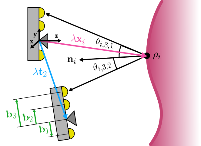
Photometry depends on the scale because of two factors: the inverse-square decay of illumination with distance from the light source, and the angle between the incident light and the surface normal in the case of light sources at a small –but non-zero– baseline from the camera optical center (Figure 1).
As photometry is involved, illumination power, camera gain, and scene albedo and normals need also to be estimated. We show how all these values can be computed from the sole input of the images gathered by the near-light camera of the standard endoscope. We follow a two-step approach: first obtain and up-to-scale reconstruction using SfM or V-SLAM, and then solve a photometric optimization to recover scale, gains and albedo.
To summarize, our contributions are:
-
•
An scale dependent near-light photometric model that can be applied to any monocular up-to-scale multi-view reconstruction.
-
•
The first ever photometric optimization method to estimate the true metric scale, scene albedo and camera gain.
-
•
An initialization method to facilitate convergence avoiding local minima.
-
•
Simulation experiments showing the scale accuracy that can be obtained in realistic endoscope settings.
II Related work
Feature-based monocular visual SLAM [4, 5], and SfM [6], recover the up-to-scale multi-view geometry by bundle adjustment, which optimizes only the geometric reprojection error while neglecting any intensity information. On the other hand, photometric monocular SLAM methods, [7, 8], optimize on the gray level detected by each camera, being able to recover the sparse multi-view geometry, albedo and camera gains. However, they are unable to recover the metric scale due to the underlying assumption of a Lambertian scene where the cameras do move while illumination remains fixed and constant. In contrast, we propose a two-step approach to recover a sparse metric map, metric camera trajectory, albedos and camera gains, from the sole input of a near-light monocular video stream. In the first step, we estimate the up-to-scale geometry by bundle adjustment. In the second step, the metric scale, albedos and camera gains are recovered through non-linear optimization of the photometric error.
The closest classical approach to our metric scale estimation is photometric stereo, which can be traced back to [9]. They use multiple orthographic images from a fixed camera under the illumination of distant light sources that are selectively turned on and off. They recover dense normals, and albedos of the scene, though still without determining the metric scale. The first photometric stereo method able to recover metric scale was [10]. This was accomplished by leveraging the inverse-square illumination decay of multiple point light sources at known positions from a fixed projective camera. Photometric stereo has been recently revisited in non-medical scenes formalizing the concept of near-field photometric stereo in [11] for the case in which the camera and the light sources are close to the imaged object. One step further in the practicality of this approach was introduced in [12] by means of the semi-calibrated near-light photometric stereo where the light positions are known while their intensities are unknown. The fact that an endoscope is a calibrated near-light photometric stereo was exploited for the first time in [13]. They use three colored light sources to combine their illumination in a single shot, being able to produce true-scale dense depth. However they need to modify the standard endoscope acquisition.
In summary, near-light photometric stereo is able to produce real-scale dense models of an scene with respect to a fixed projective camera from three or more images taken under different point light sources. But to be applied to endoscopy, the actual device has to be modified either to turn the lights on and off or by using colored lights, which is undesirable in clinical practice. By contrast, using the unmodified images of a standard endoscope, we produce not only a metric scale sparse map, but we also estimate the metric camera trajectory. Also, while photometric stereo needs a minimum of three near-light sources, we can handle any number of point light sources.
It is also worth mentioning [14] because they produce metric reconstruction from endoscopic monocular sequences without modifying the lights. However they need the camera trajectory from an EM tracker, while we are able to also estimate it.
The work [15] used illumination decay to recover 3D information from a single monocular view from an unmodified endoscope, assuming constant albedo. However, the resulting reconstructions remained up-to-scale due to three unknown factors: illumination power, camera gain, and surface albedo.
Our multi-view two-step method was first proposed in [16], however they only provided a preliminary simulation to validate the second step of the approach. Their validation assumed that the up-to-scale ground-truth geometry was available and focused on the photometric optimization to estimate the metric scale, the albedos and the cameras gain for any number of near-light point sources. In this work we go one step further proving that the method can work just from the sole input of the near-light images. For that we use photorealistic simulated images of colonoscopies, compute the up-to-scale geometry using SfM or SLAM, and then apply the near-light photometric optimization.
III Fundamentals
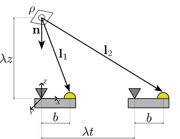
In this section we discuss the simplest multi-view near-light photometric problem [16], to show its properties. Suppose a moving monocular camera with a single point light source at a distance from the optical center, that observes a Lambertian point with albedo , that is located along the optical axis of the first camera at a depth , with the normal pointing to the camera, and a second camera pose that was translated to the right (Figure 2). Here is the unknown scale factor of a SfM or VSLAM reconstruction, that we want to compute.
Assuming the light intensity and camera gain are constant, and no gamma compression, the intensity of the point in each image will be [10]:
| (1) |
where is a scaled albedo. Please, note that what we call, for short, camera gain in a real endoscope is the result of exposure control, sensor sensitivity, and ADC gain, values not available to the user.
For the simple case in Figure 2, the intensities observed in both images should be:
| (2) | |||||
| (3) |
If albedo, light intensity and camera gain are known, the scale can be recovered from a single view. Otherwise we can eliminate to get a second-order equation on :
| (4) |
where is a known constant. This equation allows to obtain , except when , in which case simplifies away, and cannot be solved.
In conclusion, in a multi-view monocular near-light scenario, the metric scale is observable, provided that the baseline between camera and light source is non-zero. Also, we can expect the scale accuracy to degrade when the distance to the surface is too big compared with the camera-light baseline. The rest of the paper describes a practical method to obtain the scale in real endoscopic settings and studies its accuracy.
IV EndoMetric
Our proposed method, named EndoMetric, works in two steps: first obtain an up-to-scale multi-view reconstruction of the scene using any SLAM of SfM method, and then solve an optimization problem for metric scale estimation that impose albedo consistency. The key for scale estimation is leveraging a near-light photometric model that takes into account the baseline of the light sources with respect to the camera and the inverse-square law of illumination decline with distance.
IV-A Up-to-scale Multi-view Reconstruction
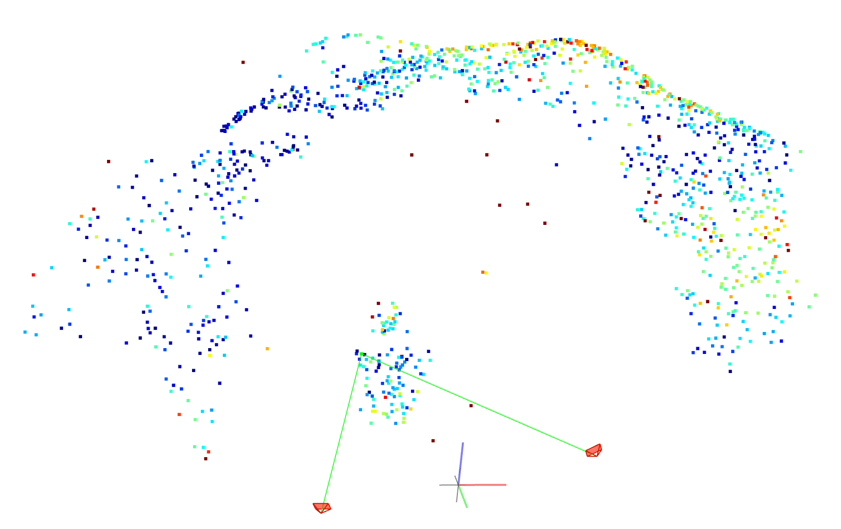
Classical multiview geometry is able to produce, from a sequence of calibrated monocular colonoscopy images (at least two), the geometry of the scene points and the camera poses up to an unknown scale factor . At the core of the estimation is the bundle adjustment, i.e. the non-linear optimization of the re-projection errors of the image points matched along the sequence. There are widely available batch approaches such as COLMAP [6] to compute multiview geometry from image collections. There are also sequential SLAM approaches able to produce in real time the multiview geometry, such as ORB-SLAM3 [4], or CudaSIFT SLAM [5] that is specialized for endoscopic imagery.
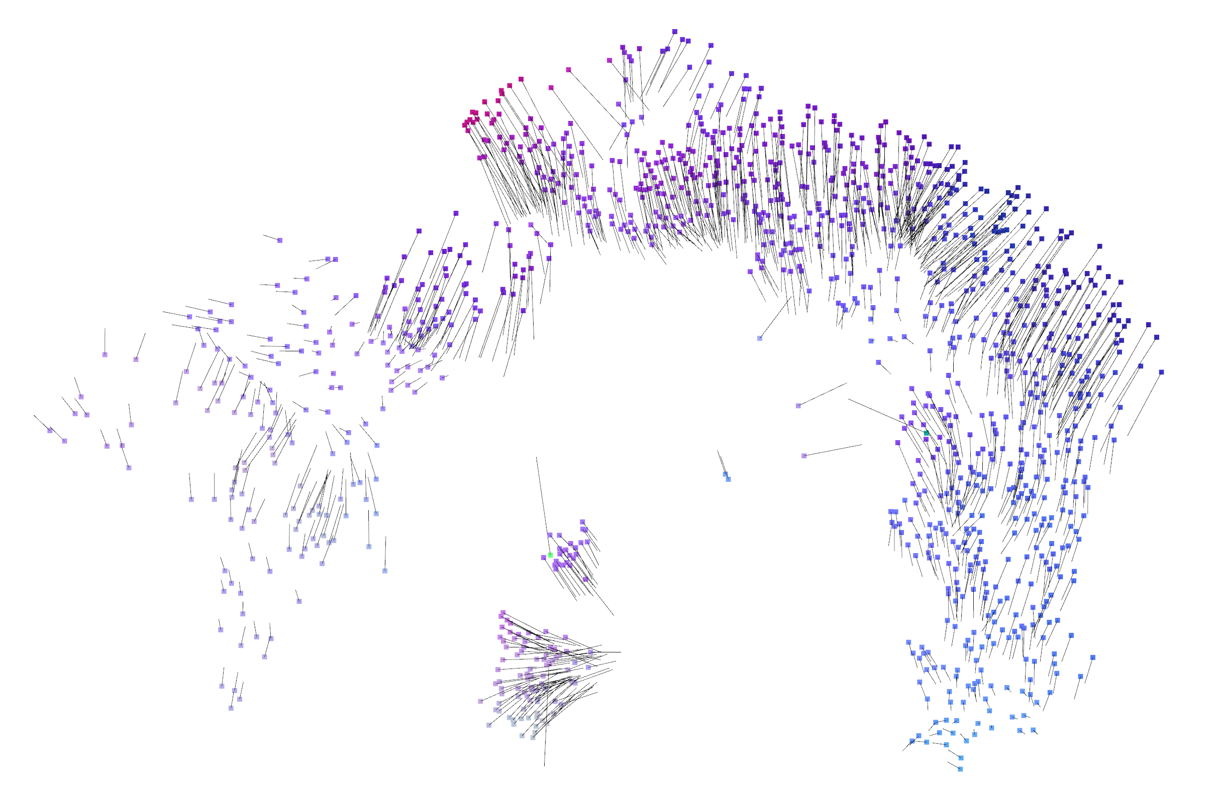
The photometric models not only need the sparse scene geometry, but also the surface normals at each scene point. We propose to compute the normals fitting a plane to the neighbors of each scene point. The result is an up-to-scale reconstruction of the 3D scene formed by camera poses , , and point coordinates and normals (Figure 3).
IV-B Near-light Photometric Model
We assume a mobile calibrated camera with attached point light sources, where the relative 3D position of each light with respect to the camera’s optical center is known, along with the camera intrinsic parameters. For simplicity in the presentation, we assume that all lights have the same intensity , uniform in all directions, and that the camera has no lens vignetting. For a more sophisticated model, see [15].
We also assume that the surface is Lambertian, with varying unknown albedo. Taking into account this assumption, we discard points that are close to specular reflections (that, in any case, are also bad points for SfM), and use robust cost functions for photometric errors.
We have grayscale images taken by the camera from poses while observing scene points. For a given point in an image the image intensity depends on the point albedo , the camera gain , the light intensity , and the gamma compression . Crucially, as illumination decreases with the inverse square of the distance from each light source to the point, the perceived radiance depends on the unknown scale factor of the multi-view reconstruction as (Figure 1):
| (5) |
| (6) |
It can be observed that albedos are always multiplied by the camera gain and . As these values are not provided by currently available endoscopes, we define two new variables that are observable from the images:
| (7) | |||||
| (8) |
where is a scaled albedo (that may be greater than 1) and represents the gain change with respect to the first image. Using them, our near-light photometric model becomes:
| (9) |
IV-C Metric Scale Estimation
Given the up-to-scale reconstruction, our near-light photometric model, and the original images , our goal is to recover the scale factor and, as a side product, the point albedos and camera gain changes . This can be achieved minimizing the photometric error of the observed points with respect to the model:
| (10) |
where is the intensity of point observed in image , and a robust Huber cost function is used to reduce the influence of spurious data. This nonlinear optimization problem can be solved using, for example, the Levenberg-Marquardt algorithm from the Ceres [17] library.
IV-D Initial Guess for the Scale
All SfM and SLAM systems use some heuristic to chose the scale of their reconstructions, for example by setting the distance between two camera poses, or the average depth of the reconstructed points in the first view. In some endoscopic settings the resulting scale factor may be close to 1, just by chance, and in other it can be arbitrarily far. This means that, unless the photometric residual is a convex function, there is no guarantee that the optimization will converge to the true scale.
To avoid local minima and achieve faster convergence it is crucial to find good initial values for the optimization variables (, , and ). However, these variables are closely related: modifying any of them requires adjusting the others to achieve the same intensity level. Therefore, instead of estimating their initial values separately as in [16], we propose to estimate them jointly.
For that, we perform an exhaustive search for the scale parameter over a logarithmic space . For each trial value , we estimate the albedo values solving Equation 9 for each point in the first image.
Then, to estimate the relative gains of the rest of images, we perform a robust regression. First, we undo the gamma compression to work in linear space. Next, we find the gain value that minimizes the difference between the real value and the estimated value of the points using the Huber cost function.
Finally, we select the value of with the lowest Huber cost:
| (11) |
V Experiments
| Optimized | Initial | % error | |||||||
| guess | |||||||||
| Fernandes [16] | GT | GT | 0.72 | 3.92 | 0.01 | ||||
| Our model | SfM | SfM | 3.30 | 3.34 | 0.03 | ||||
| Ablation study | ✗ | SfM | SfM | 71.43 | 6.03 | 1.03 | |||
| SfM | GT | 2.53 | 3.02 | 0.12 | |||||
| GT | SfM | GT | 2.47 | 3.15 | – | ||||
| GT | GT | GT | 0.86 | 3.11 | – | ||||
Current hardware limitations make it challenging to obtain real endoscopic images with ground-truth metric scale. To our knowledge, no dataset simultaneously provides both photometric calibration of the endoscope and metric scale annotations of the observed surface. As a result, we evaluated the accuracy of our method through simulations.
V-A Dataset
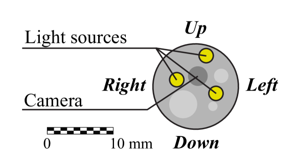
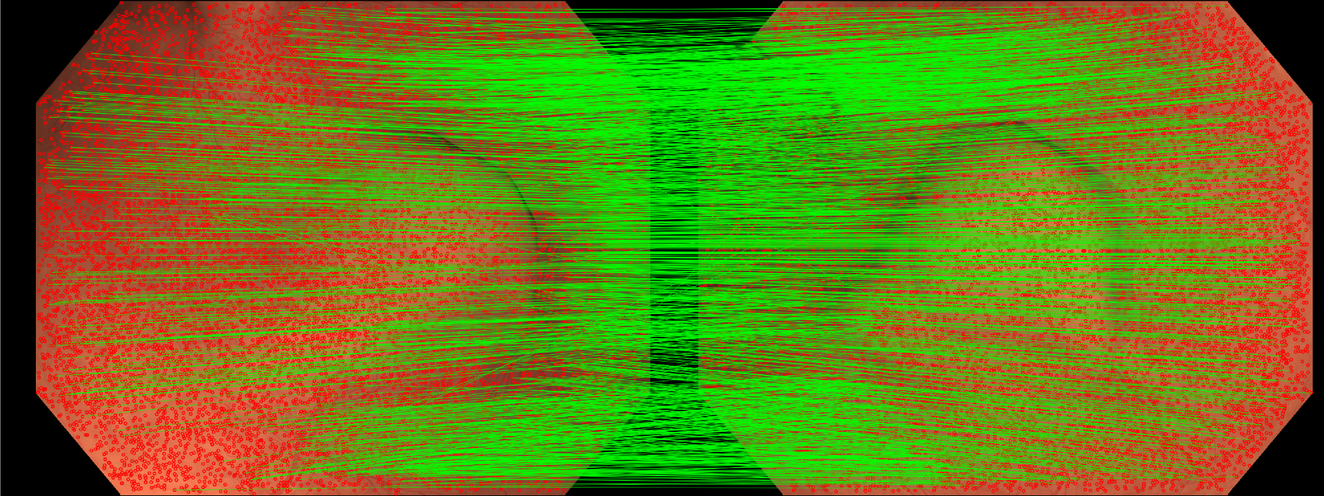
We conducted experiments using images captured by a simulated moving endoscope equipped with a monocular fish-eye camera and three surrounding light sources with a baseline of approximately 3 mm (Figure 4). The simulation was based on the real 3D mesh from [18] and employed a Lambertian illumination model.
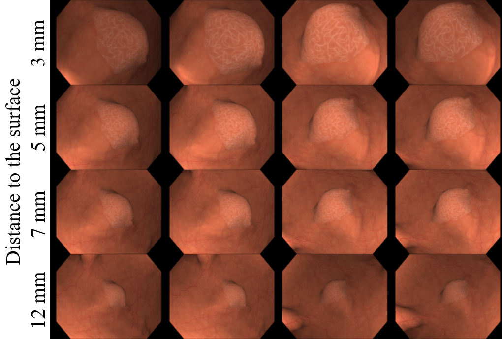
For each experiment we generated four images taken from camera positions forming a square such that the closest image point has a maximum parallax of approximately 15 degrees. The camera orientations were slightly tilted towards the center, to maximize view overlap. We tested ten different distances between the camera and the closest surface point, from 3 to 12mm, and five different orientations by rotating the four cameras around the square center. Examples of images in our dataset are provided in Figure 5.
V-B Implementation Details
V-C Accuracy as a Function of Distance to the Surface
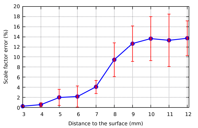
To demonstrate the practical utility and accuracy of our method, we assessed its ability to estimate the scale factor at various distances. Figure 6 shows the average scale error over the five sets of four images for the different distances tested.
We observe that the best performance, better that 1% scale error, is achieved when the closest surface point is located at 3mm from the endoscope, which corresponds approximately to the camera-light baseline. When the distance is increased, the error increases progressively. The large standard deviations shown in the plot can be explained by the fact that we are not observing a flat area, but an area containing a simulated polyp. Depending on the set of images used in each experiment, there is more polyp or more background in the images. For distances to the polyp bigger that 10mm the scale error stabilizes, probably because other colon areas closer to the camera start to appear in the images (see the last row of Figure 5), helping to improve scale accuracy.
We can see that the method works with an error of less than 10% at distances of up to 8mm, which are typical in endoscopy procedures. This confirms that the method operates well in near-field conditions, where the distances between the cameras, light sources and surface are within the same order of magnitude.
V-D Accuracy at Near-Field Ranges
To further investigate accuracy at near-field distances, Table I summarizes the mean estimation error of scale, albedo, and gain for five sets of images taken at approximately 5 mm from the surface. Previous work [16] assumed known camera poses and scene geometry. We tested this configuration with our software and achieved an error of 0.72% in estimating the real scale of the environment under these ideal conditions. In contrast, our model estimates both the camera poses and scene geometry automatically, resulting in a slightly higher error of 3.30%.
The ablation study demonstrates that scale accuracy remains similar whether or not ground truth surface normals and camera gain values are provided. However, the error decreases significantly in the scenario with the most ground truth information. This indicates that enhancing the underlying multi-view reconstruction could improve the accuracy of our scale estimation method.
V-E Impact of the Initial Guess
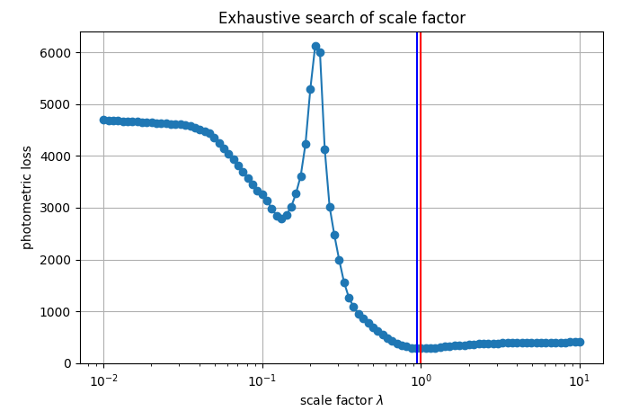
Our photometric cost function is non-linear and can exhibit multiple local minima, as illustrated in Figure 7. To ensure robustness and generality, we propose a method for computing the initial seed for non-linear optimization. Without this step, our method may get trapped in a sub-optimal solution, resulting in errors of 71% on average, as shown in Table I.
VI Conclusions
We have presented, for the first time, a fully automatic method that leverages illumination decline to obtain 3D reconstructions with real metric scale using a conventional monocular endoscope. The method assumes a calibrated light-camera setup but does not require any hardware modifications. It operates in near-field conditions, where the distances between the cameras, light sources and surface are within the same order of magnitude, which is realistic for most endoscopic situations. Our simulations show that 3% accuracy in scale recovery is achievable in practical scenarios, even with varying unknown albedo and gain.
Exploiting near-field illumination decline opens the way to real-scale visual SLAM in endoscopy. This will be crucial for many applications such as measuring polyps, stenosis, or the extent of tissues affected by a disease, and in the longer term, for automatic robotic exploration and surgery.
Our method works in two separate steps: up-to-scale geometric reconstruction using features and bundle adjustment, and photometric optimization to recover the scale. A promising direction for future research could be an integrated photometric SLAM approach that takes advantage of illumination decline to generate denser maps with true scale, even in areas of poor texture.
References
- [1] José Lamarca, Shaifali Parashar, Adrien Bartoli and José M.. Montiel “DefSLAM: Tracking and mapping of deforming scenes from monocular sequences” In IEEE Transactions on Robotics 37.1 IEEE, 2020, pp. 291–303
- [2] Ruibo Ma et al. “RNNSLAM: Reconstructing the 3D colon to visualize missing regions during a colonoscopy” In Medical image analysis 72 Elsevier, 2021, pp. 102100
- [3] José Javier Gómez-Rodríguez, José M M Montiel and J D Tardós “NR-SLAM: Non-rigid monocular SLAM” In IEEE Transactions on Robotics IEEE, 2024
- [4] Carlos Campos et al. “ORB-SLAM3: An accurate open-source library for visual, visual–inertial, and multimap SLAM” In IEEE Transactions on Robotics 37.6 IEEE, 2021, pp. 1874–1890
- [5] Richard Elvira, Juan D. Tardós and José M.. Montiel “CudaSIFT-SLAM: multiple-map visual SLAM for full procedure mapping in real human endoscopy” In arXiv preprint arXiv:2405.16932, 2024
- [6] Johannes L Schonberger and Jan-Michael Frahm “Structure-from-motion revisited” In IEEE Conference on Computer Vision and Pattern Recognition (CVPR), 2016, pp. 4104–4113
- [7] Jakob Engel, Vladlen Koltun and Daniel Cremers “Direct sparse odometry” In IEEE Transactions on Pattern Analysis and Machine Intelligence 40.3 IEEE, 2017, pp. 611–625
- [8] Jon Zubizarreta, Iker Aguinaga and Jose Maria Martinez Montiel “Direct sparse mapping” In IEEE Transactions on Robotics 36.4 IEEE, 2020, pp. 1363–1370
- [9] Robert J Woodham “Photometric method for determining surface orientation from multiple images” In Optical engineering 19.1 SPIE, 1980, pp. 139–144
- [10] Yuji Iwahori, Hidezumi Sugie and Naohiro Ishii “Reconstructing shape from shading images under point light source illumination” In IEEE International Conference on Pattern Recognition 1, 1990, pp. 83–87
- [11] Roberto Mecca, Aaron Wetzler, Alfred M Bruckstein and Ron Kimmel “Near field photometric stereo with point light sources” In SIAM Journal on Imaging Sciences 7.4 SIAM, 2014, pp. 2732–2770
- [12] Yvain Quéau, Tao Wu and Daniel Cremers “Semi-calibrated near-light photometric stereo” In Scale Space and Variational Methods in Computer Vision: 6th International Conference, SSVM 2017, Kolding, Denmark, June 4-8, 2017, Proceedings 6, 2017, pp. 656–668 Springer
- [13] Toby Collins and Adrien Bartoli “3D reconstruction in laparoscopy with close-range photometric stereo” In Int. conf. on Medical Image Computing and Computer-Assisted Intervention (MICCAI), 2012, pp. 634–642 Springer
- [14] Chenyu Wu, Srinivasa G Narasimhan and Branislav Jaramaz “A multi-image shape-from-shading framework for near-lighting perspective endoscopes” In International Journal of Computer Vision 86.2 Springer, 2010, pp. 211–228
- [15] Víctor M Batlle, José M.. Montiel and Juan D. Tardós “Photometric single-view dense 3D reconstruction in endoscopy” In IEEE/RSJ Int. Conf. on Intelligent Robots and Systems (IROS), 2022, pp. 4904–4910
- [16] Anyiel Fernandes-Araujo and José M.. Montiel “Estimación de Escala Absoluta para Structure from Motion en Endoscopio con Fuente de Luz Cercana”, 2022
- [17] Sameer Agarwal, Keir Mierle and The Ceres Solver Team “Ceres Solver”, 2023 URL: https://github.com/ceres-solver/ceres-solver
- [18] Kağan İncetan et al. “VR-Caps: a virtual environment for capsule endoscopy” In Medical image analysis 70 Elsevier, 2021, pp. 101990
- [19] Qian-Yi Zhou, Jaesik Park and Vladlen Koltun “Open3D: A modern library for 3D data processing” In arXiv preprint arXiv:1801.09847, 2018