SAMReg: SAM-enabled Image Registration with ROI-based Correspondence
Abstract
This paper describes a new spatial correspondence representation based on paired regions-of-interest (ROIs), for medical image registration. The distinct properties of the proposed ROI-based correspondence are discussed, in the context of potential benefits in clinical applications following image registration, compared with alternative correspondence-representing approaches, such as those based on sampled displacements and spatial transformation functions. These benefits include a clear connection between learning-based image registration and segmentation, which in turn motivates two cases of image registration approaches using (pre-)trained segmentation networks. Based on the segment anything model (SAM), a vision foundation model for segmentation, we develop a new registration algorithm SAMReg, which does not require any training (or training data), gradient-based fine-tuning or prompt engineering. The proposed SAMReg models are evaluated across five real-world applications, including intra-subject registration tasks with cardiac MR and lung CT, challenging inter-subject registration scenarios with prostate MR and retinal imaging, and an additional evaluation with a non-clinical example with aerial image registration. The proposed methods outperform both intensity-based iterative algorithms and DDF-predicting learning-based networks across tested metrics including Dice and target registration errors on anatomical structures, and further demonstrates competitive performance compared to weakly-supervised registration approaches that rely on fully-segmented training data. Open source code and examples are available at: https://github.com/sqhuang0103/SAMReg.git.
keywords:
Image Registration , Correspondence Representation , Segment Anything Model (SAM)1 Introduction
Both segmentation and registration are fundamental tasks in medical image analysis, enabling a wide range of clinical applications. Traditionally, a segmentation task takes one input image and searches for one or more regions-of-interest (ROIs). The segmentation is often represented by binary mask(s) or forms of ROI boundaries (e.g. point sets [1, 6] and parametric splines [36, 69]), whilst pairwise registration takes a pair of images as input and outputs a spatial that aligns one to another. Such spatial transformation can be represented by dense displacement fields (DDFs) or other parametric functions (e.g. rigid, affine and control-point-based splines).
Modern deep learning-based approaches adopted the same paradigms to learn the predictive relationships between respective input and output. Formulating segmentation and registration as machine learning tasks, recent works have increasingly exploited the inter-relationships between the two. Using multi-task learning, for example, segmentation and registration models can be jointly optimised, as two weight-sharing models (e.g. [67, 30]). The two networks can also be chained sequentially. Some used segmented ROIs as weak labels to supervise or regularise registration network training (e.g. [27, 33]). Before deep-learning was adopted in segmentation, one of the most effective segmentation methods was based on registration, which propagates segmented ROIs from segmented reference images to images-to-segment [15, 70]. The label-propagation-then-fusion idea has motivated another class of joint learning methods between the two, in which the registration was used to provide pseudo or intermediate segmentation labels, for supervising segmentation networks [26, 37]. What was commonly observed from the above-discussed work is the benefits of combining segmentation and registration, although they remain to be considered as two distinct tasks, with different forms of output, loss functions and training strategies. One of the characteristic differences, for example, is that registration often requires spatial resampling for computing unsupervised and/or weakly-supervised losses [17], whilst segmentation does not.
Previous work argued that, for a type of registration-enabled applications, predicting the ROIs on the fixed images given the ROI-labelled moving images is the practical aim of such registration, without requiring estimating unnecessary dense correspondence (those “outside” of the ROIs) or pixel-level correspondence [25]. As a result, no spatial resampling is needed in the proposed conditional segmentation algorithms. Our preliminary results summarised in a conference paper proposed a more general correspondence representation, completely based on ROI pairs, without point-to-point correspondence or those represented by parametric functions. In this work, we aim to further explore the synergy between the underlying aims of segmentation and registration, localising ROIs and correspondence, respectively.
Studying such connections between segmentation and registration facilitate many interesting discussions in the field of medical image analysis. First, we discuss in greater depth the proposed ROI-level correspondence representation, highlighting its generality and efficiency. This new representation is applicable to most if not all existing registration applications and, in its limiting cases, equivalent to DDF-predicting algorithms. It is also efficient in its versatility which allows only regions-of-registration-interest to be focused on and optimised. Further, we discuss two different approaches to estimate correspondence-representing ROI pairs and their respective probabilistic interpretations. Second, taking advantage of the recent development of vision foundation models for semantic segmentation, such as segment anything models (SAMs), it is computationally efficient to develop our proposed registration algorithms with very little to none training. Therefore, the resulting algorithms, named “SAMReg” does not require costly clinical data, with or without labels.
2 Related work
2.1 SAM-based applications other than segmentation
Unlike traditional segmentation models that require extensive training data for specific tasks, segment anything model (SAM) [34] is designed to be highly adaptable in natural image segmentation and can perform various segmentation tasks with minimal or no additional training. Its state-of-the-art performance on various benchmarks has led to its application in fields such as medical imaging [38, 13], autonomous driving [14], and remote sensing [47, 62] segmentation. Beyond its role in segmentation, SAM functions as a 2-dimensional dense prediction tool, offering broader applications in various computer vision tasks.
One notable application is its ability to extend 2D clusters into additional dimensions. For instance, by incorporating a third spatial coordinate, SAM enriches the semantic understanding of each 2D slice, enhancing tasks such as depth estimation [55, 45] and 3D reconstruction [11, 8]. Similarly, when applied to the temporal dimension, SAM’s ability to capture regions of interest (ROIs) across sequential frames aligns with the requirements of object tracking [68, 64]. Another key application lies in adaptive local processing. SAM can be used to selectively manipulate certain objects in image synthesis or to enhance local image quality, offering precise and context-aware improvements within images [20, 9]. In this paper, SAM is applied to image registration tasks, where two or more images are taken as input, and the output is a correspondence rather than semantic segmentation.
2.2 Learning-based image registration
Both rigid and deformable image registration seek to establish correspondences between two or more images to achieve alignment. These correspondences are typically represented using one of two approaches: parametric transformations or Dense Displacement Fields (DDFs) [17]. Parametric functions, such as rigid, affine, and spline-based transformations [50], describe the mapping of specific locations between images. DDFs, as a more general representation, define the transformation for each paired sample and are often learned through learning-based registration algorithms [17].
Early learning-based registration methods utilized supervised learning, where the deformation field was obtained either manually or from classical approaches [49]. VoxelMorph was among the first to introduce unsupervised learning for brain MRI registration [3], while LabelReg pioneered weakly-supervised learning for high-fidelity alignment of anatomical landmarks [27]. Subsequent research expanded these paradigms, exploring diverse network architectures [35], loss functions [10, 43], and transformations incorporating inverse consistency or symmetric constraints [33, 60, 48]. However, these learning-based methods faced challenges with hyperparameter tuning, requiring retraining for each new regularization setting, which limited their flexibility. This limitation led to the development of foundation models like UniGradICON [59], which is trained with a fixed set of hyperparameters across multiple datasets and can handle both in-distribution and out-of-distribution tasks without retraining. However, UniGradICON requires heavy pre-training efforts and specific fine-tuning when applied to unseen domains.
Recent efforts in either utilising large foundation models [28, 57, 54] and developing registration-specific foundation models [60, 66] have been reported. Majority existing work aim to predict inter-image correspondence represented by pixel-level displacements or parametric spatial transformation functions, thus requiring spatial resampling during training or fine-tuning to compute loss function values, a distinct difference from the work presented in this paper.
2.3 Our preliminary conference presentation
This work extends our preliminary results presented in MICCAI 2024. In this extended version, we adopt a novel perspective in introducing the representation and provide a more detailed algorithmic description, such as the adaptation details from 2D SAM to 3D registration with pseudo code, as well as newly added discussion and derivation on probabilistic interpretation of the segmentation-registration connection. In addition, the evaluation has been expanded to include five datasets, including both 2D and 3D data sets across various modalities such as RGB, MRI, and CT. All ablative quantitative and qualitative results have been updated and are exclusive to the current study. Moreover, a new discussion section has been included to share our experience in designing and experimenting with the new correspondence representation and its applications. In summary, our main contributions are as follows:
-
1.
We pioneer a new correspondence representation for image registration using a set of corresponding paired ROIs.
-
2.
We introduce the general-purpose SAMReg by segmenting corresponding ROIs from both images, without requiring any labels or fine-tuning. To our knowledge, it is the first application of SAM in image registration. Code and demo are available at https://github.com/sqhuang0103/SAMReg.git.
-
3.
Experimental results on multiple real clinical data sets show that our methods outperform the widely adopted registration tools and unsupervised registration algorithms, and is competitive with weakly-supervised registration approaches that require additional labeled training images.
3 Method
3.1 One Registration is Worth Two Segmentations
3.1.1 A brief perspective on ROI-based correspondence
For a given class , a segmentation task is to estimate the binary class probability vector over voxel locations, thereby forming a segmented region of interest (ROI) . When applied to two images and , the segmentator identifies the ROIs and , of the same class , in each image. These two ROIs, representing the same segmentation class, are considered corresponding, thereby locally registering and - local to the ROIs.
In multi-class segmentation, which estimates joint probability of a random vector for -class probabilities, the corresponding segmented ROI sets and , derived by binarizing the estimated class probability vectors with , exhibit a more comprehensive correspondence between and . It is a sufficiently comprehensive representation form for both local and global correspondence, with ROIs with smaller and larger areas/volumes, respectively, as well as for both sparse and dense correspondence, determined by the spatial distribution of these pairs of ROIs. In this section, we formalize such a ROI-based correspondence representation and discuss registering and by estimating this correspondence.
3.1.2 Representing correspondence in image registration
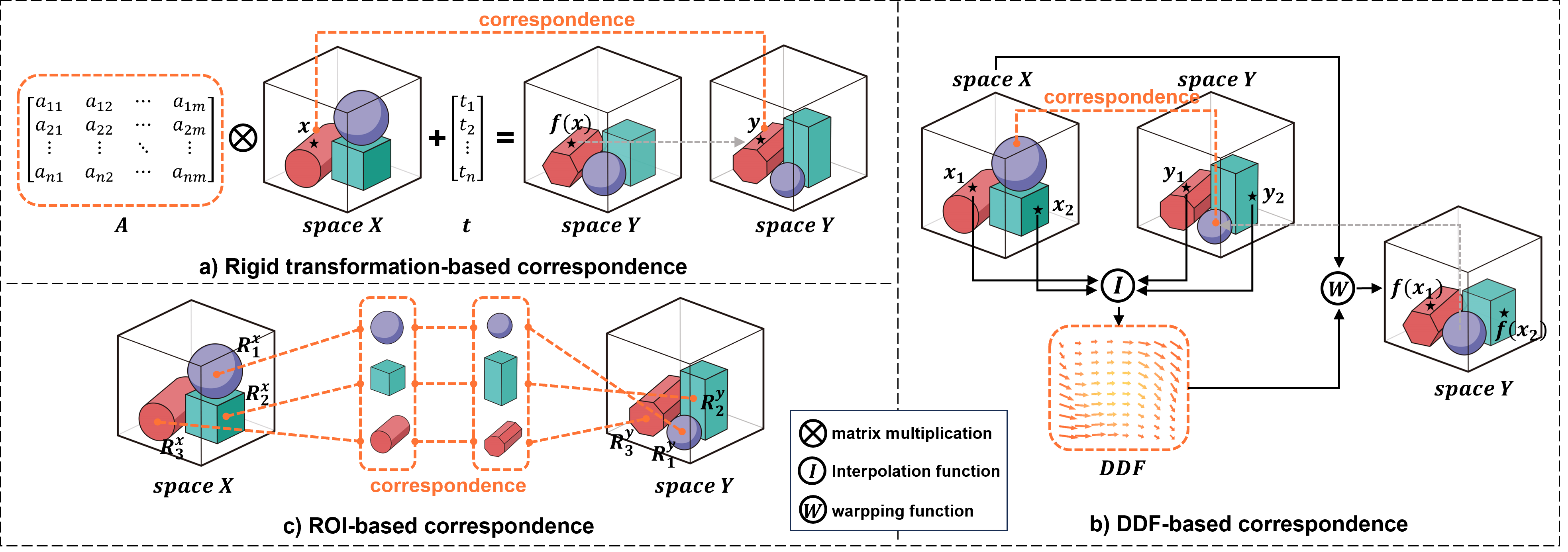
The spatial correspondence is considered to be a mapping from spatial locations X in a moving image coordinate system to locations Y in the fixed image coordinate system. For example, in 3D, and are random vectors (to estimate) containing Euclidean coordinates, in the x-, y- and z-directions.
Existing methods for representing spatial correspondence, denoted as , are categorized into two primary types: transformation functions of spatial locations or paired samples. The first category includes parametric functions such as rigid, affine, and spline-based transformations (for example, thin-plate splines [50]). These functions transform input vectors x into coordinate vectors y using the mapping . A specific example, illustrated in Fig. 1a, is the rigid transformation defined by , where is a rotation matrix and t is a translation vector. The second category involves representing correspondence through pairs of corresponding locations where . For instance, DDF (shown in Fig. 1b) is represented by where is a set of vectors defined on all voxel locations . To estimate y corresponding to x, at spatial locations other than the voxels , interpolation is often used (bi-/tri-linear interpolation in this case). Sparser samples can also be used, also known as control points, such as those in free-form deformation [53], with which denser correspondence can be interpolated by B-splines [52]. The so-called point-feature-based registration may be considered in this category, using a set of sparse and scattered (not sampled on a regular grid) control points (a.k.a. a point cloud), e.g. iterative closest point (ICP) [2] and coherent point drift (CPD) [44].
3.1.3 ROI pairs is a sufficient representation
Theorem 1
Denoting x and y as spatial locations at respective moving- and fixed image spaces (i.e. coordinate systems), it is sufficient for pairs of regions-of-interest (ROIs), denoted as , to indicate any spatial correspondence between the two image spaces, if is sufficiently large, where and are two sets of and spatial locations that represent the same (corresponding) ROIs, in the moving- and fixed image spaces, respectively.
This is intuitive in an extreme case, in which a large number () of single-voxel (i.e. ) ROIs are used, the ROI pairs can be sampled at voxels to an equivalent DDF representation .
Therefore, an informal proof for Theorem 1 can be understood by iterating the following process until sufficiency is reached: When a point-to-point correspondence that has NOT been represented by current ROI pairs , one can always sample an additional ROI pair , such that the resulting () ROI pairs suffice.
3.1.4 Properties of ROI-based correspondence
We discuss the properties of the proposed ROI-based correspondence as follows:
-
1.
Paired ROIs are practically convenient as binary mask pairs, a well-established data structure in many computational libraries;
-
2.
Paired ROIs can represent both local or global correspondence, if they are sufficiently small or large, respectively;
-
3.
Paired ROIs can represent both dense and sparse correspondence, dependent on the potentially pre-configurable spatial distribution of the ROI locations;
-
4.
If dense-level correspondence is required, denser than available ROIs, the correspondence at any spatial location can be inferred. We discuss specific algorithms in Sec. 3.2.2;
-
5.
If dense-level correspondence is not required, omitting smoothness regularization (e.g., rigidity or bending energy) during estimation may improve registration efficiency with ROI-based representation;
-
6.
Correspondence can be established between (e.g. multi-modal) images, only on identifiable and/or confidently-estimated corresponding regions, without mandating interpolation in uncertain locations;
-
7.
Paired ROIs are relatively independent of varying image resolutions;
-
8.
Overlapping ROIs facilitate one-to-many correspondence;
-
9.
Paired ROIs have potentials to readily represent uncertainty in correspondence, by using overlapping ROIs in both images or by not using any.
3.1.5 The perceived need for pixel-level correspondence
In this section, we challenge the requirement for pixel-level dense correspondence or the spatial ubiquity in parameterising correspondence, which the proposed ROI-based representation is, although capable of, often not imposing.
As discussed in the previous work [28], many clinical applications are interested in one or multiple local regions to be registered, for example, a tumour location before and during a surgical intervention and the same anatomical structures between subjects. It is debatable that, when a global transformation or a regularised local transformation is used, the goodness-of-alignment at regions-of-clinical-interest should not be a trade-off with locations elsewhere which may or may not contain more salient corresponding features that are easier to align. In these applications, such pixel-level or spatially-continuously-defined correspondences are a by-product of the adopted representation, rather than a requirement. Furthermore, enforcing these types of dense correspondence could negatively impact the registration performance measured by downstream tasks. This may partly explain the benefits observed in our experiments, presented and discussed further in Sec. 6.
For applications that indeed require denser correspondence, such as image warping and those for visualisation purposes, general-purposed algorithms are proposed for converting ROI-based correspondence to DDFs in Sec. 3.2.2.
3.1.6 Probabilistic ROI-pair-predicting image registration
In this section, we consider two cases to search corresponding ROI pairs , and their probabilistic links to two independent multi-class segmentation of and .
Case 1: Segmenting the Same ROIs from Two Images
In this case, the segmentation algorithm (e.g., one or two neural networks) is capable of segmenting the same classes C of ROIs in both images and . Following notations in Sec. 3.1.1, obtaining ROI pairs becomes to estimate the joint probability given two images , where
| (1) |
which is subject to normalization constants. This approach typically requires predefined ROI types and segmentation networks trained on ROI-labeled datasets. Notably, these segmented training datasets are exactly the same as those required for training weakly-supervised registration algorithms [27, 25].
Case 2: Matching the Segmented ROI Candidates
Assume an alternative“unsupervised” segmentation can be used to obtain two independent sets of candidate ROIs. Specifically, for image , a random vector representing -class probabilities is estimated, and the segmented ROI set is derived by thresholding the estimated class probability vectors , where . A similar process is applied to image , yielding ROIs via estimating -class probabilities , where is the class probabilities for classes. To identify corresponding ROI pairs, we search for the subset of common ROI classes C, which corresponds to the joint probability between the two conditionally independent “candidate probabilities”, i.e.,
| (2) |
In the following Sec. 3.2, we propose a specific algorithm using this strategy.
3.2 Segment Everything and Match: A Training-Free Registration Algorithm
3.2.1 Revisiting SAM
SAM [34] has become a significant milestone in computational vision, extending the capabilities of Large Language Models (LLMs) into visual analytics. SAM’s architecture, which integrates an image encoder, a prompt encoder, and a mask decoder, is adept at image segmentation without task-specific training, leading to the development of several enhanced variants [12, 31]. In recent developments, SAM has been tailored for medical imaging applications through training on extensive medical datasets [38, 13], thereby serving as a foundational model. Further refinement has been achieved by fine-tuning on specific medical datasets, incorporating additional adaptation layers [65, 21, 63]. However, as noted by various researchers, SAM’s performance in medical imaging shows variable sensitivity compared to natural images [29, 39], leading to inconsistent results in directly segmenting specific anatomical structures and pathologies without additional training.
It is interesting to argue that known anatomical significance in segmentation may not be required for medical image registration. Although correspondence between medical images may be application-dependent to some extent [16], the primary goal is to identify similar regions across images, without necessarily clearly predefined or even known anatomical or pathological definitions for these regions. This goal may be achieved by SAM’s more general-purposed feature extraction and pixel classification abilities, compared with that from registration-task-specific models. For instance, SAM effectively segments ROIs with distinct boundaries, such as the iliac internal and shared cavities between structures, which, while not typical targets, are valuable for establishing local correspondence. By utilizing SAM to delineate extensive foreground areas, our objective is to identify corresponding ROIs that encompass a variety of imaging structures and regions, even those without precise clinical meaning.
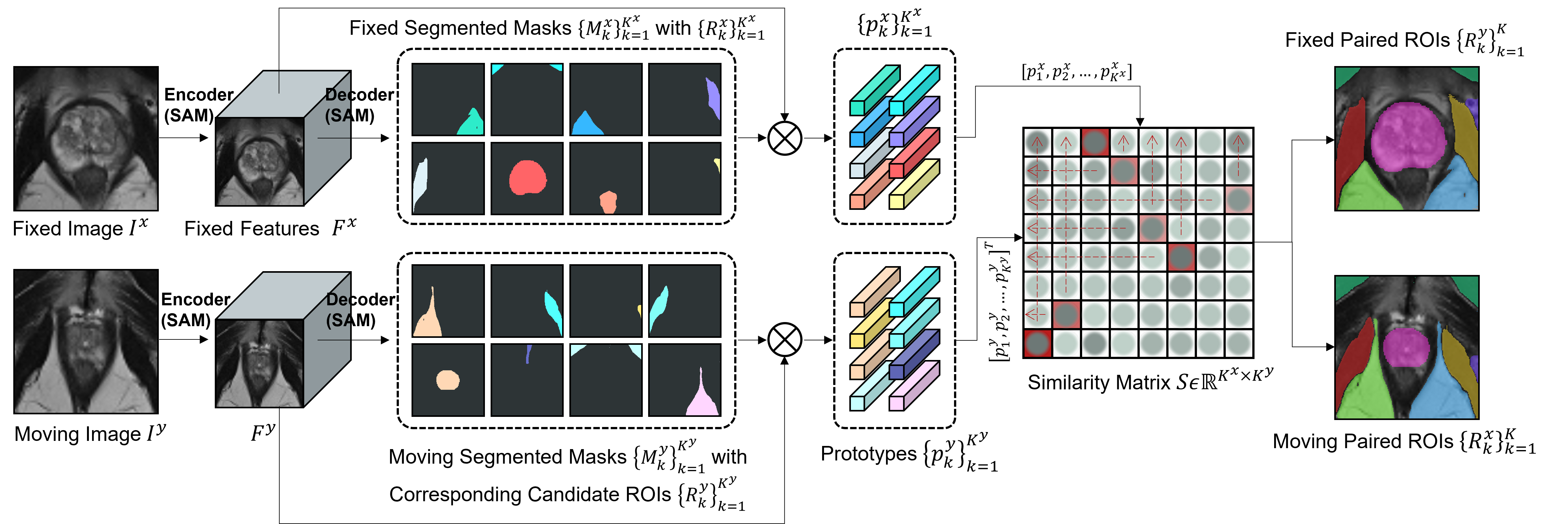
3.2.2 The SAMReg algorithm
Since the multiple ROIs generated by SAM segmentation lack explicit ‘one-to-one’ correspondence, our strategy involves embedding these ROIs before matching them in feature space, with the proposed pipeline illustrated in Fig. 2.
ROI Embedding
Leveraging the image encoder in SAM, we embed the 2D input images into features :
| (3) |
By setting the SAM decoder to the everything mode with a simple outlier-removing filter (further details in Sec. 4), we exploit its generality to produce a comprehensive array of segmentation masks for each image, obtaining mask sets , representing ROIs :
| (4) |
For 2D registration tasks, paired 2D images and yield respective mask sets and . For 3D registration tasks, as detailed in algorithm 1, paired 3D images and , each sized , are processed as a series of 2D slices and respectively. Each segmented ROI in , corresponds to an ROI potentially located in any slice . To optimize computation, we empirically limit our focus to a range of slice , sequentially processed to generate a comprehensive set , which are then matched with .
For each binary mask, we derive moving prototypes by element-wise multiplying the mask with their corresponding feature map, element being indexed by :
| (5) |
where indicates resizing to the size of as . This also applies on the fixed prototypes .
ROI Matching
Assume a similarity matrix to measure the cosine similarity between the moving- and fixed prototypes:
| (6) |
where normalises each element , and .
A set of index pairs then can be identified:
| (7) |
corresponding to pairings with maximum similarities. A further constraint is added to ensure each is chosen at most once, if overlapping ROIs segmented from SAM are filtered. is the similarity threshold and the set length
| (8) |
Using the selected index pairs , we construct two new sets of masks, and :
| (9) |
If exact ‘one-to-one’ mapping of correspondences is not strictly necessary, each may be selected repeatedly. The value of is established based on the threshold .
Further, as discussed in Sec. 3.1.6, we explore the capability of converting estimated ROI-based correspondence to its dense counterpart DDF, useful for applications such as full-image alignment. In this section, we describe a general method that iteratively refines the DDF, to optimize an objective function that combines a region-specific alignment measure with a regularization term to ensure smooth interpolation:
| (10) |
where represents parameters of the transformation function and is a regularization parameter that balances the alignment accuracy with the deformation smoothness. This may itself be considered a multi-ROI alignment algorithm but with known ROI correspondence. In this work, we use an equally-weighted MSE and Dice as and a L2-norm of DDF gradient as .
| Method | 3-Dimensional Data | 2-Dimensional Data | ||||||||
|---|---|---|---|---|---|---|---|---|---|---|
| Prostate | Cardiac | Lung | Retina | Aerial | ||||||
| Dice | TRE | Dice | TRE | Dice | TRE | Dice | TRE | Dice | TRE | |
| NiftyReg [42] | 7.68±3.98 | 4.67±3.48 | 9.93±2.21 | 3.29±2.89 | 10.93±2.02 | 4.23±1.64 | 8.77±3.29 | 5.53±3.52 | 10.21±3.02 | 4.02±2.25 |
| VoxelMorph∗ [3] | 56.84±3.41 | 3.68±1.92 | 60.10±3.95 | 3.03±2.41 | 77.98±2.72 | 3.23±0.81 | 53.12±3.46 | 4.66±3.43 | 72.73±2.41 | 3.53±1.3 |
| LabelReg∗ [27] | 77.32±3.56 | 2.72±1.23 | 78.97±2.42 | 1.73±1.34 | 83.56±2.43 | 1.52±0.86 | 69.73±3.51 | 2.69±3.21 | 83.35±2.14 | 3.51±1.47 |
| SAMReg w SAM [34] | 75.67±3.81 | 2.09±1.35 | 80.28±3.67 | 1.43±1.13 | 85.23±2.16 | 1.31±0.91 | 61.79±3.42 | 3.44±3.35 | 84.13±2.23 | 3.46±1.61 |
| SAMReg w MedSAM [38] | 70.32±3.52 | 2.11±1.66 | 78.66±3.71 | 1.59±1.21 | 86.34±2.12 | 1.27±0.83 | 64.33±3.38 | 2.89±3.27 | 82.19±2.56 | 3.99±1.52 |
| SAMReg w SAMed2D [13] | 71.23±3.77 | 2.49±1.17 | 77.52±3.75 | 1.65±1.29 | 86.16±2.15 | 1.27±0.86 | 63.10±3.35 | 2.81±3.22 | 80.72±2.98 | 3.96±1.76 |
| SAMReg w SAMSlim [12] | 70.66±3.79 | 2.48±1.24 | 78.23±3.67 | 1.57±1.20 | 84.53±2.34 | 1.42±0.90 | 60.53±3.46 | 3.40±3.31 | 83.52±2.20 | 3.49±1.62 |
| SAMReg w SAMHQ [31] | 73.83±3.69 | 2.26±1.15 | 79.11±3.62 | 1.48±1.15 | 84.78±2.29 | 1.39±0.89 | 61.87±3.39 | 3.36±3.28 | 85.17±2.18 | 3.36±1.59 |
4 Experiments
4.1 Datasets
We evaluate our method in five medical and non-medical datasets: Prostate [5, 22, 46, 18], Cardiac [4], Lung [23], Retina [32] and Aerial [40]. The first three datasets are three-dimensional and contain MR and CT imaging modalities, while the latter two are two-dimensional.
- 1.
-
2.
Cardiac: Consisting of exams acquired at the University Hospital of Dijon and proposed in Automated Cardiac Diagnosis Challenge (ACDC) challenge [4]. Each clinical exams contains pair MR cardiac images from different frames.
-
3.
CT-Lung: Consisting of pairs of FBCT images from the Learn2Reg 2021 challenge [23], this dataset provides images capturing both expiratory and inspiratory states.
-
4.
Retina: comprising retinal image pairs from Fundus Image Registration Dataset (FIRE) [24], acquired at the Papageorgiou Hospital, Aristotle University of Thessaloniki, Thessaloniki from patients.
-
5.
2D-Aerial: comprising pairs of QuickBird-acquired images of the city of Zurich [40]. Image side lengths ranges from to .
Further, to ensure a fair comparison with other supervised registration methods, 80% of the images are utilized for training purposes. Our method does not use this subset for training and is tested on the remaining data alongside other methods. For ablative experiments, the proposed algorithms are tested using all available images.
4.2 Implementation Details
For non-rigid registration [19, 58], we assess accuracy by the mean transformation accuracy of critical anatomical structures [27], with TRE calculated from the centroids of the relevant ROIs.
In configuring the SAM parameters, beyond the standard settings, we adjusted the pred-iou-thresh and stability-score-thresh to 0.90. Our ROI filtering strategy is based on area and overlapping ratio, setting a minimum of 200 and a maximum of 7000 for the ROI sizes and a maximum ratio of 0.8. The similarity threshold is set to 0.8. Our network is implemented with pytorch and MONAI [7].
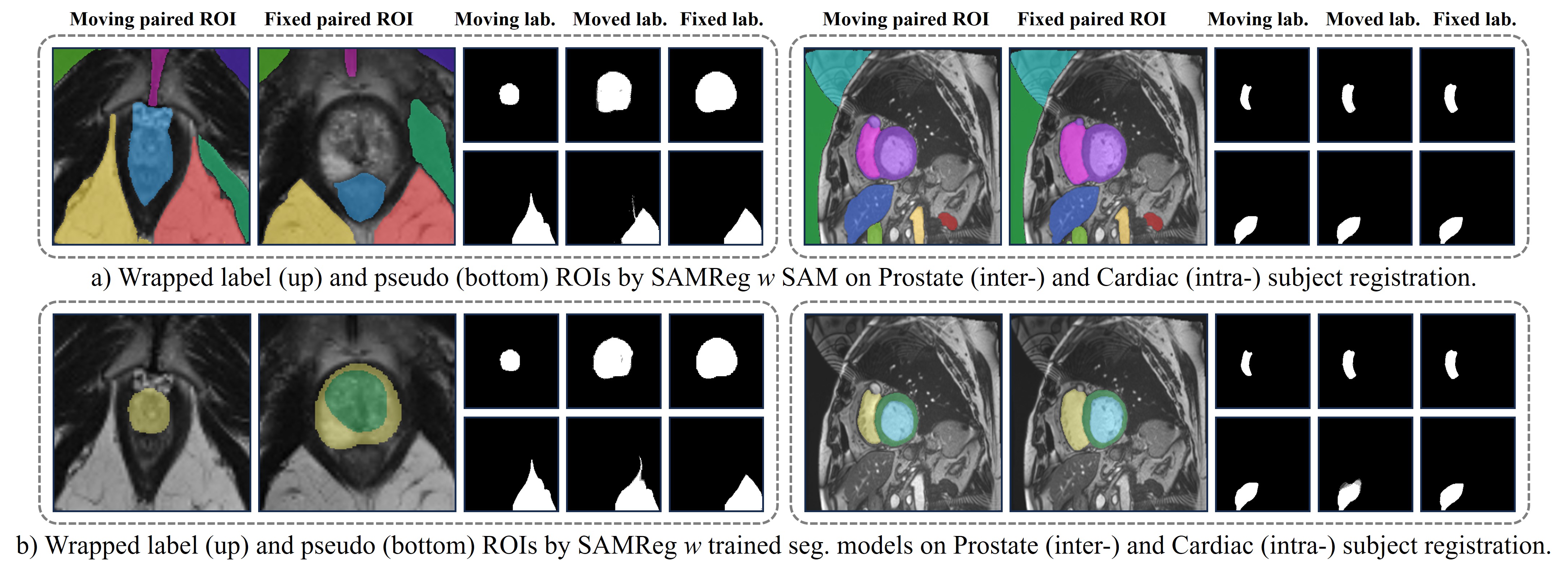
| Dataset | Segmentation Algorithm | Label ROI | Pseudo ROI | ||
| Dice | TRE | Dice | TRE | ||
| Prostate [5] | SAM [34] | 75.67±3.81 | 2.72±1.23 | 97.73±3.15 | 0.82±0.23 |
| Vnet [41] | 80.35±3.14 | 2.13±1.24 | 43.56±3.54 | 5.32±2.51 | |
| Cardiac [4] | SAM [34] | 80.28±3.67 | 1.73±1.34 | 98.87±3.61 | 1.62±0.67 |
| FCT [61] | 83.54±3.46 | 1.31±1.25 | 31.35±3.74 | 7.24±2.52 | |
| Lung [23] | SAM [34] | 85.23±2.16 | 1.31±0.91 | 98.94±3.24 | 1.31±0.63 |
| MedNeXt [51] | 89.78±3.19 | 1.10±1.45 | 50.16±3.27 | 5.01±2.33 | |
5 Results
5.1 Comparison with SOTA Registration Methods
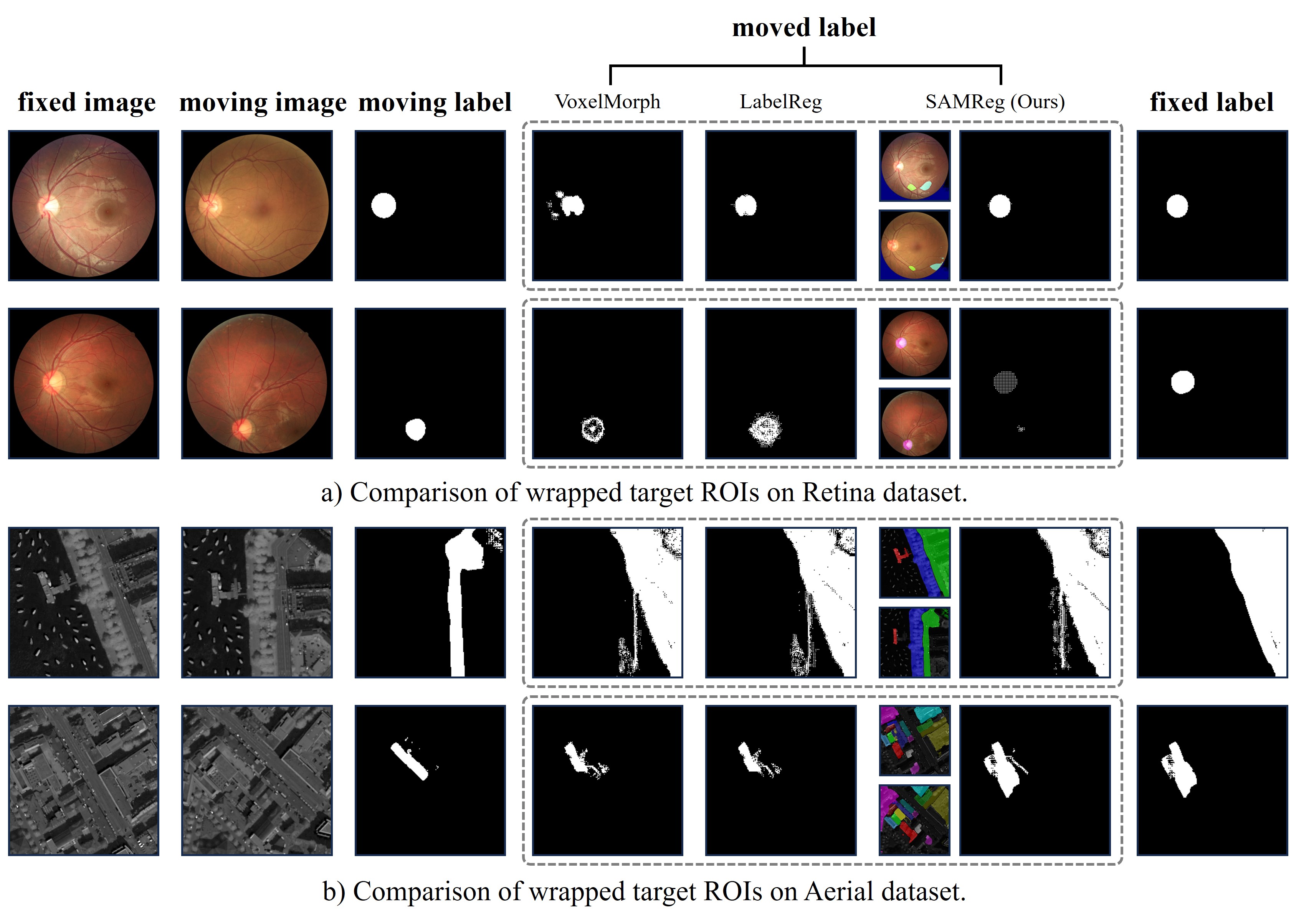
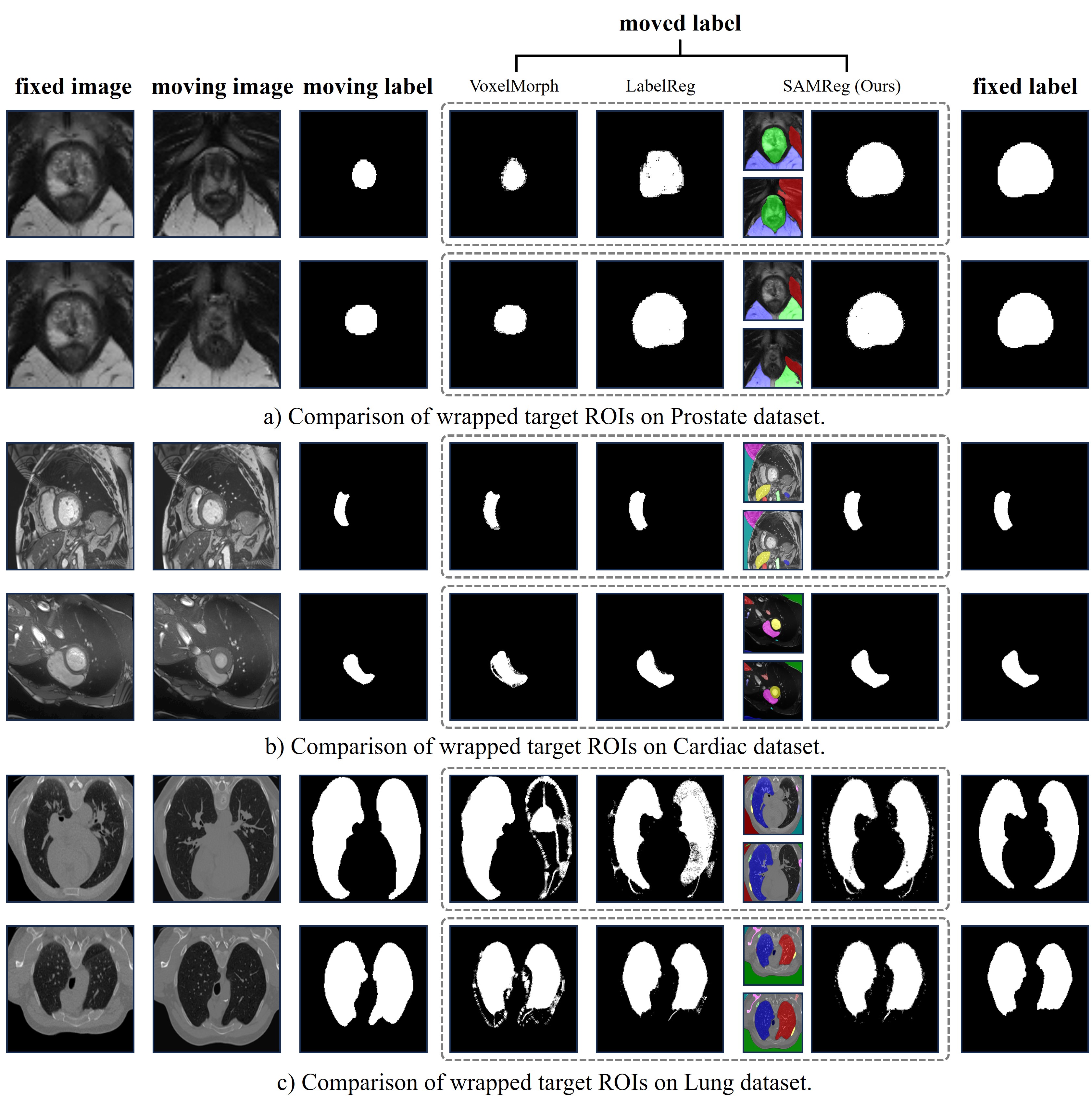
In our comprehensive evaluation (Table 1), we present comparisons between the proposed SAMReg’s performance against both non-learning and learning-based medical image registration techniques on both 2D and 3D datasets. SAMReg is fairly compared with NiftyReg [42], a well-established registration tool for non-learning iterative algorithms, without using any training data for both. It is also compared with adapted methods including VoxelMorph [3] and LabelReg [27], although these methods require training data. VoxelMorph and LabelReg are fine-tuned on the specific datasets, employing unsupervised and weakly-supervised learning respectively, with LabelReg also depending on anatomical annotations. SAMReg achieves competitive performance, particularly in intra-subject cardiac and lung registrations. While Dice scores in inter-subject prostate registrations might be marginally lower than the learning-based methods, SAMReg’s competitive TRE suggests a focus on achieving high local alignment accuracy. This might be due to the inherently larger anatomical variations present in the prostate glands compared to organs like the hearts or lungs. Notably, SAMReg’s success without dataset-specific training underscores its generalizability across different medical imaging domains. This translates to reduced complexity during clinical deployment, making SAMReg a compelling alternative for real-world registration tasks. Furthermore, SAMReg demonstrates promising performance on non-medical image registration tasks, as exemplified by aerial image datasets, suggesting that SAMReg’s ability to capture salient image features and establish correspondences relatively independent of specific domains, potentially offering broader applicability in computer vision tasks and more robust to data differences in medical imaging domain.
Qualitative comparisons of 2D and 3D registration results are shown in Figs. 3 and 4, respectively. These figures illustrate SAMReg’s capacity in transforming anatomical structures during the registration process. Compared to other registration methods, DDF derived from multiple ROI pairs enables accurate warping of the moving label to closely match the fixed label, even if the ROI is unseen by SAMReg.
5.2 Comparison of SOTA ROI Segmentation Algorithms
SAMReg’s performance can be further enhanced through the direct integration of advanced segmentation algorithms. This can be implemented in two ways: 1) dataset-specific integration – for specific datasets and tasks, SAMReg can be efficiently combined with other pre-trained segmentation models optimized for that domain. 2) general-purpose integration – for broader applicability, SAMReg can be effectively utilized with SAM-based segmentation, offering a generic solution for various registration tasks.
5.2.1 Impact of Dataset-Specific SOTA Segmentation Models
Table 2 and Fig. 5 present a comparison of ROI segmentation and subsequent registration alignment performance using SAM [34] compared to leading segmentation models in specific datasets [41, 61, 51]. While the benchmark models benefit from being specifically designed and trained on ground-truth annotations (label ROI), SAM-based SAMReg demonstrates robustness and effectiveness in registration tasks. Notably, SAM achieves competitive performance, even surpassing these models, when utilizing SAM-generated pseudo ROIs (pseudo ROI) instead of ground-truth data. Furthermore, SAM’s extensive ROI detection capabilities demonstrably enhance image analysis and strengthen the overall registration accuracy. This is particularly evident when dealing with previously unseen ROIs, highlighting SAM-based SAMReg’s interesting advantage in handling both predefined and unknown ROIs effectively.
5.2.2 Impact of SAM-Based SOTA Segmentation Models
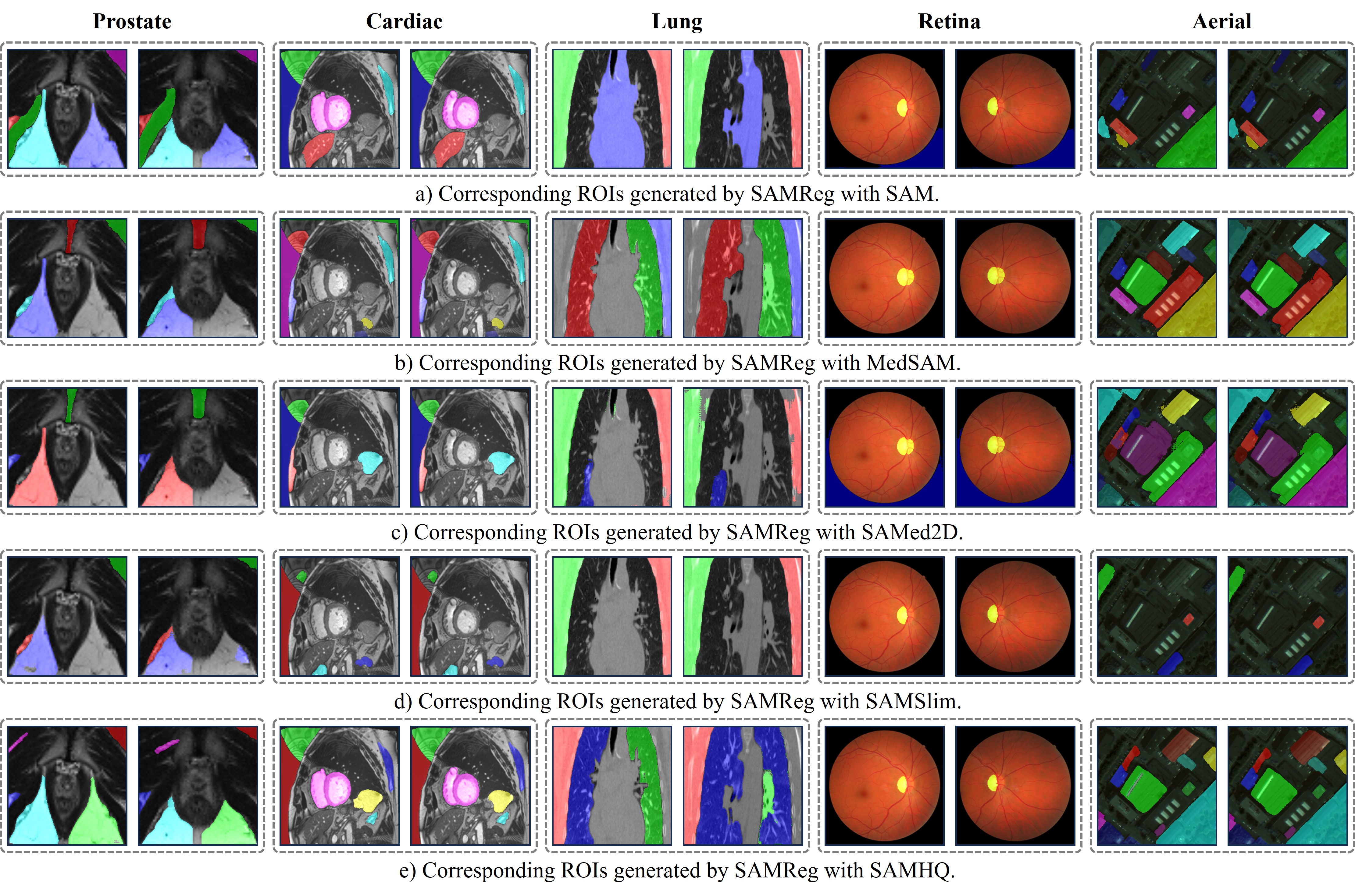
Table 1 compares the SAMReg performance with SAM [34] and its variants when applied to medical and general registration tasks. Specifically, MedSAM [38] and SAMed2D [13] are fine-tuned on large medical datasets for medical fundamental segmentation, while SAMSlim [12] and SAM_HQ [31] are modified for enhancing general segmentation performance. Examples of the segmented and paired ROIs of these SAM variants on all five datasets are visualized in Fig. 6.
The results demonstrate that medical SAM variants achieve more accurate anatomical structure segmentation compared to general SAM variants, leading to better alignment performance. Conversely, general SAM variants outperform medical SAM variants in non-medical registration tasks, i.e., aerial dataset. Interestingly, the basic SAM consistently achieves competitive results across all tasks, which may be due to the fact that it generates more paired ROIs as illustrated in Fig. 6.
| Quantity Threshold | Intra-Subject: Cardiac | Inter-Subject: Prostate | ||
|---|---|---|---|---|
| Dice | TRE | Dice | TRE | |
| 1 | 73.31±3.87 | 1.73±1.34 | 69.59±3.51 | 3.85±1.59 |
| 3 | 78.26±3.77 | 1.58±1.21 | 76.00±3.23 | 2.06±1.37 |
| 5 | 79.45±3.72 | 1.47±1.18 | 75.36±3.78 | 2.11±1.39 |
| 7 | 78.38±3.76 | 1.49±1.20 | 74.75±3.80 | 2.15±1.37 |
| 9 | 77.31±3.79 | 1.52±1.25 | 72.89±3.83 | 2.22±1.41 |
| Similarity Threshold | Intra-Subject: Cardiac | Inter-Subject: Prostate | ||
|---|---|---|---|---|
| Dice | TRE | Dice | TRE | |
| 0.7 | 78.69±3.73 | 1.70±1.29 | 74.41±3.91 | 2.19±1.39 |
| 0.75 | 80.21±3.69 | 1.54±1.16 | 75.13±3.87 | 2.13±1.36 |
| 0.8 | 80.28±3.67 | 1.43±1.13 | 75.67±3.81 | 2.09±1.35 |
| 0.85 | 79.11±3.70 | 1.65±1.20 | 75.45±3.79 | 2.11±1.33 |
| 0.9 | 77.93±3.72 | 1.72±1.23 | 74.12±3.84 | 2.12±1.34 |
| 0.95 | 77.12±3.77 | 1.73±1.21 | 73.80±3.86 | 2.17±1.38 |
| Slice Range (±) | Intra-Subject: Cardiac | Inter-Subject: Prostate | ||
|---|---|---|---|---|
| Dice | TRE | Dice | TRE | |
| 1 | 71.23±3.54 | 2.16±1.34 | 61.29±4.05 | 2.77±1.53 |
| 3 | 73.58±3.59 | 1.84±1.26 | 65.12±4.01 | 2.65±1.59 |
| 5 | 76.37±3.44 | 1.67±1.21 | 68.59±3.97 | 2.52±1.52 |
| 7 | 78.55±3.51 | 1.58±1.23 | 71.66±3.92 | 2.28±1.45 |
| 9 | 79.13±3.62 | 1.49±1.19 | 73.54±3.91 | 2.15±1.39 |
| 11 | 80.28±3.67 | 1.43±1.13 | 75.67±3.81 | 2.09±1.35 |
5.3 Ablative Study on ROI Selecting Strategies
In this section, we conduct an ablative study on ROI selecting strategies, which define the correspondence and the subsequent generated DDFs, testing different threshold values in similarity or quantity, as described in Sec. 3.2.2.
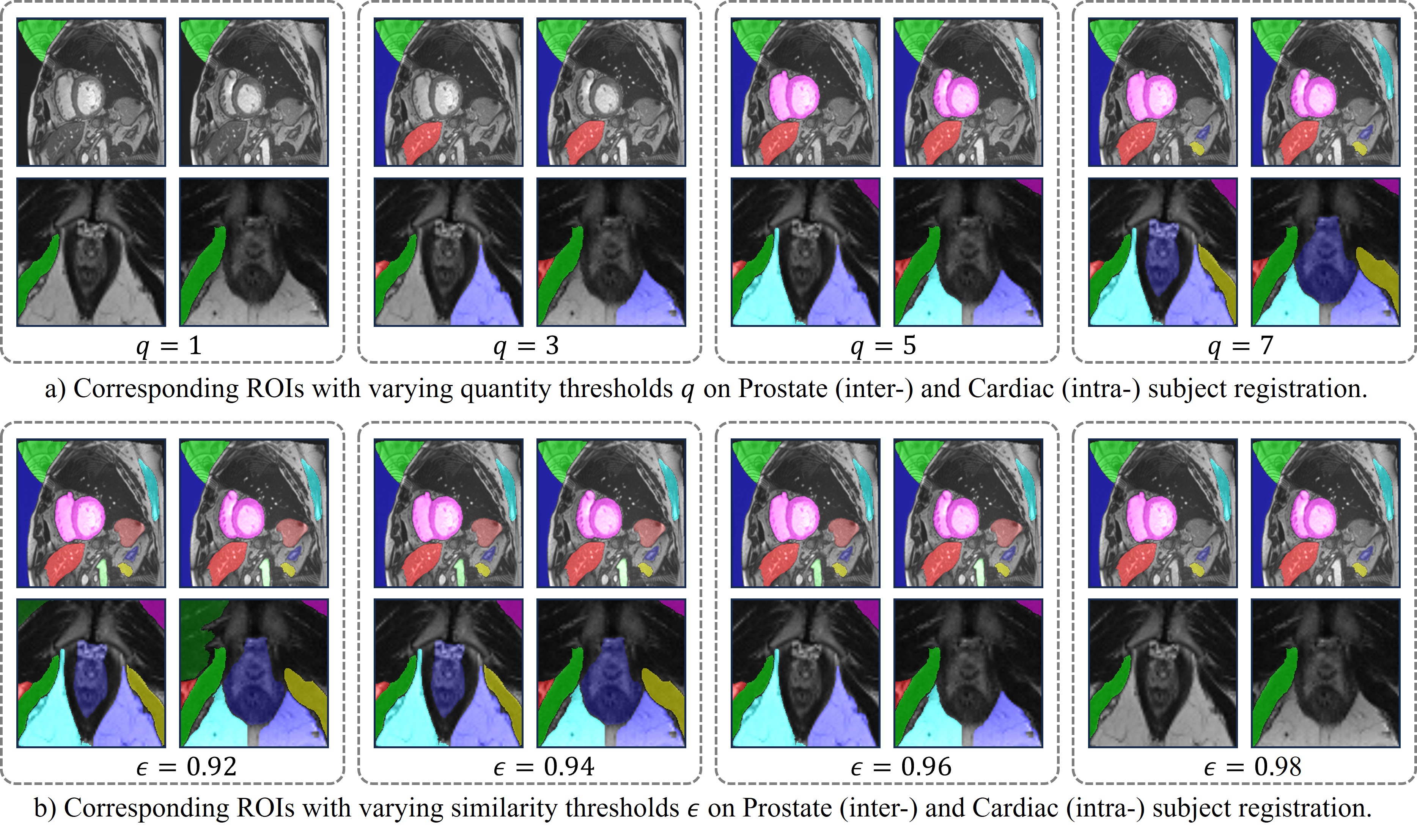
5.3.1 Impact of Quantity Threshold
The impact of the number of selected ROI pairs on registration performance is evaluated. Table 4 shows Dice and TRE scores for different quantities of ROI pairs on intra-subject (cardiac) and inter-subject (prostate) datasets. The results suggest that using up to five ROI pairs achieves the best registration performance on prostate dataset and three on cardiac dataset, suggesting that a relatively small number of well-chosen ROIs can be sufficient for achieving optimal registration. As shown in Fig. 7a, additional paired ROIs improve the entire image alignment but introduce inherent noise due to segmentation model limitations.
5.3.2 Impact of Similarity Threshold
We further investigate the effect of a similarity threshold on ROI selection. Table 4 illustrates the Dice and TRE scores for different similarity thresholds on intra-subject (cardiac) and inter-subject (prostate) datasets. The results demonstrate that a 0.8 similarity threshold leads to the best overall registration performance, indicating that using ROIs with a moderately high degree of similarity ( 0.8) between the source and target images is beneficial for registration. A higher threshold ( 0.8) reduces the number of selected ROIs, potentially leading to a lack of sufficient corresponding information for accurate registration, reflected in the higher TRE scores. Conversely, a lower threshold might incorporate too many ROIs with potentially irrelevant similarities, as illustrated in Fig. 7b, hindering the registration performance. These findings underscore the importance of striking a balance between the similarity and quality threshold of selected paired ROIs for achieving optimal registration results.
5.4 Ablative Study on ROI Mapping Strategies
The inherent uncertainty in segmented ROIs necessitates exploring appropriate correspondence representation, such as ‘one-to-one’ matching or allowing ‘one-to-many’ relationships. Furthermore, instead of identifying the ‘corresponding slices’ for 2D ROI alignment, the corresponding ROIs are searching within a range of slices in a 3D volume. The mapping strategies in 3D space are investigated in this section.
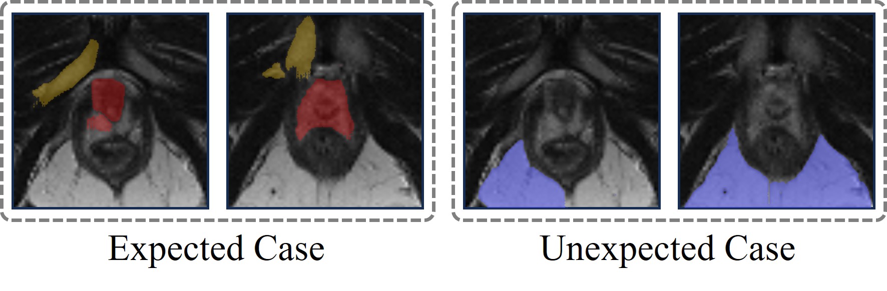
5.4.1 Comparison Between ‘One-to-One’ and ‘One-to-Many’ Correspondence
Fig. 8 illustrates expected and unexpected scenarios of ‘one-to-many’ correspondence in ROI alignment. In the expected case (left), multiple sub-structures corresponding to one complete anatomical structure contribute to ROI alignment. The unexpected case (right) shows mismatched sub-structures, leading to potentially inaccurate registration. Table 6 presents quantitative results on the performance of ‘one-to-many’ correspondence compared to ‘one-to-one’ matching for both intra-subject and inter-subject registration tasks. For intra-subject registration tasks, where image features exhibit a high degree of similarity, ‘one-to-many’ matching performs worse than the standard ‘one-to-one’ matching approach, however, for inter-subject registration tasks, where the image features exhibit a more significant gap, ‘one-to-many’ correspondence shows competitive performance with ‘one-to-one’ matching. This is likely because the presence of multiple corresponding sub-structures can help bridge the larger gap in image features between subjects, leading to more accurate registration.
5.4.2 Effect of Slice Range
Table 6 shows the ablative study on the quantity of corresponding ROI search range (number of slices ). A larger search range, indicated by a larger number of slices, leads to better registration performance. By looking at the range of anatomical structures highlighted in these ROIs, as depited in Fig. 8, we can gain insights into how the search range affects the registration process.
| Correspondence | Intra-Subject: Cardiac | Inter-Subject: Prostate | ||
|---|---|---|---|---|
| Dice | TRE | Dice | TRE | |
| ‘one-to-one’ | 80.28±3.67 | 1.43±1.13 | 75.67±3.81 | 2.09±1.35 |
| ‘one-to-many’ | 78.12±3.89 | 1.65±1.19 | 75.12±4.03 | 2.03±1.62 |

6 Discussion
As analyzed in Sec. 3.1, increasing the number or precision of ROI pairs generally improves performance up to a task-specific upper limit. However, as shown in Tables 4 and 4, there is an optimal balance between the number/precision of ROI pairs and registration performance, which falls below the theoretical maximum. Beyond this balance point, further increasing the number or precision of ROI pairs results in a performance decline.
This counter-intuitive phenomenon occurs because segmentation and alignment performance are assumed constant in Sec. 3.1. In SAMReg’s ROI matching mechanism, ROI pairs are ranked by similarity, with the most similar pairs ranked highest. Adding more pairs reduces the average similarity, introducing noise and potential mismatches. Conversely, stricter thresholds reduce the number of pairs, limiting the benefits of ROI-to-dense interpolation. Future work should explore adaptive thresholding strategies that dynamically adjust based on image similarity or segmentation confidence to optimize the number of pairs while maintaining high-quality correspondences.
In this study, ‘one-to-one’ correspondence pairs each ROI in one image with a unique ROI in the other, while ‘one-to-many’ correspondence allows for multiple ROIs from one image to correspond to a single ROI in the other.
Two key scenarios necessitate considering ‘one-to-many’ correspondence: 1) Segmentation Uncertainty: Segmentation models are not perfect. They might segment multiple sub-structures in one image while identifying a complete structure ROI in another. ‘one-to-many’ correspondence can bridge this gap by allowing multiple sub-structures to correspond to a single complete structure. 2) 2D ROIs for 3D Structures: Representing 3D structures with 2D ROIs exists inherent limitations. For instance, a single gland might be represented by 20 2D slices in one image and 40 in the other. This disparity requires ‘one-to-many’ correspondence to handle multiple-to-one correspondence scenarios.
Interestingly, as evidenced by Table 6, the performance of both ‘one-to-one’ and ‘one-to-many’ approaches is comparable in intra-subject registration tasks, where images exhibit high similarity. However, for inter-subject registration tasks with greater anatomical variability, ‘one-to-one’ correspondence performs better. This observation can be attributed to the inherent characteristics of the registration tasks. In intra-subject registration, the high paired similarity between corresponding ROIs diminishes the impact of the chosen correspondence strategy. Both methods achieve comparable ROI pairs due to the strong inherent correspondence between the structures. Conversely, inter-subject variability benefits from ‘one-to-one”s exclusion mechanism, reducing erroneous pairings and improving alignment accuracy.
Therefore, future research should focus on two main areas: refining ROI representation to enhance similarity measures and exploring a hybrid approach that adjusts indeterminacy dynamically based on factors such as image similarity or segmentation confidence.
7 Conclusion
In this study, We introduce a novel ROI-based correspondence representation. With this representation, the image registration is reformalized as two multi-class segmentation tasks, with a proposed, general-purpose and practical implementation, SAMReg. Our extensive experiments demonstrate that SAMReg delivers competitive performance in image registration, positioning it as a robust and versatile tool. The promising results suggest that this new approach may pave the way for a fresh research direction in image registration.
Acknowledgements
This work was supported by the International Alliance for Cancer Early Detection, a partnership between Cancer Research UK [C28070/A30912; C73666/A31378], Canary Center at Stanford University, the University of Cambridge, OHSU Knight Cancer Institute, University College London and the University of Manchester.
References
- Amini et al. [1998] Amini, A.A., Chen, Y., Curwen, R.W., Mani, V., Sun, J., 1998. Coupled b-snake grids and constrained thin-plate splines for analysis of 2-d tissue deformations from tagged mri. IEEE transactions on medical imaging 17, 344–356.
- Audenaert et al. [2019] Audenaert, E.A., Van Houcke, J., Almeida, D.F., Paelinck, L., Peiffer, M., Steenackers, G., Vandermeulen, D., 2019. Cascaded statistical shape model based segmentation of the full lower limb in ct. Computer methods in biomechanics and biomedical engineering 22, 644–657.
- Balakrishnan et al. [2019] Balakrishnan, G., Zhao, A., Sabuncu, M.R., Guttag, J., Dalca, A.V., 2019. Voxelmorph: a learning framework for deformable medical image registration. IEEE transactions on medical imaging 38, 1788–1800.
- Bernard et al. [2018] Bernard, O., Lalande, A., Zotti, C., Cervenansky, F., Yang, X., Heng, P.A., Cetin, I., Lekadir, K., Camara, O., Ballester, M.A.G., et al., 2018. Deep learning techniques for automatic mri cardiac multi-structures segmentation and diagnosis: is the problem solved? IEEE transactions on medical imaging 37, 2514–2525.
- Bosaily et al. [2015] Bosaily, A.E.S., Parker, C., Brown, L., Gabe, R., Hindley, R., Kaplan, R., Emberton, M., Ahmed, H., Group, P., et al., 2015. Promis—prostate mr imaging study: a paired validating cohort study evaluating the role of multi-parametric mri in men with clinical suspicion of prostate cancer. Contemporary clinical trials 42, 26–40.
- Brigger et al. [2000] Brigger, P., Hoeg, J., Unser, M., 2000. B-spline snakes: a flexible tool for parametric contour detection. IEEE Transactions on image processing 9, 1484–1496.
- Cardoso et al. [2022] Cardoso, M.J., Li, W., Brown, R., Ma, N., Kerfoot, E., Wang, Y., Murrey, B., Myronenko, A., Zhao, C., Yang, D., et al., 2022. Monai: An open-source framework for deep learning in healthcare. arXiv preprint arXiv:2211.02701 .
- Cen et al. [2023] Cen, J., Fang, J., Zhou, Z., Yang, C., Xie, L., Zhang, X., Shen, W., Tian, Q., 2023. Segment anything in 3d with radiance fields. arXiv preprint arXiv:2304.12308 .
- Chen et al. [2024a] Chen, H., Li, Y., Gu, Z., Xu, Z., Lan, J., Li, H., 2024a. Segment anything model meets image harmonization, in: ICASSP, IEEE. pp. 2630–2634.
- Chen et al. [2022] Chen, J., Frey, E.C., He, Y., Segars, W.P., Li, Y., Du, Y., 2022. Transmorph: Transformer for unsupervised medical image registration. Medical image analysis 82, 102615.
- Chen et al. [2024b] Chen, Y., He, T., Huang, D., Ye, W., Chen, S., Tang, J., Chen, X., Cai, Z., Yang, L., Yu, G., et al., 2024b. Meshanything: Artist-created mesh generation with autoregressive transformers. arXiv preprint arXiv:2406.10163 .
- Chen et al. [2023] Chen, Z., Fang, G., Ma, X., Wang, X., 2023. 0.1% data makes segment anything slim. arXiv preprint arXiv:2312.05284 .
- Cheng et al. [2023a] Cheng, J., Ye, J., Deng, Z., Chen, J., Li, T., Wang, H., Su, Y., Huang, Z., Chen, J., Sun, L.J.H., He, J., Zhang, S., Zhu, M., Qiao, Y., 2023a. Sam-med2d. arXiv:2308.16184.
- Cheng et al. [2023b] Cheng, Y., Li, L., Xu, Y., Li, X., Yang, Z., Wang, W., Yang, Y., 2023b. Segment and track anything. arXiv preprint arXiv:2305.06558 .
- Collins and Evans [1997] Collins, D.L., Evans, A.C., 1997. Animal: validation and applications of nonlinear registration-based segmentation. International journal of pattern recognition and artificial intelligence 11, 1271--1294.
- Crum et al. [2003] Crum, W.R., Griffin, L.D., Hill, D.L., Hawkes, D.J., 2003. Zen and the art of medical image registration: correspondence, homology, and quality. NeuroImage 20, 1425--1437.
- DeTone et al. [2016] DeTone, D., Malisiewicz, T., Rabinovich, A., 2016. Deep image homography estimation. arXiv preprint arXiv:1606.03798 .
- Dickinson et al. [2013] Dickinson, L., Ahmed, H.U., Kirkham, A., Allen, C., Freeman, A., Barber, J., Hindley, R.G., Leslie, T., Ogden, C., Persad, R., et al., 2013. A multi-centre prospective development study evaluating focal therapy using high intensity focused ultrasound for localised prostate cancer: the index study. Contemporary clinical trials 36, 68--80.
- Eppenhof et al. [2018] Eppenhof, K.A., Lafarge, M.W., Moeskops, P., Veta, M., Pluim, J.P., 2018. Deformable image registration using convolutional neural networks, in: Medical Imaging 2018: Image Processing, SPIE. pp. 192--197.
- Fei et al. [2024] Fei, B., Li, Y., Yang, W., Gao, H., Xu, J., Ma, L., Yang, Y., Zhou, P., 2024. Lightening anything in medical images. arXiv preprint arXiv:2406.10236 .
- Gong et al. [2023] Gong, S., Zhong, Y., Ma, W., Li, J., Wang, Z., Zhang, J., Heng, P.A., Dou, Q., 2023. 3dsam-adapter: Holistic adaptation of sam from 2d to 3d for promptable medical image segmentation. arXiv preprint arXiv:2306.13465 .
- Hamid et al. [2019] Hamid, S., Donaldson, I.A., Hu, Y., Rodell, R., Villarini, B., Bonmati, E., Tranter, P., Punwani, S., Sidhu, H.S., Willis, S., et al., 2019. The smarttarget biopsy trial: a prospective, within-person randomised, blinded trial comparing the accuracy of visual-registration and magnetic resonance imaging/ultrasound image-fusion targeted biopsies for prostate cancer risk stratification. European urology 75, 733--740.
- Hering et al. [2020] Hering, A., Murphy, K., van Ginneken, Bram, 2020. Learn2reg challenge: Ct lung registration. https://doi.org/10.5281/zenodo.3835682.
- Hernandez-Matas et al. [2017] Hernandez-Matas, C., Zabulis, X., Triantafyllou, A., Anyfanti, P., Douma, S., Argyros, A.A., 2017. Fire: fundus image registration dataset. Modeling and Artificial Intelligence in Ophthalmology 1, 16--28.
- Hu et al. [2019] Hu, Y., Gibson, E., Barratt, D.C., Emberton, M., Noble, J.A., Vercauteren, T., 2019. Conditional segmentation in lieu of image registration, in: MICCAI, Springer. pp. 401--409.
- Hu et al. [2018a] Hu, Y., Modat, M., Gibson, E., Ghavami, N., Bonmati, E., Moore, C.M., Emberton, M., Noble, J.A., Barratt, D.C., Vercauteren, T., 2018a. Label-driven weakly-supervised learning for multimodal deformable image registration, in: 2018 IEEE 15th International Symposium on Biomedical Imaging (ISBI 2018), IEEE. pp. 1070--1074.
- Hu et al. [2018b] Hu, Y., Modat, M., Gibson, E., Li, W., Ghavami, N., Bonmati, E., Wang, G., Bandula, S., Moore, C.M., Emberton, M., et al., 2018b. Weakly-supervised convolutional neural networks for multimodal image registration. Medical image analysis 49, 1--13.
- Huang et al. [2024a] Huang, S., Xu, T., Shen, Z., Saeed, S.U., Yan, W., Barratt, D., Hu, Y., 2024a. One registration is worth two segmentations. arXiv preprint arXiv:2405.10879 .
- Huang et al. [2024b] Huang, Y., Yang, X., Liu, L., Zhou, H., Chang, A., Zhou, X., Chen, R., Yu, J., Chen, J., Chen, C., et al., 2024b. Segment anything model for medical images? Medical Image Analysis 92, 103061.
- Kang et al. [2022] Kang, M., Hu, X., Huang, W., Scott, M.R., Reyes, M., 2022. Dual-stream pyramid registration network. Medical image analysis 78, 102379.
- Ke et al. [2024] Ke, L., Ye, M., Danelljan, M., Tai, Y.W., Tang, C.K., Yu, F., et al., 2024. Segment anything in high quality. Advances in Neural Information Processing Systems 36.
- Kevin et al. [2021] Kevin, E., Bin, L., Adib, K., 2021. Multimodal biomedical dataset for evaluating registration methods (full-size tma cores). https://doi.org/10.5281/zenodo.4550300.
- Kim et al. [2021] Kim, B., Kim, D.H., Park, S.H., Kim, J., Lee, J.G., Ye, J.C., 2021. Cyclemorph: cycle consistent unsupervised deformable image registration. Medical image analysis 71, 102036.
- Kirillov et al. [2023] Kirillov, A., Mintun, E., Ravi, N., Mao, H., Rolland, C., Gustafson, L., Xiao, T., Whitehead, S., Berg, A.C., Lo, W.Y., et al., 2023. Segment anything. arXiv preprint arXiv:2304.02643 .
- Krebs et al. [2017] Krebs, J., Mansi, T., Delingette, H., Zhang, L., Ghesu, F.C., Miao, S., Maier, A.K., Ayache, N., Liao, R., Kamen, A., 2017. Robust non-rigid registration through agent-based action learning, in: MICCAI, Springer. pp. 344--352.
- Li et al. [1995] Li, H., Manjunath, B., Mitra, S.K., 1995. A contour-based approach to multisensor image registration. IEEE transactions on image processing 4, 320--334.
- Liu et al. [2023] Liu, L., Aviles-Rivero, A.I., Schönlieb, C.B., 2023. Contrastive registration for unsupervised medical image segmentation. IEEE Transactions on Neural Networks and Learning Systems .
- Ma et al. [2024] Ma, J., He, Y., Li, F., Han, L., You, C., Wang, B., 2024. Segment anything in medical images. Nature Communications 15, 654.
- Mazurowski et al. [2023] Mazurowski, M.A., Dong, H., Gu, H., Yang, J., Konz, N., Zhang, Y., 2023. Segment anything model for medical image analysis: an experimental study. Medical Image Analysis 89, 102918.
- Michele and Vittorio [2022] Michele, V., Vittorio, F., 2022. Zurich summer dataset. https://doi.org/10.5281/zenodo.5914759.
- Milletari et al. [2016] Milletari, F., Navab, N., Ahmadi, S.A., 2016. V-net: Fully convolutional neural networks for volumetric medical image segmentation, in: 2016 fourth international conference on 3D vision (3DV), Ieee. pp. 565--571.
- Modat et al. [2014] Modat, M., Cash, D.M., Daga, P., Winston, G.P., Duncan, J.S., Ourselin, S., 2014. Global image registration using a symmetric block-matching approach. Journal of medical imaging 1, 024003--024003.
- Mok and Chung [2022] Mok, T.C., Chung, A., 2022. Affine medical image registration with coarse-to-fine vision transformer, in: CVPR, pp. 20835--20844.
- Myronenko and Song [2010] Myronenko, A., Song, X., 2010. Point set registration: Coherent point drift. IEEE transactions on pattern analysis and machine intelligence 32, 2262--2275.
- Oquab et al. [2023] Oquab, M., Darcet, T., Moutakanni, T., Vo, H., Szafraniec, M., Khalidov, V., Fernandez, P., Haziza, D., Massa, F., El-Nouby, A., et al., 2023. Dinov2: Learning robust visual features without supervision. arXiv preprint arXiv:2304.07193 .
- Orczyk et al. [2021] Orczyk, C., Barratt, D., Brew-Graves, C., Peng Hu, Y., Freeman, A., McCartan, N., Potyka, I., Ramachandran, N., Rodell, R., Williams, N.R., et al., 2021. Prostate radiofrequency focal ablation (proraft) trial: a prospective development study evaluating a bipolar radiofrequency device to treat prostate cancer. The Journal of Urology 205, 1090--1099.
- Osco et al. [2023] Osco, L.P., Wu, Q., de Lemos, E.L., Gonçalves, W.N., Ramos, A.P.M., Li, J., Junior, J.M., 2023. The segment anything model (sam) for remote sensing applications: From zero to one shot. International Journal of Applied Earth Observation and Geoinformation 124, 103540.
- Qiu et al. [2021] Qiu, H., Qin, C., Schuh, A., Hammernik, K., Rueckert, D., 2021. Learning diffeomorphic and modality-invariant registration using b-splines, in: Medical imaging with deep learning.
- Rohé et al. [2017] Rohé, M.M., Datar, M., Heimann, T., Sermesant, M., Pennec, X., 2017. Svf-net: learning deformable image registration using shape matching, in: MICCAI, Springer. pp. 266--274.
- Rohr et al. [2001] Rohr, K., Stiehl, H.S., Sprengel, R., Buzug, T.M., Weese, J., Kuhn, M., 2001. Landmark-based elastic registration using approximating thin-plate splines. IEEE Transactions on medical imaging 20, 526--534.
- Roy et al. [2023] Roy, S., Koehler, G., Ulrich, C., Baumgartner, M., Petersen, J., Isensee, F., Jaeger, P.F., Maier-Hein, K.H., 2023. Mednext: transformer-driven scaling of convnets for medical image segmentation, in: MICCAI, Springer. pp. 405--415.
- Rueckert et al. [1999] Rueckert, D., Sonoda, L.I., Hayes, C., Hill, D.L., Leach, M.O., Hawkes, D.J., 1999. Nonrigid registration using free-form deformations: application to breast mr images. IEEE transactions on medical imaging 18, 712--721.
- Schnabel et al. [2001] Schnabel, J.A., Rueckert, D., Quist, M., Blackall, J.M., Castellano-Smith, A.D., Hartkens, T., Penney, G.P., Hall, W.A., Liu, H., Truwit, C.L., et al., 2001. A generic framework for non-rigid registration based on non-uniform multi-level free-form deformations, in: MICCAI, Springer. pp. 573--581.
- Shi et al. [2024] Shi, Y., Gottipati, A., Yan, Y., 2024. Path-ct image registration with self-supervised vision transformer for lung cancer, in: ISBI, IEEE. pp. 1--5.
- Shvets et al. [2024] Shvets, M., Zhao, D., Niethammer, M., Sengupta, R., Berg, A.C., 2024. Joint depth prediction and semantic segmentation with multi-view sam, in: WACV, pp. 1328--1338.
- Simmons et al. [2014] Simmons, L.A., Ahmed, H.U., Moore, C.M., Punwani, S., Freeman, A., Hu, Y., Barratt, D., Charman, S.C., Van der Meulen, J., Emberton, M., 2014. The picture study—prostate imaging (multi-parametric mri and prostate histoscanning™) compared to transperineal ultrasound guided biopsy for significant prostate cancer risk evaluation. Contemporary clinical trials 37, 69--83.
- Song et al. [2024] Song, X., Xu, X., Yan, P., 2024. Dino-reg: General purpose image encoder for training-free multi-modal deformable medical image registration, in: MICCAI, Springer. pp. 608--617.
- Sotiras et al. [2013] Sotiras, A., Davatzikos, C., Paragios, N., 2013. Deformable medical image registration: A survey. IEEE transactions on medical imaging 32, 1153--1190.
- Tian et al. [2024] Tian, L., Greer, H., Kwitt, R., Vialard, F.X., Estepar, R.S.J., Bouix, S., Rushmore, R., Niethammer, M., 2024. unigradicon: A foundation model for medical image registration. arXiv preprint arXiv:2403.05780 .
- Tian et al. [2023] Tian, L., Greer, H., Vialard, F.X., Kwitt, R., Estépar, R.S.J., Rushmore, R.J., Makris, N., Bouix, S., Niethammer, M., 2023. Gradicon: Approximate diffeomorphisms via gradient inverse consistency, in: CVPR, pp. 18084--18094.
- Tragakis et al. [2023] Tragakis, A., Kaul, C., Murray-Smith, R., Husmeier, D., 2023. The fully convolutional transformer for medical image segmentation, in: WACV, pp. 3660--3669.
- Wang et al. [2024] Wang, D., Zhang, J., Du, B., Xu, M., Liu, L., Tao, D., Zhang, L., 2024. Samrs: Scaling-up remote sensing segmentation dataset with segment anything model. Advances in Neural Information Processing Systems 36.
- Wang et al. [2023a] Wang, H., Guo, S., Ye, J., Deng, Z., Cheng, J., Li, T., Chen, J., Su, Y., Huang, Z., Shen, Y., Fu, B., Zhang, S., He, J., Qiao, Y., 2023a. Sam-med3d. arXiv:2310.15161.
- Wang et al. [2023b] Wang, Q., Chang, Y.Y., Cai, R., Li, Z., Hariharan, B., Holynski, A., Snavely, N., 2023b. Tracking everything everywhere all at once, in: ICCV, pp. 19795--19806.
- Wu et al. [2023] Wu, J., Fu, R., Fang, H., Liu, Y., Wang, Z., Xu, Y., Jin, Y., Arbel, T., 2023. Medical sam adapter: Adapting segment anything model for medical image segmentation. arXiv preprint arXiv:2304.12620 .
- Wu et al. [2024] Wu, Q., Jiang, H., Luo, L., Li, J., Ding, Y., Xie, J., Yang, J., 2024. Diff-reg v1: Diffusion matching model for registration problem. arXiv preprint arXiv:2403.19919 .
- Xu and Niethammer [2019] Xu, Z., Niethammer, M., 2019. Deepatlas: Joint semi-supervised learning of image registration and segmentation, in: MICCAI, Springer. pp. 420--429.
- Yang et al. [2023] Yang, J., Gao, M., Li, Z., Gao, S., Wang, F., Zheng, F., 2023. Track anything: Segment anything meets videos. arXiv preprint arXiv:2304.11968 .
- Zhu et al. [2008] Zhu, Q., Wang, L., Wu, Y., Shi, J., 2008. Contour context selection for object detection: A set-to-set contour matching approach, in: ECCV, Springer. pp. 774--787.
- Zhuang et al. [2010] Zhuang, X., Rhode, K.S., Razavi, R.S., Hawkes, D.J., Ourselin, S., 2010. A registration-based propagation framework for automatic whole heart segmentation of cardiac mri. IEEE transactions on medical imaging 29, 1612--1625.