Vortex Interference Enables optimal 3D Interferometric Nanoscopy
Abstract
Super-resolution imaging methods that combine interferometric (z) analysis with single-molecule localization microscopy (iSMLM) have achieved ultra-high 3D precision and contributed to the elucidation of important biological ultrastructures. However, their dependence on imaging multiple phase-shifted output channels necessitates complex instrumentation and operation. To solve this problem, we develop an interferometric super-resolution microscope capable of optimal direct axial nanoscopy, termed VILM (Vortex Interference Localization Microscopy). Using a pair of vortex phase plates with opposite orientation for each dual-opposed objective lenses, the detection point-spread functions (PSFs) adopt a bilobed profile whose rotation encodes the axial position. Thus, direct 3D single-molecule coordinate determination can be achieved with a single output image. By reducing the number of output channels to as few as one and utilizing a simple 50:50 beamsplitter, the imaging system is significantly streamlined, while the optimal iSMLM imaging performance is retained, with axial resolution 2 times better than the lateral. The capability of VILM is demonstrated by resolving the architecture of microtubules and probing the organization of tyrosine-phosphorylated signalling proteins in integrin-based cell adhesions.
Intensive development in super-resolution fluorescence microscopy in recent years have enabled their applications to facilitate a broad range of biological discoveries[1, 2, 3, 4]. Among super-resolution microscopy techniques widely utilized in bioimaging, single-molecule localization microscopy (SMLM) depends on the stochastic spatiotemporal control of the density of the emitting fluorophores and their subsequent localization[2, 5, 6, 7, 8]. However, while SMLM can readily approach molecular-scale lateral (xy) precision especially with recent improvements in data analysis and labelling strategies, achieving comparable axial (z) precision is significantly more challenging. Commonly used approaches to realize 3D super-resolution in SMLM such as astigmatic imaging offers optical simplicity[9, 10] but at the cost of at least 2-3 times poorer z-resolution compared to the lateral (xy) resolution. Greater extent of axial resolution enhancement generally requires interferometric techniques, among which the configurations based on 4Pi geometry have been established to provide ultra-high 3D resolution. Interferometric SMLM (iSMLM) techniques such as iPALM[11], 4Pi-SMS[12], W-4PiSMSN[13, 14], and 4Pi-STORM[15], provide a major gain in axial resolution, averaging up to twice the lateral precision over the focus range, and are theoretically reaching the intrinsic quantum limit of spatial resolution[16]. The ultra-high resolution of iSMLM methods have been instrumental in elucidating major biological insights[3, 13, 17, 18]. However, in significant part due to the highly complex optical instrumentation, calibration, and operation[11, 12, 13], these techniques have remained confined to just a handful of laboratories world-wide, even after more than a decade since their emergence. To enable broader accessible to the research community thus requires the simplification of its operation while retaining its high optical performance.
In current iSMLM methods, the 4Pi geometry encodes high-precision z-position information via self-interference of fluorescence emission. The axial position of a single-molecule fluorophore is then retrieved primarily via multi-phase interferometry[11, 12, 13]. However, at least three detection channels are needed to maximize the full imaging depth allowed by interferometry[11]. As a result, complex optical configuration is required to realize three or four detection channels. For example, in iPALM, a custom-designed 3-phase beam splitter is used to achieve three detection channels[11], while in 4Pi-SMS or 4Pi-STORM customized phase modulation devices are employed to realize four polarization-dependent channels[12, 13]. In practice, these compound the alignment and calibration complexity. By reducing the number of detection channels to one or two, and utilizing a commercial beam splitter, the system’s complexity can be significantly reduced. However, the problem of optimizing interferometric nanoscopy using a single beamsplitter has been relatively underexplored.
To solve this problem, here we developed Vortex Interference Localization Microscopy (VILM), which is demonstrated to enable optimal direct 3D interfereometric nanoscopy. By harnessing vortex beam interference, 3D spatial information of a single-molecule fluorophore can be retrieved using just one output image. By placement of a vortex phase mask in each 4Pi optical arm with opposite chirality, the self-interfered PSFs generated by a simple 50:50 beamsplitter adopt a bilobed profile with its orientation encoding the z-coordinate. 3D information is thus readily extracted using one channel. Our numerical analysis established that PSFs generated by VILM perform at the optimal limit for a single beamsplitter interferometric system. Biological imaging performance of VILM is validated by resolving the architecture of microtubules as well as signalling components of integrin-based cell adhesions.
Theoretical foundation The interference of emissions from a fluorescent molecule with itself can occur even over short coherence lengths. In a 4Pi configuration utilizing two opposed objective lenses, interference detection captures nearly the full solid angle, resulting in a detection PSF with an axial fringe structure having three or four times finer feature. In order to determine the axial position, multiple intensity measurements are required (see also Supplementary Notes). This can be achieved by various multi-phase projection methods which enable multiple output channels to be simultaneously imaged. Such multi-phase projection optics impose additional complexity and cost, however. Towards simplifying iSMLM, we realized that a significant design simplification can be achieved using vortex beam interference generated by commercially available vortex phase masks.
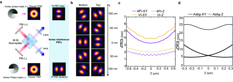
In super-resolution microscopy, the vortex phase mask is best known as the optical component that generates the “donut” beam used in STED or MINFLUX microscopy[7, 5]. Since the vortex phase ramp is chiral, , depending on the orientation of the vortex phase mask, the donut beam is also chiral. Therefore, when 2 donuts with opposite handedness interfere, this gives rise to a pair of counter-rotating bi lobes (see also Supplementary Notes I, Supplementary Movie 1). This specific type of interference is referred to as ”vortex interference” and has recently been utilized for various high-precision optical measurements[19, 20]. As illustrated in Fig.1(a), vortex interference can be readily incorporated into a 4Pi geometry. With a simple 50:50 beamsplitter, the detection PSFs for such configuration can be approximated as:
| (1) |
where correspond to the polar coordinates in the image plane, and denotes the phase shifts between the light collected by the two channels. Here, and fwhm are constants defined by the numerical aperture of the imaging system, while denotes the amplitude. Accordingly, the PSF appears as a bilobed profile with two maxima and two minima, which azimuthally rotate as a function of the axial position (z) (Fig. 1(b)). The range for a complete rotation corresponds to approximately where is the refractive index of the specimen. Importantly, the axial position of the fluorophore can be directly calculated from the rotation angle of a bilobed PSF. While two channels are generated as outputs, their information content is largely redundant, and duplex channel summation primarily improved precision due to higher photon counts but is otherwise dispensable (Supplementary Fig. S1(d)). This configuration, here termed VILM (Vortex Interference Localization Microscopy), thus enables interferometric monoplex 3D coordinate read-out with simplified optical construction.
To evaluate the performance of VILM, we calculate the corresponding Cramer-Rao Lower Bound (CRLB) theoretical localization precision in comparison to other super-resolution microscopy modalities as displayed in Fig. 1(c) (see also Supplementary Notes III)[21]. Our analysis showed that theoretical axial resolution of VILM is expected to be 2-3 times superior to lateral resolution, similar to other iSMLM techniques[11, 12, 13]. While comparison to 4Pi-SMLM with unmodified PSF shows that both the axial and lateral resolutions of VILM are slightly reduced by approximately 1.2 times, VILM still provides significant resolution enhancement over non-interference-based 3D SMLM methods (Fig. 1(d)) [9]. For example, for equivalent photon number of 2000 the theoretical axial and lateral resolution for monoplex VILM is 3 and 7 nm, respectively, whereas for astigmatic imaging, the axial and lateral resolution are 20 nm and 7 nm, being significantly worse axially as described earlier [9] (Supplementary Fig. S1(c-d)). Duplex VILM provides further resolution enhancement with axial and lateral resolution of 2 and 4 nm, respectively. Compared to astigmatism-based SMLM,VILM achieves nearly the same 3D precision as conventional iSMLM using only one beam splitter configuration.
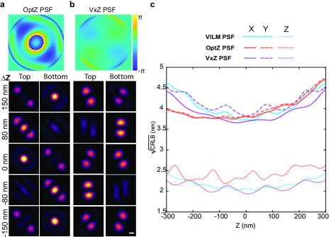
Vortex beam interference enables optimal direct axial nanoscopy Next we further evaluate the performance of VILM following a design strategy previously developed to identify the optimal 3D SMLM pupil function that generates a PSF with the best Fisher information properties, based on Zernike mode composition[22]. We first calculated the optimal Zernike modes for realizing iSMLM with a single beamsplitter, with the emphasis on optimizing the axial resolution as much as possible, while not seeking to extend the axial range. The optimization problem is to minimize the weighted mean precision defined as follows:
| (2) |
with the last term introduced to mitigate fluctuations in the axial resolution and emphasize attaining the best possible axial resolution. In addition to the optimal pupil function () based on solely linear combination of Zernike modes, we also calculated the optimal pupil function based on the linear combination of the vortex phase and Zernike modes. The two optimized pupil functions and corresponding z-depth-dependent PSFs are presented in Fig. 2(a-b), denoted OptZ PSF and VxZ PSF, respectively (see also Supplementary Notes IV). Theoretical precision for these optimized PSFs as well as for the PSF of VILM over the upper and lower outputs of the 50:50 beamsplitter are displayed in Fig. 2(c) and Supplementary Movies 1-3. Analogous to VILM PSFs whose intensity variations are transferred to the azimuthal direction, OptZ PSFs exhibit intensity variation along the lateral direction as a function of z-position. In the case of VxZ PSFs, the axial rotation is also maintained, along with additional intensity variation between the two channels. Compared to OptZ PSFs, we found that VILM PSFs offer comparable precision overall, with slightly better performance in axial (z) dimension. The axial precision of VILM PSF also vary more smoothly as a function of z-position compared to the greater undulation in OptZ PSFs Fig. 2(c). Comparison between VILM PSFs and VxZ PSFs reveals a similar trend. Notably, while VxZ PSFs achieves better axial precision for certain ranges of z-position, this is accompanied by greater undulation in axial precision and anisotropy in lateral precision, primarily due to the intensity imbalance between the two channels in VxZ PSFs. Furthermore, while VILM enable single-channel detection due to the uniform intensity variation over the axial range, in practice both OptZ and VxZ would require dual-channel detection due to the intensity oscillation between the upper and lower output channels (see also Supplementary Notes II, Supplementary Movies 1-3). Taken together, we conclude that VILM theoretically attains the optimal super-resolution performance under the simplest single beamsplitter condition.
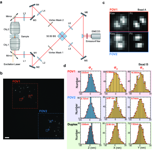
Optical implementation and imaging performance validation. To validate our design, we next constructed a VILM imaging system according to an optical configuration illustrated in Fig. 3(a) (see also Supplementary Fig. S2 and Supplementary Table S1). The system is based on a 4Pi geometry with dual opposed objective lenses aligned co-axially above and below the specimen plane. The specimen is mounted on a piezoelectric sample holder that can axially translate in precise nanometric step for calibration and alignment. Subsequent to each objective lens, an assembly of relay lenses and mirrors are used to map the pupil plane to a vortex phase mask. Opposite orientation of the vortex phase mask for the upper and lower arms then generates vortex beam interference as described above. A simple 50:50 non-polarizing beam splitter (BS, Fig. 3(a)) is positioned to project the interfered emission signals toward the detection EMCCD camera, with monoplex or duplex detection modes selectable by the placement of either a mirror or a prism in front of the camera. Detailed steps in optical instrumentation, alignment, calibration, and operation of VILM are described in Supplementary Methods section. We first image nanoparticle fluorescent fiducials to evaluate the imaging performance of VILM, using the duplex imaging mode to assess whether designed PSFs behave as expected. As shown in Fig. 3(b-c), over both field-of-views (FOVs) each fiducial can be seen to exhibit bilobed shape consistent with the theoretical prediction. The bilobed PSF orientations within each FOV are largely uniform since the substrate-affixed fiducials are co-planar in z. The PSFs corresponding to each fiducial are perpendicularly oriented between FOV1 and FOV2 as expected. As the specimen is axially translated by the piezo sample holder, the rotation of bilobed PSFs are apparent (Supplementary Movies 4-5) with a complete rotation over half of the emission wavelength, as shown in Supplementary Fig. S4. Altogether, these results demonstrate that vortex interference effectively convert z-coordinate information into azimuthal rotation of the bilobed PSFs, hence encoding 3D coordinate information in a single-image.
To determine the 3D coordinate from the bilobed PSF, we made use of cubic spline fitting [23, 24, 15] based on theoretical PSFs calculated using the system parameters. As shown in Fig. 3(c), the bilobed PSF can be fitted well with theoretical model. For monoplex imaging mode, the 3D coordinate of a single FOV is used, while in duplex imaging mode, the 3D coordinate is first separately determined for both FOVs. Subsequently the FOV1 lateral coordinates are mirrored and axial position inversed, and then aligned with the FOV2 coordinates using an affine transformation. The mean positions of FOV1 and FOV2 are then taken as the final coordinates (see also Supplementary Figure S3). The histograms of the 3D coordinates of a representative fiducial, with typical photon number of 1100-1200 in each channel, analyzed by either monoplex or duplex detection are presented in Fig. 3(d). The monoplex axial precision () for FOV1 and FOV2 are 5.02 nm and 5.12 nm, respectively. In duplex mode, the axial precision is enhanced (3.66 nm) as the number of photons is roughly doubled. The monoplex lateral precision of FOV1 and FOV2 are (12.99 nm, 10.55 nm) and (9.97 nm, 9.14 nm) respectively. Combining two channels together, the duplex lateral precision is enhanced accordingly (8.44 nm, 7.02 nm). For each channel, the monoplex axial resolution is about 2 times better than the lateral precision as defined by . Similarly, the duplex axial precision is around two times better than the corresponding lateral precision. Altogether, our results demonstrated that VILM enable ultra-high precision SMLM imaging with significant axial precision enhancement exceeding the lateral resolution. Our analysis over an extended imaging depth (Supplementary Fig. S4) shows that the axial precision is largely uniform within the central period. Similar to other iSMLM methods [11, 12, 13], periodicity in z-coordinate determination can be expected and thus phase-wrapping effect is observed beyond the half-wavelength period ( 252 nm for 670 nm emission). Nevertheless, we note that beyond the central period, the VILM PSFs appear to gain chiral tails, adopting a “yin-yang” pattern, which can be used to distinguish the higher-harmonic PSF from the pure bilobed shape of the central period (Supplementary Movies 1).
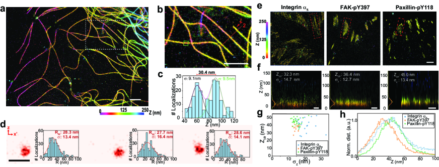
To demonstrate its performance in realizing direct axial nanoscopy, monoplex mode is used to image cellular structures. The microtubule cytoskeletons are hollow protein polymers with a known nanoscale dimension, commonly used as a resolution benchmark in super-resolution microscopy. When labelled by immunostaining using unlabelled primary antibody and fluorophore-labelled secondary antibody, microtubule have been imaged as a hollow structure[12, 13]. Compared to iPALM which required a long period of calibration and alignment for each imaging site, the simplified procedure of VILM enabled a cell to be imaged within min after identification. As shown in Fig. 4(a-b), VILM imaging of a fixed COS7 cell with microtubules immunostained using AlexaFluor 647conjugated secondary antibody reveals clear 3D profiles of the filament networks. From the histogram of the axial-coordinates within the green box in Fig. 4(b), two distinct peaks, separated by 30.4 nm, can be observed with of 9.1 nm and 9.5 nm, respectively, as shown in Fig. 4(c). The tubular architecture of microtubules can be discerned when the transverse cross-section reconstructed images along the red dashed line in Fig. 4(b) are viewed, with examples shown in Fig. 4(d).
We also made use of VILM to map z-position of proteins in integrin-based focal adhesions. The high axial resolution of iSMLM techniques such as iPALM has earlier been harnessed to elucidate the axial compartmentalization of distinct proteins or to map different conformation of the integrin transmembrane receptors[3, 18, 17]. In previous iPALM study of focal adhesions, photoactivatable fluorescent protein (PA-FP) fusions were used due to the small effective probe dimension[3]. However, PA-FP labelling is unsuitable for probing either endogenous proteins or signalling events such as phosphorylation, both of which could otherwise be readily detected by immunostaining. Earlier, based on the localization of signalling proteins such as focal adhesion kinase (FAK) and paxillin in close proximity with integrin, their organization were termed the integrin signalling layer (ISL) nanoscale compartment. However, whether endogenous proteins or actual signalling events such as protein tyrosine phosphorylation are located in such compartment has not been directly determined. Hence, we employed VILM to probe the nanoscale localization of key indicators of integrin-dependent signalling such the phosphorylation of Tyrosine 397 in FAK (FAK-pY397) and Tyrosine 118 in Paxillin (Paxillin-pY118). As shown in Fig.4(e-h), monoplex VILM imaging reveals that Integrin 51, FAK-pY397, and Paxillin-pY118 form thin plaques in close proximity to the substrate plane and one another. Reconstructed images of the longitudinal projection reveal axially well-defined distribution, with a standard deviation of nm (Fig. 4(f-g) that reflect the compound axial precision and effective size of the primary/secondary antibody complexes. The analysis of the z-position histograms of adhesion regions revealed that z-center positions are distributed in the 35-45 nm range, consistent with earlier studies[3]. The axial distribution of phosphorylated FAK and phosphorylated paxillin overlap well with integrin, with FAK being slightly lower, potentially due to its integrin binding site locating proximal to plasma membrane. Therefore, VILM imaging revealed that the organization of endogenous focal adhesion proteins as well as the signalling-dependent post-translational modifications in close proximity to integrin, corroborating the earlier designation of ISL compartment in focal adhesions[3]. Taken together, these results also established the utility of VILM in resolving nanoscale organization in cells with high axial resolution necessary for analyzing 3D cellular ultrastructure.
In this study, we demonstrate how vortex interference can greatly simplify the optical implementation and operation of 3D interferometric nanoscopy. By encoding axial coordinate information into a rotating bilobed point spread function (PSF), we show that a single output image is sufficient for determining 3D single-molecule coordinates. Our theoretical analysis reveals that the VILM PSF achieves optimal performance with a single beamsplitter configuration. Finally, imaging of biological specimens showcases its effectiveness in performing direct interferometric 3D nanoscopy.
We acknowledge funding support by Quantum Engineering Programme (QEP-P7), Ministry of Education Academic Research Fund Tier 3 (MOE-T3-2020-0001), Mechanobiology Institute intramural support, and Singapore National Research Foundation Mid-Sized Grant (NRF-MSG-2023-0001) to P.K. H.L. is supported by MBI Graduate Scholarship.
References
- Betzig et al. [2006] E. Betzig, G. H. Patterson, R. Sougrat, O. W. Lindwasser, S. Olenych, J. S. Bonifacino, M. W. Davidson, J. Lippincott-Schwartz, and H. F. Hess, Science 313, 1642 (2006).
- Rust et al. [2006] M. J. Rust, M. Bates, and X. Zhuang, Nat. Methods 3, 793 (2006).
- Kanchanawong et al. [2010] P. Kanchanawong, G. Shtengel, A. M. Pasapera, E. B. Ramko, M. W. Davidson, H. F. Hess, and C. M. Waterman, Nature 468, 580 (2010).
- Xu et al. [2013] K. Xu, G. Zhong, and X. Zhuang, Science 339, 452 (2013).
- Balzarotti et al. [2017] F. Balzarotti, Y. Eilers, K. C. Gwosch, A. H. Gynnå, V. Westphal, F. D. Stefani, J. Elf, and S. W. Hell, Science 355, 606 (2017).
- Hell and Wichmann [1994] S. W. Hell and J. Wichmann, Opt. Lett 19, 780 (1994).
- Westphal and Hell [2005] V. Westphal and S. W. Hell, Phys. Rev. Lett 94, 143903 (2005).
- Hao et al. [2021] X. Hao, E. S. Allgeyer, D.-R. Lee, J. Antonello, K. Watters, J. A. Gerdes, L. K. Schroeder, F. Bottanelli, J. Zhao, P. Kidd, et al., Nat. Methods 18, 688 (2021).
- Huang et al. [2008] B. Huang, W. Wang, M. Bates, and X. Zhuang, Science 319, 810 (2008).
- Pavani et al. [2009] S. R. P. Pavani, M. A. Thompson, J. S. Biteen, S. J. Lord, N. Liu, R. J. Twieg, R. Piestun, and W. E. Moerner, Proc. Natl Acad. Sci 106, 2995 (2009).
- Shtengel et al. [2009] G. Shtengel, J. A. Galbraith, C. G. Galbraith, J. Lippincott-Schwartz, J. M. Gillette, S. Manley, R. Sougrat, C. M. Waterman, P. Kanchanawong, M. W. Davidson, et al., Proc. Natl Acad. Sci 106, 3125 (2009).
- Aquino et al. [2011] D. Aquino, A. Schönle, C. Geisler, C. V. Middendorff, C. A. Wurm, Y. Okamura, T. Lang, S. W. Hell, and A. Egner, Nat. Methods 8, 353 (2011).
- Huang et al. [2016] F. Huang, G. Sirinakis, E. S. Allgeyer, L. K. Schroeder, W. C. Duim, E. B. Kromann, T. Phan, F. E. Rivera-Molina, J. R. Myers, I. Irnov, et al., Cell 166, 1028 (2016).
- Zhang et al. [2020] Y. Zhang, L. K. Schroeder, M. D. Lessard, P. Kidd, J. Chung, Y. Song, L. Benedetti, Y. Li, J. Ries, J. B. Grimm, et al., Nature methods 17, 225 (2020).
- Bates et al. [2022] M. Bates, J. Keller-Findeisen, A. Przybylski, A. Hüper, T. Stephan, P. Ilgen, A. R. Cereceda Delgado, E. D’Este, A. Egner, S. Jakobs, et al., Nat. Methods 19, 603 (2022).
- Backlund et al. [2018] M. P. Backlund, Y. Shechtman, and R. L. Walsworth, Phys. Rev. Letters 121, 023904 (2018).
- Bertocchi et al. [2017] C. Bertocchi, Y. Wang, A. Ravasio, Y. Hara, Y. Wu, T. Sailov, M. A. Baird, M. W. Davidson, R. Zaidel-Bar, Y. Toyama, et al., Nat. Cell. Biology 19, 28 (2017).
- Case et al. [2015] L. B. Case, M. A. Baird, G. Shtengel, S. L. Campbell, H. F. Hess, M. W. Davidson, and C. M. Waterman, Nat. Cell Biology 17, 880 (2015).
- Wisniewski-Barker et al. [2014] E. Wisniewski-Barker, G. M. Gibson, S. Franke-Arnold, R. W. Boyd, and M. J. Padgett, Opt. Express 22, 11690 (2014).
- Guo et al. [2016] J. Guo, B. Guo, R. Fan, W. Zhang, Y. Wang, L. Zhang, and P. Zhang, Opt. Engineering 55, 035104 (2016).
- Ober et al. [2004] R. J. Ober, S. Ram, and E. S. Ward, Biophysical. J 86, 1185 (2004).
- Shechtman et al. [2014] Y. Shechtman, S. J. Sahl, A. S. Backer, and W. E. Moerner, Phys. Rev. Lett. 113, 133902 (2014).
- Babcock and Zhuang [2017] H. P. Babcock and X. Zhuang, Sci. Rep 7, 552 (2017).
- Li et al. [2018] Y. Li, M. Mund, P. Hoess, J. Deschamps, U. Matti, B. Nijmeijer, V. J. Sabinina, J. Ellenberg, I. Schoen, and J. Ries, Nat. Methods 15, 367 (2018).