Real-time observation of frustrated ultrafast recovery from ionisation in nanostructured SiO2 using laser driven accelerators.
Abstract
Ionising radiation interactions in matter can trigger a cascade of processes that underpin long-lived damage in the medium. To date, however, a lack of suitable methodologies has precluded our ability to understand the role that material nanostructure plays in this cascade. Here, we use transient photoabsorption to track the lifetime of free electrons () in bulk and nanostructured SiO2 (aerogel) irradiated by picosecond-scale ( s) bursts of X-rays and protons from a laser-driven accelerator. Optical streaking reveals a sharp increase in from 1 ps to 50 ps over a narrow average density () range spanning the expected phonon-fracton crossover in aerogels. Numerical modelling suggests that this discontinuity can be understood by a quenching of rapid, phonon-assisted recovery in irradiated nanostructured SiO2. This is shown to lead to an extended period of enhanced energy density in the excited electron population. Overall, these results open a direct route to tracking how low-level processes in complex systems can underpin macroscopically observed phenomena and, importantly, the conditions that permit them to emerge.
As the size and complexity of a system grows, hierarchical sets of laws can emerge that define behaviour over different resolutions [1]. An excellent illustration of this occurs in many-body physics. As the long-range order of atoms, or a lattice, is established it can spontaneously break the symmetry of free space leading to the emergence of phonons [2]. Gaining direct insight into how such a hierarchy can underpin a system’s response is particularly relevant for radiation interactions in matter. This is because the evolution of deposited energy can span spatiotemporal scales ranging from Å( m) to cm and fs to ns and encompass everything from physical to chemical and biological processes [1, 2, 3, 4, 5, 6, 7, 8, 9, 10].
A common starting point for models tracking this transition from excitation to equilibrium is the mean free path between collisions :
| (1) |
where is number density and is the interaction cross section. While Eq.1 implies that scales inversely proportional with , the possibility of a non-uniform density [8, 11, 12] calls into question the assumption of being independent of . For example, a heterogeneous distribution can have fundamentally different nanoscopic structure to that of a homogenous density, yet have the same average [8, 12]. This leads to an unavoidable question -– how does the heterogeneity, or granularity, of a medium on the nanoscale affect the ultrafast processes that underpin recovery post-irradiation? As most media are not homogenous on these spatial scales (defects, disorder, grain boundaries etc), developing methods to test the impact of heterogeneity on is central to our ability to realise a comprehensive model of nanoscopic energy transport in irradiated matter [7, 8, 9, 10, 11, 12, 13, 14, 15]. From engineering carrier lifetimes in nanostructured electronics deployed in radiation harsh environments [3, 4, 5, 10] to investigating novel modalities for radiotherapy in healthcare [6, 13], our ability to transform existing technologies will rely on predicting and controlling [3] the evolution of dose on the shortest spatial and fastest temporal scales post-irradiation.
In general, multiple models based on are cascaded to interpret this dynamic phase. The initial dose distribution is obtained from the linear energy transfer (or stopping power) of the irradiating species [18, 19]. Next, thermal spike model [10, 20, 21] can be invoked for the subsequent energy density evolution and thermalisation with the background material. A key element for describing these dynamics is the role of collective medium response. This can be approximated using the phonon gas model (PGM) which assumes a quasi-infinite periodic lattice to support delocalised plane-wave vibrational modes, or phonons [2, 9, 22]. At the same time, for disordered media, discrepancies between PGM predictions and observations for thermal conductivities and vibrational spectra are addressed by invoking phonon localisation [23, 24, 25].
Unfortunately, these observables typically encompass a broad range of processes averaged over macroscopic regions. This makes their use in understanding how localisation affects material response and recovery to ionising radiation a challenge due to the nascent action of ionising species in matter occurring on ultrafast and nanoscopic spatiotemporal scales. To overcome this challenge we track recovery time (free carrier lifetime), , in bulk and nanostructured SiO2 irradiated by ps pulses of X-rays and protons. As they transfer their energy, these ionising species generate electron spectra with distinctly different average energies that, as a result, interrogate the material response over distinctly different nm length scales. This duality provides a unique tool with which to study how localisation of vibrational modes impacts and, ultimately, represents a test of how changing complexity on the nanoscale affects macroscopic response.
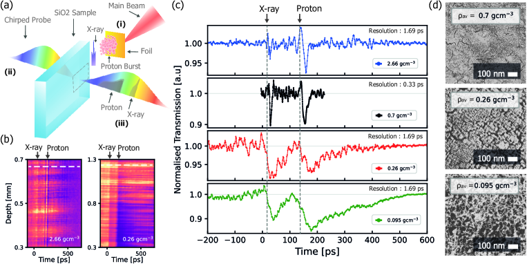
When a relativistically intense laser pulse ( Wcm-2) is incident at an oblique angle (30∘ - 45∘) onto a m-scale thick metal foil target, electrons from the front surface are driven through the foil by the strong electric field of the driving laser. This provides the basis for two highly synchronous ultrafast bursts of radiation from a single high-power laser pulse - a bright broadband pulse of Bremsstrahlung X-ray radiation and burst of protons via the Target Normal Sheath Acceleration (TNSA) mechanism [16]. While Bremsstrahlung from laser matter interactions is a well-established source of keV X-rays with ps scale duration, only recently have experiments verified that TNSA pulse durations as short as ps are possible from the same interactions [26]. Monte Carlo modelling performed using Geant4 [27] shows that protons ( MeV) generate dense nanotracks of ionisation with average electron energy, , eV. Conversely, X-rays ( keV, keV) undergo multiple high momentum transfer scattering events that homogenise the initial dose distribution and result in on the order of keV (See Supplemental Material [17]). In Fig.1(c) we track following the interaction of these ps-scale pulses in bulk and nanostructured SiO2 with a range of average densities () via single-shot optical streaking using the laser-driven accelerator at the TARANIS facility in Queen’s University Belfast [28, 26]. Silica aerogels [29, 30, 31, 32, 33, 34, 35] were used which comprise of a nanostructured matrix of solid density SiO2 nanoparticles with an average size of nm and tuneable average pore size (See Fig.1(d)). These nanostructured media can be described as a percolating fractal network across spatial scales from up to an acoustic correlation length (where ) [32, 33, 34]. The acoustic correlation length can be estimated as [35]. In SiO2, Longitudinal Optical (LO) phonon emission [36] and polaronic stopping (POL) [37, 38] represent the primary mechanisms of energy loss for electrons with average energies () within the sub-excitation range in SiO2. As such, aerogels provide control over heterogeneity on spatial scales relevant for scattering of sub-excitation energy electrons from LO phonons () in SiO2 [36].
Optical streaks obtained for X-rays followed by slower protons interacting in bulk and a range of nanostructured SiO2 samples are shown in Fig.1. These materials and densities straddle the transition expected for fractal behaviour in nanostructured SiO2 [35]. As can be seen there is a sharp increase in for both the X-rays and protons interacting in the 0.26 gcm-3 aerogel sample. Based on the simplistic scaling, one would expect a moderate times increase in . Instead, we observe factors of times longer in for both X-rays and protons interacting in aerogel. As mentioned above, for electrons due to MeV proton stopping in SiO2 is on the order 10’s of eV. From Fig.SM 2.1 in [17] (based on [36]), for these electrons in bulk SiO2 is nm. The implication is that the dose due to MeV proton stopping directly interrogates the nanoscopic heterogeneity in aerogel, which is at bulk density. In contrast, due to a bremsstrahlung source with a keV temp is 10’s of keV. From [17], for these electrons in bulk SiO2 is nm and considerably larger in the lower density samples (dashed line in Fig.SM 2.1). This is significantly larger than predicted by the scaling from [35]. This implies that these electrons probe the medium over lengths scales on which homogeneous behaviour is expected.
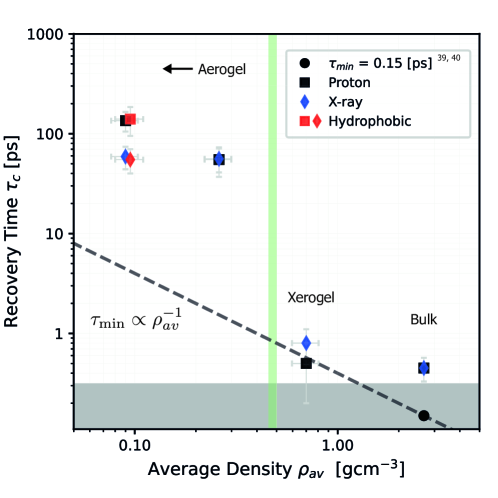
Fig.2 shows dependence of the recovery time on average density () which reveals a sharp discontinuity in the scaling. As the material structure changes from root mean square (rms) pore size nm to rms pore size nm (Fig.1(d)) the ultrafast response in SiO2 due to self-trapped exciton formation (STE) [39] is observed to quench, with ps growing to ps. Importantly, this switching is observed for recovery due to irradiation from both X-rays () and protons (). While this is a strong indication that ultrafast STE formation is being inhibited in aerogel, it is important to note that this does not preclude STE formation in general. It simply implies that the time taken to meet the trapping criterion put forward by Martin et al. [40] is significantly longer than would be expected if the scaling is applied to the result of Audebert et al. [39] (grey dashed line, Fig.2).
To help interpret these observations, we implement a energy transfer model [21, 20, 41] to track the early stage evolution of the thermal electron population caused by a single proton stopping in bulk SiO2 and aerogel with gcm-3. First, we consider the situation where the cross sections for interactions are independent of structure on the nanoscale. This is given by the solid traces in Fig.3(a) and 3(b), with red for bulk and blue for nanostructured SiO2
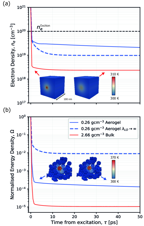
Fig.3(a) shows that following a rapid diffusion phase ( ps), there is a clear effect of the nanostructure in slowing the evolution of electron density, , in aerogel compared to that in bulk SiO2, with for extending to ps. However, for ps in both materials, is below cm-3. This is the maximum free carrier density observed for STE formation in SiO2 from experiments [42]. Accordingly, confinement of a near-solid density population of hot electrons in the nanostructure can be ruled out as the source of enhanced in aerogel. Similarly, Fig.3(b) reveals a corresponding increase in the kinetic energy density () for the electron population in nanostructured SiO2. This increase is largely reconciled by the increase in . Therefore, for these conditions, similar is expected for electrons ionised by protons stopping in both bulk and nanostructured SiO2. This suggests that the trapping criterion of Martin et al. [40] should not be violated in aerogel for conditions where ultrafast trapping is observed in the bulk sample. Our initial assumption of constant fails to expain for the enhanced observed in aerogel. To reconcile this discrepancy, we test how the nanoscopic heterogeneity of the aerogel may lead to the suppression of loss mechanisms for sub-excitation energy electrons. Assuming the cross section for LO phonon emission () becomes vanishingly small, i.e. the above picture changes considerably. For ps our model suggests times increase in over that expected in bulk SiO2 (blue dash line, Fig.3(b)). This implies that the requirement for to approach the thermal energy of the lattice for trapping via excitonic states is no longer met on ultrafast ( ps) timescales.
The assumption that in aerogel is supported in several ways. Firstly, it does not suggest that vibrations cease to exist in aerogel. The hierarchy of fractons and complex surface modes of the nanoparticles comprising the aerogel substructure can play a key role in the carrier dynamics post irradiation [29, 30, 31, 32, 43]. However, when it is considered that electrons with eV (electron affinity in SiO2) [37] are essentially confined in the nanoscopic network (i.e. Fig.3(a)) this can lead to a non-equilibrium phonon distribution due to a locally enhanced . For these conditions the cross section for reabsorption of optical phonons () approaches approximating to (hot phonon bottleneck) [44]. Secondly, LO phonons decay into two acoustic phonons on ps timescales [45]. While long-wavelength acoustic modes () can still propagate in aerogel, albeit at considerably reduced velocity to their bulk counterparts [34], recent work has begun to question the relative stopping powers for optical and short-wavelength acoustic phonons in bulk materials [46]. In aerogel these short wavelength acoustic modes will manifest as fractons with short localisation length for (). This implies a significantly reduced spatial overlap with free carriers in aerogel over bulk, subsequently reducing the probability of energy loss [12]. Finally, it is also important to recognise the significance of the quasi-infinite periodic lattice assumed in the PGM. As outlined earlier, the periodicity of the lattice spontaneously breaks the Galilean symmetries of free space leading to the emergence of Higgs and Goldstone modes, or optical and acoustic phonons respectively [2]. Conversely, the nanoscopic self-similarity of fractal networks implies that these systems do not possess the long-range translational symmetry of a crystal lattice i.e. the assumption of quasi-infinite periodicity is invalid. In this sense, Galilean symmetries are not spontaneously broken in self-similar/fractal materials as they are not translationally invariant [31, 32] and the assumption that in aerogels can be grounded in basic symmetry arguments. Although the modelling in Fig.3 provides insight into the excited electron population dynamics over short distances, the rigorous analysis required to fully interrogate recombination and trapping dynamics for the excited electron population in a numerical framework for this phase is beyond the scope of the work here [47].
In conclusion, this work further supports the idea that the fundamental nature of atomic scale vibrations, and what may be termed coherent phonon modes must be considered carefully in the context of disordered and nanostructured materials. Our work shows that this is particularly relevant for understanding the evolution of deposited energy due to ionising radiation in such media. The observation of a sharp discontinuity in the scaling of for a heterogeneous reduction in demonstrates that the efficient mechanisms for ultrafast relaxation of sub-excitation energy electrons via LO phonon emission is not dominant in aerogels over length scales where both strongly localised () and delocalised () phonon modes are expected. These length scales are probed by primary electrons due to proton and X-ray stopping respectively. This leads to the conclusion that ultrafast decay of free carriers in SiO2 emerges as the nanoscale structure of the material is modified to that of a non-fractal medium with long-range ( nm) periodic lattice. Furthermore, this work demonstrates how the multi-species capabilities of laser-driven accelerators can provide a platform for future investigations on the role of anomalous diffusion on fractal networks and in disordered systems in real time. It also provides the potential for highly synchronised harmonic probe beams with attosecond resolution [48] for investigating the fundamental nature of energy loss via electron-phonon interactions (i.e. LO versus acoustic modes) [46] in irradiated materials on the primary excitation timescales.
Acknowledgements.
We would like to thank V. P. Efremov for supplying aerogel samples. Work is funded through EPSCR grants (EP/P010059/1, EP/P016960/1 and EP/W017245/1). We are also grateful for use of the computing resources from the Northern Ireland High Performance Computing (NI-HPC) service funded by EPSRC (EP/T022175). L.S. also acknowledges funding from the European Union’s Horizon 2020 research and innovation programme under the Marie Skłodowska-Curie Actions staff exchange Grant agreement No. 823897 (MSCA-RISE ”ATLANTIC”). L.S. and J.S. also acknowledgments support from the COST Action CA17126 ’TUMIEE’.References
- Anderson [1972] P. W. Anderson, Science 177, 393 (1972).
- Vallone [2020] M. Vallone, Physica Status Solidi (b) 257, 1900443 (2020).
- Ackland [2010] G. Ackland, Science 327, 1587 (2010).
- Coughlan et al. [2020] M. Coughlan, H. Donnelly, N. Breslin, C. Arthur, G. Nersisyan, M. Yeung, B. Villagomez-Bernabe, M. Afshari, F. Currell, M. Zepf, et al., New Journal of Physics 22, 103023 (2020).
- Mo et al. [2019] M. Mo, S. Murphy, Z. Chen, P. Fossati, R. Li, Y. Wang, X. Wang, and S. Glenzer, Science Advances 5, eaaw0392 (2019).
- de Kruijff [2020] R. M. de Kruijff, International Journal of Radiation Biology 96, 419 (2020), pMID: 31829765.
- Cahill et al. [2014] D. G. Cahill, P. V. Braun, G. Chen, D. R. Clarke, S. Fan, K. E. Goodson, P. Keblinski, W. P. King, G. D. Mahan, A. Majumdar, et al., Applied Physics Reviews 1, 011305 (2014), ISSN 1931-9401.
- Wingert et al. [2016] M. C. Wingert, J. Zheng, S. Kwon, and R. Chen, Semiconductor Science and Technology 31, 113003 (2016).
- Reid [1976] J. S. Reid, Physics Education 11, 348 (1976).
- Toulemonde et al. [2012] M. Toulemonde, W. Assmann, C. Dufour, A. Meftah, and C. Trautmann, Nuclear Instruments and Methods in Physics Research Section B: Beam Interactions with Materials and Atoms 277, 28 (2012), ISSN 0168-583X, basic Research on Ionic-Covalent Materials for Nuclear Applications.
- Siddiqui and Arif [2016] M. U. Siddiqui and A. F. M. Arif, Materials 9 (2016), ISSN 1996-1944.
- Orbach [1986] R. Orbach, Science 231, 814 (1986).
- Surdutovich and Solov’yov [2019] E. Surdutovich and A. V. Solov’yov, Cancer Nanotechnology 10, 1 (2019).
- Tagliabue et al. [2020] G. Tagliabue, J. S. DuChene, M. Abdellah, A. Habib, D. J. Gosztola, Y. Hattori, W.-H. Cheng, K. Zheng, S. E. Canton, R. Sundararaman, et al., Nature Materials 19, 1312 (2020).
- Siemens et al. [2010] M. E. Siemens, Q. Li, R. Yang, K. A. Nelson, E. H. Anderson, M. M. Murnane, and H. C. Kapteyn, Nature materials 9, 26 (2010).
- Macchi et al. [2013] A. Macchi, M. Borghesi, and M. Passoni, Rev. Mod. Phys. 85, 751 (2013).
- [17] See supplemental material at, URL_will_be_inserted_by_publisher, for additional information.
- Henke et al. [1993] B. Henke, E. Gullikson, and J. Davis, Atomic Data and Nuclear Data Tables 54, 181 (1993), ISSN 0092-640X.
- Ziegler [1999] J. F. Ziegler, Journal of Applied Physics 85, 1249 (1999), ISSN 0021-8979.
- Klaumünzer [2006] S. Klaumünzer, Matematisk-fysiske meddelelser 52, 293 (2006).
- Kaganov [1957] M. Kaganov, Sov. Phys. JETP 4, 173 (1957).
- Lv and Henry [2016] W. Lv and A. Henry, Scientific reports 6, 37675 (2016).
- Anderson [1958] P. W. Anderson, Phys. Rev. 109, 1492 (1958).
- Graebner and Golding [1986] J. E. Graebner and B. Golding, Phys. Rev. B 34, 5788 (1986).
- Luckyanova et al. [2018] M. N. Luckyanova, J. Mendoza, H. Lu, B. Song, S. Huang, J. Zhou, M. Li, Y. Dong, H. Zhou, J. Garlow, et al., Science Advances 4, eaat9460 (2018).
- Dromey et al. [2016] B. Dromey, M. Coughlan, L. Senje, M. Taylor, S. Kuschel, B. Villagomez-Bernabe, R. Stefanuik, G. Nersisyan, L. Stella, J. Kohanoff, et al., Nature communications 7, 10642 (2016).
- Agostinelli et al. [2003] S. Agostinelli, J. Allison, K. Amako, J. Apostolakis, H. Araujo, P. Arce, M. Asai, D. Axen, S. Banerjee, G. Barrand, et al., Nuclear Instruments and Methods in Physics Research Section A: Accelerators, Spectrometers, Detectors and Associated Equipment 506, 250 (2003), ISSN 0168-9002.
- Dzelzainis et al. [2010] T. Dzelzainis, G. Nersisyan, D. Riley, L. Romagnani, H. Ahmed, A. Bigongiari, M. Borghesi, D. Doria, B. Dromey, M. Makita, et al., Laser and Particle Beams 28, 451–461 (2010).
- Schaefer and Keefer [1986] D. W. Schaefer and K. D. Keefer, Phys. Rev. Lett. 56, 2199 (1986).
- Courtens et al. [1987] E. Courtens, J. Pelous, J. Phalippou, R. Vacher, and T. Woignier, Phys. Rev. Lett. 58, 128 (1987).
- Anglaret et al. [1994] E. Anglaret, A. Hasmy, E. Courtens, J. Pelous, and R. Vacher, Europhysics Letters 28, 591 (1994).
- Nakayama and Yakubo [2003] T. Nakayama and K. Yakubo, Fractal concepts in condensed matter physics, vol. 140 (Springer Science & Business Media, 2003).
- Gefen et al. [1983] Y. Gefen, A. Aharony, and S. Alexander, Phys. Rev. Lett. 50, 77 (1983).
- Caponi et al. [2004] S. Caponi, G. Carini, G. D’Angelo, A. Fontana, O. Pilla, F. Rossi, F. Terki, G. Tripodo, and T. Woignier, Phys. Rev. B 70, 214204 (2004).
- Courtens et al. [1988] E. Courtens, R. Vacher, J. Pelous, and T. Woignier, Europhysics Letters 6, 245 (1988).
- Schreiber and Fitting [2002] E. Schreiber and H.-J. Fitting, Journal of Electron Spectroscopy and Related Phenomena 124, 25 (2002), ISSN 0368-2048.
- Stepanov [1995] S. V. Stepanov, Radiation Physics and Chemistry 46, 29 (1995), ISSN 0969-806X.
- Dapor [2012] M. Dapor, Journal of Physics: Conference Series 402, 012003 (2012).
- Audebert et al. [1994] P. Audebert, P. Daguzan, A. Dos Santos, J. C. Gauthier, J. P. Geindre, S. Guizard, G. Hamoniaux, K. Krastev, P. Martin, G. Petite, et al., Phys. Rev. Lett. 73, 1990 (1994).
- Martin et al. [1997] P. Martin, S. Guizard, P. Daguzan, G. Petite, P. D’Oliveira, P. Meynadier, and M. Perdrix, Phys. Rev. B 55, 5799 (1997).
- Grasser et al. [2003] T. Grasser, T.-W. Tang, H. Kosina, and S. Selberherr, Proceedings of the IEEE 91, 251 (2003).
- Mauclair et al. [2016] C. Mauclair, A. Mermillod-Blondin, K. Mishchik, J. Bonse, A. Rosenfeld, J.-P. Colombier, and R. Stoian, High Power Laser Science and Engineering 4, e46 (2016).
- Ramayya et al. [2008] E. B. Ramayya, D. Vasileska, S. M. Goodnick, and I. Knezevic, Journal of Applied Physics 104, 063711 (2008), ISSN 0021-8979.
- Yang et al. [2016] Y. Yang, D. P. Ostrowski, R. M. France, K. Zhu, J. Van De Lagemaat, J. M. Luther, and M. C. Beard, Nature Photonics 10, 53 (2016).
- Klemens [1966] P. G. Klemens, Phys. Rev. 148, 845 (1966).
- Fischetti et al. [2019] M. V. Fischetti, P. D. Yoder, M. M. Khatami, G. Gaddemane, and M. L. Van de Put, Applied Physics Letters 114, 222104 (2019), ISSN 0003-6951.
- Stella et al. [2021] L. Stella, J. Smyth, B. Dromey, and J. Kohanoff, The European Physical Journal D 75, 1 (2021).
- Seiffert et al. [2017] L. Seiffert, Q. Liu, S. Zherebtsov, A. Trabattoni, P. Rupp, M. Castrovilli, M. Galli, F. Süßmann, K. Wintersperger, J. Stierle, et al., Nature physics 13, 766 (2017).