Real-Time Boron Concentration Measurement in BNCT Using Compton Imaging
Abstract
Dosimetry in BNCT poses significant challenges due to the indirect effect of neutrons interacting with elements within the body and uncertainties associated with the uptake of boron compounds used in clinical practice. Current treatment planning relies on unconventional estimates of boron tumor uptake derived from prior PET scans and thus, an online boron-uptake monitor would be highly convenient. This work presents the first pilot experiments carried out at ILL-Grenoble with the high-efficiency Compton camera i-TED, hereby aiming at demonstrating its applicability for BNCT dosimetry by introducing real-time measurement of the boron concentration and imaging capabilities of spatial dose distribution. In this experiment, we measured the 10B uptake of different cancer cells of tongue squamous cell carcinoma, malignant melanoma and glioblastoma treated with BPA (80 ppm of 10B). The samples were irradiated with the thermal neutron spectrum of ILL-Grenoble and the 478keV -rays from the 7Li de-excitation after the neutron-boron reaction were registered both with the Compton imager and the high-sensitivity FIPPS HPGe array. These series of measurements allowed us to demonstrate the imaging capabilities of the Compton imaging device for this type of application, as well as to assess its sensitivity, which was found to be below 1 g of 10B.
keywords:
BNCT , dosimetry , Compton imaging , 10B concentration[1]organization=Instituto de Física Corpuscular, CSIC-Universitat de València,country=Spain \affiliation[2]organization=Institut Laue-Langevin,country=France \affiliation[3]organization=Instituto de Biopatología y Medicina Regenerativa, Centro de Investigación Biomédica. Universidad de Granada,country=Spain \affiliation[4]organization=Departamento de Bioquímica y Biología Molecular III e Inmunología, Universidad de Granada,country=Spain \affiliation[5]organization=Departamento de Física Atómica, Molecular y Nuclear, Universidad de Granada,country=Spain
1 Introduction
Boron neutron capture therapy (BNCT) is a promising cancer treatment technique, which exploits the high probability of a stable-boron nucleus (10B) to capture thermal (25 meV) or epithermal neutrons (1 eV to 10 keV) [1, 2]. The cross section of 10B for absorbing neutrons is significantly greater than those of the elements in the human body, thus the accumulation of 10B in tumor cells along with the external irradiation of neutrons lead to high dose concentrations in the tumor. Additionally, the products of this reaction (7Li nuclides and particles) have high-linear energy transfer (LET) and high relative biological effectiveness (RBE). These characteristics make BNCT unique among external radiotherapies in its cellular-level selectivity [3].
BNCT has been clinically investigated primarily for unresectable, locally advanced, and recurrent cancers, including glioblastoma multiforme, meningioma, head and neck cancers, lung cancers, and breast cancers [3]. BNCT is undergoing significant expansion, marked by the establishment of new in-hospital, accelerator-based facilities worldwide [4]. However, dosimetry in BNCT poses considerable challenges due to the indirect effect of neutrons with the elements present in the body and the unpredictable uptake of the boron compound 4‐borono‐L‐phenylalanine (BPA) used in clinical practice. As a result, treatment planning in BNCT is unconventional, relying primarily on estimates of boron uptake in tumors. These estimates are obtained through PET scans of patients using 18F-BPA, which provide an estimation of the tumor dose [5, 6]. Additionally, the dose to organs at risk is estimated based on boron analytics in the blood [7].
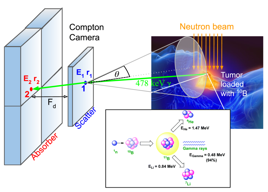
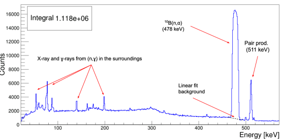
A breakthrough in the field could be accomplished if one could measure — in real time — the boron concentration at the tumor and at organs of risk during treatment, as it is well known that the boron concentration varies with time. This would lead to an accurate dosimetry and more efficient treatments. A very interesting possibility comes from the detection, quantification and imaging of the prompt 478 keV -rays emitted in 94% of neutron-capture reactions in 10B (see Fig. 1).
In this context, extensive research has been conducted on imaging using conventional gamma cameras (collimator-based) and single-photon emission computed tomography (SPECT) systems [8, 9, 10, 11, 12, 13]. These systems depend on two-dimensional (2D) collimation of -rays to produce images, which restricts the range of detectable energy (i.e., below 300 keV) and affects the attainable efficiency and spatial resolution [14]. Notably, significant background noise arises from the interaction of neutrons with the collimator material and the secondary 511 keV radiation generated by pair production [10]. As a promising alternative, Compton imaging has been proposed. A Compton camera localizes the emission point of gamma rays by analyzing the kinematics of its Compton scattering in a multi-layer detector. This device comprises at least two sensitive detectors — a scatter detector and an absorber detector — that record the position and energy loss of the Compton reaction, as illustrated in Fig. 1. In recent years, several research groups have investigated the potential of Compton imaging for BNCT using Monte Carlo simulations [15, 14, 16, 17]. However, experimental studies remain limited. For instance, a study using a commercial Si/CdTe Compton camera [18] reported a very low efficiency of 210-6, which may constrain its applicability for real-time dose assessment in BNCT.
In order to provide further experimental evidence of the potential of Compton imaging for real-time determination of the boron distribution in BNCT, we present in this work the first pilot experiments carried out with i-TED, a high-efficiency Compton-based imager developed at Instituto de Física Corpuscular (IFIC) within the HYMNS-ERC project [19]. The i-TED device and its Compton imaging performance are briefly introduced in Sec. 2. Sec. 3 describes the experiment carried out at ILL, in which several BPA and cell samples were irradiated with a thermal neutron spectrum and the 478keV -rays were registered both with the Compton imager and the high-sensitivity FIPPS HPGe array. The analysis of this pilot experiment, whose results are discussed in Sec. 4, aims at studying the sensitivity to measure low 10B concentrations with i-TED compared to the a high-resolution low-background setup of FIPPS, and providing the first experimental insight on the ability and performance of the i-TED imaging system for the 478 keV -ray of interest for BNCT.
2 Compton imaging with i-TED
2.1 The i-TED Compton imager
In the pilot experiments presented in this work we evaluated the performance of one of the Compton cameras of i-TED, an array of four high-efficiency Compton cameras [20, 21]. The original aim of this Compton camera was the enhancement of the sensitivity in high-resolution neutron TOF experiments of key stellar reactions [20, 21, 22]. After the development and characterization of the first demonstrator [23, 24, 25, 26], the full array has been assembled and characterized in recent years [27] and used for experiments of astrophysical relevance at the CERN-n_TOF-Facility [22, 28]. A picture of the full i-TED Compton imaging array installed in the neutron beam of CERN-n_TOF is shown in Fig. 2. Moreover, the applicability of the imaging capability of i-TED to range verification in Hadron Therapy by means of Prompt Gamma Imaging and Dual PET-Compton imaging has been explored with promising results [29, 30], as well as its application for nuclear waste characterization [31].
Each of the i-TED Compton modules encompasses five position-sensitive detectors (PSDs) distributed in two parallel detection planes, Scatter (S) and Absorber (A), as shown in Figs. 1- 2. The innovative i-TED design comprises a geometry where the four-fold size of the A-detector with respect to the S-detector enhances its efficiency, especially for low-energy -rays characterized by large scattering angles (see Fig.2 in Ref. [26]). Each PSD contains a LaCl3(Ce) monolithic crystal with a square-cuboid shape and a base surface of 5050 mm2. The LaCl3 is hygroscopic and thus it is encapsulated in an aluminum housing. Each crystal base is coupled to a 2 mm thick quartz window, which is optically joined to a silicon photomultiplier (SiPM) from SensL (ArrayJ-60035-64P-PCB). The photosensor features 88 pixels over a surface of 5050 mm2. A 15 mm thick crystal is used for the PSD in the S-plane. Four 25 mm thick crystals are utilized for the PSDs placed in the A-plane (see Fig. 2). In total, 320 SiPM channels per Compton module are biased and readout by means of front-end and processing PETsys TOFPET2 ASIC electronics [32], which allow acquiring event-rates of up to 500 kHz per individual detector. In order to minimize gain shifts due to changes in the temperature of the experimental hall, every ASIC is thermally coupled to a refrigeration system composed by a Peltier cell, a heat-sink and a small-size fan (see Ref. [26] for further details). More recently, a water-cooled system has been installed to further improve the temperature stability and, consequently, the energy resolution and the overall imaging performance.
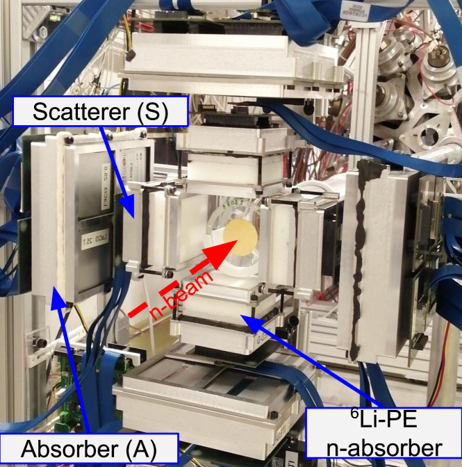
An important feature for the application of i-TED to BNCT relies in its relatively low neutron sensitivity. To accomplish the latter goal LaCl3(Ce) was preferred versus other options, owing to the relatively small integral capture cross section of Chlorine, and the small contribution of resonances in the keV-energy range of relevance. The Compton modules are supplemented with 6Li-loaded neutron-absorber pads of 20 mm thickness for further reducing the intrinsic neutron sensitivity of the array [33]. In addition, the successful implementation of ASIC-based TOF-PET readout electronics for Compton imaging has led to a rather compact and cost-effective system, when compared to other Compton imagers [34, 35].
2.2 Compton imaging technique and performance
The Compton imaging technique is applied with i-TED using its high resolution position [24, 25] and energy [23] sensitive radiation detectors operated in time coincidence. In order to reconstruct the Compton image, energy depositions (E1 and E2 in Fig. 1) and 3D-localisation of the -ray hits (r1 and r2) in both layers are reconstructed for each coincidence event. Details on the reconstruction methods can be found elsewhere [26, 21, 30]. From the energies and positions, one can trace a cone, whose central axis corresponds to the straight line defined by the -ray interaction position in the two layers and its aperture is obtained from the measured energies using the Compton scattering formula, see Fig. 1. The i-TED Compton modules embed the so-called dynamic electronic collimation technique [26], that allows one to remotely vary the distance between the A- and S-planes,Fd in Fig. 1, thereby optimizing the balance between efficiency and imaging resolution [26].
Most of the studies published to date on the Compton imaging performance of i-TED [26, 21] have been based on an adaptation of the fast back-projection (BP) method of Ref. [36]. In order to boost the performance of i-TED for medical [29, 30] and industrial [31] imaging applications, more evolved imaging algorithms have been recently implemented. This algorithms comprise the Stochastic Origin Ensemble (SOE) method [37], the Maximum-Likelihood Expectation-Maximization (MLEM) [38] and the Analytical Algorithm (AA) of Tomitani et al. [39]. More details on the implementation of the algorithms and a comparative study of their performance are given in Refs. [29, 40, 30].
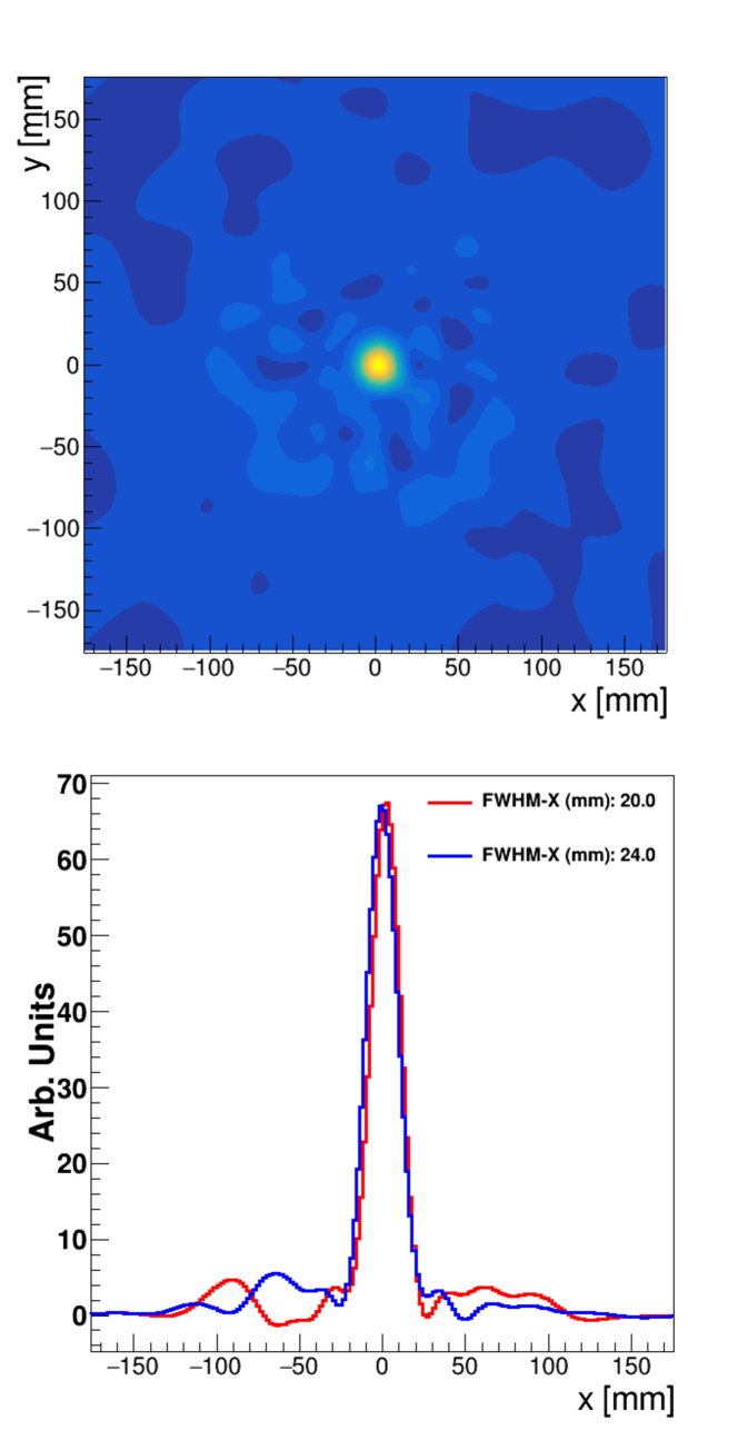
To illustrate the state-of-the-art imaging performance of i-TED, in Fig. 3 we present a Compton image reconstructed using the best performing AA algorithm. To generate this image a 22Na source with an activity of 230 kBq was placed at 100 mm from one of the i-TED Compton cameras and measured for 900 s. The focal distance between the two PSD planes of the Compton camera (Fd in Fig. 1) was set to 50 mm, to optimize the angular resolution [26]. Last, coincidence events corresponding to the 1274 keV -ray emitted in the decay of a 22Na source where selected. The bottom panel of the same figure shows the projection of the image onto the X and Y axis around the maximum. The projections show the accurate reproduction of the source coordinates (0,0) and allow one to quantify the spatial resolution, which is found to be 20 mm (FWHM), significantly better than the 40-50 mm obtained with an early prototype of i-TED at the same distance from the source using the back-projection algorithm [21]. The spatial resolution determined from the projections in Fig. 1 corresponds to an angular resolution of about 5∘ to the level of one standard deviation (), clearly enhanced with respect to the early demonstrator of the current Compton cameras [26]. These results demonstrate that the recent upgrades of i-TED [25, 27], combined with more advanced imaging-algorithms, have boosted its performance to a level, which is particularly well suited for this type of medical applications.
At variance with mechanically-collimated systems, one of the main challenges to implement electronic (Compton) collimation for real-time dosimetry is the computing time required with advanced image-reconstruction algorithms, which scales with the complexity of the involved models. According to our recent benchmark study [29], the fastest algorithm is BP followed by SOE and AA, being the latest about a factor 500 slower than BP. To overcome this limitation and really profit from its clearly superior performance, a GPU-boosted implementation of the AA algorithm based on the CUDA 11.1 toolkit was developed. The CUDA version of the Analytical Algorithm speeds-up the image reconstruction in a factor of 120 with respect to the singled-threaded CPU version, and brings it back reconstruction times comparable to the SOE algorithm and only a factor 5 slower than the fast back-projection. For the technical details on this work, the reader is referred to [29, 30].
3 First pilot experiment at ILL
3.1 Neutron beam at ILL-FIPPS
The experiment discussed herein was carried out at the FIPPS instrument, which exploits the thermal neutron beam of the H22 guide exiting the research reactor of Institut Laue-Langevin (ILL). This reactor produces the most intense continuous neutron flux in the world in the moderator region, 1.51015n/cm2/s, with a thermal power of 58.3 MW.
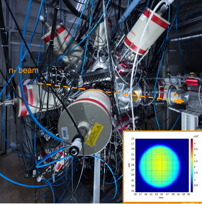
The thermal neutron beam of the H22 guide is collimated in vacuum up to the target position, located at the center of the FIPPS array (see Fig. 4). The complex collimation system, which aims at reducing the beam-related -ray background in the detector, is composed of B4C apertures followed by 5 cm lead absorbers and enriched 6LiF apertures. The latter are placed in the closest position to the FIPPS detectors [41]. The neutron beam at the target position, shown in the insert of Fig. 4, has a diameter of 1.5 cm and a flux of 5107n/cm2/s [41].
3.2 Experimental setup
The FIPPS detector array, shown in Fig. 4, consists on eight germanium clover detectors arranged in the plane perpendicular to the neutron beam at the target position. Eight additional clover detectors with their anti-Compton (AC) shields from the IFIN collaboration are mounted at 45∘ with respect to this plane [42]. A Li-loaded tube mounted around the neutron beam is used to place the target in vacuum in the center of the setup and absorb scattered neutrons, thus minimizing the neutron-induced -ray background. FIPPS features a very high energy resolution of 2 keV at 1.3 MeV (60Co) which, combined to the high efficiency of the 16 clovers (8% at 1.4 MeV and 18% at 487 keV [41]) and the optimized background conditions, lead to a unique sensitivity to detect neutron-induced -rays, such as the 478 keV of the 10B(n,) reaction. However, it is worth noting that such a detection system would not be compatible with a clinical BNCT treatment room.
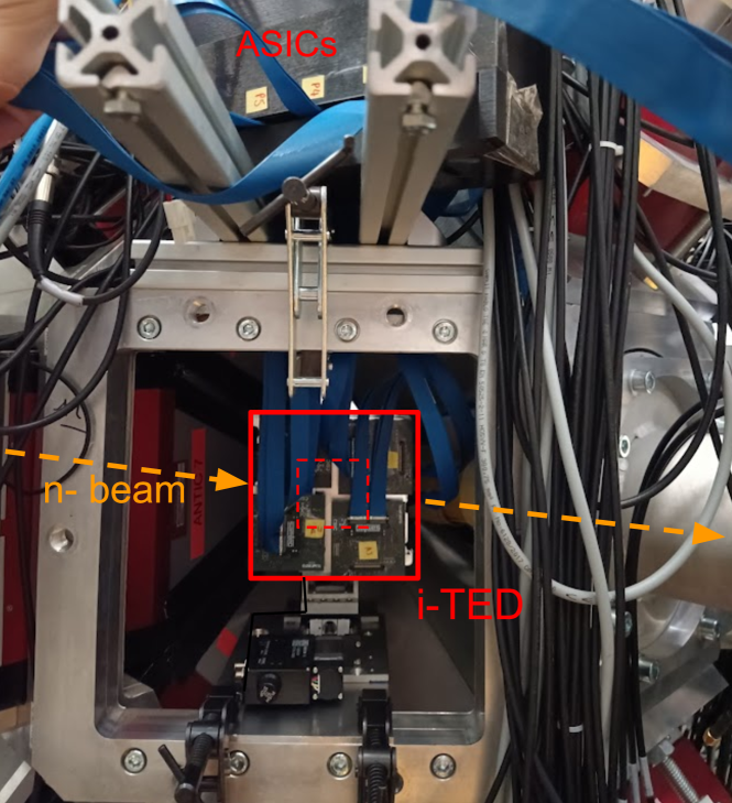
For this experiment, one of the Compton Cameras of i-TED was integrated in the FIPPS setup with the aim of comparing the sensitivity of the two setups. The bottom panel of Fig. 4 shows the Compton imager installed in the gap left by one of the FIPPS clover detectors, that was taken out from the array. The Compton imager was vertically and horizontally aligned with the target and placed 150 mm away from its center at an angle of 45∘ with respect to the beam. The compactness of the device and electronics were key to adapt the setup to the limited space within the FIPPS array.
3.3 Samples and measurements
The campaign at ILL can be divided in two main experimental blocks, with different sample characteristics. A summary of all the samples, their main characteristics and irradiation times can be found in Table 1.
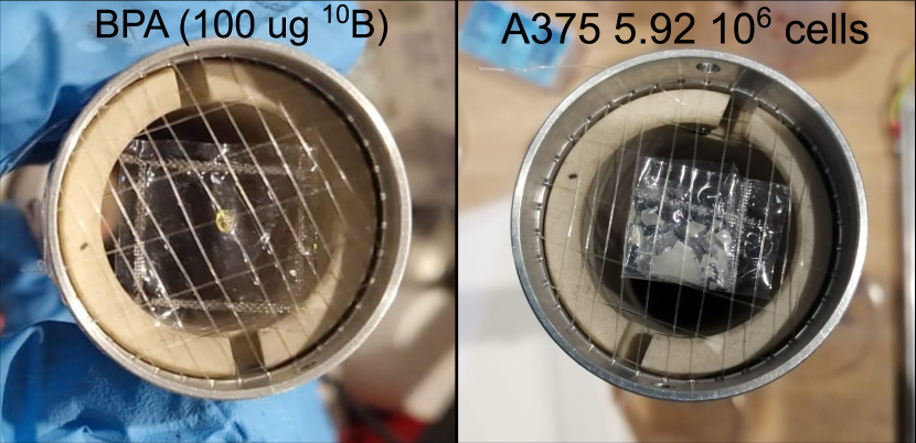
In the first part, a series of reference samples made out of a evaporated solution of fructose with BPA, with 10B equivalent contents spanning between roughly 300 ng and 500 g, were irradiated in the thermal neutron beam. This allowed us to calibrate the detection response of each apparatus as a function of the 10B content, study their sensitivity and evaluate the Compton imaging performance of i-TED for a point-like sample of 487 keV -rays. The left panel of Fig. 5 shows the sample containing 100 g of 10B sealed in a Teflon bag and mounted on the target holder of FIPPS.
After the previous characterization study, in the second block of the campaign a series of biological 10B-loaded samples were irradiated with thermal neutrons. Samples of tumor cell lines CAL33 (head and neck cancer), A172 (Glioblastoma) and A375 (melanoma) containing 4–18106 cells were treated with BPA (80 ppm of 10B in the medium) before drying them and depositing a monolayer of cells on a Teflon foil that is fully intercepted by the neutron beam (see right panel of Fig. 5). The irradiation time of each sample, listed in Table 1, was estimated to achieve sufficient statistics with i-TED in S&A coincidence — 100 kcounts — for the imaging of the 478 keV -rays.
| Sample | 10B mass (g) | TIrrad (min) |
|---|---|---|
| BPA + fructose | 500–0.6 | 5–900 |
| Fructose (bckg.) | 0 | 30 |
| Cell Line | Cells (x106) | TIrrad (min) |
| CAL33 | 17.4 / 16.1 | 660 / 450 |
| A172 | 14.0 / 5.92 | 570 / 1080 |
| A375 | 18.4 / 11.2 | 435 / 360 |
Besides the boron-loaded samples and cells, ancillary irradiations were carried out to assess the beam-related background with no BPA, by measuring a pure fructose sample deposited on the same Teflon bags. Moreover, the flux was measured by gold-foil activation. Last, ancillary calibration measurements with a 152Eu source were taken to calibrate in energy both the HPGe detectors of FIPPS and the five individual LaCl3 detectors of the Compton imager.
4 Results and discussion
4.1 Sensitivity to 10B uptake
The aims of the sensitivity study were the determination of the 10B uptake in various cell lines treated with BPA and the comparison between the sensitivity of the high-resolution spectrometer FIPPS versus the medium-resolution Compton imager. Prior to this, we had to calibrate the detection response to the absolute 10B content of the samples and assess its linearity. To this end, we analyzed the spectra registered with both setups during the irradiation of BPA samples with known 10B concentration and studied the correlation between the 478 keV peak count rate and the 10B mass.
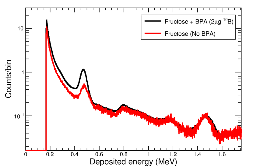
In the case of i-TED, the response of the individual detectors composing the Compton camera was individually calibrated to the 10B mass. To this aim, the energy spectra registered for each of irradiation were reconstructed applying the individual energy calibrations obtained from the measurement of a 152Eu sample and a temperature-dependent gain correction. Then, the count rate associated to the 478 keV line was determined from the excess of counts in each measurement with respect to the background counts. Fig. 6 shows the deposited energy spectra of a detector of the absorber plane obtained in the irradiation of the BPA sample containing 2g of 10B (black) compared to the measurement with no BPA content (red). To properly account for the underlying continuum, the 478 keV photo-peak was fitted in all the measurements to a Gaussian function and a linear background and the count rate was determined from the integral of the Gaussian fit.
.
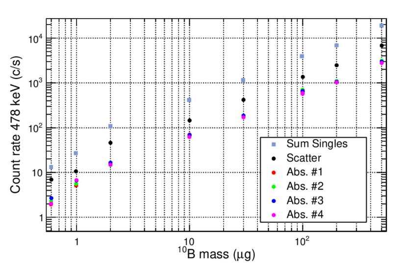
The background-subtracted count rates resulting from the analysis of the different BPA samples irradiated with thermal neutrons are displayed in Fig. 7. In this figure, the correlation between the 478 keV peak count rate and the 10B content of the irradiated samples is shown for the five individual crystals and the sum of all of them. Despite the large gamma-ray emission yields originated in the 10B(n,) reaction, expected to be 106 gamma-rays per second for the 500g sample, the count rate in single i-TED detectors does not exceed 10 kHz and, thus, no dead-time effects were noticed. Focusing in the low mass range (see Fig. 7), the low background conditions within the FIPPS setup and the low neutron sensitivity of i-TED have pushed the sensitivity of i-TED down to at least 600 ng of 10B.
As for FIPPS, the signals were processed with digital electronics (CAEN V1724 cards) with a sampling frequency of 100 MHz. The events were built in a 200 ns wide window from the data recorded in list-mode [41]. The data reduction chain merged the spectra of the full array of clover detectors and generated a energy-calibrated spectra, like the one displayed in Fig 8. The unique spectroscopic performance of the HPGe allows to perfectly separate the annihilation peak at 511 keV from the 478 keV peak. The latter has a broad rectangular line shape that is 8–10 times larger than that of normal -rays peaks as a result of Doppler broadening [43]. In order to build the calibration curve with FIPPS, the count-rate of the 478 keV photo-peak was determined after fitting the background with a linear fit (see Fig. 8) and subtracting its contribution. The resulting calibration showed a good linearity down to the lowest measured mass of 600 ng, as expected from the high sensitivity of FIPPS. On the contrary, samples of 100 g and beyond, leading to count rates in the individual HPGe 10 KHz, were discarded from the calibration, due to the significant dead time losses. The latter limitation would represent a true additional drawback towards the implementation of HPGe detectors for dose monitoring in BNCT.
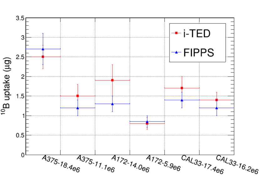
Once the response of both setups had been calibrated, the 10B uptake of the different dried cell samples (see Table 1) was determined from their irradiation in the thermal neutron beam of FIPPS. The background-subtracted counting rate of the 478 keV peak registered during the irradiation was transformed into 10B content, by means of a simple linear interpolation of the calibration curves. The resulting values for the 10B uptake of the six cell samples is presented in Fig. 9. This figure shows a consistent agreement — within 1 for most cell samples, and below 1.5 for all of them— for the results obtained with FIPPS and i-TED. The uncertainties in the results of Fig. 9 include the systematic uncertainty in the 10B masses of the calibration samples, estimated to be 10 %. In the case of i-TED, the final value for the 10B mass was computed as the average of the individual detectors, and the standard deviation of the individual results was incorporated as an additional systematic uncertainty. In summary, these results validate the capability of i-TED to perform absolute measurements of small boron concentrations in cells. Indeed, the results of 10B uptake of the cell lines in this work are being used in the pre-clinical cell survival study of Ref. [44].
4.2 Compton imaging in BNCT
The second goal of the pilot experiment discussed herein was to address for the first time the potential of Compton imaging with i-TED in BNCT. To evaluate the imaging performance of this Compton imager, we profited from the same irradiations used for the sensitivity study.
In order to reconstruct the images, only events in time coincidence between the S- and A-layers were considered. This selection reduces the efficiency to about 15% relative to that of the Scatter detector [21]. As introduced in Sec. 2, the Compton imaging algorithms make use of the energy depositions and 3D-localisation of the -ray hits in both S- and A- position sensitive layers to compute the Compton cones and reconstruct the images. In order to image a single -ray energy, a selection is performed in the sum-energy value in time coincidence, referred to as add-back energy. As an example, the i-TED add-back spectrum for the BPA sample equivalent to 10 g of 10B is shown in Fig. 10, where it is also compared to a background run with no 10B. Signals below the 478 keV peak correspond to non-full-energy deposition events.
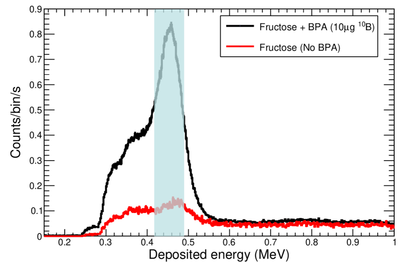
The main experimental challenges for the Compton imaging of such low -ray energies arise from the poor energy resolution at low energy depositions and the efficiency and angular restrictions due to the low energy thresholds [26]. As observed in Fig. 10, add-back deposited energies go down to 240-280 keV, associated to a low energy detection threshold of about 100 keV in the S- detector and 140-180 keV in the A-cristal. Moreover, the fair add-back energy resolution at this energy enables the clear distinction of the peak from the Compton continuum at lower add-back energies (see Fig. 10).
The coincident events falling inside the add-back energy window 0.42–0.5 MeV were selected to perform Compton imaging (see shadowed area in Fig. 10). Compton images were reconstructed on an image plane parallel to the detector planes and located in the sample position, 15 cm away from the Compton imager. Finally, the Compton images shown hereafter were obtained by implementing the Analytical Algorithm of Tomitani et al. [39], introduced in Sec. 2.
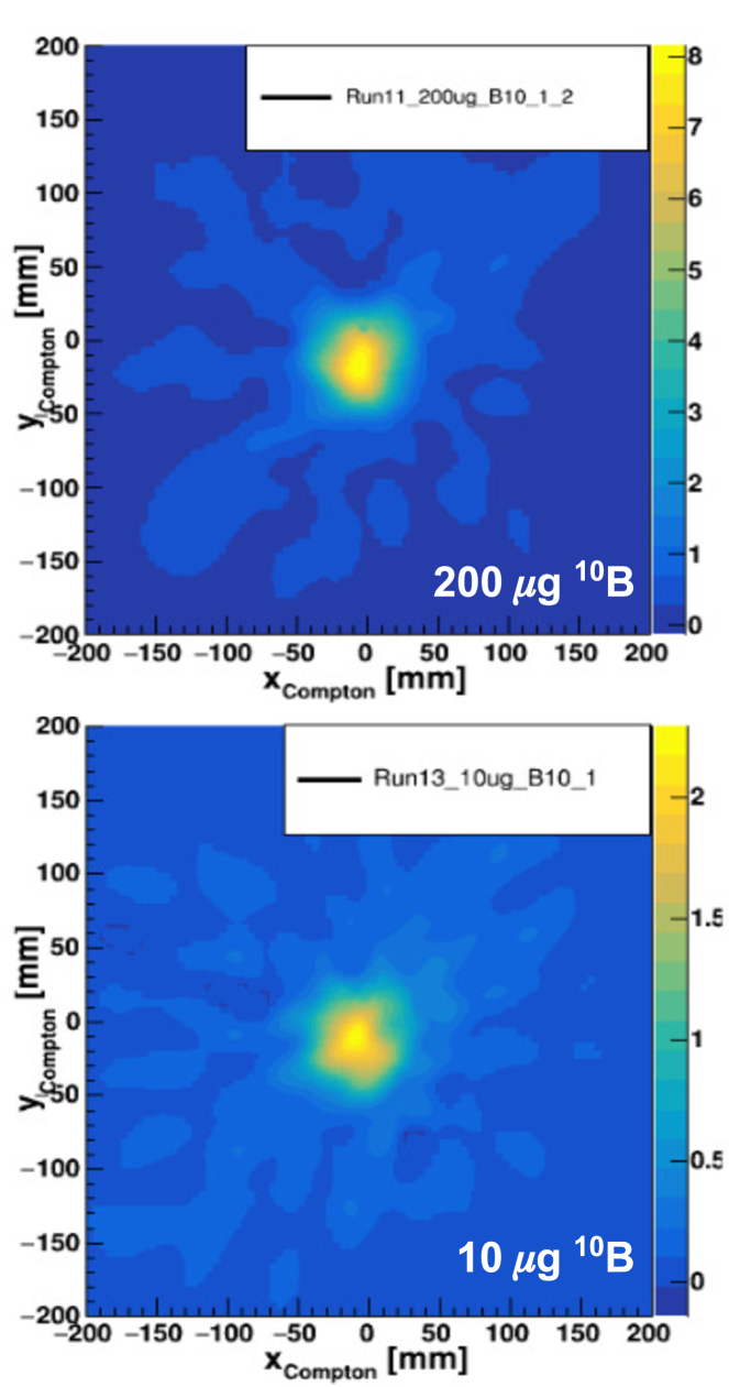
To demonstrate the 2D Compton imaging reconstruction capability and resolution, we profited from the measurements of the point-like 10B calibration samples. Fig. 11 shows the resulting images obtained for two samples with very different 10B content, 200 and 10 g. These images were obtained during irradiations of, respectively, 15 min and 180 min in the thermal beam of the H22 guide. The analysis of the Compton images of Fig. 11 indicates that the spatial resolution achieved at 478 keV is 49–54 mm (FWHM), corresponding to an angular resolution of 8-9∘ at the level of one standard deviation. The worsening of the resolution from the state-of-the-art performance of Fig. 3 is expected from the impact of the reduced energy resolution at lower gamma-ray energy [26]. Comparing the two 2D images of Fig. 11, one observes the different peak to background ratio obtained with 200 and 10 g of 10B. The latter effect can be explained from the different relative contribution of the 478 keV -rays present in the neutron induced background, see Figs 6 and 10.
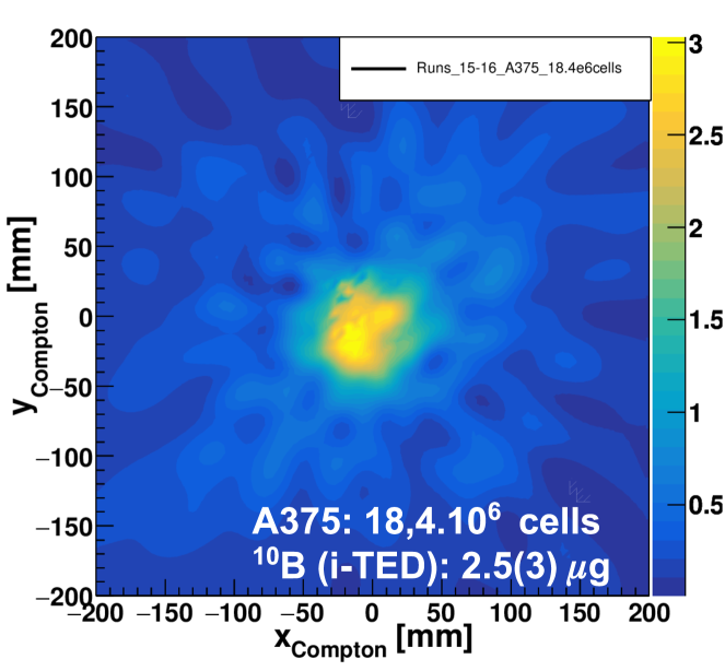
To provide a clearer insight on the ability and performance of the i-TED imaging system in a realistic situation with extended biological samples previously loaded with boron, 2D Compton images were also reconstructed for the different cell samples listed in Table 1. The Compton image reconstructed from the irradiation of a dried monolayer of A375 melanoma cells is displayed in Fig. 12. This sample, containing 18.4106 cells, was treated with 80 ppm BPA with an uptake of 2.5(3) g of 10B (see Fig. 9), and then irradiated in the FIPPS setup for 435 min. The 2D image of Fig. 12 shows a relatively broad spatial resolution of 55–60 mm (FWHM) and less symmetric shape than the images of point-like BPA samples. This may reflect the uneven distribution of the pieces of dried cells in the neutron beam, as shown in Fig. 5. The image of a sample containing only 2.5g of 10B, even though neutron induced background on Boron is present, shows the sensitivity of the device for the spatial localization of dose in BNCT. After this successful first pilot experiment, an optimization of the i-TED imaging system based on Monte Carlo simulations of a clinical scenario and more realistic tests have been undertaken [33].
5 Conclusions and outlook
Online boron-uptake monitoring and imaging of the 478 keV line would be of great value for real-time dosimetry in BNCT. In this work, we have presented the first pilot experiment of the attainable sensitivity and imaging performance of i-TED, an array of four high-efficiency Compton cameras based on large LaCl3 crystals, that is fully developed and has demonstrated its imaging capability in a wide range of fields. Its compactness, high count rate capability, low neutron sensitivity and GPU-Boosted algorithms make it particularly well suited for its application to real-time imaging in BNCT.
The experiment presented herein was carried out at ILL-Grenoble using the very high thermal neutron flux of 5107n/cm2/s and using the FIPPS instrument as reference. One of the i-TED Compton imagers was embedded in the FIPPS HPGe array and both setups were run in parallel to detect the 478 keV -rays originated in 10B(n,)7Li in BPA samples with known 10B content and tumoral cell samples (CAL33, A172, A375) treated with BPA (80 ppm 10B). The results show that, in the low-background conditions of the FIPPS setup, i-TED is sensitive enough to measure 10B concentrations 1 g and is capable to determine absolute 10B concentrations in cell samples, obtaining consistent results to the high-sensitivity FIPPS array. These results make i-TED a promising non-destructive tool for pre-clinical in-vitro studies and open the door to future online dosimetry in clinical treatment. Last, this work represents the first experimental demonstration of the Compton imaging performance of i-TED for 478 keV -rays. 2D Compton images of cell samples of few g have been reconstructed, despite the presence of boron-related background.
The successful pilot experiments described herein represent the first milestone towards the future applicability of large field-of-view Compton imaging in a more realistic BNCT preclinical scenario, where a broad neutron beam is irradiating a large biological sample loaded with 10B. The on-going work and future prospects, discussed in a recent publication [33], include a new experimental campaign of the full i-TED array at ILL with more realistic phantoms, the optimization of the Compton camera in a Monte Carlo based clinical scenario, the development of 3D Compton algorithms, and, finally, future experimental campaigns with clinical beams. Last, besides the development of the Compton-based monitoring in BNCT, we aim at evaluating the performance in this field of a novel device which complements the -ray Compton technique with the imaging of the outgoing, mostly thermalized neutrons [45, 46].
Declaration of competing interest
The authors declare that they have no known competing financial interests or personal relationships that could have appeared to influence the work reported in this paper.
CRediT authorship contribution statement
J. Lerendegui-Marco: Investigation, Methodology, Formal analysis, Data curation, Visualization, Writing - original draft. J. Balibrea-Correa: Investigation, Methodology, Formal analysis, Data curation, Visualization, software. P. Álvarez-Rodríguez: Investigation. V. Babiano-Suárez: Investigation, Methodology, software. B. Gameiro: Investigation, Methodology, software. I. Ladarescu: Investigation, Software. C. Méndez-Malagón: Investigation. C. Michelagnoli: Investigation, Resources. I. Porras: Investigation, Resources, Project administration, Funding acquisition, Writing - review & editing. M. Porras-Quesada: Investigation. C. Ruiz-Ruiz: Investigation, Resources, Writing - review & editing. P. Torres-Sánchez: Investigation, Methodology, Writing - review & editing. C. Domingo-Pardo: Conceptualization, Investigation, Methodology, Supervision, Project administration, Funding acquisition, Writing - review & editing.
Acknowledgment
This work builds upon research conducted under the ERC Consolidator Grant project HYMNS (grant agreement No. 681740) and has been supported by the ERC Proof-of-Concept Grant project AMA (grant agreement No. 101137646). We also acknowledge funding from the Spanish Ministerio de Ciencia e Innovación under grants PID2022-138297NB-C21 and PID2019-104714GB-C21, as well as from CSIC under grant CSIC-2023-AEP128 and the Instituto de Salud Carlos III under grant DTS22/00147. Additionally, the authors thank the support provided by postdoctoral grants FJC2020-044688-I and ICJ220-045122-I, funded by MCIN/AEI/10.13039/501100011033 and the European Union NextGenerationEU/PRTR; postdoctoral grant CIAPOS/2022/020 funded by the Generalitat Valenciana and the European Social Fund and a PhD grant PRE2023 from CSIC. Financial support from the Institut Laue-Langevin during the experimental campaign is also gratefully acknowledged.
References
- [1] W. A. G. Sauerwein. Principles and Roots of Neutron Capture Therapy, pp. 1–16. Springer Berlin Heidelberg, Berlin, Heidelberg, 2012.
- [2] W. H. Jin et al. A review of boron neutron capture therapy: Its history and current challenges. International Journal of Particle Therapy, 9(1):71–82, 2022.
- [3] T. D. Malouff et al. Boron neutron capture therapy: A review of clinical applications. Frontiers in Oncology, 11, 2021.
- [4] Y. Kiyanagi et al. Status of accelerator-based BNCT projects worldwide. AIP Conference Proceedings, 2160(1):050012, 10 2019.
- [5] G. W. Kabalka et al. Evaluation of fluorine-18-BPA-fructose for boron neutron capture treatment planning. J Nucl Med, 38(11):1762–1767, November 1997.
- [6] M. Balcerzyk et al. Quantification of boron compound concentration for bnct using positron emission tomography. Cells, 9(9), 2020.
- [7] H. Koivunoro et al. Boron neutron capture therapy for locally recurrent head and neck squamous cell carcinoma: An analysis of dose response and survival. Radiotherapy and Oncology, 137:153–158, 2019.
- [8] T. Kobayashi et al. A noninvasive dose estimation system for clinical BNCT based on PG-SPECT—conceptual study and fundamental experiments using hpge and cdte semiconductor detectors. Medical Physics, 27(9):2124–2132, 2000.
- [9] P. M. af Rosenschöld et al. Prompt gamma tomography during BNCT—a feasibility study. Journal of Instrumentation, 1(05):P05003, may 2006.
- [10] D. Minsky et al. First tomographic image of neutron capture rate in a BNCT facility. Applied Radiation and Isotopes, 69(12):1858–1861, 2011. Special Issue: 14th International Conference on Neutron Capture Therapy.
- [11] D.-K. Yoon et al. Tomographic image of prompt gamma ray from boron neutron capture therapy: A Monte Carlo simulation study. Applied Physics Letters, 104(8):083521, 02 2014.
- [12] S. Fatemi et al. Innovative 3d sensitive CdZnTe solid state detector for dose monitoring in Boron Neutron Capture Therapy (BNCT). Nuclear Instruments and Methods in Physics Research Section A: Accelerators, Spectrometers, Detectors and Associated Equipment, 936:50–51, 2019. Frontier Detectors for Frontier Physics: 14th Pisa Meeting on Advanced Detectors.
- [13] A. Caracciolo et al. A spectroscopic and imaging gamma-detector prototype towards dose monitoring in bnct. Nuclear Instruments and Methods in Physics Research Section A: Accelerators, Spectrometers, Detectors and Associated Equipment, 1048:168019, 2023.
- [14] C. hui Gong et al. Optimization of the compton camera for measuring prompt gamma rays in boron neutron capture therapy. Applied Radiation and Isotopes, 124:62–67, 2017.
- [15] T. Lee et al. Monitoring the distribution of prompt gamma rays in boron neutron capture therapy using a multiple-scattering compton camera: A monte carlo simulation study. Nuclear Instruments and Methods in Physics Research Section A: Accelerators, Spectrometers, Detectors and Associated Equipment, 798:135–139, 2015.
- [16] Z. Hou et al. Boron concentration prediction from compton camera image for boron neutron capture therapy based on generative adversarial network. Applied Radiation and Isotopes, 186:110302, 2022.
- [17] D. Ramos López et al. Study of alternative imaging methods for in vivo boron neutron capture therapy. Cancers (Basel), 15(14), July 2023.
- [18] M. Sakai et al. Experimental study on compton camera for boron neutron capture therapy applications. Scientific Reports, 13(1):22883, Dec 2023.
- [19] High-sensitivitY Measurements of key stellar Nucleo-Synthesis reactions (HYMNS). ERC Consolidator Grant, Agreement No. 681740, PI C. Domingo-Pardo.
- [20] C. Domingo-Pardo. i-TED: A novel concept for high-sensitivity (n,) cross-section measurements. Nuclear Instruments and Methods in Physics Research A, 825:78–86, July 2016.
- [21] V. Babiano et al. Imaging neutron capture cross sections: i-TED proof-of-concept and future prospects based on machine-learning techniques. The European Physical Journal A, 57:197, 2021.
- [22] Lerendegui-Marco, J. et al. New detection systems for an enhanced sensitivity in key stellar (n,) measurements EPJ Web Conf., 279:13001, 2023.
- [23] P. Olleros et al. On the performance of large monolithic LaCl3(Ce) crystals coupled to pixelated silicon photosensors. Journal of Instrumentation, 13(03):P03014–P03014, mar 2018.
- [24] V. Babiano et al. -Ray position reconstruction in large monolithic LaCl3(Ce) crystals with SiPM readout. Nuclear Instruments and Methods in Physics Research A, 931:1–22, July 2019.
- [25] J. Balibrea-Correa et al. Machine Learning aided 3D-position reconstruction in large LaCl3 crystals. Nuclear Instruments and Methods in Physics Research Section A: Accelerators, Spectrometers, Detectors and Associated Equipment, 1001:165249, 2021.
- [26] V. Babiano et al. First i-TED demonstrator: A Compton imager with Dynamic Electronic Collimation. Nuclear Instruments and Methods in Physics Research A, 953:163228, 2020.
- [27] B. Gameiro. Optimization of imaging techniques for background suppression of stellar nucleo-synthesis reactions with i-TED, 2023.
- [28] C. Domingo-Pardo et al. Advances and new ideas for neutron-capture astrophysics experiments at cern n_tof. The European Physical Journal A, 59(1):8, Jan 2023.
- [29] J. Lerendegui-Marco et al. Towards machine learning aided real-time range imaging in proton therapy. Scientific Reports, 12(1):2735, Feb 2022.
- [30] J. Balibrea-Correa et al. Hybrid in-beam pet- and compton prompt-gamma imaging aimed at enhanced proton-range verification. Eur. Phys. J. Plus, 137(11):1258, 2022.
- [31] V. Babiano et al. i-ted for nuclear waste characterization at el cabril. Nuclear Instruments and Methods, (in preparation), 2024.
- [32] A. Di Francesco et al. TOFPET 2: A high-performance circuit for PET time-of-flight. Nuclear Instruments and Methods in Physics Research A, 824:194–195, July 2016.
- [33] P. Torres-Sánchez et al. The i-ted compton camera array for real-time boron imaging and determination during treatments in boron neutron capture therapy. Applied Radiation and Isotopes, (submitted), 2024.
- [34] J. Kataoka et al. Handy Compton camera using 3D position-sensitive scintillators coupled with large-area monolithic MPPC arrays. Nuclear Instruments and Methods in Physics Research A, 732:403–407, December 2013.
- [35] Y. Nagao et al. Development of a cost-effective Compton camera using a positron emission tomography data acquisition system. Nuclear Instruments and Methods in Physics Research A, 912:20–23, December 2018.
- [36] S. Wilderman et al. Fast algorithm for list mode back-projection of compton scatter camera data. IEEE Transactions on Nuclear Science, 45(3):957–962, 1998.
- [37] A. Andreyev et al. Resolution recovery for Compton camera using origin ensemble algorithm. Medical Physics, 43:4866, 2016.
- [38] S. Wilderman et al. List-Mode Maximum Likelihood reconstruction of Compton scatter camera images in nuclear medicine. In 1998 IEEE Nuclear Science Symposium Conference Record. 1998 IEEE Nuclear Science Symposium and Medical Imaging Conference (Cat. No.98CH36255), volume 3, pp. 1716–1720 vol.3, 1998.
- [39] T. Tomitani and M. Hirasawa. Image reconstruction from limited angle Compton camera data. Physics in Medicine and Biology, 47(12):2129–2145, jun 2002.
- [40] J. Lerendegui-Marco et al. i-TED: Compton imaging and Machine-Learning techniques for enhanced sensitivity neutron capture time-of-flight measurements. pp. 1–7, 2021.
- [41] Michelagnoli, Caterina et al. Fipps (fission product prompt -ray spectrometer) and its first experimental campaign. EPJ Web Conf., 193:04009, 2018.
- [42] F. Kandzia et al. Development of a liquid scintillator based active fission target for FIPPS. The European Physical Journal A, 56(8):207, Aug 2020.
- [43] M. Magara and C. Yonezawa. Decomposition of prompt gamma-ray spectra including the doppler-broadened peak for boron determination. Nuclear Instruments and Methods in Physics Research Section A: Accelerators, Spectrometers, Detectors and Associated Equipment, 411(1):130–136, 1998.
- [44] P. Álvarez Rodríguez et al. A comparative study of the biological response to both bpa administration and neutron irradiation in head and neck carcinoma cells versus glioblastoma cells. Biology, (submitted), 2024.
- [45] J. Lerendegui-Marco et al. Simultaneous gamma-neutron vision device: a portable and versatile tool for nuclear inspections. EPJ Techniques and Instrumentation, 11(1):2, Feb 2024.
- [46] J. Lerendegui-Marco et al. Device for the simultaneous detection, identification, quantification and/or location of gamma radiation and neutron sources, 2021. International Patent WO2021229132A1.