Visual Prompt Engineering for Medical Vision Language Models in Radiology
Abstract
Medical image classification in radiology faces significant challenges, particularly in generalizing to unseen pathologies. In contrast, CLIP offers a promising solution by leveraging multimodal learning to improve zero-shot classification performance. However, in the medical domain, lesions can be small and might not be well represented in the embedding space. Therefore, in this paper, we explore the potential of visual prompt engineering to enhance the capabilities of Vision Language Models (VLMs) in radiology. Leveraging BiomedCLIP, trained on extensive biomedical image-text pairs, we investigate the impact of embedding visual markers directly within radiological images to guide the model’s attention to critical regions. Our evaluation on the JSRT dataset, focusing on lung nodule malignancy classification, demonstrates that incorporating visual prompts — such as arrows, circles, and contours — significantly improves classification metrics including AUROC, AUPRC, F1 score, and accuracy. Moreover, the study provides attention maps, showcasing enhanced model interpretability and focus on clinically relevant areas. These findings underscore the efficacy of visual prompt engineering as a straightforward yet powerful approach to advance VLM performance in medical image analysis.
1 Introduction
Medical image classification remains a long-standing and critical problem in the field of healthcare. Despite the advances in automatic classification approaches, these methods are typically limited to a few specific pathologies, thus restricting their utility in clinical practice where the ability to generalize to unseen pathologies is essential [4]. This limitation is particularly pronounced due to the vast variety of potential pathologies and the insufficient availability of comprehensive training data.
Current state-of-the-art approaches, such as CLIP [7], tackle this problem by contrastively training on a diverse range of image-text pairs. While CLIP captures the content of the entire image, it is also crucial to focus on the regions of interest to enable a more finegrained understanding. This is especially relevant in the radiology, since pathological structures are often small and subtle. Furthermore, radiologist often recognize an abnormality but wish for support in classifying it [15]. An intuitive approach to let the VLM focus on a specific region would be to crop the image. However, this would lose the global context of the pathology, therefore might harm classification performance. While there are approaches which actively model this focusing capabilities [11], this is only of limited applicability due to the lack of datasets in the medical domain. However, a simple approach which does not require any additional training is to draw markers directly into the image. Recent works in the natural image domain investigated this approach leading to state-of-the-art results in zero-shot tasks [10, 14, 8]. They hypothesize, that the model has seen the chosen visual markers during training, therefore understands the meaning behind those concepts. However, they also indicate that this behavior is more likely to be learned from large datasets and high-capacity models, given the scarcity of such visual markers in the training data.
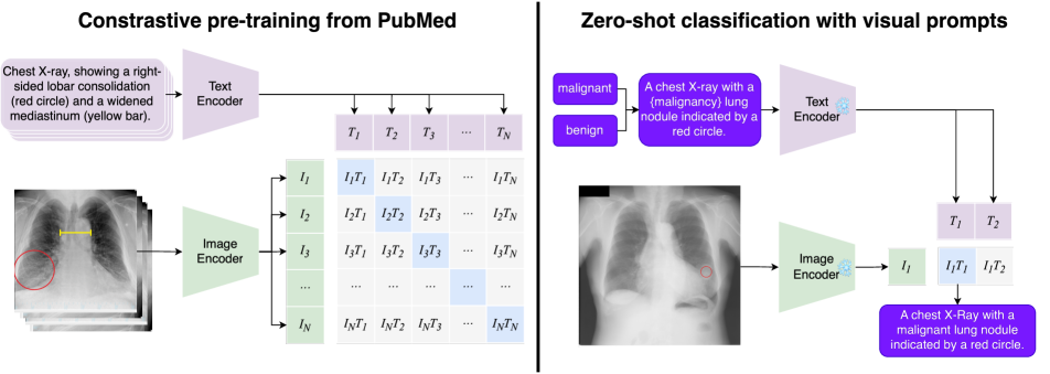
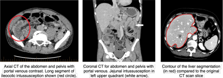
In the radiological domain, due to limited data availability, a common strategy for training VLMs involves utilizing public research articles [3, 16, 17]. BiomedCLIP [16, 17], the current state of the art in zero-shot classification in radiology was trained on the PMC-15M dataset. This dataset contains 15 million biomedical image-text pairs collected from 4.4 million scientific articles. Since visual markers are very prevalent in scientific images (see examples in Fig. 1), we hypothesize that BiomedCLIP, although significantly smaller than its natural image counterparts, also possesses the ability to recognize markers in the image domain. Consequently, this work evaluates whether visual prompt engineering — providing markers within the imaging domain — enhances zero-shot classification performance. We test our hypothesis on the challenging task of lung nodule malignancy classification using the JSRT [9] dataset and provide both quantitative analysis and attention maps to verify if the model truly recognizes the visual markers. To our knowledge, this is the first study to investigate visual prompt engineering of VLMs in the medical domain.
2 Methods
2.1 Zero-shot Classification with CLIP
Our goal is to evaluate the visual prompting ability of medical Vision Language Models (VLMs). VLMs solve prediction tasks that involve jointly processing text and images. For example, models such as CLIP are trained to match text and image samples. The input to such a VLM is an image and text , where is an alphabet. The output is a score that expresses the degree of compatibility between the supplied image and text [10, 7].
To utilize CLIP for zero-shot classification, a set of class descriptions for is fed in. The similarity score for the image and each text description is computed as . The probability distribution over the classes is then given by applying the softmax function to these similarities:
2.2 Visual Prompt Engineering
The usual way of encoding location information in a visual prompt is to crop the image around the desired location. However, for radiological images this might be problematic due to loosing the global context. Therefore, we follow the approach from [10] and draw the visual prompts directly in the image. We study a range of visual prompts in shape and color, inspired by common highlighting techniques in the medical literature. We start with simple arrows, which point at the target object, and circle which are positioned with the target object in the center. Furthermore, we also generate a visual prompt using the lesions contours. To this extend, we employ the medical imaging segmentation foundation model MedSAM [5]. This model has seen more than 1.5 million image-mask pairs during training, covering 10 imaging modalities. We utilize the generated segmentation mask, create a contour from it and draw it on the image. Those modified images are then fed into the image encoder and classified using the previously described approach (Sec. 2.1).
3 Experiments
3.1 Dataset
To conduct our experiments, we need a radiological dataset which is open source and has location annotations available for the pathologies. These location information can either be coordinates and the size of the lesion or segmentation masks. Furthermore, this dataset has to be multi-class but not multi-label for fair evaluation. To the best of our knowledge the only dataset which fulfills those criteria is the JSRT Database [9]. The database includes 154 conventional chest X-Rays (2048 2048 pixels) with a lung nodule (100 malignant and 54 benign nodules). The database also includes the required additional information diagnosis (malignant or benign), X and Y coordinates of nodule, and the size of the nodule [9].
3.2 Implementation Details
Visual Prompts.
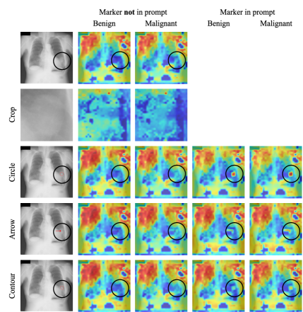
We use the lung nodules’ size, X and Y coordinates to construct the visual prompts. For cropping, we square crop around the lung nodule 224 224 pixels. For the circle prompt, we draw a red circle around the lung nodules’ X and Y coordinates with a diameter of five times the nodule size to prevent drawing on relevant context. In the same manner, we also construct the bounding box which is utilized as box prompt within MedSAM [5] to generate the lesion contour. For the arrow, we draw a red arrow always pointing to the right with an offset of the nodule diameter.
Text Prompts.
The text prompts are ”A chest x-ray with a {class} lung nodule”, with {class} being either benign or malignant. We also do an ablation study on the effect if a description of the visual prompt is mentioned in the text prompt by adding ”indicated by a red {annotation}”, with {annotation} being circle, arrow or contour.
CLIP Processing.
3.3 Evaluation
Quantitative Evaluation
We quantitatively evaluate the effect on malignancy classification performance providing visual prompts using AUROC, AUPRC, F1, Precision, Recall, Accuracy, Balanced Accuracy and MCC.
Explainability.
To investigate, if not only the performance improved by providing visual prompts, but also if the model is actually attending on the visual cues, we add LeGrad [1] as explainability method. LeGrad computes the gradient with respect to the attention maps of ViT layers, considering the gradient itself as the explainability signal. LeGrad aggregates the signal over all layers, combining the activations of the last as well as intermediate tokens to produce the merged explainability map. It showcased superior spatial fidelity and robustness to perturbations compared to other state-of-the-art explainability methods [1].
4 Results
| Visual Prompt | Marker in prompt | AUROC | AUPRC | F1 | Precision | Recall | Accuracy | Balanced Accuracy | MCC |
| 0.5450 | 0.6895 | 0.3182 | 0.6563 | 0.2100 | 0.4156 | 0.5031 | 0.0074 | ||
| Crop | 0.6230 | 0.7644 | 0.5839 | 0.7705 | 0.4700 | 0.5649 | 0.6054 | 0.2056 | |
| Arrow | 0.5087 | 0.6746 | 0.5465 | 0.6528 | 0.4700 | 0.4935 | 0.5035 | 0.0067 | |
| Arrow | 0.5472 | 0.6866 | 0.7289 | 0.6560 | 0.8200 | 0.6039 | 0.5119 | 0.0289 | |
| Circle | 0.6385 | 0.7219 | 0.5854 | 0.7500 | 0.4800 | 0.5584 | 0.5919 | 0.1779 | |
| Circle | 0.6581 | 0.7626 | 0.6974 | 0.7158 | 0.6800 | 0.6169 | 0.5900 | 0.1767 | |
| Contour | 0.6009 | 0.7341 | 0.6364 | 0.7368 | 0.5600 | 0.5844 | 0.5948 | 0.1810 | |
| Contour | 0.6400 | 0.7647 | 0.7819 | 0.6643 | 0.9500 | 0.6558 | 0.5306 | 0.1132 |
4.1 Quantitative Results
Our quantitative results show that visual prompts almost always improve the performance across metrics (Tab. 1). This suggests that explicitly guiding the model’s attention to the region of interest enhances its discriminative capabilities. While cropping achieves the best performance for precision, balanced accuracy and MCC, the MedSAM contours with mentioning the marker in the prompt achieves the highest performance for AUPRC, F1, Recall and Accuracy. The circle prompt leads to the highest AUROC. The performance was notably better when the circle prompt was mentioned in the text, achieving high F1 scores (0.6974) and accuracy (0.6169). This indicates that circles are an effective and intuitive way to highlight areas of interest. This is particularly interesting since, drawing a circle, doesn’t require costly manual contour drawing or inference with MedSAM. The arrow annotation, while often being used in medical images to highlight regions of interest, performs worse than a circle or contour.
In general, the incorporation of visual marker descriptions in the text prompts further enhanced performance. This synergy between visual and textual information appears to guide the model more effectively, aligning the visual attention with the provided context. For instance, specifying the marker in the text prompt led to improved AUROC, AUPRC, and accuracy across various visual prompts, highlighting the importance of multi-modal guidance in medical image classification.
4.2 Explainability
To better understand the effect of adding visual prompts, we employ an explainability method, namely LeGrad [1]. The attention maps (visualized in Fig. 3) revealed that when visual markers were mentioned in the text prompt, the model’s focus was more aligned with the regions of interest. This suggests that visual prompts not only improve quantitative performance but also enhance the interpretability of the model’s decisions by ensuring it attends to clinically relevant areas.
4.3 Limitations
Despite the promising results, there are limitations to our study. The JSRT dataset, while useful, is relatively small and may not fully represent the complexity of real-world medical imaging scenarios. Additionally, the downscaling of high-resolution images to 224 224 pixels could lead to loss of critical information, particularly for small lesions. Future research should explore larger and more diverse datasets, higher resolution models, and additional foundation models to validate and extend our findings. Moreover, developing more sophisticated visual prompt designs, such as [8] and combining them with advanced text descriptions could further enhance VLM performance in clinical applications.
5 Conclusion
This study demonstrates that visual prompt engineering significantly enhances the zero-shot classification performance of Vision Language Models (VLMs) for radiological images, particularly in identifying lung nodule malignancies. By employing various visual prompts such as arrows, circles, and lesion contours, and integrating these with corresponding text prompts, we observed notable improvements in classification metrics including AUROC, AUPRC, F1 score, and accuracy. Our findings indicate that visual prompts not only improve quantitative performance but also enhance the model’s attention to clinically relevant areas, as indicated by the attention maps. Despite limitations such as dataset size and image resolution, our results highlight the potential of visual prompt engineering as a simple yet effective approach to advance VLM capabilities in medical image analysis.
References
- Bousselham et al. [2024] Walid Bousselham, Angie Boggust, Sofian Chaybouti, Hendrik Strobelt, and Hilde Kuehne. Legrad: An explainability method for vision transformers via feature formation sensitivity. arXiv preprint arXiv:2404.03214, 2024.
- Chowdhury et al. [2018] Waliul Chowdhury, Muhammad Uzair Lodhi, Aaron R Kuzel, Peter Johnson, Umar Rahim, and Mustafa Rahim. Management of persistent hyponatremia induced by long-acting injectable risperidone therapy. Cureus, 10(5), 2018.
- Eslami et al. [2023] Sedigheh Eslami, Christoph Meinel, and Gerard De Melo. Pubmedclip: How much does clip benefit visual question answering in the medical domain? In Findings of the Association for Computational Linguistics: EACL 2023, pages 1181–1193, 2023.
- Langlotz [2023] Curtis P Langlotz. The future of ai and informatics in radiology: 10 predictions, 2023.
- Ma et al. [2024] Jun Ma, Yuting He, Feifei Li, Lin Han, Chenyu You, and Bo Wang. Segment anything in medical images. Nature Communications, 15(1):654, 2024.
- Massoptier and Casciaro [2008] Laurent Massoptier and Sergio Casciaro. A new fully automatic and robust algorithm for fast segmentation of liver tissue and tumors from ct scans. European radiology, 18(8):1658–1665, 2008.
- Radford et al. [2021] Alec Radford, Jong Wook Kim, Chris Hallacy, Aditya Ramesh, Gabriel Goh, Sandhini Agarwal, Girish Sastry, Amanda Askell, Pamela Mishkin, Jack Clark, et al. Learning transferable visual models from natural language supervision. In International conference on machine learning, pages 8748–8763. PMLR, 2021.
- Rezaei et al. [2024] Razieh Rezaei, Masoud Jalili Sabet, Jindong Gu, Daniel Rueckert, Philip Torr, and Ashkan Khakzar. Learning visual prompts for guiding the attention of vision transformers. arXiv preprint arXiv:2406.03303, 2024.
- Shiraishi et al. [2000] Junji Shiraishi, Shigehiko Katsuragawa, Junpei Ikezoe, Tsuneo Matsumoto, Takeshi Kobayashi, Ken-ichi Komatsu, Mitate Matsui, Hiroshi Fujita, Yoshie Kodera, and Kunio Doi. Development of a digital image database for chest radiographs with and without a lung nodule: receiver operating characteristic analysis of radiologists’ detection of pulmonary nodules. American journal of roentgenology, 174(1):71–74, 2000.
- Shtedritski et al. [2023] Aleksandar Shtedritski, Christian Rupprecht, and Andrea Vedaldi. What does clip know about a red circle? visual prompt engineering for vlms. In Proceedings of the IEEE/CVF International Conference on Computer Vision, pages 11987–11997, 2023.
- Sun et al. [2024] Zeyi Sun, Ye Fang, Tong Wu, Pan Zhang, Yuhang Zang, Shu Kong, Yuanjun Xiong, Dahua Lin, and Jiaqi Wang. Alpha-clip: A clip model focusing on wherever you want. In Proceedings of the IEEE/CVF Conference on Computer Vision and Pattern Recognition, pages 13019–13029, 2024.
- Turco [2024] Jennifer Turco. Synchronous intussusception with primary neuroendocrine tumour in an adult. Journal of Surgical Case Reports, 2024(3):rjae128, 2024.
- Wolf et al. [2019] Thomas Wolf, Lysandre Debut, Victor Sanh, Julien Chaumond, Clement Delangue, Anthony Moi, Pierric Cistac, Tim Rault, Rémi Louf, Morgan Funtowicz, et al. Huggingface’s transformers: State-of-the-art natural language processing. arXiv preprint arXiv:1910.03771, 2019.
- Yang et al. [2024] Lingfeng Yang, Yueze Wang, Xiang Li, Xinlong Wang, and Jian Yang. Fine-grained visual prompting. Advances in Neural Information Processing Systems, 36, 2024.
- Yildirim et al. [2024] Nur Yildirim, Hannah Richardson, Maria Teodora Wetscherek, Junaid Bajwa, Joseph Jacob, Mark Ames Pinnock, Stephen Harris, Daniel Coelho De Castro, Shruthi Bannur, Stephanie Hyland, et al. Multimodal healthcare ai: identifying and designing clinically relevant vision-language applications for radiology. In Proceedings of the CHI Conference on Human Factors in Computing Systems, pages 1–22, 2024.
- Zhang et al. [2023a] Sheng Zhang, Yanbo Xu, Naoto Usuyama, Jaspreet Bagga, Robert Tinn, Sam Preston, Rajesh Rao, Mu Wei, Naveen Valluri, Cliff Wong, et al. Large-scale domain-specific pretraining for biomedical vision-language processing. arXiv preprint arXiv:2303.00915, 2(3):6, 2023a.
- Zhang et al. [2023b] Sheng Zhang, Yanbo Xu, Naoto Usuyama, Hanwen Xu, Jaspreet Bagga, Robert Tinn, Sam Preston, Rajesh Rao, Mu Wei, Naveen Valluri, et al. Biomedclip: a multimodal biomedical foundation model pretrained from fifteen million scientific image-text pairs. arXiv preprint arXiv:2303.00915, 2023b.