Capsule Vision 2024 Challenge: Multi-Class Abnormality Classification for Video Capsule Endoscopy
Abstract
We present the Capsule Vision 2024 Challenge: Multi-Class Abnormality Classification for Video Capsule Endoscopy. It is being virtually organized by the Research Center for Medical Image Analysis and Artificial Intelligence (MIAAI), Department of Medicine, Danube Private University, Krems, Austria and Medical Imaging and Signal Analysis Hub (MISAHUB) in collaboration with the 9th International Conference on Computer Vision & Image Processing (CVIP 2024) being organized by the Indian Institute of Information Technology, Design and Manufacturing (IIITDM) Kancheepuram, Chennai, India. This document describes the overview of the challenge, its registration and rules, submission format, and the description of the utilized datasets.
Index Terms:
Video Capsule Endoscopy, Abnormality Classification, Biomedical Challenge, Medical Image AnalysisI Overview of the Challenge
A significant rise in the burden of Gastro-Intestinal (GI) and liver diseases across the globe has been observed due to varying environmental factors brought on by industrialization, changes in nutrition, and increased use of antibiotics [1, 2, 3]. Different endoscopic techniques are utilized in the diagnosis, treatment, and management of these diseases. Video Capsule Endoscopy (VCE) is one such technique which allows direct visualization of the GI tract and travels with the help of a disposable capsule-shaped device [4]. The device comprises an optical dome, a battery, an illuminator, an imaging sensor, and a transmitter.
VCE is a non-invasive technique, which does not suffer from any sedation-related complications and has improved a physician’s ability to find different anomalies in the GI tract - especially small bowel related diseases such as Crohn’s disease, Celiac disease, and intestinal cancer, among others [4, 5]. It is often considered as an alternative to conventional endoscopy techniques [5, 6].
The true potential of VCE technology is hindered by the longer reading time of the video frames without compromising the quality of the report and cost of the capsule [7]. During 6-8 hours of VCE procedure, a video of the GI tract trajectory is recorded on a device attached to the patient’s belt which produces about 57,000-1,000,000 frames [8]. These video frames are analysed retrospectively by experienced gastroenterologists. Presently, an experienced gastroenterologist takes approximately 2–3 hours to inspect the captured VCE video through a frame-by-frame analysis [8]. This analysis is subject to human bias and high false-positive rates due to bubbles, debris, intestinal fluid, foreign objects, and chyme (food) among other factors negatively affecting the mucosal frames [9]. Further, the inadequate doctor-to-patient ratio across the globe delays this process. Other hardware related technological limitations include capsule retention, battery limitations, and bowel obstructions [6, 7, 8].
Artificial intelligence (AI) is predicted to have profound effects on the future of VCE technology in the context of abnormality classification [10]. There is a need for investigation and state-of-the-art development of robust, user-friendly, interpretable AI-based models for multi-class abnormality classification that can aid in reducing the burden on gastroenterologists and save their valuable time by reducing the inspection time of VCE frames while maintaining high diagnostic precision.
I-A Significance of the Challenge
-
•
The aim of the challenge is to provide an opportunity for the development, testing, and evaluation of AI models for automatic classification of abnormalities captured in VCE video frames.
-
•
This challenge consists of distinct training, validation, and test datasets for training, internal validation, and external validation purposes.
-
•
It promotes the development of vendor-independent and generalized AI-based models for an automatic abnormality classification pipeline with 10 class labels, namely: angioectasia, bleeding, erosion, erythema, foreign body, lymphangiectasia, polyp, ulcer, worms, and normal.
I-B Prizes
-
•
Cash prize sponsored by the 9th International Conference on Computer Vision & Image Processing (CVIP 2024) .
-
–
1st Prize: 200 €
-
–
2nd Prize: 150 €
-
–
3rd Prize: 100 €
-
–
-
•
E-certificate to top three winning teams.
-
•
Co-authorship in the challenge summary paper for top 4 teams.
-
•
Opportunity to showcase work at CVIP 2024.
I-C Challenge relevant dates
-
•
Launch of the challenge: August 15, 2024
-
•
Registration open: August 15, 2024 - October 10, 2024
-
•
Release of Training Data: August 15, 2024
-
•
Release of Test Data and Result submission open: October 11, 2024 - October 25, 2024
-
•
Result analysis by the organizing team: October 26, 2024 - November 24, 2024
-
•
Announcement of results for all teams: November 25, 2024
II Registration and Rules
II-A Rules for participation
-
•
This challenge will be open to all students (B. Tech/ M. Tech/ Ph.D. of all branches), faculty members, researchers, clinicians, and industry professionals across the globe for free.
-
•
It will take place in full-virtual mode.
-
•
Participants will either register as a solo participant or can form a team.
-
•
The results will be submitted in a specific format over email to ask.misahub@gmail.com. The template for submission has been provided.
-
•
In the case of ties, the organizing committee may rank teams based on the method’s novelty and readability of codes. The organizing committee’s decision in this regard will be final.
II-B Rules for Team Formation
-
•
A team can have a maximum of 4 participants.
-
•
Team members can be from the same or different organizations/ affiliations.
-
•
A participant can only be a part of a single team.
-
•
Only one member from the team must register for the challenge.
-
•
Each team can only have a single registration. Multiple registrations per team can lead to disqualification.
-
•
There are no limitations on the number of teams from the same organizations/ affiliations. (However, each participant can only be part of a single team.)
II-C Rules for use of Training and Validation Dataset
-
•
Download the dataset. It consists of the labelled training and validation dataset.
-
•
Develop a model to classify 10 class labels using ONLY the training and validation dataset.
-
•
Store the model, associated weights and files. Compute the predictions and evaluation metrics on the validation dataset (internal validation) as per the provided sample code on our github repository.
-
•
The participants are allowed to perform augmentations, resize the data for saving computational resources, and can use existing machine learning, meta-learning, ensembles, any pre-trained models, and deep learning models.
-
•
A sample code has been provided on our github how to load the dataset.
II-D Rules for use of Testing Dataset
-
•
Download the dataset. It consists of the un-labelled test dataset.
-
•
Compute the predictions (external validation) as per the provided sample code on our github repository.
-
•
Store the developed excel sheet and submit it as a part of your submission.
-
•
Prepare the report and github repository as per the submission format.
II-E Submission format
Each team is required to submit their results in an EMAIL with the following structure to ask.misahub@gmail.com.
-
•
The email should contain:
-
–
Challenge name and Team name as the SUBJECT LINE.
-
–
Team member names and affiliation in the BODY OF THE EMAIL.
-
–
Contact number and email address in the BODY OF THE EMAIL.
-
–
A link of the github repository in public mode in the BODY OF THE EMAIL.
-
–
A link of their report on any open preprint server of their choice (ArXiv, Authorea, BioRxiv, Figshare etc) in the BODY OF THE EMAIL.
-
–
Generated excel sheet (in xlsx format) as an attachment.
-
–
-
•
The github repository in public mode should contain the following:
-
–
Developed code for training, validation, and testing in .py / .mat etc in readable format with comments.
-
–
Stored model, associated weights or files
(optional). -
–
Any utils or assets or config. or checkpoints.
-
–
II-F Important Notes
-
•
The challenge dates and the time zones will be as per the Central European Summer Time (CEST; UTC+02:00, Austria). They will be counted as per end of the day 23:59.
-
•
The github repository should be PUBLIC. Repositories which require access will NOT be considered for evaluation.
-
•
The submitted repository MUST be readable, documented properly, and interactive.
-
•
The participants are requested to STRICTLY follow the submission format.
-
•
The participants are NOT allowed to utilize any other dataset for training/ validation/ testing in this challenge.
-
•
The participants are strictly advised to use the overleaf template for developing their report. The zip file is also available at our github repository.
-
•
The participants are requested to acknowledge and cite the datasets in their reports and for any other research purposes. Use of the datasets for commercial products or any purpose other than research is STRICTLY NOT ALLOWED.
-
•
The participants are encouraged to expand their novel AI pipeline to full-length manuscripts for consideration in prestigious conferences like CVIP 2024 and journals.
-
•
All participating teams allow the organizing team to summarize their developed AI pipelines in the form of a challenge summary paper.
-
•
Any submitted excel sheet with missing data will be evaluated but given zero marks.
-
•
For the final test set result submission, both training and validation datasets may be used for training.
-
•
For the final test set result submission, only one submission is allowed. In case of multiple submissions, only the last one will be considered.
-
•
Our team has conducted a preliminary analysis utilizing a variety of machine learning models. The sample codes are available on our github repository. We will release a standardized benchmarking pipeline along with its performance results on the validation dataset on our github repository coinciding with the release of the test datasets. Participants are requested to review these benchmarks, compare their own results accordingly, and include this comparative analysis in their submissions.
II-G Criteria of judging a submission
-
•
All emails received before the result submission closing date will be considered for evaluation.
-
•
The github repository, report on any open-source pre-print server, and excel sheet received for all entries
will be downloaded by the organizing team. -
•
A common data file will be prepared to compare the evaluation metrics achieved for testing the dataset.
-
•
A semi-automated python script file will be used to achieve the best evaluation metrics received among all entries.
-
•
The following checklist will be used to select the top three winning teams:
-
–
Best evaluation metrics (especially mean AUC) on testing dataset.
-
–
In case of ties:
-
*
Model uniqueness and reproducibility.
-
*
Readability of the submitted report and github repository.
-
*
Unique methods of handling class-imbalance problems in the datasets.
-
*
-
–
III Datasets
III-A Training, validation, and test dataset description
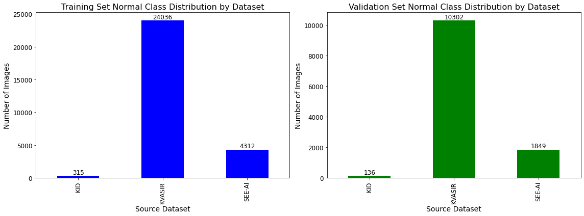
The training and validation dataset has been developed using three publicly available (SEE-AI project dataset [11], KID [12], and Kvasir-Capsule dataset [13]) and one private (AIIMS [8]) VCE datasets. The training and validation datasets consist of 37,607 and 16,132 VCE frames, respectively, mapped to 10 class labels namely angioectasia, bleeding, erosion, erythema, foreign body, lymphangiectasia, polyp, ulcer, worms, and normal. Fig. 1 and Fig. 2 depict the class distribution of the training and validation as per the source datasets. The source datasets in the training and validation dataset have ONLY been extracted, cropped, resized, and split for the development of a large dataset in this challenge.
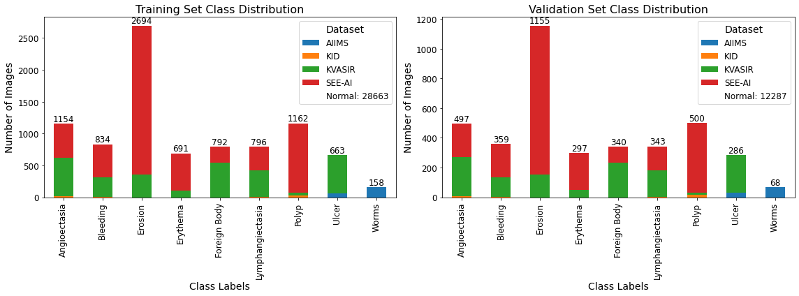
| Type of Data | Source Dataset | Angioectasia | Bleeding | Erosion | Erythema | Foreign Body | Lymphangiectasia | Normal | Polyp | Ulcer | Worms |
|---|---|---|---|---|---|---|---|---|---|---|---|
| KID | 18 | 3 | 0 | 0 | 0 | 6 | 315 | 34 | 0 | 0 | |
| KVASIR | 606 | 312 | 354 | 111 | 543 | 414 | 24036 | 38 | 597 | 0 | |
| Training | SEE-AI | 530 | 519 | 2340 | 580 | 249 | 376 | 4312 | 1090 | 0 | 0 |
| AIIMS | 0 | 0 | 0 | 0 | 0 | 0 | 0 | 0 | 66 | 158 | |
| Total Frames | 1154 | 834 | 2694 | 691 | 792 | 796 | 28663 | 1162 | 663 | 158 | |
| KID | 9 | 2 | 0 | 0 | 0 | 3 | 136 | 15 | 0 | 0 | |
| KVASIR | 260 | 134 | 152 | 48 | 233 | 178 | 10302 | 17 | 257 | 0 | |
| Validation | SEE-AI | 228 | 223 | 1003 | 249 | 107 | 162 | 1849 | 468 | 0 | 0 |
| AIIMS | 0 | 0 | 0 | 0 | 0 | 0 | 0 | 0 | 29 | 68 | |
| Total Frames | 497 | 359 | 1155 | 297 | 340 | 343 | 12287 | 500 | 286 | 68 |
The testing dataset has been developed using a high-quality, medically annotated private dataset from the Department of Gastroenterology and HNU, All India Institute of Medical Sciences Delhi, India. It consists of 4,385 VCE frames which have been collected from more than 70 patient VCE video with abnormalities namely angioectasia, bleeding, erosion, erythema, foreign body, lymphangiectasia, polyp, ulcer, and worms. The images are de-identified and do not contain any patient information. The data collection for this AI study was done according to Helsinki declarations. It was approved by the Department of Gastroenterology and HNU, All India Institute of Medical Sciences Delhi, India ethics committee (Ref. No.: IEC-666/05.08.2022). Further it is part of the objectives of the Core Research Grant Project (CRG/2022/001755). Upon completion of the challenge, the test set will be open-sourced with its ground truths.
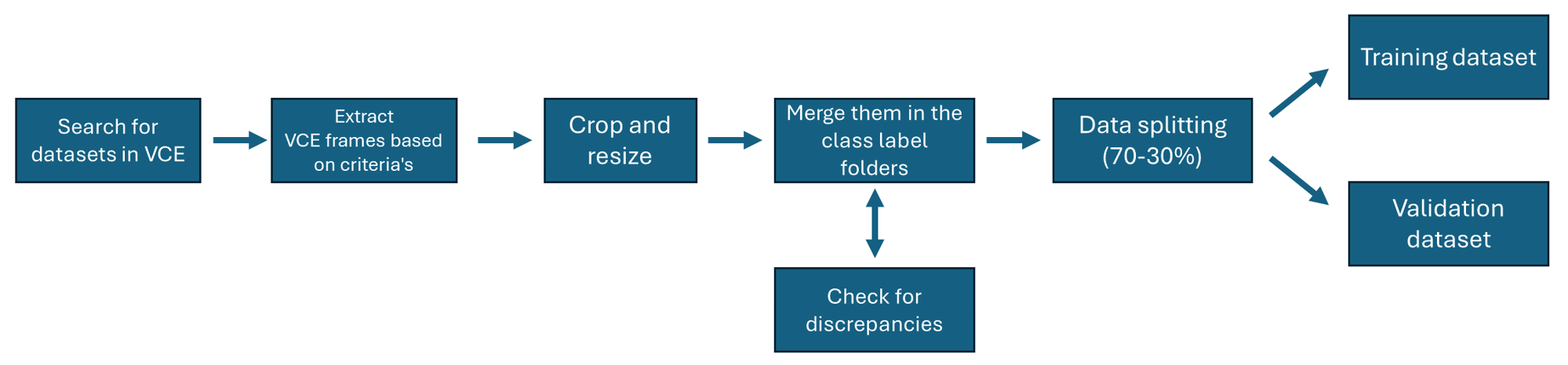
III-B Development of the Training and Validation Dataset
A standard pipeline was followed to develop the training and validation dataset for this challenge (see Fig. 3). At first, all the publicly available datasets in VCE research were searched and collected. The datasets found were kvasir-capsule [13], SEE-AI project [11], EndoSLAM [14], annotated bleeding dataset [15], rhodes island [16], WCEBleedGen [17], AutoWCEBleedGen-Test [17], AI-KODA [18], KID [12], and red lesion endoscopy [19].
EndoSLAM, AI-KODA, and rhode island dataset were not considered as they do not contain abnormalities and focus on virtual capsule reconstruction, cleanliness assessment, and organ classification respectively. Annotated bleeding dataset and red lesion endoscopy dataset were not considered as they only contain red lesions. WCEBleedGen was not considered as it contains frames from KID, kvasir-capsule, annotated bleeding dataset and only focuses on bleeding classification, detection, and segmentation. AutoWCEBleedGen-Test has been developed by the co-authors of this challenge and hence was considered as a part of the test dataset. All the frames have been anonymized and renamed.
Finally, the KID, kvasir-capsule, and SEE-AI project were considered. Then the VCE frames and their abnormalities in each of these datasets were downloaded, and extracted based on the inclusion and exclusion criteria.
-
•
Inclusion criteria:
-
–
VCE frames with one abnormality in each frame.
-
–
Normal VCE frames.
-
–
-
•
Exclusion criteria:
-
–
VCE frames with multiple abnormalities and duplicates.
-
–
A new class label of worms with 226 VCE frames has been introduced in the training and validation dataset. VCE frames containing 95 ulcer VCE frames have also been added. The ulcer and worm VCE frames have been collected from three and three patients, respectively, from the department of gastroenterology, HNU, All India Institute of Medical Sciences Delhi, India and were utilized in the study conducted by Goel et al. [8].
All the frames were cropped and resized to . The frames were merged as per their source and class label folders and checked for discrepancies. Checks for discrepancies included that all VCE frames should be:
-
•
Correctly labelled as per their class name and in the right source folder.
-
•
Of resolution and in .jpg format.
-
•
Properly cropped from the four-side black boundaries.
-
•
NOT have any patient information or annotation markings.
-
•
NOT have multiple abnormalities.
-
•
NOT have duplicates.
After a thorough check, the main folder was subjected to a random data split of 70-30% for the development of the training and validation dataset. Table I details the number of VCE frames with respect to their respective class labels in each of the datasets.
IV Conclusion
The Capsule Vision 2024 Challenge seeks to advance VCE through a multi-class abnormality classification. By offering unique training, validation, and test datasets, the challenge promotes the development of efficient models to aid gastroenterologists in reducing analysis time while maintaining high diagnostic accuracy. We invite participants to use these resources, strictly follow submission guidelines, and contribute to enhancing VCE abnormality classification. This challenge is a platform for innovation and collaboration in medical imaging, aiming for impactful advancements of AI in VCE.
References
- [1] M. Arnold, C. C. Abnet, R. E. Neale, J. Vignat, E. L. Giovannucci, K. A. McGlynn, and F. Bray, “Global burden of 5 major types of gastrointestinal cancer,” Gastroenterology, vol. 159, no. 1, pp. 335–349, 2020.
- [2] D. Shah, G. K. Makharia, U. C. Ghoshal, S. Varma, V. Ahuja, and S. Hutfless, “Burden of gastrointestinal and liver diseases in india, 1990–2016,” Indian Journal of Gastroenterology, vol. 37, pp. 439–445, 2018.
- [3] Z. Yu, X. Bai, R. Zhou, G. Ruan, M. Guo, W. Han, S. Jiang, and H. Yang, “Differences in the incidence and mortality of digestive cancer between global cancer observatory 2020 and global burden of disease 2019,” International Journal of Cancer, vol. 154, no. 4, pp. 615–625, 2024.
- [4] G. Iddan, G. Meron, A. Glukhovsky, and P. Swain, “Wireless capsule endoscopy,” Nature, vol. 405, no. 6785, pp. 417–417, 2000.
- [5] P. A. Thwaites, C. K. Yao, E. P. Halmos, J. G. Muir, R. E. Burgell, K. J. Berean, K. Kalantar-zadeh, and P. R. Gibson, “Current status and future directions of ingestible electronic devices in gastroenterology,” Alimentary Pharmacology & Therapeutics, vol. 59, no. 4, pp. 459–474, 2024.
- [6] E. Rondonotti, J. M. Herrerias, M. Pennazio, A. Caunedo, M. Mascarenhas-Saraiva, and R. de Franchis, “Complications, limitations, and failures of capsule endoscopy: a review of 733 cases,” Gastrointestinal endoscopy, vol. 62, no. 5, pp. 712–716, 2005.
- [7] R. Gupta, “The journey of capsule endoscopy in india,” Journal of Digestive Endoscopy, vol. 9, no. 04, p. 183, 2018.
- [8] N. Goel, S. Kaur, D. Gunjan, and S. Mahapatra, “Dilated cnn for abnormality detection in wireless capsule endoscopy images,” Soft Computing, pp. 1–17, 2022.
- [9] L.-D. Lazaridis, G. Tziatzios, E. Toth, H. Beaumont, X. Dray, R. Eliakim, P. Ellul, I. Fernandez-Urien, M. Keuchel, S. Panter et al., “Implementation of european society of gastrointestinal endoscopy (esge) recommendations for small-bowel capsule endoscopy into clinical practice: results of an official esge survey,” Endoscopy, vol. 53, no. 09, pp. 970–980, 2021.
- [10] H. Messmann, R. Bisschops, G. Antonelli, D. Libânio, P. Sinonquel, M. Abdelrahim, O. F. Ahmad, M. Areia, J. J. Bergman, P. Bhandari et al., “Expected value of artificial intelligence in gastrointestinal endoscopy: European society of gastrointestinal endoscopy (esge) position statement,” Endoscopy, vol. 54, no. 12, pp. 1211–1231, 2022.
- [11] A. Yokote, J. Umeno, K. Kawasaki, S. Fujioka, Y. Fuyuno, Y. Matsuno, Y. Yoshida, N. Imazu, S. Miyazono, T. Moriyama et al., “Small bowel capsule endoscopy examination and open access database with artificial intelligence: The see-artificial intelligence project,” DEN open, vol. 4, no. 1, p. e258, 2024.
- [12] A. Koulaouzidis, D. K. Iakovidis, D. E. Yung, E. Rondonotti, U. Kopylov, J. N. Plevris, E. Toth, A. Eliakim, G. W. Johansson, W. Marlicz et al., “Kid project: an internet-based digital video atlas of capsule endoscopy for research purposes,” Endoscopy international open, vol. 5, no. 06, pp. E477–E483, 2017.
- [13] P. H. Smedsrud, V. Thambawita, S. A. Hicks, H. Gjestang, O. O. Nedrejord, E. Næss, H. Borgli, D. Jha, T. J. D. Berstad, S. L. Eskeland et al., “Kvasir-capsule, a video capsule endoscopy dataset,” Scientific Data, vol. 8, no. 1, p. 142, 2021.
- [14] K. B. Ozyoruk, G. I. Gokceler, T. L. Bobrow, G. Coskun, K. Incetan, Y. Almalioglu, F. Mahmood, E. Curto, L. Perdigoto, M. Oliveira et al., “Endoslam dataset and an unsupervised monocular visual odometry and depth estimation approach for endoscopic videos,” Medical image analysis, vol. 71, p. 102058, 2021.
- [15] F. Deeba, F. M. Bui, and K. A. Wahid, “Automated growcut for segmentation of endoscopic images,” in 2016 International Joint Conference on Neural Networks (IJCNN). IEEE, 2016, pp. 4650–4657.
- [16] A. Charoen, A. Guo, P. Fangsaard, S. Taweechainaruemitr, N. Wiwatwattana, T. Charoenpong, and H. G. Rich, “Rhode island gastroenterology video capsule endoscopy data set,” Scientific Data, vol. 9, no. 1, p. 602, 2022.
- [17] P. Handa, D. Nautiyal, D. Chhabra, M. Dhir, A. Saini, S. Jha, H. Mangotra, N. Pandey, A. Thakur et al., “Auto-wcebleedgen version v1 and v2: Challenge, datasets and evaluation,” Authorea Preprints, 2024.
- [18] P. Handa, N. Goel, S. Indu, and D. Gunjan, “Comprehensive evaluation of a new automatic scoring system for cleanliness assessment in video capsule endoscopy,” International Journal of Imaging Systems and Technology, vol. 34, no. 3, p. e23097, 2024.
- [19] R. Leenhardt, C. Li, J.-P. Le Mouel, G. Rahmi, J. C. Saurin, F. Cholet, A. Boureille, X. Amiot, M. Delvaux, C. Duburque et al., “Cad-cap: a 25,000-image database serving the development of artificial intelligence for capsule endoscopy,” Endoscopy international open, vol. 8, no. 03, pp. E415–E420, 2020.