Quantifying variabilities in cardiac digital twin models of the electrocardiogram
Abstract
Cardiac digital twins (CDTs) of human cardiac electrophysiology (EP) are digital replicas of patient hearts that match like-for-like clinical observations. The electro-cardiogram (ECG), as the most prevalent non-invasive observation of cardiac electrophysiology, is considered an ideal target for CDT calibration. Recent advanced CDT calibration methods have demonstrated their ability to minimize discrepancies between simulated and measured ECG signals, effectively replicating all key morphological features relevant to diagnostics. However, due to the inherent nature of clinical data acquisition and CDT model generation pipelines, discrepancies inevitably arise between the real physical electrophysiology in a patient and the simulated virtual electrophysiology in a CDT.
In this study, we aim to qualitatively and quantitatively analyze the impact of these uncertainties on ECG morphology and diagnostic markers. We analyze residual beat-to-beat variability in ECG recordings obtained from healthy subjects and patients. Using a biophysically detailed and anatomically accurate computational model of whole-heart electrophysiology combined with a detailed torso model calibrated to closely replicate measured ECG signals, we vary anatomical factors (heart location, orientation, size), heterogeneity in electrical conductivities in the heart and torso, and electrode placements across ECG leads to assess their qualitative impact on ECG morphology.
Our study demonstrates that diagnostically relevant ECG features and overall morphology appear relatively robust against the investigated uncertainties. This resilience is consistent with the narrow distribution of ECG due to residual beat-to-beat variability observed in both healthy subjects and patients.
keywords:
Electrocardiograms, Computational Cardiology, Beat-to-beat variability, Anatomical Uncertainty, Lead Placement Uncertainty1 Introduction
Computational modeling of cardiac EP is an established important research tool for analyzing experimental or clinical data [1, 2, 3, 4, 5], and is now increasingly considered in industrial applications such as medical device design [6, 7], as well as in clinical applications for diagnosis, stratification, and therapy planning [8, 9, 10, 11]. Unlike in basic research, where generic representations of cardiac anatomy and EP are used to gain generic mechanistic insights, a more specific individualized modeling approach is required in industrial and clinical applications [12, 13, 14, 15]. There, models must be calibrated to cover the variability of a given patient population, in the form of a virtual cohort [9], or, even to represent anatomy and EP of individual patients. Such high-fidelity digital replicas of patient hearts, derived from clinical data and calibrated to match like-for-like clinical observations, are often referred to as patient-specific models or CDTs [7, 16, 17, 18].
Owing to its broad clinical availability and non-invasive nature, ECG recordings appear to be a most natural choice as a target for model calibration [19, 20, 21]. Advanced CDT calibration methods have been developed that are, in principle, able to minimize the discrepancy between simulated and measured ECG [19, 22, 23], and have been shown to replicate all morphological key features relevant for diagnostics [22, 24]. These methods have demonstrated the ability to replicate all key morphological features relevant for diagnostics [22, 24]. However, inconsistencies inevitably arise between the real physiological electrical activity in a patient and the simulated virtual electrical activity in a CDT, due to the nature of clinical data acquisition and CDT model generation pipelines. Major sources of these inconsistencies include: i) the fundamental issue of non-uniqueness in the models used to solve the forward problem of electrocardiography – different activation and repolarization sequences can produce identical ECG patterns [25]; ii) residual variability in heartbeat, leading to beat-to-beat variations in ECG morphology [26, 27, 28], which limits the fidelity of a CDT calibrated using a single ECG; and iii) significant observational uncertainties due to technical limitations in anatomical imaging and ECG recording, which hinder accurate simultaneous measurement of all model parameters contributing to ECG generation [29, 30]. Therefore, all factors involved in anatomical and electrophysiological modeling must be considered as uncertainty, and the measured ECG itself as one signal of an envelope of ECG s.
Residual ECG variability arises from physiological sources, such as beat-to-beat variations in the activation sequence [31, 32], and anatomical variations of the heart due to factors like breathing [33, 34, 35, 36, 37, 35] and body posture [38, 39], which alter the shape, position, and orientation of the heart relative to ECG recording locations on the torso [40, 41, 42]. In addition to anatomical factors, there are significant uncertainties in the torso structure due to electrical heterogeneities [43, 44], and related model parameters that cannot be directly measured in vivo, only estimated. Furthermore, despite standardization of ECG lead placement, operator variability in electrode positioning remains non-negligible [45, 46, 47, 48, 49]. If variability in any of these factors translates into marked variability in ECG morphology, it could render ECG-based calibration ambiguous and unreliable.
In this study, we aim to qualitatively and quantitatively analyze the impact of the latter two sources of uncertainties on ECG morphology and diagnostic markers, and to investigate their role in the ECG-based calibration of CDT. To this end, we investigate residual beat-to-beat variability in ECG recordings obtained from healthy subjects and patients undergoing treatment for atrial fibrillation or ventricular tachycardia ablation therapy. As a computational reference, a biophysically detailed anatomically accurate computational whole heart-torso model of one of the analyzed healthy subjects is calibrated to replicate the measured mean ECG with high fidelity. Keeping constant the electrical activation and repolarization sequences, we vary the position, orientation, and size of the heart, heterogeneity in electrical conductivities in the torso, and electrode placement in individual ECG leads, to compare their impact on the ECG. Our findings indicate that diagnostically relevant ECG features and overall morphology remain relatively robust against the investigated uncertainties. This resilience aligns with the narrow distribution of ECG due to residual beat-to-beat variability observed in both healthy subjects and patients. Therefore, our results suggest that an accurate inference of CDT electrical activation sequences from the ECG is feasible, and not impeded by residual beat-to-beat variability or inevitable model inconsistencies.
2 Methods
2.1 Residual Variability
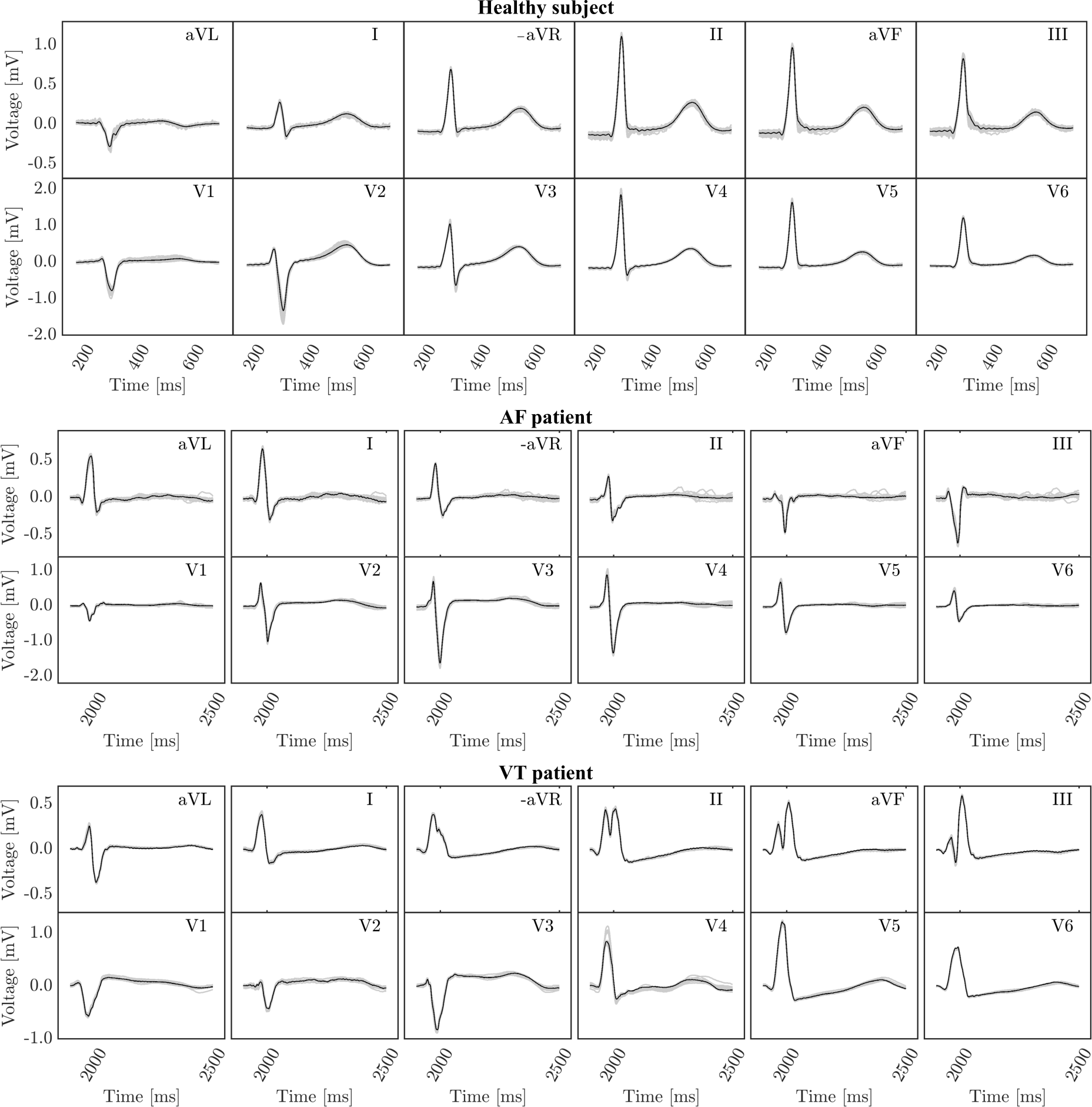
We analyzed beat-to-beat residual variability in the 12-lead ECG for a cohort of 14 healthy subjects, and patients treated for ventricular tachycardia (VT) and atrial fibrillation (AF). To provide a relative margin of uncertainty, over ECGs were recorded. The ECG data were then filtered with a 150 Hz low pass filter, a 50 Hz bandstop filter, and a high pass filter of 0.05 Hz to reduce noise. Individual beats were detected using a modified Pan Tompkins algorithm for R-wave detection [50] and stored in a beat matrix. For each matrix, the average ECG beat was computed and plotted against all beats in the matrix.
Normal sinus rhythm ECGs were recorded from 18 AF and 17 VT patients during ablation procedures using an electro-anatomical mapping system (Carto XP). Ablation procedures were carried out at the University Hospital of Graz, Graz, Austria, and included in the local ablation registry approved by the ethics committee of the Medical University of Graz (reference number 31-036 ex 18/19 for the VT patients, and reference number 26-217 ex 13/14 for the AF patients). All patients gave written informed consent. ECGs of were recorded using an electro-anatomical mapping system (Carto XP) at each position visited by the mapping catheter. The ECG corresponding to the beat selected by the mapping system for deriving an instance of local activation was chosen. Depending on the density of the constructed maps, hundreds to thousands of ECGs were recorded, analyzed [51], and stored per patient in a beat matrix. As for the healthy subjects, an average ECG was computed and plotted against the entire beat matrix.
2.2 ECG modeling
The reference baseline forward ECG model has been described previously in great detail elsewhere [22] with updates as detailed in [52] and [53]. Briefly, an anatomically accurate heart-torso model of the subject was generated from clinical magnetic resonance imaging [54]. Images of the heart were segmented using an automated tool [55], and the segmentation was semi-automatically refined using seg3D [56]. The segmented patient-specific anatomy included a whole heart embedded in a torso with cardiac blood pools, lungs, bones, liver, fat, and skin labeled as different volumes. Computational meshes were generated from segmentation labels [57] using [58], at an average resolution of in the heart, and a coarser resolution in other tissues and torso, with an average resolution of on the torso surface. Myocardial fibers were implemented based on rule-based algorithms within the atria [59] and ventricles [60]. Universal coordinates were computed within the atria [61] and the ventricles [22, 62].
The model was calibrated to faithfully replicate the measured ECG from this subject across all 12 leads. The atria were assigned generic electrophysiology that gave a realistic P-wave as detailed with activation stemming from a sino-atrial node on the right atrial roof. An atrio-ventricular node within the right atria was connected to a physiologically detailed His-Purkinje system that facilitated ventricular activation. Initially, a simplified ventricular conduction system was assumed to compromise 5 fascicles rooted in the endocardium – 3 in the left ventricle, one in the right-ventricular septum, and one in the right ventricular moderator band, combined with a fast-conducting endocardial layer. Fascicular locations were varied through sampling to minimize the mismatch in QRS morphology in the ECG. The optimized five-fascicle conduction system was subsequently replaced by a topologically realistic model of the His-Purkinje system (HPS) that produced the same actThe model was calibrated to faithfully replicate the measured ECG from this subject across all 12 leads.ivation sequence and retained an equally good match in the ECG [53]. Action potential duration was spatially varied using a linear mapping with activation to obtain heterogeneous ventricular repolarization patterns satisfyingly matching the T-wave morphology [52]. All other electrophysiological parameters are described in further detail in [52].
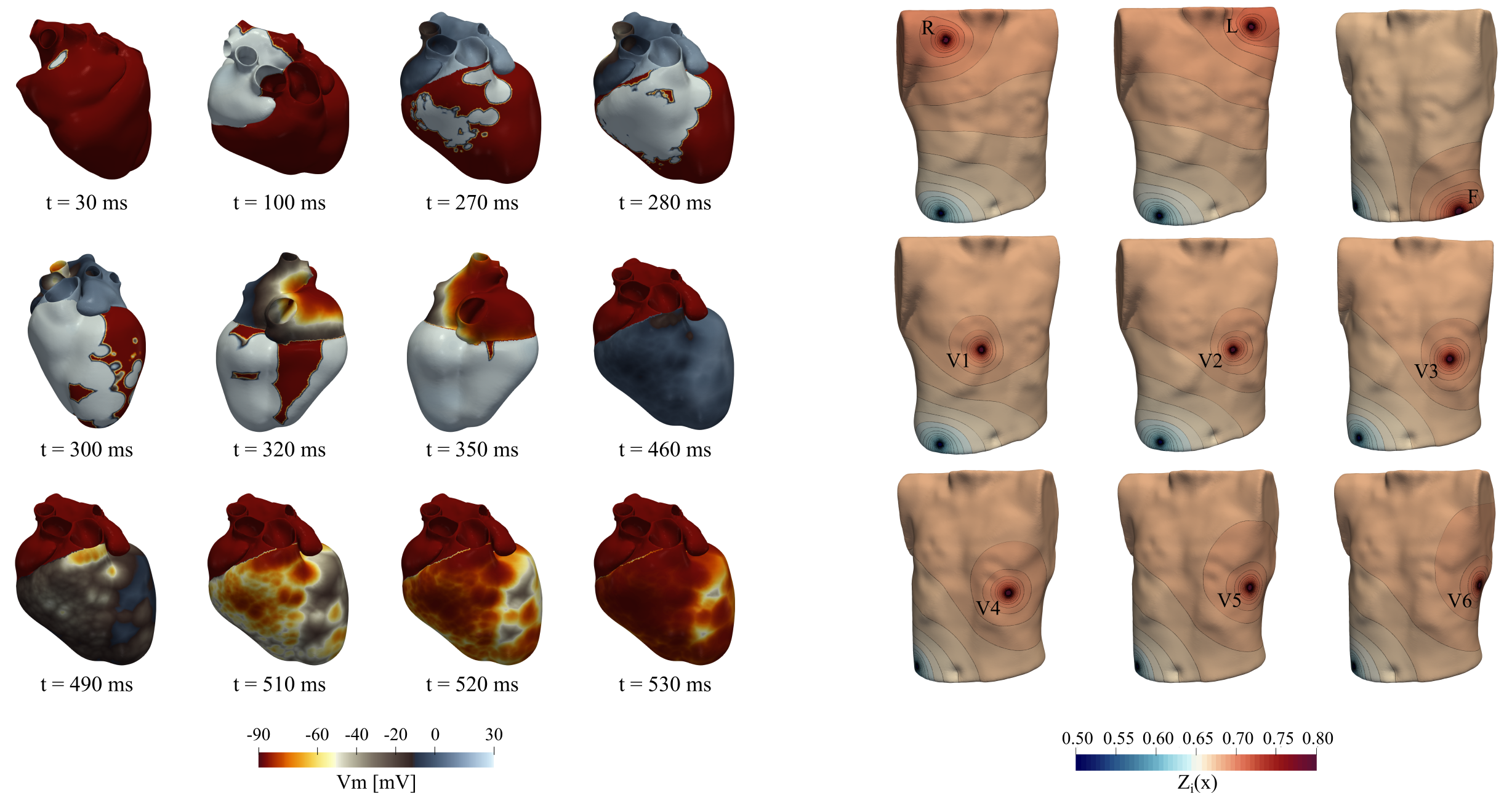
The reaction-Eikonal model in monodomain mode was used to describe wavefront propagation and the associated electrical sources in the form of transmembrane voltages, (referred to Fig.1, left), generating the ECG [63]. For all ECG recording locations, , the Lead field solutions, (referred to Fig.1, right), have been computed and used to accurately compute extracellular potential differences, , between electrode locations corresponding to the ECG [64].
2.3 Cardiac anatomical variation
The role of anatomical variation of the heart in position, orientation, and size, mediated due to breathing and body posture, upon the ECG is investigated by defining a set of heart-torso anatomies while preserving the EP settings and simulation from the calibrated reference model. Starting from the reference cardiac and torso geometry, anatomical uncertainty was introduced by altering the heart’s position, orientation, and size. The external shape of the torso and the electrode placements were kept constant for all anatomical models. To prevent intersections between the heart and other organs or tissues, a homogeneous torso model was used for this investigation.
To circumvent the need for complete remeshing of the torso with each new cardiac configuration, we introduced a spherical halo surrounding the heart (referred to Fig. 2). The volumetric meshes extending from the surface of the torso to the halo, as well as the heart mesh itself, remained unchanged for all anatomical models, with only the smaller volume between the halo and each new heart position, orientation, and shape requiring remeshing. To streamline the generation of the anatomical set, an efficient and semi-automatic pipeline was implemented.
Variations in position, orientation, and size of the heart were modeled by rigid translations, rotations, and scaling of the heart. To cover the full range of possible anatomical cahnges[37, 35], translational directions and rotational axes were built following the work of Odille et al. [65], where the authors present a cardiac reference system that aids the statistical investigation of the variation of the heart position in the human population. Three main translational directions, each with corresponding positive and negative vectors, were defined, along with three rotational axes and their positive and negative rotational angles (see Fig. 2). To ascertain the exploration of the entire space of anatomical variability of the heart, we employed translational, rotational, and scaling magnitudes well beyond the physiological ranges as detailed in the following.
For all changes in the heart geometry, the electrical source distribution over the entire myocardial volume was kept constant, while the lead field solutions were recomputed, thus minimizing any potential impact of spatial discretization upon depolarization and repolarization pattern on the ECG prediction.
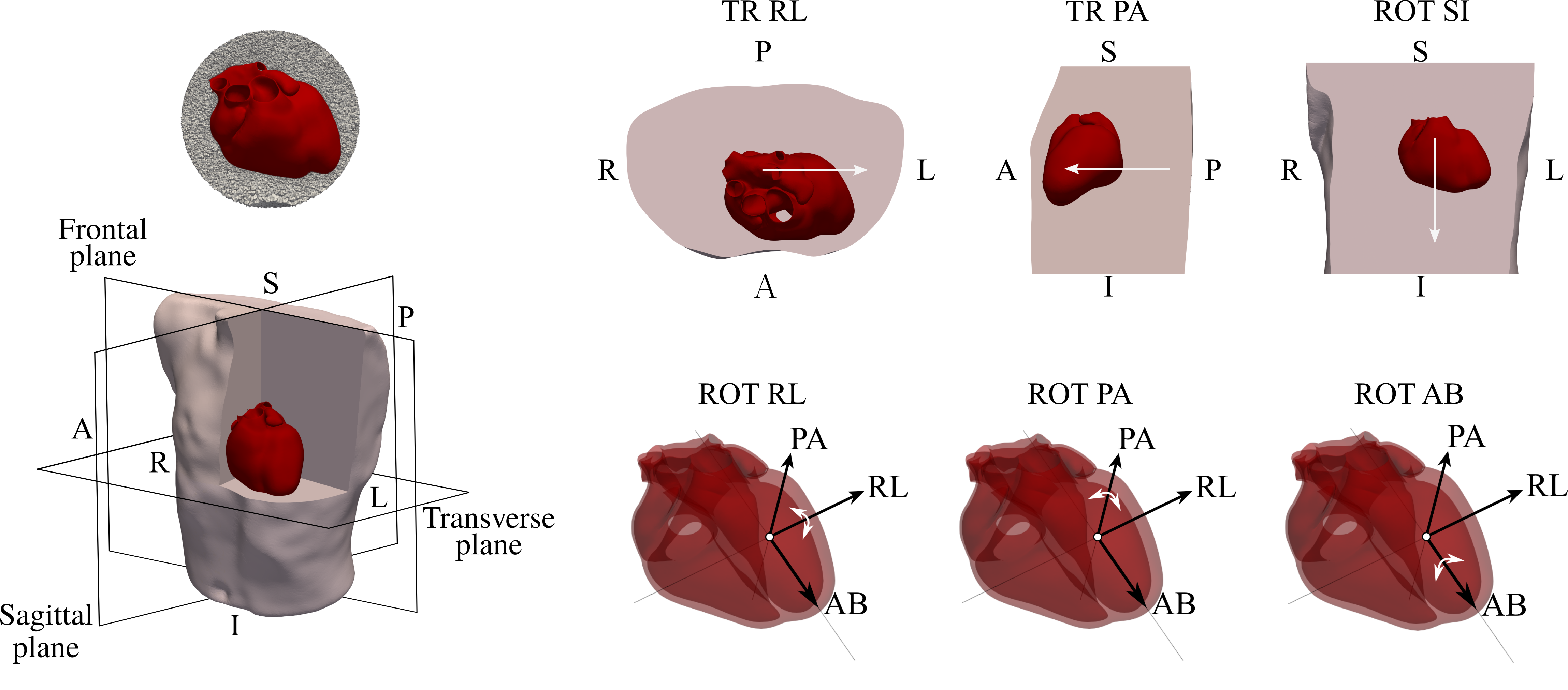
Position uncertainty was accounted for by translating the heart within the torso along three orthogonal directions. These were determined using the cardiac center of mass as origin, and axes aligned perpendicular to the frontal, sagittal, and transverse planes of the torso (referred to Figure 2). The three axes were labeled according to their anatomical orientation within the torso as follows: perpendicular to the sagittal plane as right-left (RL), to the frontal plane as posterior-anterior (PA), and to the transverse plane as superior-inferior (SI). The variations of the cardiac position were then probed by moving the heart along RL, and SI axis. Translation along the PA axis was further restricted to to prevent any intersections of the heart with the torso. Hereon, we will denote the translations as translation superior-inferior (TR-SI), translation right-left (TR-RL), and translation posterior-anterior (TR-PA) where a translation is counted positive if it is along an axis as indicated in Figure 2).
Orientation uncertainty was systematically implemented by rotating the heart around an orthogonal set of eigenaxes derived from the cardiac anatomy. Eigenaxes were obtained by employing a principal component analysis algorithm on the cardiac mesh, and the Gram-Schmidt orthogonalization. The first axis, defined as apico-basal (AB) axis, was determined as the eigenvector associated with the largest eigenvalue obtained with the principal component analysis. The RL axis was computed by applying the Gram-Schmidt orthogonalization to the vector connecting the centers of the left and right blood pools, whereas the PA axis was determined as the vector orthogonal to the plane spanned by the AB and RL axes (referred to Fig. 2). The midpoint of the left ventricular blood pool was considered the origin of this orthogonal system. The variations of the cardiac orientation were then explored by rotating the heart by around the axes RL, PA and AB, respectively (see Fig. 2). The rotational transformations were denoted then as rotation apico-basal (ROT-AB), rotation right-left (ROT-RL) and rotation posterior-anterior (ROT-PA), respectively.
Uncertainty in size as induced by imaging uncertainty or breathing, which modulates intra-thoracic pressure and, thus, the filling state and the size of the heart, have been shown to remain under 10% [33, 34]. The associated variability in size was mimicked by applying 10% scaling factors to the heart while keeping its center of mass unaltered.
We sought to discriminate the contribution of sheer changes in myocardial volume which scales the cardiac sources without affecting the propagation of the depolarization wave fonts, from the changes in the activation and repolarization pattern due to the impact of the myocardial volume size. We thus investigated ECG variability due to heart scaling by prescribing i) the activation and repolarization sequence given by the spatially scaled source distribution , to mirror that of the reference simulation, and ii) the orthotropic conduction velocities associated with the electrophysiological parameter settings that yield an altered source distribution, . In the latter case, prescribed conduction velocities may slow down or accelerate the epicardial activation, with epicardial breakthroughs being retarded as the myocardial volume and transmural wall width increase, or with precipitated epicardial breakthroughs when the size of the heart is reduced. Combined with a larger/slower epicardial surface slowing down/hastening total epicardial activation, the ensuing activation and repolarization pattern will be altered, and, consequently, the ECGs.
Data analysis We quantitatively analyzed the variations in the ECG depending on the anatomical transformations by comparing their impact upon the ECG amplitude on the single R, S, and T peaks for each lead, in terms of both absolute and relative variations. In the former case, we compute:
while in the latter case, we derive
where and represent the reference signal and the signal computed with the anatomical transformation , respectively, at the instant of each peak , and . While absvar expresses the same change in peak amplitude that can be observed by plotting the ECG, the relative variation value relvar points to the geometrical transformations that caused real major variations in the ECG, as it identifies these variations relative to the reference scaled amplitude of the ECG signal.
2.4 Uncertainty in electrical conductivities
For a given distribution of cardiac electric sources, the potential field generated in the torso and the corresponding ECG are determined by the tissue-specific electrical conduction inhomogeneities within the torso itself. The conductivity within the torso is indeed highly heterogeneous, with substantial differences between compartments (including organs or distinct tissues such as bones, fat, and skin) [66, 67]. Moreover, obtaining precise measures of tissue conduction properties is a difficult endeavor, usually necessitating the use of approximations based on literature values[68, 69].
To investigate the variability in ECG caused by torso heterogeneities, we remove individual tissue-specific conduction inhomogeneities from a reference fully-heterogeneous torso. Conductive properties of ventricular blood masses, bones, lungs, liver, skin, and subcutaneous fat were taken into account for this study. The respective conductivities were taken from [68, 70] and are summarized in Table 1.
| Blood | Lungs | Bones | Liver | Fat | Skin | Torso tissue |
| 0.66 | 0.06 | 0.006 | 0.35 | 0.037 | 0.01 | 0.25 |
Data analysis The impact of torso conduction inhomogeneities on the amplitude of the ECG was quantified by computing the relative root mean square error (rmse) [68, 71] for each lead , expressed by:
where and represent the ECG signals obtained with reference and varied conductivity settings, respectively, and refers to the number of time samples of the ECG signals. Variations of ECG morphology in each lead, , was instead characterized by the correlation coefficient (CC) [68, 71] , computed by:
where and represent the standard deviations of and over time, and and are the corresponding arithmetic mean values over time. Both rmse and CC were then averaged over all twelve leads.
2.5 Lead placement uncertainty
To assess the impact of electrode displacement in the 12-lead ECG system, we systematically perturbed the known electrode sites within our calibrated anatomical model. Primary tissues such as lungs, atria, blood pools, and general tissue conductivities were used within the model with conductivity values reported in Table 2 as defined as the nominal values within [72].
| Atria | Blood | Lungs | Bones | Fat | Skin | Torso tissue |
| 0.0537 | 0.7 | 0.0389 | 0.006 | 0.037 | 0.01 | 0.22 |
To streamline the automated prescription of electrode positions, we utilized an abstract reference framework defined by universal torso coordinates (UTCs), retrofitted to our torso model [22]. All electrode positions were adjusted from the baseline configuration by % along the superior-inferior axis and % circumferentially with respect to UTCs. Precordial electrodes V1 through V4 were additionally shifted upwards, in the superior direction, by % of their original position to replicate observed clinical patterns in electrode placement, as reported in [73]. For each new position of one of the 9 electrodes, a corresponding new configuration of the 12-lead ECG was computed.
TData analysis o quantitatively characterize morphological changes in the 12-lead ECG arising from each electrode movement, we computed the time averages of the norm for each lead :
where is the time interval over which the ECG lead is computed, and and are the signals obtained with the reference and varied position of the electrodes. The resulting values were then averaged across all leads. Furthermore, we normalized the norm to the highest observed values under the given electrode configurations.
3 Results
3.1 Residual Variability
The relative margin of uncertainty introduced by residual beat-to-beat variability of the ECG was investigated in both healthy subjects and patients treated for AF and VT.
As expected, in all 14 healthy subjects considered the observed residual beat-to-beat variability in the 12-lead ECG s was very minor. The mean ECG beat, representative of a single subject, was plotted relative to the envelope formed by all beats in the beat matrix recorded over a time window, as shown in Fig. 3. The margin of the envelope in the limb leads was mostly noise-related. Minor variability was seen in the precordial leads V1 and V2 which are located closest to the heart. No noticeable variability was witnessed in leads V3-V6.
Similarly, minor beat-to-beat variability was observed in the majority of VT and AF patients. For such instances, the ECGs were recorded under an intrinsic sinus rhythm over extended acquisition periods lasting at the order of minutes, necessary for map construction during the electro-anatomical mapping procedures. Representative examples are shown in the VT and the AF panels in Fig. 3. Only minor noise-related margins appeared during repolarization in the AF case, and a minor variability was observed in the R peak of V4 in the VT case. Overall, in most scenarios residual variability in the ECG can be considered negligible, and, thus, using a median or mean ECG as an objective for model calibration appears suitable and justified.
3.2 Cardiac anatomical variation - position and orientation
The distribution of ECGs due to cardiac anatomical variations is qualitatively shown as an envelope around the reference ECG in Fig. 4. Varying position and orientation of the heart led to variations in both ECG morphology and peak amplitudes. In leads where the lead field axis was closer co-aligned to the maximum dipole, with smaller angle deviations, – these are lead II or V5 for this vertical-to-normal electrical axis type – morphology was largely unaffected under all transformations, while only minor changes in peak amplitudes were observed. In leads where the lead field axis was oriented rather orthogonal to the maximum dipole vector, such as the limb leads aVL and III, or the precordial leads V1-V4 closest to the cardiac surfaces, larger morphological variations were witnessed. Specifically, a high relative change in amplitude was observed in aVL, resulting in the appearance of an R wave, and a notable variation in the magnitude and the shape of the S wave. Finally, changes in the T wave were very minor in all leads, as expected.
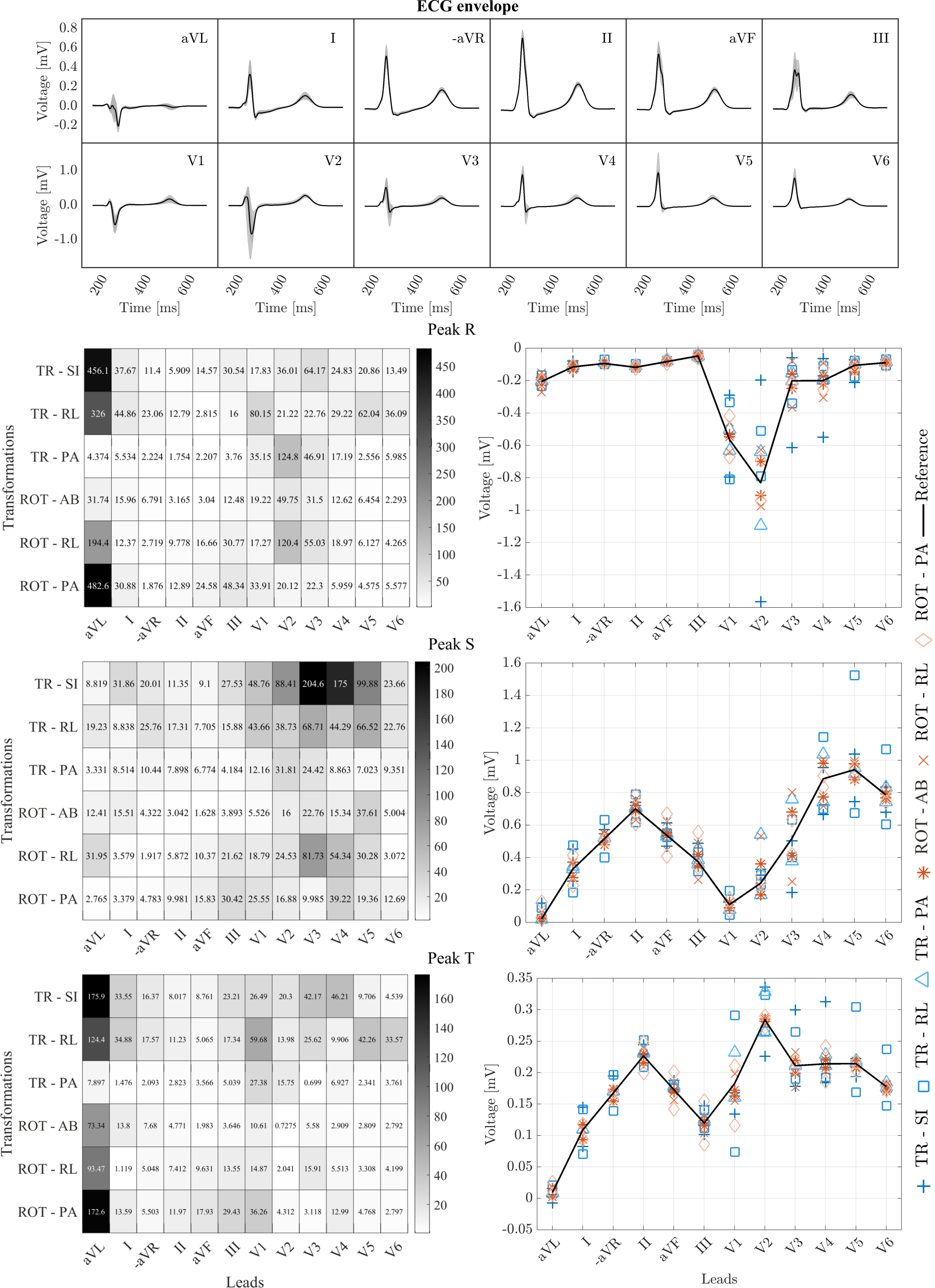
A quantitative analysis of relative and absolute amplitude variations of each peak across all leads was performed to identify leads where ECG morphology is most sensitive to anatomical uncertainty. As illustrated in Fig. 4 (left), a significant change in the amplitude of the R peak relative to baseline values was observed in lead aVL when the heart was translated by TR-SI or TR-RL, or rotated by ROT-PA and ROT-RL. Similarly, substantial relative variations of the R peak were noticeable in lead V2 under the transformations TR-PA and ROT-RL. Regarding the absolute variation in R peak magnitude, the most significant variations occurred in the precordial leads V2, V3, V4, V5, and V6 (see Fig. 4 (second row-right))
The highest changes in peak S were witnessed in the precordial leads V1 to V4. These were predominantly mediated by the transformations TR-SI, TR-RL, and ROT-RL. The largest absolute variation in peak S was induced in lead V2 by the transformations TR-SI which changed peak S from a maximum negative value of down to (see Fig. 4 Peak S, right panel).
3.3 Cardiac anatomical variation - size
3.3.1 Variable size with prescribed activation sequence
The ECG obtained by scaling the heart while prescribing the activation sequence as in the reference cardiac anatomy is shown in Fig. 5 (top). Prescribing the same activation sequence to a smaller/larger heart is inherently equivalent to assume a slower/faster conduction velocity. As expected, the main effect on the ECG was a scaling of signal amplitudes. As illustrated in Fig. 5, a significant change in the amplitude of the S peak relative to the baseline values was especially observed in precordial leads V2-V5. Specifically, the transition zone of the S wave – in the normal healthy case located between V3-V4 – was affected. In the smaller heart the transition zone of the S wave shifted towards V2-V3, whereas in the larger heat a shift towards V4-V5 occurred (see top panel of Fig. 5). The range of magnitude scaling in the remaining leads was comparable to those observed for translation and rotation of the heart. Also, similar morphological changes such as the appearance of the R wave in lead aVL were witnessed. However, the overall ECG morphology remained largely unaltered, with only minor variations of R and S wave shape and duration in leads V2 and V3, and an altered T wave duration in lead V3. These morphological effects are mediated by the change in distance between cardiac sources and the lead positions of V2 and V3, which are located in the immediate vicinity of the heart.
3.3.2 Variable size with prescribed conduction properties
Changes in ECG signal magnitudes and morphology obtained by altering the size and prescribing the conduction properties were comparable to those obtained from prescribing the activation sequence (see Fig. 5). However, in contrast, a noticeable time shift of about was witnessed here for the entire trace, mostly corresponding to an earlier/later epicardial breakthrough when the cardiac model was scaled to -10%/+10% of its dimension, respectively.
3.4 Uncertainty in electrical conductivities
The ECG envelope resulting from varying the conductive properties of distinct tissues within the torso is illustrated in Fig. 6. Removing conductive inhomogeneities from the reference heterogeneous torso resulted in minor variations in peak amplitudes in all leads, with the exception of the limb leads aVL and I, and the precordial leads V4-V6 where a rather pronounced increase in the R wave amplitude was observed. The T wave consistently remained largely unaffected across all leads, with the exception of a very modest increase in the T wave duration noticed in the limb leads II, aVF, and III, a slight.

A quantitative assessment of the ECG variation due to tissue-specific conductive inhomogeneity against a fully heterogeneous torso was assessed by computing the rmse and the CC averaged over all leads, as shown in Fig. 6 (bottom panel). The blood mass was observed to have a major role in affecting the amplitudes of the QRS complex, with a rmse of 37%, and the T wave with a a rmse of 20%, followed by fat, skin, bones and lungs. The conductive properties of the liver had a negligible impact upon the ECG. Finally, the CC reported on the bottom-right panel of Fig. 6 showed a negligible sensitivity of the ECG morphology to a variation in conductive properties of all tissues, only a minor reduction in CC was noticed when the blood mass inhomogeneity was omitted.
3.5 Lead placement uncertainty
The effect of lead placement uncertainty on ECG amplitudes and morphology is illustrated for each electrode placement in the form of the norm computed over all the ECG traces, and the corresponding 12-lead ECG (see Fig. 7). Most significant lead placement effects were witnessed in the precordial leads closest to the heart, i.e. V1-V4. Noticeable morphological changes in the ECG traces stemmed from a higher placement of the electrodes V1 and V2, causing an increasingly apparent RSR pattern and a T-wave inversion in leads V1 and V2, and a pronounced S wave in leads V3 and V4. As expected, limb leads appeared relatively robust against electrode movement. This was also the case with the precordial lead V6. Finally, slight S-wave slurring and R amplitude elevation were obtained within -aVR and lead II, as well as small morphological variations in leads aVL and I.
4 Discussion
Advanced CDT generation and calibration methods have been developed for finding model parameters that minimize the discrepancy between simulated and measured ECG [19, 22, 23]. While feasible, in principle, the fundamental issue of non-uniqueness of the solution persists, that is, more than one parameter set may exist that calibrates the model equally well to the observed ECG. Moreover, due to the nature of the clinical data acquisition process and the computational workflows for the generation of CDT models, inconsistencies between real and virtual CDT replicas of anatomy and physics in a patient, inevitably arise. These lead to discrepancies between real and simulated EP, although the observations used for calibration – the ECG– might be very similar or identical.
In our study, we investigate the impact of the major sources of inconsistencies upon the simulated ECG. These comprise i) the residual beat-to-beat variability reflected in morphological alterations of the ECG ii) observational uncertainties due to technical limitations in anatomical imaging and ECG recording, impeding an accurate synchronous measurement of all model parameters contributing to the genesis of the ECG [29, 30]. The latter includes the generation of computational cardiac and torso anatomies from tomographic images, which are modulated by the subject/patient breathing, the inability to measure electrical model parameters, such as the heterogeneity of electrical conductivities throughout the torso, and variability in the lead placement. If the uncertainty in the observed ECG is high, and the ECG predicted by the computational representation is overly sensitive to these inaccuracies in anatomical and electrical parameters, the calibration of a model based on a measured ECG is effectively impeded, or may be unreliable.
Here, we aim to provide a general view of the impact of these uncertainties on the simulated ECG, and to investigate to which extent they may affect the ability to calibrate a computational model of cardiac EP. Our qualitative analysis of residual variability in both healthy subjects and AF and VT patients showed a rather tight envelope in the ECG waveforms, indicating that using an arbitrarily chosen ECG beat, or a mean ECG beat, is admissible, and does not introduce any significant uncertainty. Further, the effects on ECG morphology and derived diagnostic markers of uncertainties in location, orientation, and size of the heart, in electrical torso conductivities, and lead placement, were computed. We moreover considered a worst-case scenario, defining uncertainty outputs falling outside the level of accuracy obtainable with careful dedicated clinical data acquisition. While all these factors were shown to impact the ECG waveforms, they primarily contributed to generating ECG traces that retained the most important morphological features and diagnostic markers, and closely adhered to the ground truth ECG.
Overall, our results suggest that an accurate calibration of a CDT is not impeded by observational uncertainties and model inconsistencies. Whether a set of parameters that optimally calibrates a model to the ECG is uniquely identifiable remains an open question, beyond the scope of our study.
4.1 Residual variability
The calibration of a CDT to replicate the patient heart’s intrinsic activation and repolarization pattern based on the ECG, relies upon a stable rhythm. However, beat-to-beat alterations cause residual variability in repetitive measurements of the ECG, leading to an ensemble of ECG waveforms, and the problem of selecting a representative beat in the ECG to be used for calibration.
Beat-to-beat variability is a well-known and extensively studied phenomenon. In healthy subjects, the heart rate is known to be always variable, even under resting conditions [26, 74, 28, 75]. However, heart rate variability per se is not a relevant factor as only the temporal onset of the heart beat is modulated, but the electrical activation and repolarization sequence remains largely unaffected, that is, the ECG waveform does not change. With respect to CDT calibration, only beat-to-beat alterations that affect the ECG waveform noticeably enough to indicate a change in the cardiac activation or repolarization pattern, are considered of interest. Such alterations in morphology, including very small amplitude variations at a scale [76, 27, 28, 77, 78, 28, 79, 80, 81], are often utilized as a clinical biomarker, indicative of a propensity for arrhythmias to occur.
In our study, we investigate beat-to-beat ECG morphological variability in cohorts of healthy subjects, AF and VT patients. In the healthy subjects, the ECG envelope caused by the residual variability was narrow, suggesting that such variability could be negligible for an ECG based calibration of a CDT.
This finding also held true for the patient cohorts treated with AF or VT ablation therapy. Our analysis of ECGs recorded during interventions in 36 AF and 17 VT patients showed very limited variability in most cases. This was expected, as ECGs were recorded during mapping studies where stable activation patterns are required for map construction. In cases where more noticeable changes in ECG morphology occurred, clustering of ECG morphologies would be necessary, and CDTs would need to be re-calibrated for each identified cluster. As high-fidelity ECG calibration becomes increasingly feasible and streamlined [19], facilitating calibration to an ensemble of ECGs, the identified model parameters leading to a good match may offer insights into the underlying physiological mechanisms.
4.2 Cardiac anatomical variation
Clinical ECG recording and anatomical image acquisition are not synchronous. Therefore, the precise anatomical configuration of the heart at the time of ECG recording is generally unknown. Moreover, a 3D whole heart anatomical MRI scan, as used in this study, is typically acquired during diastasis, as this phase of the cardiac cycle provides the longest window with minimal cardiac motion. However, during diastasis, the ventricles are smaller than their end-diastolic configuration, when electrical depolarization associated with the QRS complex occurs, and larger than their end-systolic configuration, when repolarization associated with the T-wave takes place [82, 83]. Other anatomical and technical factors during image acquisition are moreover responsible for the increase of uncertainty in the cardiac anatomy reconstruction, and its relation to the recorded ECG.
For instance, discrepancies between cardiac anatomy in a patient and its representation in an anatomical model inevitably arise due to a combination of physiological and technical factors. Physiologically, the position, orientation, size, and shape of the heart are highly variable, influenced by factors such as breathing [33, 34, 35, 36, 37, 35], body posture [38, 39], as well as cardiac motion and deformation over a heartbeat itself [84, 85]. Breathing alters the relative location and orientation of the heart within the torso, while the size of the heart is specifically affected by maneuvers during acquisition, such as a breath hold, which changes the intra-thoracic pressure and, thus, the blood volume in the cavities [86, 87]. Moreover, prolonged time intervals often occur between imaging and ECG recording. This can result in significant changes in the heart’s size due to variations in heart rate or the patient’s fluid status.
Technical factors include limited spatial resolution and contrast in the images, hindering the accurate segmentation of all relevant structures. Additional challenges include the displacement between slices in sequential 2D image acquisitions, which are temporally registered using a navigator or ECG gating, the registration errors between the heart and the whole torso imaging, and the device artifacts that partially obscure the cardiac anatomy [88, 89, 90, 91].
All these factors combined contribute to cardiac anatomical uncertainty, affecting the heart’s shape, size, and position not only anatomically but also as an electrical source relative to the ECG recording site. This uncertainty effectively alters the ECG waveform.
The influence of the heart’s position and orientation on the ECG has been explored in vivo and with real patients in several studies. In [41, 42], the authors estimate the heart’s position relative to the torsos of 25 subjects by comparing body surface potential maps with the cardiac and torso geometry obtained from MRI scans, and then compute the corresponding ECG through computational models. In [41], the pericardial position of each subject is inferred through an electrocardiographic inverse solution. These 25 solutions are then used to compute the statistical variability caused by geometric error on the ECG. However, the authors are unable to separate the influence of physiological and geometric variability on the ECG. A direct measurement of the heart’s position compared to the torso shape and the position of the lead electrodes is performed in [42], where the authors used MRI images of both the heart and the torso to create 3D triangular meshes. The solid angle of the heart relative to the torso is then computed, along with other thoracic indices, and these are accounted for in the analysis of ECG variability. However, this study does not isolate the effect of cardiac size and position variability within a single subject, but rather represents a population study. Single-subject ECG variability due to heart position is addressed in [40], where the authors analyze ECG changes due to heart position using an instrumented, isolated heart suspended in a torso-shaped electrolytic tank. Changes in heart position are accounted for by moving the heart in a three-axis orthogonal system built into the torso, to match uncertainty in cardiac position due to the MRI imaging process of human subjects. While the applied cardiac transformations resemble our heart translations in both direction and magnitude, the hearts used are from animals, the torso is not realistic, and the ECG s and their variations are not clearly represented.
A more in-depth analysis of ECG variations in silico has been previously investigated in a small number of studies. In [92], the authors exploited the effect of the cardiac position on pathological QRS complex in five patients. The simulated ECG s of the reference patients were initially validated against clinical data and then compared with the same ECG signals obtained by displacing the cardiac domain using upper-lower and right-left rigid translations and horizontal-vertical rigid rotations. Variability of the QRS complex due to heart-torso geometrical uncertainty was also analyzed in [93]. In this study, starting with 5 cardiac and 5 torso geometries, a cohort of 625 heart-torso anatomical models was created by combining the cardiac and torso models, and defining two rigid translational directions and two rigid rotational directions for the heart. The ECG s were compared, and a qualitative assessment of the reliability of the computed ECG s against clinical data was conducted. However, in combining the heart and torso geometries, additional factors such as electrophysiological activation and variations in cardiac shape between anatomies contribute to ECG generation. Moreover, both studies focus solely on the QRS complex and consider a limited number of translations and rotations. Additionally, the heart’s size is not included as a factor affecting the ECG, and the set of simulated ECG s are compared against a single reference signal for each patient, without considering beat-to-beat variability.
Our work aims to model all possible transformations of the heart based on in vivo analysis, including heart scaling, by exploring the limit values of these transformations in a single patient. This approach effectively defines the range of variations in the ECG signals without introducing additional factors. Additionally, we extend the analysis to the T wave and compare the results with the beat-to-beat variability of the reference ECG. Our results showed that ECG shape variations are overall minor. Furthermore, we observed different sensitivities of the R, S, and T peaks to cardiac geometrical transformations in different leads, with aVL, V3, and V4 being the most affected. The most influential transformations were translations in the superior-inferior direction, rotations in the postero-anterior direction, and heart scaling.
We highlight the potential for increasing and decreasing depolarization and repolarization duration due to changes in heart size when prescribing conduction parameters, as opposed to a time shift of the ECG signals but with the same wave duration when prescribing the activation sequence. This underscores the prominent role of cardiac activation in ECG generation.
Overall, our results suggest that the dimension, position, and orientation of the heart have a limited influence on the calibration of CDT s.
4.3 Uncertainty in conductive properties
"While efficient, anatomically detailed segmentation of tissues and organs of the torso from high-quality clinical images is becoming increasingly feasible [94], measuring their respective electrical properties is challenging and infrequently undertaken. Consequently, modeling studies often rely on values reported in the literature [22, 68] or public databases, with potential ad-hoc adjustments to better fit the ECG signal [69].
Computational analysis of the effect of tissue-specific conduction properties within the torso on the ECG has been conducted previously in [66, 72]. In [66] the effect of each of the lungs, skeletal muscle, and subcutaneous fat inhomogeneities was investigated in a single patient model. While quantitative indices were taken into consideration, a highly simplistic lumped dipole model was used, and the effect of low skin conductivity was not considered. Similarly, in [68], the effect of major tissue-specific inhomogeneities on the ECG was comprehensively analyzed using a simplified model of the cardiac sources. These sources were represented by a monodomain model in the ventricles and a cellular automaton in the atria, interpolated onto a background torso mesh, and used as a volumetric source for solving the forward problem. Consistent with our study, the conductivity of the blood pool was identified as the most significant factor. Keller et al. also highlighted the importance of fat, ranking it closely behind blood conductivity, and the negligible effect of the liver. However, unlike our findings, they observed that lungs exerted a greater influence than bones. Nevertheless, a limitation of both studies is the lack of a reference clinical ECG for comparison with the simulated results and the absence of simulated clinical 12-lead ECG s. Without these, an analysis of morphological effects and their diagnostic interpretation is precluded, and the assessment of the validity and fidelity of the source model is limited.
Overall, our findings indicate a rather marginal impact of conductive inhomogeneities on the 12-lead ECG (see Fig. 6, suggesting that their consideration – with the exception of the blood pool conductivity – is not essential for the calibration of CDTs to the ECG.
4.4 Lead placement uncertainty
In clinical practice, variations in the morphological patterns of recorded 12-lead ECG systems can result from electrode positioning discrepancies, a phenomenon noted even among trained practitioners [73, 95]. Wenger et al. [96] demonstrated that precordial electrodes are typically positioned approximately away from the standard clinical placements, with deviations exceeding observed upwards in 50% of V1 and V2 placements, and leftward and downward in up to 50% of V4 and V6 placements. Recent clinical studies have sought to assess the impact of electrode positioning by manually adjusting electrode positions or selecting specific subsets from body surface potential maps (BSPMs) recordings [45, 46]. Nevertheless, these studies often encounter challenges such as low or inconsistent spatial resolution due to manual placement or reliance on artificial signal interpolation from BSPMs recordings. Additionally, the accuracy of information regarding the actual underlying cardiac electrical activity is frequently limited
An in silico study investigating the effect of electrode displacement on five patients was conducted in [92]. While this study provided a consistent experimental setup with precise electrode placement and simulated physiological conditions, its probe is restricted to displacing solely the precordial electrodes in downward directions, therefore not exploring the entirety of potential electrode sites.
Our study aims to explore a consistent part of the thoracic surface, by moving the electrodes on the longitudinal axis of the torso, both upwards and downwards, and circumferentially with respect to the UTCss, thus covering right and left sites, including the average displacement found in [96]. Furthermore, all electrodes were displaced, including the limb leads. Our findings indicate that electrode displacement primarily affects the R wave, particularly in the precordial leads, as well as in leads I and III. While the ECG morphology remains largely intact across most leads, we observed slight to more pronounced variations in amplitude in the limb leads. Among the precordial leads, V2 is notably sensitive to upward and downward shifts of its corresponding electrodes, particularly affecting the amplitude of the R wave. However, significant changes in the waveform, such as the appearance of an RSR pattern and inverted T waves, are only observed with an extreme displacement of the V2 electrode. When electrodes are moderately displaced, variations in the ECG resemble those induced by shifting the heart in TR-SI directions, as reported in [92]. Our findings affirm that the distance between the heart and electrode placement, especially for precordial leads closest to cardiac sources, is crucial in determining ECG morphology.
With respect to the CDT s, we demonstrated that electrode displacement should be considered but does not fundamentally define the ECG morphology or overall calibration.
5 Limitations
"A major limitation of this study is the use of a single-patient anatomical model. Given that the shape of the heart, the cardiac conduction system, and the torso are crucial factors influencing the spatio-temporal evolution of cardiac sources and their manifestation as torso potentials, some aspects of our findings may not be generalizable. Specifically, the modeled subject had a vertical electrical axis, suggesting that changes in ECG morphology may not manifest similarly in other subjects. Nevertheless, our overall observation regarding the minimal impact of model inconsistencies on ECG morphology should remain applicable, regardless of the electrical axis type.
While the selected set of cardiac anatomical transformations covers the complete range of possible anatomical variations of the heart within a single patient, our experiments only consider individual transformations. The potential effects on the ECG resulting from combined anatomical variations are not explored. However, considering the impact of anatomical variations, which result in a relatively narrow distribution, it seems unlikely that combined anatomical transformations would significantly widen the range of ECG variations.
In examining the ECG dependence on electrical tissue properties, we tested only a single conductivity value for each organ, although measurements suggest a certain variability. Additionally, the conductivities of the heart and their anisotropy ratios were held constant, despite their known high uncertainty [97], which can significantly influence ECG morphology [98, 99].
6 Conclusions
Our results indicate that uncertainties related to anatomical position, orientation, size, electrical conductivity heterogeneity of tissues and organs in the torso, as well as lead placement, have limited impact on ECG morphology and diagnostically relevant ECG features. This finding aligns with the narrow distribution of ECG due to residual beat-to-beat variability observed in both healthy subjects and patients. Our findings suggest that the ECG morphology is robustly defined by the electrical activation and repolarization pattern, and, to a much lesser extent, by the considered inconsistencies. Consequently, these factors do not significantly impede ECG-based CDT calibration. However, the core challenge of calibration – the unique identification of model parameters – remains a challenge, although it was beyond the scope of our study.
Acknowledgments
This research was supported by grants from the Austrian Science Fund (FWF) grants no. I6467-B to G.P. and 10.55776/ESP592 to K.G., the European Union’s Horizon 2020 research and innovation program under the Marie Skłdowska-Curie grant TwinCare-AF agreement no. 101148636 to E.Z., and in part the Austrian Research Promotion Agency (FFG) grant FO999891133 to M.A.F.G. E. Z. acknowledges her membership to INdAM GNCS - Gruppo Nazionale per il Calcolo Scientifico (National Group for Scientific Computing, Italy).
![[Uncaptioned image]](/html/2407.17146/assets/Images/eu_funded_en.jpg)
References
- Cluitmans et al. [2023] M. Cluitmans, R. Walton, G. Plank, Editorial: Computational methods in cardiac electrophysiology, Frontiers in Physiology 14 (2023).
- Monaci et al. [2022] S. Monaci, S. Qian, K. Gillette, E. Puyol-Antón, R. Mukherjee, M. K. Elliott, J. Whitaker, R. Rajani, M. O’Neill, C. A. Rinaldi, G. Plank, A. P. King, M. J. Bishop, Non-invasive localization of post-infarct ventricular tachycardia exit sites to guide ablation planning: a computational deep learning platform utilizing the 12-lead electrocardiogram and intracardiac electrograms from implanted devices, EP Europace 25 (2022) 469–477.
- Heijman et al. [2021] J. Heijman, H. Sutanto, H. J. G. M. Crijns, S. Nattel, N. A. Trayanova, Computational models of atrial fibrillation: achievements, challenges, and perspectives for improving clinical care, Cardiovascular Research 117 (2021) 1682–1699.
- Aronis et al. [2019] K. N. Aronis, R. L. Ali, J. A. Liang, S. Zhou, N. A. Trayanova, Understanding af mechanisms through computational modelling and simulations, Arrhythmia & Electrophysiology Review 8 (2019) 210.
- Kabra et al. [2022] R. Kabra, S. Israni, B. Vijay, C. Baru, R. Mendu, M. Fellman, A. Sridhar, P. Mason, J. W. Cheung, L. DiBiase, S. Mahapatra, J. Kalifa, S. A. Lubitz, P. A. Noseworthy, R. Navara, D. D. McManus, M. Cohen, M. K. Chung, N. Trayanova, R. Gopinathannair, D. Lakkireddy, Emerging role of artificial intelligence in cardiac electrophysiology, Cardiovascular Digital Health Journal 3 (2022) 263–275.
- Swenson et al. [2020] D. J. Swenson, R. T. Taepke, J. J. E. Blauer, E. Kwan, E. Ghafoori, G. Plank, E. Vigmond, R. S. MacLeod, P. DeGroot, R. Ranjan, Direct comparison of a novel antitachycardia pacing algorithm against present methods using virtual patient modeling., Heart rhythm (2020).
- Corral-Acero et al. [2020] J. Corral-Acero, F. Margara, M. Marciniak, C. Rodero, F. Loncaric, Y. Feng, A. Gilbert, J. F. Fernandes, H. A. Bukhari, A. Wajdan, et al., The ‘digital twin’ to enable the vision of precision cardiology, European heart journal 41 (2020) 4556–4564.
- Peirlinck et al. [2021] M. Peirlinck, F. S. Costabal, J. Yao, J. Guccione, S. Tripathy, Y. Wang, D. Ozturk, P. Segars, T. Morrison, S. Levine, et al., Precision medicine in human heart modeling: Perspectives, challenges, and opportunities, Biomechanics and modeling in mechanobiology 20 (2021) 803–831.
- Niederer et al. [2019] S. A. Niederer, J. Lumens, N. A. Trayanova, Computational models in cardiology, Nature Reviews Cardiology 16 (2019) 100–111.
- Cartoski et al. [2019] M. J. Cartoski, P. P. Nikolov, A. Prakosa, P. M. Boyle, P. J. Spevak, N. A. Trayanova, Computational identification of ventricular arrhythmia risk in pediatric myocarditis, Pediatric cardiology 40 (2019) 857–864.
- Arevalo et al. [2016] H. J. Arevalo, F. Vadakkumpadan, E. Guallar, A. Jebb, P. Malamas, K. C. Wu, N. A. Trayanova, Arrhythmia risk stratification of patients after myocardial infarction using personalized heart models, Nature communications 7 (2016) 11437.
- Trayanova et al. [2023] N. A. Trayanova, A. Lyon, J. Shade, J. Heijman, Computational modeling of cardiac electrophysiology and arrhythmogenesis, Physiological Reviews (2023).
- Lopez-Perez et al. [2019] A. Lopez-Perez, R. Sebastian, M. Izquierdo, R. Ruiz, M. Bishop, J. M. Ferrero, Personalized cardiac computational models: from clinical data to simulation of infarct-related ventricular tachycardia, Frontiers in physiology 10 (2019) 580.
- Gray and Pathmanathan [2018] R. A. Gray, P. Pathmanathan, Patient-specific cardiovascular computational modeling: diversity of personalization and challenges, Journal of cardiovascular translational research 11 (2018) 80–88.
- Viceconti et al. [2016] M. Viceconti, A. Henney, E. Morley-Fletcher, In silico clinical trials: how computer simulation will transform the biomedical industry, International Journal of Clinical Trials 3 (2016) 37–46.
- Trayanova and Prakosa [2024] N. A. Trayanova, A. Prakosa, Up digital and personal: How heart digital twins can transform heart patient care, Heart Rhythm 21 (2024) 89–99.
- Gsell et al. [2023] M. A. Gsell, L. Azzolin, K. K. Gillette, A. Neic, T. Schrotter, F. Thaler, G. Plank, Towards the development of virtual heart technology for creating digital twins of cardiac electrophysiology, in: 2023 Computing in Cardiology (CinC), volume 50, IEEE, 2023, pp. 1–4.
- Jung et al. [2022] A. Jung, M. A. Gsell, C. M. Augustin, G. Plank, An integrated workflow for building digital twins of cardiac electromechanics—a multi-fidelity approach for personalising active mechanics, Mathematics 10 (2022) 823.
- Grandits et al. [2023] T. Grandits, J. Verhülsdonk, G. Haase, A. Effland, S. Pezzuto, Digital twinning of cardiac electrophysiology models from the surface ECG: a geodesic backpropagation approach, IEEE Transactions on Biomedical Engineering (2023).
- Qiao et al. [2023] L. Qiao, S. Hu, B. Xiao, X. Bi, W. Li, X. Gao, A dual self-calibrating framework for non-invasive fetal ECG R-peak detection, IEEE Internet of Things Journal (2023).
- Camps et al. [2023] J. Camps, L. A. Berg, Z. J. Wang, R. Sebastian, L. L. Riebel, R. Doste, X. Zhou, R. Sachetto, J. Coleman, B. Lawson, et al., Digital twinning of the human ventricular activation sequence to clinical 12-lead ECGs and magnetic resonance imaging using realistic Purkinje networks for in silico clinical trials, arXiv preprint arXiv:2306.13740 (2023).
- Gillette et al. [2021] K. Gillette, M. A. F. Gsell, A. J. Prassl, E. Karabelas, U. Reiter, G. Reiter, T. Grandits, C. Payer, D. Štern, M. Urschler, J. D. Bayer, C. M. Augustin, A. Neic, T. Pock, E. J. Vigmond, G. Plank, A Framework for the generation of digital twins of cardiac electrophysiology from clinical 12-leads ECGs., Medical image analysis 71 (2021) 102080. Publisher: Elsevier B.V.
- Camps et al. [2024] J. Camps, L. A. Berg, Z. J. Wang, R. Sebastian, L. L. Riebel, R. Doste, X. Zhou, R. Sachetto, J. Coleman, B. Lawson, V. Grau, K. Burrage, A. Bueno-Orovio, R. Weber dos Santos, B. Rodriguez, Digital twinning of the human ventricular activation sequence to clinical 12-lead ECGs and magnetic resonance imaging using realistic Purkinje networks for in silico clinical trials, Medical Image Analysis (2024) 103108.
- Gillette et al. [2023] K. Gillette, M. A. Gsell, C. Nagel, J. Bender, B. Winkler, S. E. Williams, M. Bär, T. Schäffter, O. Dössel, G. Plank, et al., MedalCare-XL: 16,900 healthy and pathological synthetic 12 lead ECGs from electrophysiological simulations, Scientific Data 10 (2023) 531.
- Xanthis et al. [2007] C. G. Xanthis, P. Bonovas, G. A. Kyriacou, Inverse problem of ECG for different equivalent cardiac sources, Piers Online 3 (2007) 1222–1227.
- Hasan et al. [2012] M. A. Hasan, D. Abbott, M. Baumert, Relation between beat-to-beat QT interval variability and T-wave amplitude in healthy subjects, Annals of Noninvasive Electrocardiology 17 (2012) 195–203.
- Frljak et al. [2003] S. Frljak, V. Avbelj, R. Trobec, B. Meglic, T. Ujiie, B. Gersak, Beat-to-beat QT interval variability before and after cardiac surgery, Computers in Biology and Medicine 33 (2003) 267–276. Human Heart in the Focus of Computer Power.
- Appel et al. [1989] M. L. Appel, R. D. Berger, J. Saul, J. M. Smith, R. J. Cohen, Beat to beat variability in cardiovascular variables: Noise or music?, Journal of the American College of Cardiology 14 (1989) 1139–1148.
- Keall et al. [2006] P. J. Keall, G. S. Mageras, J. M. Balter, R. S. Emery, K. M. Forster, S. B. Jiang, J. M. Kapatoes, D. A. Low, M. J. Murphy, B. R. Murray, et al., The management of respiratory motion in radiation oncology report of aapm task group 76 a, Medical physics 33 (2006) 3874–3900.
- Hawkes et al. [2005] D. J. Hawkes, D. Barratt, J. M. Blackall, C. Chan, P. J. Edwards, K. Rhode, G. P. Penney, J. McClelland, D. L. Hill, Tissue deformation and shape models in image-guided interventions: a discussion paper, Medical Image Analysis 9 (2005) 163–175.
- Monfredi et al. [2013] O. Monfredi, L. A. Maltseva, H. A. Spurgeon, M. R. Boyett, E. G. Lakatta, V. A. Maltsev, Beat-to-beat variation in periodicity of local calcium releases contributes to intrinsic variations of spontaneous cycle length in isolated single sinoatrial node cells, PLOS ONE 8 (2013) 1–13.
- Zaniboni et al. [2000] M. Zaniboni, A. E. Pollard, L. Yang, K. W. Spitzer, Beat-to-beat repolarization variability in ventricular myocytes and its suppression by electrical coupling, American Journal of Physiology-Heart and Circulatory Physiology 278 (2000) H677–H687.
- Holst et al. [2018] K. Holst, M. Ugander, A. Sigfridsson, Respiratory variation in left ventricular cardiac function with 3D double golden-angle whole-heart cine imaging, Magnetic Resonance in Medicine 79 (2018) 2693–2701.
- Claessen et al. [2014] G. Claessen, P. Claus, M. Delcroix, J. Bogaert, A. L. Gerche, H. Heidbuchel, Interaction between respiration and right versus left ventricular volumes at rest and during exercise: a real-time cardiac magnetic resonance study, American Journal of Physiology-Heart and Circulatory Physiology 306 (2014) H816–H824.
- Shechter et al. [2004] G. Shechter, C. Ozturk, J. Resar, E. McVeigh, Respiratory motion of the heart from free breathing coronary angiograms, IEEE Transactions on Medical Imaging 23 (2004) 1046–1056.
- Lendrum et al. [1979] B. L. Lendrum, A. M. Mondkar, J. B. Harris, B. Smulevitz, I. Carr, Respiratory variation in echocardiographic dimensions of left and right ventricles in normal children: The role of the interventricular septum, Pediatric Cardiology 1 (1979) 39–45.
- Jagsi et al. [2007] R. Jagsi, J. M. Moran, M. L. Kessler, R. B. Marsh, J. M. Balter, L. J. Pierce, Respiratory motion of the heart and positional reproducibility under active breathing control, International Journal of Radiation Oncology*Biology*Physics 68 (2007) 253–258.
- Rodeheffer et al. [1986] R. J. Rodeheffer, G. Gerstenblith, E. Beard, J. L. Fleg, L. C. Becker, M. L. Weisfeldt, E. G. Lakatta, Postural changes in cardiac volumes in men in relation to adult age, Experimental gerontology 21 (1986) 367–378.
- Rapaport et al. [1966] E. Rapaport, M. Wong, E. E. Escobar, G. Martinez, The effect of upright posture on right ventricular volumes in patients with and without heart failure, American Heart Journal 71 (1966) 146–152.
- MacLeod et al. [2000] R. S. MacLeod, Q. Ni, B. Punske, P. R. Ershler, B. Yilmaz, B. Taccardi, Effects of heart position on the body-surface electrocardiogram, Journal of Electrocardiology 33 (2000) 229–237. Research and Technology Transfer in Computerized Electrocardiology.
- Hoekema et al. [2001] R. Hoekema, G. J. Uijen, A. Van Oosterom, Geometrical aspects of the interindividual variability of multilead ECGrecordings, IEEE Transactions on Biomedical Engineering 48 (2001) 551–559.
- Van Oosterom et al. [2000] A. Van Oosterom, R. Hoekema, G. Uijen, Geometrical factors affecting the interindividual variability of the ECGand the vcg, Journal of electrocardiology 33 (2000) 219–227.
- Ramanathan and Rudy [2001] C. Ramanathan, Y. Rudy, Electrocardiographic imaging: I. effect of torso inhomogeneities on body surface electrocardiographic potentials, Journal of cardiovascular electrophysiology 12 (2001) 229–240.
- Benjamin Jr et al. [1950] J. M. Benjamin Jr, H. Schwan, C. F. Kay, J. H. Hafkenschiel, The electrical conductivity of living tissues as it pertains to electrocardiography: I. review of the problem of homogeneity vs. nonhomogeneity, an outline of the technical aspects of tissue resistivity measurements, and a critical and experimental analysis of certain pertinent experiments, Circulation 2 (1950) 321–335.
- Medani et al. [2018] S. A. Medani, M. Hensey, N. Caples, P. Owens, Accuracy in precordial ECG lead placement: improving performance through a peer-led educational intervention, Journal of Electrocardiology 51 (2018) 50–54.
- Kania et al. [2014] M. Kania, H. Rix, M. Fereniec, H. Zavala-Fernandez, D. Janusek, T. Mroczka, G. Stix, R. Maniewski, The effect of precordial lead displacement on ECG morphology, Medical & biological engineering & computing 52 (2014) 109–119.
- Lateef et al. [2003] F. Lateef, N. Nimbkar, Z. K. Da, F. R. Min, Vertical displacement of the precordial leads alters electrocardiographic morphology, Indian Heart Journal 55 (2003) 339–343.
- Hill and Goodman [1987] N. Hill, J. Goodman, Importance of accurate placement of precordial leads in the 12-lead electrocardiogram, Heart and Lung: Journal of Critical Care 16 (1987) 561 – 566.
- Kerwin et al. [1960] A. Kerwin, R. McLean, H. Tegelaar, A method for the accurate placement of chest electrodes in the taking of serial electrocardiographic tracings, Canadian Medical Association Journal 82 (1960) 258.
- Porr et al. [2023] B. Porr, L. Howell, I. Stournaras, Y. Nir, Popular ECG R peak detectors written in python, 2023. URL: https://doi.org/10.5281/zenodo.7652725. doi:10.5281/zenodo.7652725.
- Arnold et al. [2024] R. Arnold, A. J. Prassl, A. Neic, F. Thaler, C. M. Augustin, M. A. Gsell, K. Gillette, M. Manninger, D. Scherr, G. Plank, pyCEPS: A cross-platform electroanatomic mapping data to computational model conversion platform for the calibration of digital twin models of cardiac electrophysiology, Computer Methods and Programs in Biomedicine 254 (2024) 108299.
- Gillette et al. [2022] K. Gillette, M. A. F. Gsell, M. Strocchi, T. Grandits, A. Neic, M. Manninger, D. Scherr, C. H. Roney, A. J. Prassl, C. M. Augustin, E. J. Vigmond, G. Plank, A personalized real-time virtual model of whole heart electrophysiology, Frontiers in Physiology 13 (2022) 1–14.
- Gillette et al. [2021] K. Gillette, M. A. F. Gsell, J. Bouyssier, A. J. Prassl, A. Neic, E. J. Vigmond, G. Plank, Automated Framework for the Inclusion of a His-Purkinje System in Cardiac Digital Twins of Ventricular Electrophysiology., Annals of biomedical engineering 49 (2021) 3143–3153.
- Crozier et al. [2016] A. Crozier, C. M. Augustin, A. Neic, A. J. Prassl, M. Holler, T. E. Fastl, A. Hennemuth, K. Bredies, T. Kuehne, M. J. Bishop, S. A. Niederer, G. Plank, Image-Based Personalization of Cardiac Anatomy for Coupled Electromechanical Modeling, Annals of Biomedical Engineering 44 (2016) 58–70. ISBN: 0090-6964.
- Payer et al. [2018] C. Payer, D. Štern, H. Bischof, M. Urschler, Multi-label whole heart segmentation using CNNs and anatomical label configurations, Lecture Notes in Computer Science (including subseries Lecture Notes in Artificial Intelligence and Lecture Notes in Bioinformatics) 10663 LNCS (2018) 190–198. ISBN: 9783319755403.
- CIBC [2016] CIBC, 2016. Seg3D: Volumetric Image Segmentation and Visualization. Scientific Computing and Imaging Institute (SCI), Download from: http://www.seg3d.org.
- Prassl et al. [2009] A. J. Prassl, F. Kickinger, H. Ahammer, V. Grau, J. E. Schneider, E. Hofer, E. J. Vigmond, N. a. Trayanova, G. Plank, Automatically generated, anatomically accurate meshes for cardiac electrophysiology problems., IEEE transactions on bio-medical engineering 56 (2009) 1318–30.
- Neic et al. [2020] A. Neic, M. A. F. Gsell, E. Karabelas, A. J. Prassl, G. Plank, Automating image-based mesh generation and manipulation tasks in cardiac modeling workflows using Meshtool., SoftwareX 11 (2020) 100454. Publisher: Elsevier B.V.
- Roney et al. [2021] C. H. Roney, R. Bendikas, F. Pashakhanloo, C. Corrado, E. J. Vigmond, E. R. McVeigh, N. A. Trayanova, S. A. Niederer, Constructing a human atrial fibre atlas, Annals of biomedical engineering 49 (2021) 233–250.
- Bayer et al. [2012] J. Bayer, R. Blake, G. Plank, N. Trayanova, A novel rule-based algorithm for assigning myocardial fiber orientation to computational heart models, Annals of biomedical engineering 40 (2012) 2243–54.
- Roney et al. [2019] C. H. Roney, A. Pashaei, M. Meo, R. Dubois, P. M. Boyle, N. A. Trayanova, H. Cochet, S. A. Niederer, E. J. Vigmond, Universal atrial coordinates applied to visualisation, registration and construction of patient specific meshes, Medical image analysis 55 (2019) 65–75.
- Bayer et al. [2018] J. Bayer, A. J. Prassl, A. Pashaei, J. F. Gomez, A. Frontera, A. Neic, G. Plank, E. J. Vigmond, Universal ventricular coordinates: A generic framework for describing position within the heart and transferring data, Medical image analysis 45 (2018) 83–93.
- Neic et al. [2017] A. Neic, F. O. Campos, A. J. Prassl, S. A. Niederer, M. J. Bishop, E. J. Vigmond, G. Plank, Efficient computation of electrograms and ECGs in human whole heart simulations using a reaction-eikonal model, Journal of Computational Physics 346 (2017) 191–211. Publisher: Elsevier Inc.
- Potse [2018] M. Potse, Scalable and accurate ECG simulation for reaction-diffusion models of the human heart, Frontiers in physiology 9 (2018) 349898.
- Odille et al. [2017] F. Odille, S. Liu, P. van Dam, J. Felblinger, Statistical variations of heart orientation in healthy adults, in: 2017 Computing in Cardiology (CinC), IEEE, 2017, pp. 1–4.
- Bradley et al. [2000] C. Bradley, A. Pullan, P. Hunter, Effects of material properties and geometry on electrocardiographic forward simulations, Annals of biomedical engineering 28 (2000) 721–741.
- Sánchez et al. [2018] C. Sánchez, G. d’Ambrosio, F. Maffessanti, E. G. Caiani, F. W. Prinzen, R. Krause, A. Auricchio, M. Potse, Sensitivity analysis of ventricular activation and electrocardiogram in tailored models of heart-failure patients, Medical & biological engineering & computing 56 (2018) 491–504.
- Keller et al. [2010] D. Keller, F. M. Weber, G. Seemann, O. Dössel, Ranking the influence of tissue conductivities on forward-calculated ECGs, IEEE Transactions on Biomedical Engineering 57 (2010) 1568–1576.
- Bear et al. [2016] L. Bear, R. Dubois, N. Zemzemi, Optimization of organ conductivity for the forward problem of electrocardiography, in: 2016 Computing in Cardiology Conference (CinC), IEEE, 2016, pp. 385–388.
- Hasgall et al. [2022] P. Hasgall, F. Di Gennaro, C. Baumgartner, E. Neufeld, B. Lloyd, M. Gosselin, D. Payne, A. Klingenböck, N. Kuster, IT’IS database for thermal and electromagnetic parameters of biological tissues, itis.swiss/database, 2022. doi:10.13099/VIP21000-04-1.
- Zappon et al. [2024] E. Zappon, A. Manzoni, A. Quarteroni, A non-conforming-in-space numerical framework for realistic cardiac electrophysiological outputs, Journal of Computational Physics 502 (2024) 112815.
- Keller et al. [2010] D. U. Keller, F. M. Weber, G. Seemann, O. Dössel, Ranking the influence of tissue conductivities on forward-calculated ECGs, IEEE Transactions on Biomedical Engineering 57 (2010) 1568–1576.
- Rajaganeshan et al. [2008] R. Rajaganeshan, C. Ludlam, D. Francis, S. Parasramka, R. Sutton, Accuracy in ECG lead placement among technicians, nurses, general physicians and cardiologists, International journal of clinical practice 62 (2008) 65–70.
- Yeragani et al. [2000] V. K. Yeragani, R. Pohl, V. Jampala, R. Balon, J. Kay, G. Igel, Effect of posture and isoproterenol on beat-to-beat heart rate and QT variability, Neuropsychobiology 41 (2000) 113–123.
- Šipinková et al. [1997] I. Šipinková, G. Hahn, M. Meyer, M. Tadlanek, J. Hajek, Effect of respiration and posture on heart rate variability, Physiol. Res 46 (1997) 173–179.
- Eckberg [2003] D. L. Eckberg, Topical review: The human respiratory gate, The Journal of physiology 548 (2003) 339–352.
- Atiga et al. [1998] W. L. Atiga, H. Calkins, J. H. Lawrence, G. F. Tomaselli, J. M. Smith, R. D. Berger, Beat-to-beat repolarization lability identifies patients at risk for sudden cardiac death, Journal of cardiovascular electrophysiology 9 (1998) 899–908.
- Pueyo et al. [2016] E. Pueyo, C. E. Dangerfield, O. J. Britton, L. Virág, K. Kistamás, N. Szentandrassy, N. Jost, A. Varró, P. P. Nánási, K. Burrage, et al., Experimentally-based computational investigation into beat-to-beat variability in ventricular repolarization and its response to ionic current inhibition, PloS one 11 (2016) e0151461.
- Huikuri et al. [1996] H. V. Huikuri, T. Seppänen, M. J. Koistinen, K. J. Airaksinen, M. Ikaheimo, A. Castellanos, R. J. Myerburg, Abnormalities in beat-to-beat dynamics of heart rate before the spontaneous onset of life-threatening ventricular tachyarrhythmias in patients with prior myocardial infarction, Circulation 93 (1996) 1836–1844.
- Konta et al. [1990] T. Konta, K. Ikeda, M. Yamaki, K. Nakamura, K. Honma, I. Kubota, S. Yasui, Significance of discordant ST alternans in ventricular fibrillation., Circulation 82 (1990) 2185–2189.
- Kay et al. [1983] G. N. Kay, et al., Torsade de pointes: the long-short initiating sequence and other clinical features: observations in 32 patients, Journal of the American College of Cardiology 2 (1983) 806–817.
- Klein et al. [2017] R. Klein, E. S. Ametepe, Y. Yam, G. Dwivedi, B. J. Chow, Cardiac CT assessment of left ventricular mass in mid-diastasis and its prognostic value, European Heart Journal-Cardiovascular Imaging 18 (2017) 95–102.
- Budoff et al. [2009] M. J. Budoff, N. Ahmadi, G. Sarraf, Y. Gao, D. Chow, F. Flores, S. S. Mao, Determination of left ventricular mass on cardiac computed tomographic angiography1, Academic Radiology 16 (2009) 726–732.
- Ashikaga et al. [2008] H. Ashikaga, B. A. Coppola, K. G. Yamazaki, F. J. Villarreal, J. H. Omens, J. W. Covell, Changes in regional myocardial volume during the cardiac cycle: implications for transmural blood flow and cardiac structure, American Journal of Physiology-Heart and Circulatory Physiology 295 (2008) H610–H618.
- Carlsson et al. [2004] M. Carlsson, P. Cain, C. Holmqvist, F. Stahlberg, S. Lundback, H. Arheden, Total heart volume variation throughout the cardiac cycle in humans, American Journal of Physiology-Heart and Circulatory Physiology 287 (2004) H243–H250.
- Noseworthy et al. [2005] P. A. Noseworthy, Z. J. Malchano, J. Ahmed, G. Holmvang, J. N. Ruskin, V. Y. Reddy, The impact of respiration on left atrial and pulmonary venous anatomy: implications for image-guided intervention, Heart Rhythm 2 (2005) 1173–1178.
- Sakuma et al. [2001] H. Sakuma, N. Kawada, H. Kubo, Y. Nishide, K. Takano, N. Kato, K. Takeda, Effect of breath holding on blood flow measurement using fast velocity encoded cine MRI, Magnetic Resonance in Medicine: An Official Journal of the International Society for Magnetic Resonance in Medicine 45 (2001) 346–348.
- Counseller and Aboelkassem [2023] Q. Counseller, Y. Aboelkassem, Recent technologies in cardiac imaging, Frontiers in Medical Technology 4 (2023) 984492.
- Kalisz et al. [2016] K. Kalisz, J. Buethe, S. S. Saboo, S. Abbara, S. Halliburton, P. Rajiah, Artifacts at cardiac CT: physics and solutions, Radiographics 36 (2016) 2064–2083.
- Scott et al. [2009] A. D. Scott, J. Keegan, D. N. Firmin, Motion in cardiovascular MR imaging, Radiology 250 (2009) 331–351.
- Gamper et al. [2007] U. Gamper, P. Boesiger, S. Kozerke, Diffusion imaging of the in vivo heart using spin echoes–considerations on bulk motion sensitivity, Magnetic Resonance in Medicine: An Official Journal of the International Society for Magnetic Resonance in Medicine 57 (2007) 331–337.
- Nguyên et al. [2015] U. C. Nguyên, M. Potse, F. Regoli, M. L. Caputo, G. Conte, R. Murzilli, S. Muzzarelli, T. Moccetti, E. G. Caiani, F. W. Prinzen, et al., An in-silico analysis of the effect of heart position and orientation on the ECG morphology and vectorcardiogram parameters in patients with heart failure and intraventricular conduction defects, Journal of electrocardiology 48 (2015) 617–625.
- Mincholé et al. [2019] A. Mincholé, E. Zacur, R. Ariga, V. Grau, B. Rodriguez, MRI-based computational torso/biventricular multiscale models to investigate the impact of anatomical variability on the ECG QRS complex, Frontiers in physiology 10 (2019) 458916.
- Wasserthal et al. [2023] J. Wasserthal, H.-C. Breit, M. T. Meyer, M. Pradella, D. Hinck, A. W. Sauter, T. Heye, D. T. Boll, J. Cyriac, S. Yang, M. Bach, M. Segeroth, TotalSegmentator: Robust Segmentation of 104 Anatomic Structures in CT Images, Radiology: Artificial Intelligence 5 (2023) e230024. Publisher: Radiological Society of North America.
- Schijvenaars et al. [2008] B. J. Schijvenaars, G. van Herpen, J. A. Kors, Intraindividual variability in electrocardiograms, Journal of Electrocardiology 41 (2008) 190–196.
- Wenger and Kligfield [1996] W. Wenger, P. Kligfield, Variability of precordial electrode placement during routine electrocardiography, Journal of electrocardiology 29 (1996) 179–184.
- Roth [1997] B. J. Roth, Electrical conductivity values used with the bidomain model of cardiac tissue., IEEE transactions on bio-medical engineering 44 (1997) 326–8.
- Ushenin et al. [2021] K. Ushenin, V. Kalinin, S. Gitinova, O. Sopov, O. Solovyova, Parameter variations in personalized electrophysiological models of human heart ventricles, Plos one 16 (2021) e0249062.
- Sebastian et al. [2008] R. Sebastian, S. Ordas, G. Plank, B. Rodriguez, E. Vigmond, A. Frangi, Assessing influence of conductivity in heart modelling with the aim of studying cardiovascular diseases, Proc SPIE 6916 (2008).