Impact of Co-Excipient Selection on Hydrophobic Polymer Folding: Insights for Optimal Formulation Design
Abstract
The stabilization of liquid biological products is a complex task that depends on the chemical composition of both the active ingredient and any excipients in solution. Frequently, a large number of unique excipients are required to stabilize biologics, though it is not well-known how these excipients interact with one another. To probe these excipient-excipient interactions, we performed molecular dynamics simulations of arginine – a widely used excipient with unique properties – in solution either alone or with equimolar lysine or glutamate. We studied the effects of these mixtures on a hydrophobic polymer model to isolate excipient mechanisms on hydrophobic interactions, relevant to both protein folding and biomolecular self-assembly. We observed that arginine is the most effective single excipient in stabilizing hydrophobic polymer collapse, and its effectiveness can be augmented by lysine or glutamate addition. We utilized a decomposition of the potential of mean force to identify that the key source of arginine-lysine and arginine-glutamate synergy on polymer collapse is a reduction in attractive polymer-excipient direct interactions. Further, we applied principles from network theory to characterize the local solvent network that embeds the hydrophobic polymer. Through this approach, we found that arginine enables a more highly connected and stable network than in pure water, lysine, or glutamate solutions. Importantly, these network properties are preserved when lysine or glutamate are added to arginine solutions. Overall, we highlight the importance of identifying key molecular consequences of co-excipient selection, aiding in the establishment of rational formulation design rules.
UMN]Department of Chemistry, University of Minnesota, Minneapolis, MN 55455, USA \alsoaffiliation[CTC]Chemical Theory Center, University of Minnesota, Minneapolis, MN 55455, USA CEMS]Department of Chemical Engineering and Materials Science, University of Minnesota, Minneapolis, MN 55455, USA \alsoaffiliation[CTC]Chemical Theory Center, University of Minnesota, Minneapolis, MN 55455, USA MTU] Department of Chemical Engineering, Michigan Technological University, Houghton, MI 49931, USA UMA] Department of Chemical Engineering, University of Massachusetts Amherst, MA 01003, USA UMN]Department of Chemistry, University of Minnesota, Minneapolis, MN 55455, USA \alsoaffiliation[CTC]Chemical Theory Center, University of Minnesota, Minneapolis, MN 55455, USA
1 Introduction
Biologics are complex pharmaceuticals including proteins, vaccines, plasma products, gene therapy vectors, and biological tissues.1, 2 The stability of biologics is primarily concerned with physical denaturation related to protein unfolding and aggregation, among other factors.3, 4, 5, 6, 7, 8, 9, 10, 11, 12, 13, 14, 15 These formulations are especially susceptible to degradation when exposed to environmental stressors such as elevated temperatures, often necessitating refrigerated storage.16, 17, 18, 19 The logistics workflow known as the cold chain maintains biologics at cold temperatures from manufacturing, to long term storage, to distribution and outreach programs. However, equipment breakdowns and improper training at any point along the cold chain can disrupt entire batches of biologics, adverself affecting the accessibility of these products.20, 21, 19
A promising strategy to relieve the cold chain burden involves designing biological formulations with additives known as excipients.22, 23, 24, 25, 26, 27, 28 Excipients are often small molecules such as amino acids or sugars. Studies of excipient effects on the stability of biologics are generally high-throughput and empirical in nature, often lacking the resolution required to understand the molecular mechanism at play. Additionally, excipients are, on average, deployed in combinations of four or more unique molecules.28 This results in a vast design space and a generally poor understanding of individual excipient properties and combinatorial effects. We hypothesize that understanding the molecular details of excipient mechanisms will aid in predicting optimal excipient selections for novel formulations.
Motivated to understand the molecular-level effects of excipient combinations in biologics stability, we utilized molecular dynamics (MD) simulations of a hydrophobic polymer as a model for protein folding/unfolding. Hydrophobic polymer models are effective in decoupling hydrophobic interactions from other forces involved in protein folding (e.g., electrostatic, van der Waals, and hydrogen bonding interactions),29, 30, 31, 32, 33, 34, 35 a daunting task experimentally. By isolating hydrophobic interactions, fundamental insights into excipient mechanisms on protein stability can be obtained.36, 37, 38, 33, 30, 39, 40, 35 For example, the denaturant urea weakens hydrophobic interactions,41, 42, 37, 43, 44, 33, 30, 45 while the stabilizing osmolyte trimethylamine N-oxide (TMAO) negligibly affects or strengthens these effects.38, 46, 47, 48, 49, 50
As a starting point towards establishing excipient design rules, we turned our attention to a widely used excipient, arginine (Arg). Arg is a versatile excipient with a wide range of reported effects on the stability of biologics. Arg is frequently used in protein and vaccine storage, purification techniques, and as an aggregation reducer.51, 23, 52, 53, 54, 55 However, in some contexts, Arg has been found to denature proteins,56, 57, 58 accelerate aggregation,59, 60, 61 and inactivate viruses.62, 63, 35 In situations where Arg is denaturing, addition of charged co-excipients has been observed to reverse the denaturing properties of Arg.57 In other contexts, synergy has been observed between Arg and glutamate (Glu) in the solubilization of model proteins.64 Recently, we proposed that the multi-faceted effects of Arg arise from its positioning at the edge of a mechanistic flip between indirect- and direct-dominated mechanisms on hydrophobic interactions.35 Due to its placement at this edge, we aimed to understand whether subtle changes in the formulation environment alters the stabilizing properties of arginine. To this end, we investigated hydrophobic polymer folding in lysine (Lys), Glu, and Arg solutions, as well as equimolar formulations of Arg/Lys, Arg/Glu, and Lys/Glu. We discovered that, adding Lys or Glu to Arg solutions enhances hydrophobic polymer stability – underscoring the importance of co-excipient selection of biologics formulation design.
2 Methods
2.1 System Setup and Molecular Dynamics Simulations
To probe the effects of Arg, Lys, Glu, and binary excipient mixtures on hydrophobic interactions, we completed MD simulations of a hydrophobic polymer in excipient solutions at various concentrations. Replica exchange umbrella sampling (REUS)65 simulations were utilized to calculate the potential of mean force (PMF) of hydrophobic polymer folding in different excipient solutions. The hydrophobic polymer was modeled as a linear coarse-grained chain with 26 monomers. Each monomer represents a methane-like unit, with Lennard-Jones parameters and .38 The polymer-water parameter was modified to achieve an approximately even distribution of folded and unfolded polymer states in pure water.35 Box dimensions were defined such that 1.5 nm of space separated the fully elongated polymer from the nearest box edge. All systems were solvated with TIP4P/2005 water.66 The salt forms of all excipients (Arg/Cl, Lys/Cl, Glu/Na) under study were added to the box until the desired concentration was reached (Table S1). The CHARMM22 force field was used to describe excipient molecules and ions.67, 68 With this protocol, we generated systems comprised of 0.25 M, 0.5 M, and 1.0 M Arg, Lys, Glu, Arg/Lys, Arg/Glu, and Lys/Glu. In binary excipient solutions, equimolar concentrations were used. In excipient solutions with no polymer present, the same box size as in the polymer systems was used.
All simulations were initially subject to energy minimization using the steepest descent algorithm. 1 ns NVT equilibration simulations were carried out at 300 K, followed by 1 ns NPT equilibration simulations at 300 K and 1 atm. During equilibration, temperature was controlled according to the V-rescale thermostat69, while pressure was controlled via the Berendsen barostat.70 Following equilibration, NPT production runs were completed using the Nosé-Hoover thermostat ( = 5 ) 71 and Parrinello-Rahman barostat ( = 25 ).72 Production runs were 20 ns long for excipient/water systems, and between 50-250 ns per window for excipient/polymer/water REUS simulations (Table S1). In all simulations, the Particle Mesh Ewald (PME) algorithm was used for electrostatic interactions with a cut-off of 1 . A reciprocal grid of 42 x 42 x 42 cells was used with order B-spline interpolation. A single cut-off of 1 was used for Van der Waals interactions. The neighbor search was performed every 10 steps. Lorentz-Berthelot mixing rules73, 74 were used to calculate non-bonded interactions between different atom types. All simulations were run in GROMACS 2021.4.75
2.2 Replica Exchange Umbrella Sampling
REUS65 simulations were completed to sample the hydrophobic polymer conformational landscape in excipient solutions. REUS simulations were completed using GROMACS 2021.475 with the PLUMED 2.8.0 76, 77 patch applied. The radius of gyration () of the hydrophobic polymer was used as a reaction coordinate, placing 12 umbrella potential centers evenly between = 0.3 and = 0.9 nm. A force constant of = 5000 kJ/mol/nm2 was used in all windows, with the exception of the window centered at = 0.45, which used = 1000 kJ/mol/nm2.35
The potential of mean force (PMF) of polymer folding/unfolding was calculated as . Biased probability distributions were reweighted according to the Weighted Histogram Analysis Method (WHAM).78 The free energy of polymer unfolding () was calculated according to:
| (1) |
where was determined as the point between the folded and unfolded states where .
Following the methods outlined by several others,79, 80, 40, 81, 35 the PMF was decomposed as
| (2) |
captures intrapolymer degrees of freedom and was obtained from independent REUS simulations of the polymer in vacuum. , , and are average polymer-water, polymer-additive, and polymer-counterion interaction energies, respectively. The remaining term is , which is the cavitation component and quantifies the energetic cost of forming a polymer-sized cavity in solution.
2.3 Preferential Interaction Coefficients
Distribution of water and excipient molecules with respect to any solute can be quantified via the preferential interaction coefficient, .82, 83, 84 This parameter is calculated in simulations using the two-domain formula:85, 86, 87
| (3) |
where denotes the polymer, represents an additive species (Arg, Lys, Glu, Na+, or Cl-), and denotes water. represents the number of molecules of a given species, while angular brackets denote an ensemble average. The local and bulk domain was separated by a cutoff distance from the polymer. gives a measure of the relative accumulation or depletion of an additive in the local domain of the hydrophobic polymer, with indicating relative accumulation (preferential interaction) and indicating relative depletion (preferential exclusion).
| (4) |
Here, we use this relationship to connect preferential interactions in the unfolded () and folded () ensembles As a result of this relationship, denaturants are expected to have a greater preferential interaction coefficient in the unfolded ensemble, while stabilizing osmolytes have a greater preferential interaction coefficient in the folded ensemble.39, 32, 91, 92
2.4 Arginine Clustering
Several studies have identified the importance of Arg clustering in the variable effects of the excipient. 93, 94, 95, 96, 97, 98, 99, 63 As a free molecule, Arg forms self-associated clusters via three primary interactions: (i) backbone-backbone (COO-–NH), (ii) backbone-sidechain (Gdm+–COO-), and (iii) sidechain-sidechain (Gdm+–Gdm+). To quantify the extent of Arg cluster formation, we applied the following geometric criteria for the interactions defined above between pairs of molecules and , where : (i) at least one COO- oxygen from within 2.0 Å of an NH hydrogen from , (ii) at least one COO- oxygen from within 2.0 Å of a Gdm+ hydrogen from , and (iii) at least one Gdm+ carbon from within 4.0 Å of a Gdm+ carbon from .
For binary excipient solutions, criteria (i) and (ii) may be met via the sidechains of Glu and Lys, which introduce additional COO- and NH groups into the system, respectively. Criterion (iii) may only be achieved via two interacting Arg molecules. For every excipient molecule in solution, we iteratively searched over every other excipient molecule . Molecules found to match the criteria outlined above were used to construct individual graphs, , with a central node positioned on molecule and edges connecting to all interacting residues . NetworkX100 was used to merge any individual graphs with shared edges into clusters, . The largest cluster size is reported as the maximum value of elements within any of the constructed clusters.
To characterize excipient clusters according to interaction types, we computed an interaction efficiency metric, , according to:
| (5) |
where is the number of contacts observed that match criteria , while is the total number of all excipient molecules that can participate in criteria . For criteria (i) and (ii), denotes the total number of excipient molecules in solution, while for (iii), denotes the total number of arginine molecules, as only Arg molecules can satisfy this criterion.
2.5 Network Analysis
Several techniques from network theory were applied to quantify the solvent structure of the local polymer domain. Graphs, , were constructed for a configuration at time using NetworkX.100 Nodes were defined as any solvent molecule center-of-mass within 0.7 nm of the hydrophobic polymer. An edge was constructed between nodes if a pair of heavy atoms and , belonging to nodes representing residues and , were within 0.35 nm of each other. Configurations were taken every 100 ps from the completed REUS trajectories.
We used the Wasserman and Faust improved formula to calculate closeness centrality for all nodes in the graph:101, 102
| (6) |
where is the number of reachable nodes from node , and is the total number of nodes in the graph, . is the shortest distance between node and reachable node . A reachable node refers to any node that is accessible to node through a continuous sequence of adjacent nodes. Betweenness centrality was measured as:102, 103, 104
| (7) |
where is the number of shortest paths between nodes and through the connected network, and is the number of such paths that pass through node . For each centrality measurement, the average value in each graph was reported and plotted in a distribution that captures all frames of the trajectory. Further, by assigning nodes as either belonging to excipient molecules or water molecules, we decomposed these quantities into water centrality () and excipient centrality () measurements.
We measured graph stability by computing a fragmentation threshold, . In this approach, we began with all complete graphs for which the number of independent graphs was equal to 1. Iteratively, we randomly removed individual nodes and any associated edges from the graph. At each step, the number of disconnected graphs was computed, and the point at which this value changed from 1 to 2 was recorded as the fragmentation threshold. In practice, this value is reported as the fraction of nodes removed, , where is the number removed and is the total number of nodes in the graph.
2.6 Hydration Shell Dynamics
The rotational dynamics of water was measured by computing the characteristic reorientation time of the water dipole, .105, 106, 99 This dipole was taken as the vector connecting the oxygen atom and the center of the two hydrogen atoms of water molecule . The time evolution of this vector was monitored by computing the time correlation function:
| (8) |
where is the dipole vector of the th water molecule at time . Water molecules were considered for analysis according to the following protocol (Scheme S1): (i) the Cartesian coordinates of the oxygen water falls within to at , (ii) if a water molecule moves into a buffer region, spanning to , its position is flagged and tracked over time, and (iii) a water molecule is removed from consideration if the tracked molecule exits the buffer region without re-entry into the to shell, or persists within the buffer region for at least 2 ps. In the above criteria, denotes the minimum distance from the hydrophobic polymer, is the width of the hydration layer under consideration, and is the width of the buffer region.
To ensure accurate sampling, we used three distinct starting configurations per system for water dynamics analysis. Polymer configurations were clustered using HDBSCAN107, where clusters representing the free energy minima of the hydrophobic polymer PMF were identified (Fig. S1, Fig. S2) Configurations with the highest cluster membership probability were selected as starting configurations. From these starting points, 300 ps NPT production runs were completed, saving configurations every 0.1 ps.
3 Results and Discussion
The goal of this study is to elucidate the mechanisms underpinning the use of amino acids in the stabilization of biologics. To this end, we examined the effect of both single solutions of the amino acids Arg, Lys, Glu, and binary mixtures of these species on the stability of a hydrophobic polymer model.
3.1 Hydrophobic Polymer Collapse is Favorable in Excipient Solutions
The free energy of hydrophobic polymer folding in excipient solutions is reported in (Fig. 1). At 0.25 M concentrations, hydrophobic polymer collapse is favored in Arg solutions, while unfolding is favored in Lys and Glu solutions. At 0.5 M and 1.0 M concentrations, Arg, Lys, and Glu solutions individually favor hydrophobic polymer collapse, relative to pure water. At this concentration, Arg is the most effective single excipient in stabilizing the folded hydrophobic polymer state. Lys is the next most effective excipient, while Glu is the least effective. Given the rank-ordering of individual excipients provided here (Arg>Lys>Glu), positive amino acid charge (Arg, Lys) may be an important feature over negatively charged amino acids (Glu). Previous studies have similarly highlighted the integral role of ions in the salting-out phenomenon associated with hydrophobic interaction stabilization.108
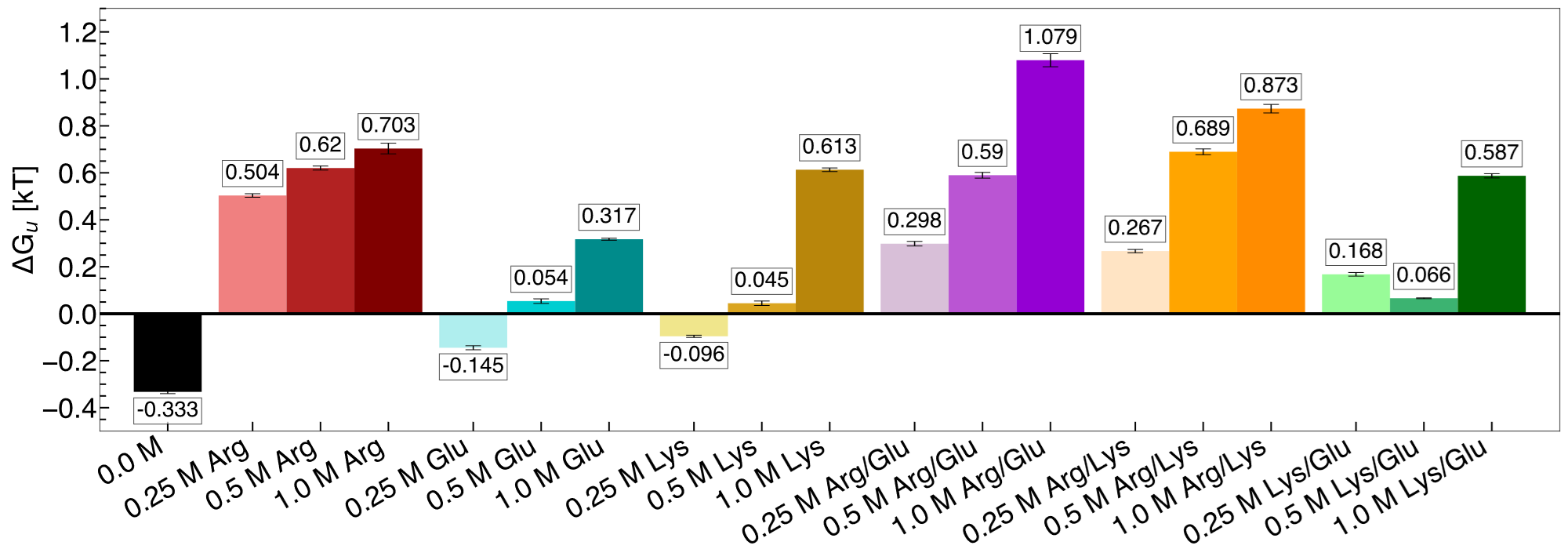
Among binary excipient mixtures, both Arg/Glu and Arg/Lys significantly stabilized the folded polymer state, particularly at 1.0 M (Fig. 1). Further, we consider 1.0 M Arg/Lys and Arg/Glu mixtures to be synergistic relative to their individual components, as the free energy of polymer folding in these solutions is more favorable than in any single excipient solution. In Lys/Glu mixtures, hydrophobic polymer folding is favored at 0.25 M, whereas the unfolded polymer is favored in Lys or Glu solutions individually at this concentration. Hence, we identify Lys/Glu mixtures to be synergistic as well at 0.25 M, although not at higher concentrations.
3.2 Thermodynamic Components of Hydrophobic Polymer Collapse in Excipient Solutions
To explore the thermodynamic origins of excipient effects on hydrophobic polymer collapse, we decomposed the PMF into individual components. Fig. 2 shows the change in each component upon unfolding in excipient solution relative to that observed in water. The first arises from the difference between folded and unfolded states (e.g., ), and the second arises from the free energy difference between excipient solution () and water () (e.g., ). In all cases, we found , as does not depend on the solvent. Additionally, the change in polymer-counterion interaction energy, , was observed to be near 0 in all cases. Hence, these terms were omitted from Fig. 2, for clarity.
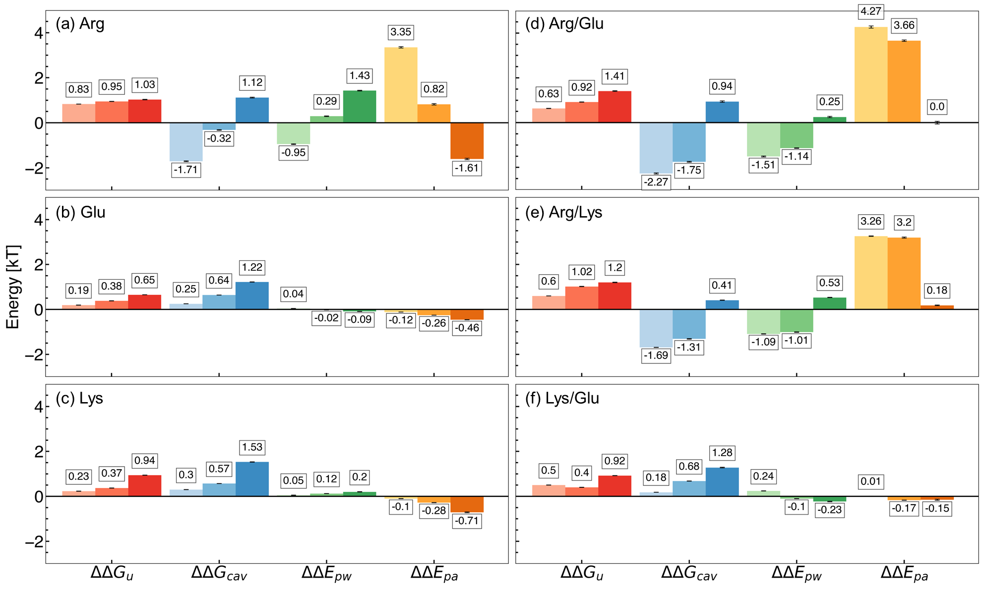
As we have previously reported,35 in Arg solutions, the favorability of individual components is concentration dependent (Fig. 2a). In particular, we observe that at low concentrations, direct polymer-Arg interactions favor collapse, while at high concentrations, the cavity component and polymer-water interactions drive folded state stability.
For Glu (Fig. 2b) and Lys (Fig. 2c) solutions, a monotonic increase in the overall favorability of polymer collapse is observed with increasing concentration, which appears to be driven primarily by a favorable cavity component. At the same time, polymer-Glu and polymer-Lys interactions oppose collapse, while the polymer-water component is negligible.
For binary mixtures Arg/Glu (Fig. 2d) and Arg/Lys (Fig. 2e), direct polymer-additive interactions dominate the free energy of polymer collapse at 0.25 M and 0.5 M, while at 1.0 M, this component is negligible. In contrast, the cavity component and polymer-water interactions favor polymer unfolding at 0.25 M and 0.5 M, while these components favor polymer collapse at 1.0 M.
In the case of Lys/Glu (Fig. 2f) solutions, stabilization of the folded polymer is nearly completely determined by the cavity component. A monotonic increase in this component is observed with increasing Lys/Glu concentration, while polymer-water and polymer-additive interactions are negligible.
3.3 Excipient Synergy is Driven by Changes in Direct Interactions
To quantify the extent of synergy in binary excipient mixtures, we computed the unfolding free energy difference associated with changing from the average of two single-excipient solutions to one binary-excipient solution (), at a given total excipient concentration. This quantity is computed as:
| (9) |
for excipients and . In cases where , the binary excipient mixtures have a more favorable effect on hydrophobic polymer collapse than did the individual components. A mechanistic understanding of this synergy may be obtained by detailing the balance of interactions involved. Specifically, we investigate both direct interactions (polymer-additive and polymer-counterion; ) and indirect effects (cavitation and polymer-water; ) associated with each solution (Fig. 3).
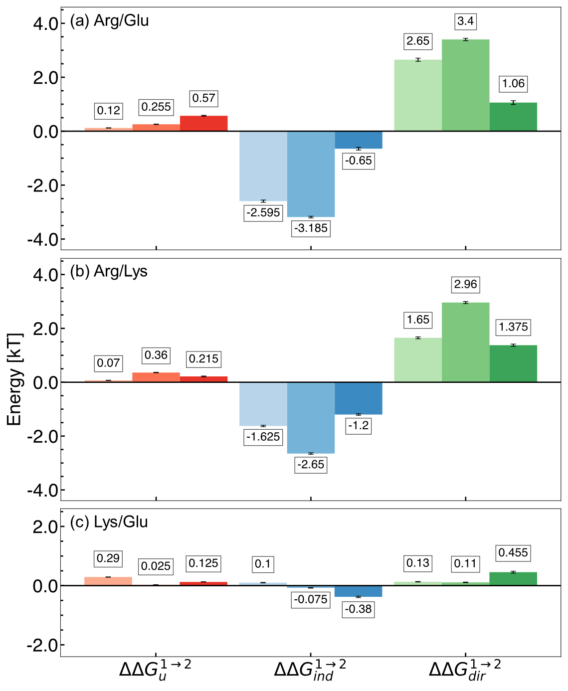
In all binary excipient solutions, we observe favorable changes in the free energy of polymer folding, relative to single excipient solutions. In Arg/Glu (Fig. 3a) and Arg/Lys (Fig. 3b) solutions, this increased favorability is associated with a favorable change in direct polymer-additive interactions. At the same time, folded state stability is opposed by unfavorable indirect components, relative to in single excipient solutions. The same observations are made for Lys/Glu (Fig. 3c) solutions, albeit to a lesser extent. Interestingly, a different optimal concentration (with maximal ) is observed for each pair of excipients – 1.0 M for Arg/Glu, 0.5 M for Arg/Lys, and 0.25 M for Lys/Glu.
The manifestation of the observed synergy describes a mechanism for improving the effectiveness of Arg-containing solutions. We observe that, while Arg is effective in stabilizing hydrophobic polymer collapse, attractive polymer-Arg interactions drive unfolding at 1.0 M concentration. In the presence of Lys or Glu, this opposition to collapse is eliminated. Hence, co-excipient addition is a suitable strategy to manipulate the underlying mechanism of Arg in hydrophobic polymer collapse.
We hypothesize that the primary source for co-excipient synergy results in a change in the balance of polymer-water-excipient interactions. Preferential interaction coefficients, , is a powerful metric for quantifying this balance. indicates relative accumulation of an excipient in the local polymer domain, while indicates relative depletion. In Fig. 4, we report for Arg, Lys, and Glu solutions. This analysis reveals that Arg preferentially interacts with the hydrophobic polymer (Fig. 4a), while Glu (Fig. 4b) and Lys (Fig. 4c) are preferentially excluded. Further, these trends were observed to increase with concentration.
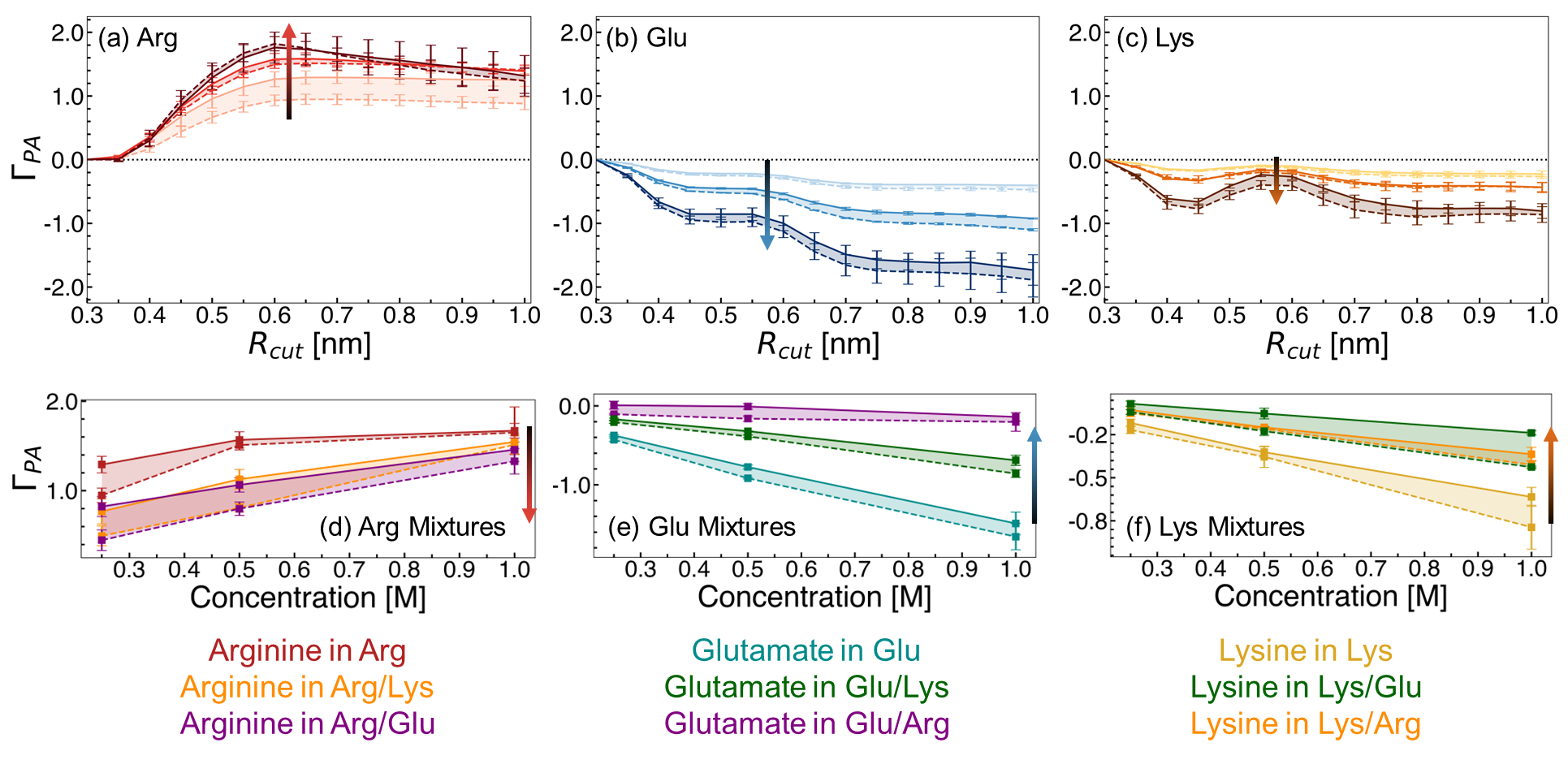
We explored the change in excipient distribution by considering of an excipient when alone versus in a binary excipient solution. In Fig. 4d-f, we show ensemble averaged values using an value of 0.7 nm. This value is selected as a cut-off distance because, beyond this distance, no significant changes in are observed for the excipients. From this perspective, the preferential accumulation of Arg near the polymer is reduced in Arg/Lys or Arg/Glu solutions, relative to in Arg solutions alone (Fig. 4d). We have previously highlighted that, at 1.0 M Arg concentration, direct polymer-Arg interactions drive unfolding.35 Hence, we hypothesize that reduction in polymer-Arg interactions upon Lys or Glu incorporation in the solution results in net stabilization of the folded hydrophobic polymer.
Alongside the changes in Arg distribution, the relative accumulation of Lys or Glu is increased in Arg/Lys and Arg/Glu solutions, relative to in single excipient solutions (Fig. 4e,f). Overall, these findings imply a mutual recruitment of co-excipient A into the preferred domain of co-excipient B.
The change in observed in binary excipient solutions describes, in part, the effects observed in Fig. 3. In Arg/Glu and Arg/Lys solutions, there is a depletion in relative to in Arg solutions alone, resulting in a net reduction of polymer-additive interactions. Correspondingly, a favorable change in the direct component, , arises in Arg/Glu and Arg/Lys solutions, conferring increased stabilization of hydrophobic polymer collapse.
3.4 Stabilizing Co-Excipients Preserve the Network Effects of Arg
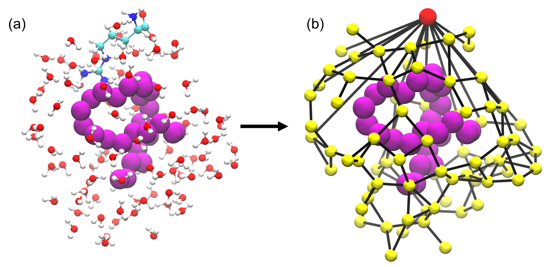
Networks of excipient-water interactions embedding the hydrophobic polymer were analyzed using principles of network theory. In this approach, we treat the center-of-mass of all excipient and water molecules within 0.7 nm of the polymer as nodes, while edges are constructed between nodes where any pair of heavy atoms are located within 0.4 nm of one another (Fig. 5). To quantify the flow of molecular interactions within the solvent network, we measured closeness centrality, , and betweenness centrality, . Closeness centrality can be regarded as a measure of how long it takes to spread information from node to all other nodes sequentially. Correspondingly, this quantity is a measure of how close a node is to the center of the network. Betweenness centrality, on the other hand, quantifies the number of times a node acts as a bridge along the shortest path between two other nodes. In other words, this quantity measures the propensity for a node to act as a ”hub” of information propagation. For both measurements, we resolve centrality from all water or excipient nodes.
Distributions of centrality measurements obtained from folded state configurations are shown in Fig. 6. Distributions from unfolded configurations result in qualitatively similar trends (Fig. S4, Fig. S5), and hence were omitted from Fig. 6, for clarity. In Arg solutions, we observe an increase in water closeness centrality () (Fig. 6a) and a decrease in water betweenness centrality () (Fig. 6b). The increase in indicates shorter distances from water nodes to all other nodes – in other words, in Arg solutions, connectivity is increased among water molecules. This observation supports previous attempts to describe excipient effects via a network-based approach, which found that proteins in solution with stabilizing excipients have more compact interaction networks, relative to those in the presence of denaturants.109
On the other hand, the decrease in suggests less information is transferred through water molecules. Correspondingly, relatively high excipient betweenness () distributions are observed for Arg solutions (Fig. 6d). Such a finding conveys that Arg molecules integrate well into the local polymer environment, acting as central hubs for information transfer in the local solvent structure. Elsewhere, it has been reported that stabilizing osmolytes have similarly high betweenness centrality values,110 implying, together with our results, that integration into the local solvent environment with minimal disruption to the solvent interaction network may be a general phenomenon for excipient stability.
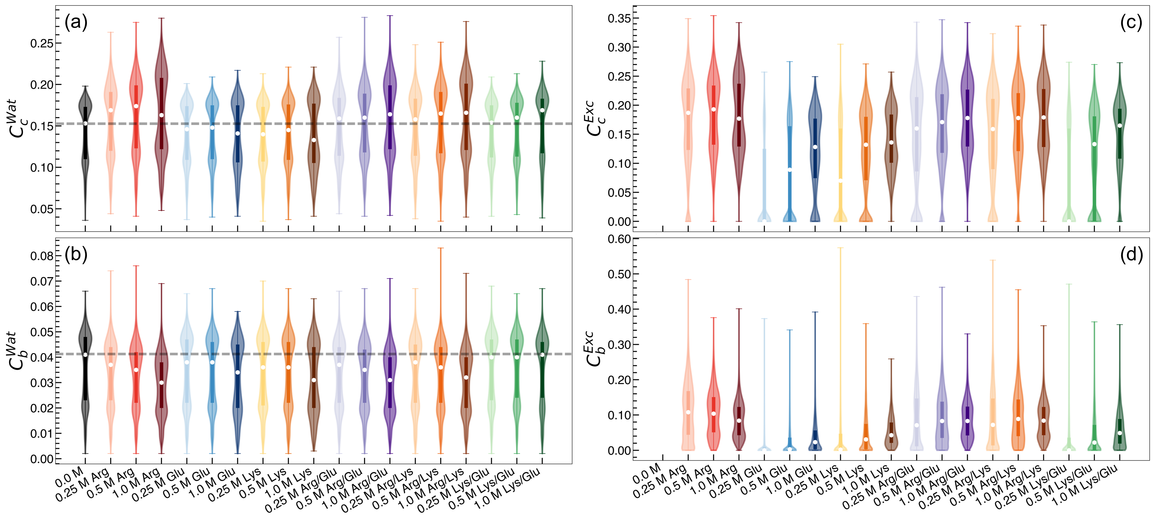
Relative to the centrality measurements associated with Arg solutions, network analysis reveals several key differences between solutions containing Lys or Glu. In solutions containing Lys or Glu as the only excipients, both and are found to decrease, or at least resulted in negligible changes relative to pure water. This finding suggests that water molecules become less connected within the local environment network (Fig. 6a), while also becoming less essential hubs for information propagation (Fig. 6b). In contrast to Arg solutions, and are both relatively low, implying that Lys and Glu do not integrate into the local solvent environment as well as Arg does (Fig. 6c,d).
These centrality measurements reflect strongly connected networks in Arg solutions, and networks with poor connectivity in Lys or Glu solutions. In binary excipient solutions containing Arg (Arg/Lys and Arg/Glu), we observe an overall preservation of the strong connectivity identified in Arg solutions alone. Specifically, is found to increase in Arg/Lys and Arg/Glu solutions relative to pure water, reflecting increased connectivity among water molecules (Fig. 6a). Similar to Arg solutions, is observed to decrease with a concomitant increase in , reflecting favorable integration of excipient molecules into the local polymer environment in Arg/Lys and Arg/Glu solutions (Fig. 6b,d). Finally, is consistently higher in Arg/Lys and Arg/Glu solutions relative to Lys or Glu alone, marking an increase in local excipient connectivity in these binary excipient solutions (Fig. 6c). In general, the Lys/Glu binary excipient solution reflected network properties similar to Lys or Glu solutions alone.
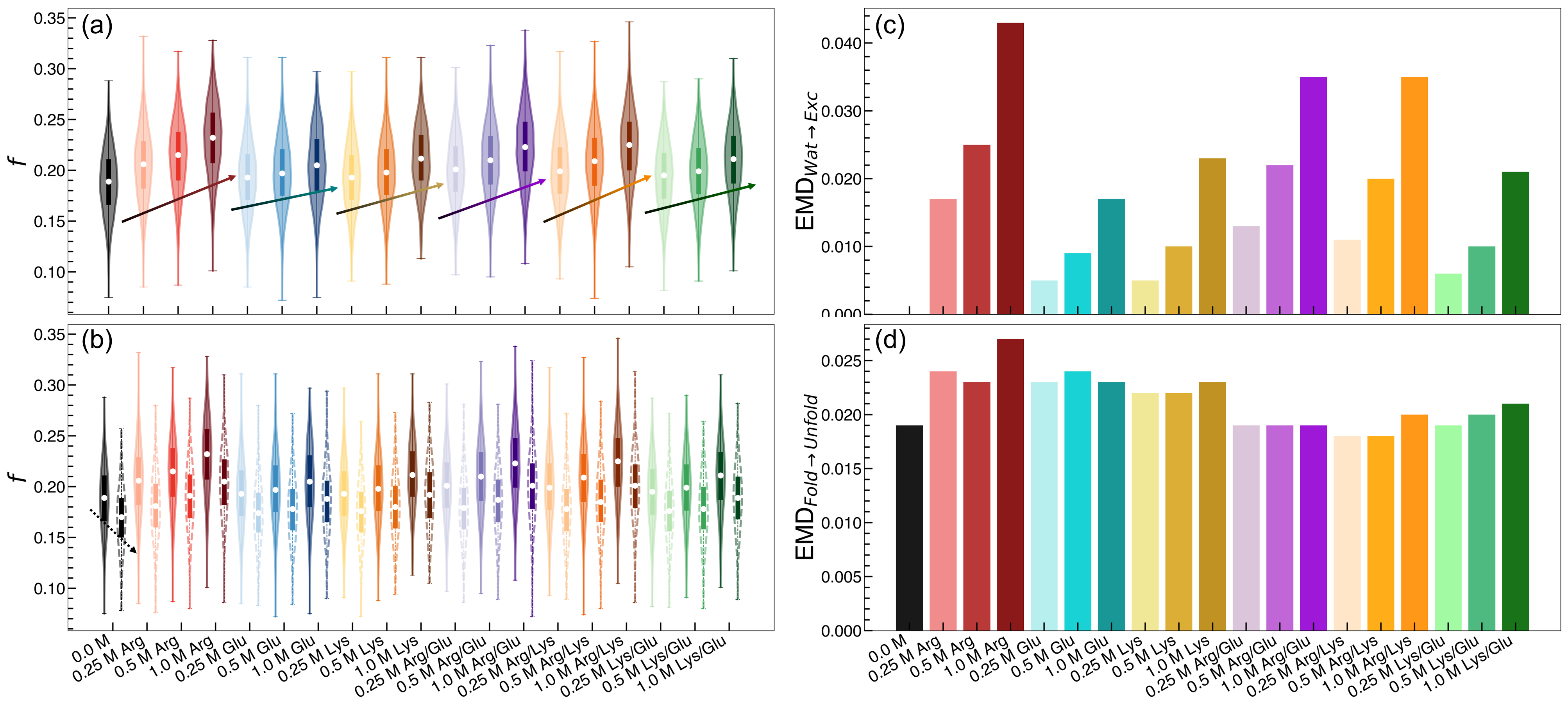
We hypothesize that solutions with stronger connectivity in the local polymer environment may confer greater stability to the network itself. To assess this, we compute a fragmentation metric, , which measures the average number of nodes that must be removed to form two independent, disconnected graphs. Such an approach is inspired by percolation theory, and has been used elsewhere to measure the stability of large biomolecular assemblies, including viral capsids.111, 112 To better quantify differences in distribution, we computed the Earth Mover’s Distance (EMD)113 as a measure of distribution dissimilarity, where higher EMD values indicate less overlap between two distributions. On average, local polymer environments are more resistant to fragmentation in Arg, Arg/Lys, and Arg/Glu solutions (Fig. 7a,c), as indicated by increasing distributions and relatively large EMD values. For all solutions, values are higher in the local environments of folded polymer states, relative to unfolded polymer states (Fig. 7b). However, this appears to be a general trend, and the differences between folded state and unfolded state graph stability does not depend on solution identity (Fig. 7d).
3.5 Rationalizing Co-Excipient Synergy
Overall, we have uncovered that Arg stabilizes hydrophobic polymer collapse to a greater extent than Lys, Glu, or Lys/Glu solutions. Further, addition of Lys or Glu to Arg solutions give rise to synergistic effects, as the resulting Arg/Lys and Arg/Glu solutions are the most effective stabilizing solutions we have investigated. Further, these synergies are associated with stabilizing indirect effects at high concentration and a dramatic reduction in destabilizing direct interactions. Correspondingly, the solvent distribution, as characterized by preferential interaction coefficient analysis, reflects a reduction in Arg accumulation in the local hydrophobic polymer domain.
Our network analysis implies that Arg integrates well into the local polymer environment, increases connectivity among water molecules, and increases the stability of the solvent network embedding the hydrophobic polymer. Importantly, these key network properties persist for Arg solutions to a greater extent than Lys or Glu solutions. Moreover, addition of Lys or Glu to Arg solution (Arg/Lys, Arg/Glu) preserves the network properties of Arg alone, which may be a key aspect giving rise to the observed synergy among these excipients.
We hypothesize that addition of Lys or Glu to Arg solutions results in the formation of the same stable, highly connected local polymer environment as in solutions containing only Arg, while simultaneously reducing the penalty associated with direct polymer-Arg interactions. While our findings support the hypothesis described above, we aimed to uncover key molecular features or signatures that drive these changes in polymer stability. To this end, we carried out additional analyses to explore whether Lys and Glu undergo unique molecular mechanisms to arrive at similar synergistic effects with Arg.
3.5.1 Rationalizing Arg/Lys Synergy: The Sticky Guanidinium Hypothesis
Given the importance of excipient-excipient interactions in dictating solution structure, we aimed to explore these molecular interactions further. In particular, clustering has been implicated as a key feature associated with the excipient effects of Arg.93, 94, 95, 96, 97, 98, 99, 63 Among amino acid excipients, these molecules interact in solution via three primary modes of interaction: (i) backbone-backbone (COO--NH), (ii) backbone-sidechain (Gdm+-COO-), and (iii) sidechain-sidechain (Gdm+-Gdm+). Using geometric criteria for identifying these specific interactions, we carried out an analysis of excipient cluster formation in different excipient solutions.
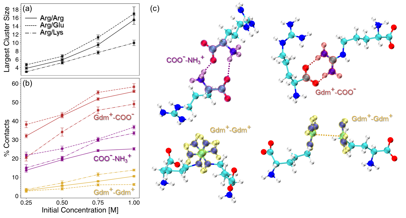
Overall, the extent of cluster formation, as measured by the average largest cluster size, is greater in Arg and Arg/Glu solutions than in Arg/Lys (Fig. 8a). We attribute this finding to favorable electrostatic interactions between Arg and Glu, as well as the unique properties of the Gdm+ sidechain of Arg that enables favorable like-charge interactions.114, 115, 95, 116, 117 The presence of cluster formation in all Arg-containing solutions is primarily driven by Gdm+-COO- interactions (Fig. 8b,c), a finding consistent with previous work detailing the importance of Arg ”head-to-tail” stacking.95 Interestingly, while excipient clustering is reduced overall in Arg/Lys solutions relative to Arg alone, the extent of Gdm+-Gdm+ pairing is increased in Arg/Lys solutions.
To probe the importance of these interactions relevant to excipient clustering, we carried out two additional REUS simulations of hydrophobic polymer folding with modified Arg-Arg interaction parameters (Fig. 9). To simulate increased Gdm+-Gdm+ pairing among Arg molecules (ArgGG), we scaled the interaction strength between Gdm+ carbons by 150% (Fig. 9b). Similarly, to simulate increased Gdm+-COO- head-to-tail pairing among Arg molecules (ArgHTT), the interaction strength between COO- oxygens and Gdm+ hydrogens was scaled by 150% (Fig. 9c). From these simulations, the free energy of polymer folding in ArgGG solution becomes significantly more favorable relative to unmodified Arg solution, while ArgHTT results in only a small increase in folded state stability (Fig. 9a).
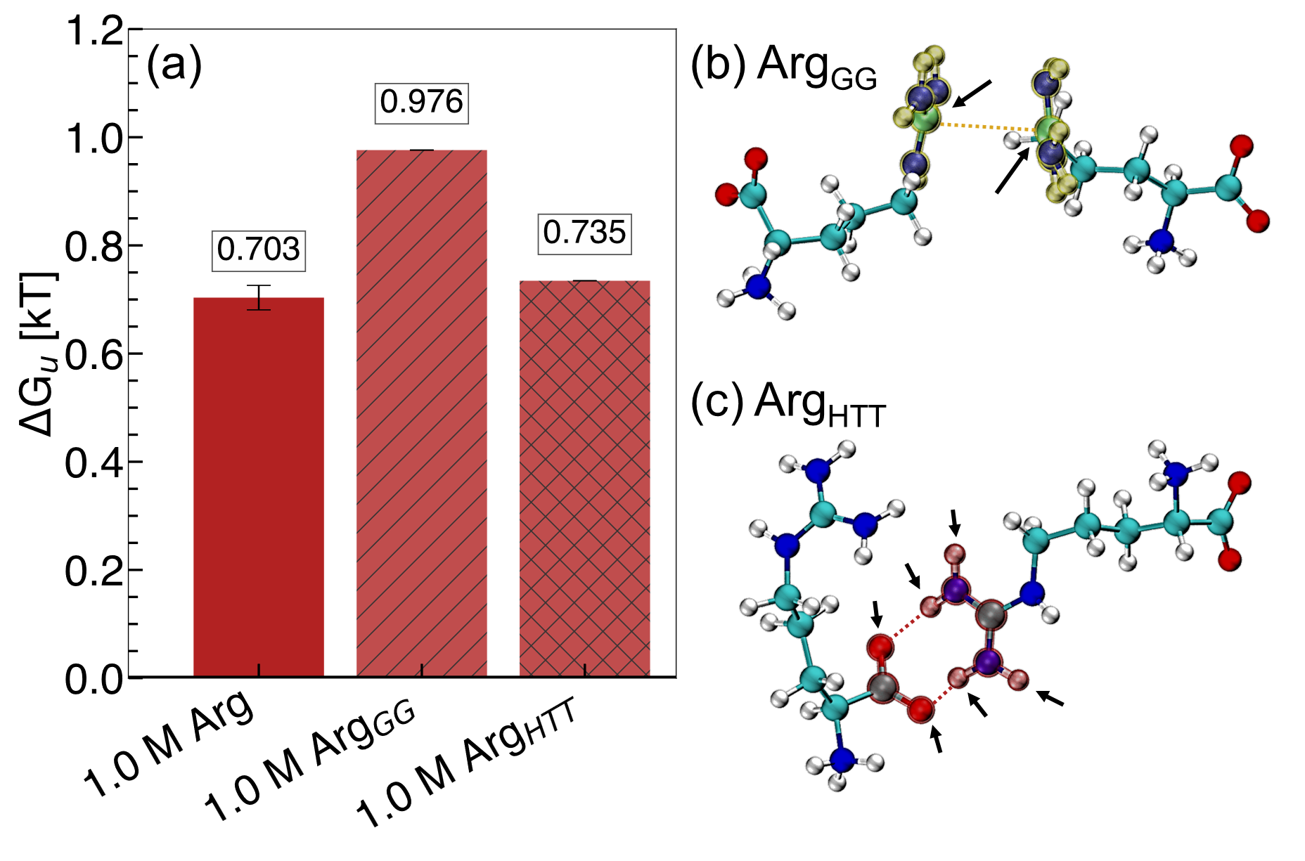
These results demonstrate the importance of the Gdm+ sidechain of Arg in hydrophobic polymer collapse. Gdm+ is a known protein denaturant, and elsewhere, has been shown to drive unfolding of an elongated hydrophobic polymer at high concentrations.80 Further, we have shown previously that direct interactions between the Gdm+ sidechain of Arg and the hydrophobic polymer favors polymer folding at lower concentrations.35 Overall, our mechanistic explanation for Arg/Lys synergy involves a Lys-mediated increase in Gdm+-Gdm+ ”stickiness” among Arg molecules. We hypothesize that upon addition of Lys, the increase in Gdm+-Gdm+ pairing among Arg molecules gives rise to the favorable change in observed in Arg/Lys solutions. This is achieved by limiting the number of Gdm+ interaction sites available to interact with the polymer, resulting in a relative depletion of Arg from the local polymer domain.
3.5.2 Rationalizing Arg/Glu Synergy: The Dynamics Reducing Hypothesis
While Arg/Lys synergy appears to be associated with changes in Gdm+-Gdm+ pairing among Arg molecules, we did not observe the same changes in excipient clustering in Arg/Glu solutions. Hence, to explain molecular-level changes linked to Arg/Glu synergy, we turned our attention to the behavior of water molecules in the local polymer environment.
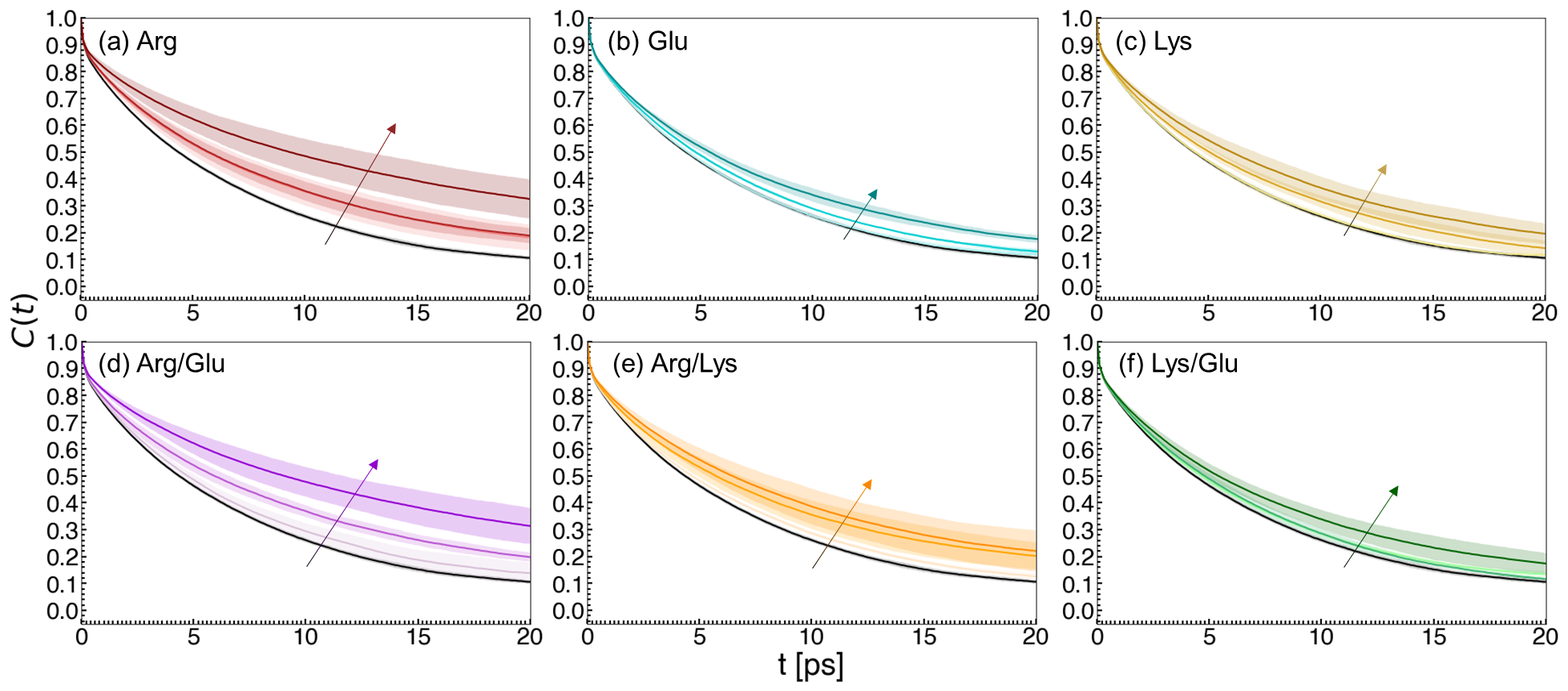
To this end, we computed water reorientation dynamics in the local hydrophobic polymer domain for our excipient solutions (Fig. 10). The characteristic reorientation time for each solution was computed by fitting water dipole correlation functions to an exponential decay. With increasing excipient concentration, the reorientation time () of local water molecules was observed to increase, indicating a slowing of water dynamics (Table 1). This effect is most pronounced in Arg and Arg/Glu solutions.
| Concentration (M) | Arg | Lys | Glu | Arg/Glu | Arg/Lys | Lys/Glu |
|---|---|---|---|---|---|---|
| 0.0 M | 6.54 (0.02) | - | - | - | - | - |
| 0.25 M | 8.52 (1.2) | 6.46 (0.1) | 7.07 (0.5) | 7.24 (0.1) | 6.9 (0.1) | |
| 0.5 M | 8.64 (0.4) | 7.85 (0.5) | 7.17 (0.1) | 8.86 (0.4) | 8.56 (1.4) | 7.17 (0.3) |
| 1.0 M | 12.6 (1.0) | 9.29 (1.4) | 8.4 (0.4) | 12.37 (0.8) | 8.81 (0.9) | 8.48 (0.6) |
Similar reductions in hydration shell dynamics have been associated with an increase in melting temperature of proteins. A recent study has proposed that stabilizing osmolytes slow down the dynamics of water, while denaturants accelerate water dynamics, inducing a pseudo-temperature change experienced by the protein.118 Other studies have highlighted that osmolytes increase hydrogen bond relaxation time among water molecules and reduce rotational, translational, and tumbling motions of water.119, 106, 120, 121, 122, 123
We hypothesize that the key consequence associated with this phenomenon is the formation of a rigid solvent network embedding the hydrophobic polymer in Arg and Arg/Glu solutions. In the case of Arg/Glu, Glu may provide an advantage relative to Arg alone due to a reduction of Gdm+ sidechain accumulation in the local polymer domain, as reflected by . This results in the favorable change in , similar to Arg/Lys, while retaining the reduced water dynamics and stable local network associated with Arg solutions.
4 Conclusions
A growing body of literature has contributed to understanding the effects of Arg on biomolecular stability. In some cases, addition of co-excipients to formulations that include Arg has resulted in reversals57 or synergistic64 effects. Recently, we demonstrated that the peculiar placement of Arg at the edge of a mechanistic flip between indirect- and direct-dominated effects on hydrophobic interactions may be a key feature that enables tunable properties of Arg.35 Our findings here show that, not only is Arg a more effective stabilizer of hydrophobic interactions than its Lys or Glu counterparts, but addition of these less effective excipients augments the effectiveness of Arg solution stability.
We observed that the primary mechanism associated with Arg/Glu and Arg/Lys synergy is a substantial reduction in direct polymer-excipient interactions that oppose collapse at high Arg concentration. Through preferential interaction coefficient analysis, we further identified that Lys and Glu are both effective at reducing the relative accumulation of Arg molecules in the local polymer domain, providing a mechanistic explanation for the favorable change in .
Analysis of the solvent network embedding the hydrophobic polymer provides an explanation for how excipients alter the local polymer environment. We found that Arg increases connectivity among water molecules, integrates favorably into the local environment, and increases the stability of the network by delaying the onset of simulated graph fragmentation. These features are more pronounced in Arg solutions than in Lys, Glu, or Lys/Glu solutions. Importantly, we identified these same features in Arg/Lys and Arg/Glu solutions, indicating that stabilizing co-excipients preserve the network effects of Arg.
Finally, we derived two hypotheses for Arg/Lys and Arg/Glu synergy. In the case of Arg/Lys, there is an increase in Gdm+-Gdm+ pairing among Arg molecules, potentially reducing the number of available Gdm+ sidechains that drive polymer unfolding at high concentrations. From simulations of increased Gdm+-Gdm+ interaction strength between Arg molecules, we found this change to be sufficient for increasing the favorability of polymer collapse. In the case of Arg/Glu, we did not find increased Gdm+-COO- paring in solution was sufficient to drive co-excipient synergy. However, we observed a reduction in the dynamics of water molecules local to the hydrophobic polymer in these solutions. Similar reductions in local water dynamics has been linked to increased melting temperature of proteins in the presence of stabilizing osmolytes.119, 106, 120, 121, 122, 123
Here, we demonstrated that changes in excipient composition alter hydrophobic interactions, the dominant force associated with several biologically-important processes including protein folding and self-assembly. As it pertains to formulations, this is an important factor in preventing protein denaturation and aggregation. Due to its placement on the edge of a mechanistic flip, formulations containing Arg as an excipient may be improved by shifting the balance of direct- and indirect-mediated effects. To this end, we increased the relative stability of hydrophobic polymer collapse by reducing destabilizing direct effects via co-excipient addition of either Lys or Glu. Overall, these results highlight the investigation of molecular-level insights of excipient mechanisms as an important endeavor in the rational design of stable biologics.
Conflicts of interest
There are no conflicts to declare.
Acknowledgements
This material is based upon work supported by the National Science Foundation under DMREF Grant Nos. 2118788, 2118693, and 2118638. Computing resources were provided by the Minnesota Supercomputing Institute (MSI).
References
- Strassburg 1982 Strassburg, M. A. The global eradication of smallpox. American Journal of Infection Control 1982, 10, 53–59
- Zhang et al. 2018 Zhang, W.-W.; Li, L.; Li, D.; Liu, J.; Li, X.; Li, W.; Xu, X.; Zhang, M. J.; Chandler, L. A.; Lin, H.; Hu, A.; Xu, W.; Lam, D. M.-K. The First Approved Gene Therapy Product for Cancer Ad- p53 (Gendicine): 12 Years in the Clinic. Human Gene Therapy 2018, 29, 160–179
- Privalov 1990 Privalov, P. L. Cold Denaturation of Protein. Critical Reviews in Biochemistry and Molecular Biology 1990, 25, 281–306
- Heremans 1982 Heremans, K. High Pressure Effects on Proteins and other Biomolecules. Annual Review of Biophysics and Bioengineering 1982, 11, 1–21
- Sarupria et al. 2010 Sarupria, S.; Ghosh, T.; García, A. E.; Garde, S. Studying pressure denaturation of a protein by molecular dynamics simulations. Proteins: Structure, Function, and Bioinformatics 2010, NA–NA
- Kauzmann 1959 Kauzmann, W. Advances in Protein Chemistry; Elsevier, 1959; Vol. 14; pp 1–63
- Frank and Evans 1945 Frank, H. S.; Evans, M. W. Free Volume and Entropy in Condensed Systems III. Entropy in Binary Liquid Mixtures; Partial Molal Entropy in Dilute Solutions; Structure and Thermodynamics in Aqueous Electrolytes. The Journal of Chemical Physics 1945, 13, 507–532
- Christensen 1952 Christensen, L. K. Denaturation and enzymatic hydrolysis of lactoglobulin; with some observations on the hydrolysis of egg albumin. Comptes Rendus Des Travaux Du Laboratoire Carlsberg. Serie Chimique 1952, 28, 37–69
- Hummer et al. 1998 Hummer, G.; Garde, S.; García, A. E.; Paulaitis, M. E.; Pratt, L. R. The pressure dependence of hydrophobic interactions is consistent with the observed pressure denaturation of proteins. Proceedings of the National Academy of Sciences 1998, 95, 1552–1555
- Ghosh et al. 2001 Ghosh, T.; García, A. E.; Garde, S. Molecular Dynamics Simulations of Pressure Effects on Hydrophobic Interactions. Journal of the American Chemical Society 2001, 123, 10997–11003
- Le Basle et al. 2020 Le Basle, Y.; Chennell, P.; Tokhadze, N.; Astier, A.; Sautou, V. Physicochemical Stability of Monoclonal Antibodies: A Review. Journal of Pharmaceutical Sciences 2020, 109, 169–190
- Authelin et al. 2020 Authelin, J.-R.; Rodrigues, M. A.; Tchessalov, S.; Singh, S. K.; McCoy, T.; Wang, S.; Shalaev, E. Freezing of Biologicals Revisited: Scale, Stability, Excipients, and Degradation Stresses. Journal of Pharmaceutical Sciences 2020, 109, 44–61
- Crommelin et al. 2024 Crommelin, D. J. A.; Hawe, A.; Jiskoot, W. In Pharmaceutical Biotechnology; Crommelin, D. J. A., Sindelar, R. D., Meibohm, B., Eds.; Springer International Publishing: Cham, 2024; pp 95–117
- Clarkson et al. 2016 Clarkson, B. R.; Schön, A.; Freire, E. Conformational stability and self-association equilibrium in biologics. Drug Discovery Today 2016, 21, 342–347
- Mazzeo and Carpenter 2009 Mazzeo, A.; Carpenter, P. In Handbook of Stability Testing in Pharmaceutical Development; Huynh-Ba, K., Ed.; Springer New York: New York, NY, 2009; pp 353–369
- Organization 2006 Organization, W. H. Temperature sensitivity of vaccines. 2006
- Hanson et al. 2017 Hanson, C. M.; George, A. M.; Sawadogo, A.; Schreiber, B. Is freezing in the vaccine cold chain an ongoing issue? A literature review. Vaccine 2017, 35, 2127–2133
- Yu et al. 2021 Yu, Y. B.; Briggs, K. T.; Taraban, M. B.; Brinson, R. G.; Marino, J. P. Grand Challenges in Pharmaceutical Research Series: Ridding the Cold Chain for Biologics. Pharm Res 2021, 38, 3–7
- Crommelin et al. 2021 Crommelin, D. J.; Anchordoquy, T. J.; Volkin, D. B.; Jiskoot, W.; Mastrobattista, E. Addressing the Cold Reality of mRNA Vaccine Stability. Journal of Pharmaceutical Sciences 2021, 110, 997–1001
- Thielmann et al. 2019 Thielmann, A.; Puth, M.-T.; Weltermann, B. Visual inspection of vaccine storage conditions in general practices: A study of 75 vaccine refrigerators. PLoS ONE 2019, 14, e0225764
- Thielmann et al. 2020 Thielmann, A.; Puth, M.-T.; Weltermann, B. Improving knowledge on vaccine storage management in general practices: Learning effectiveness of an online-based program. Vaccine 2020, 38, 7551–7557
- Kamerzell et al. 2011 Kamerzell, T. J.; Esfandiary, R.; Joshi, S. B.; Middaugh, C. R.; Volkin, D. B. Protein–excipient interactions: Mechanisms and biophysical characterization applied to protein formulation development. Advanced Drug Delivery Reviews 2011, 63, 1118–1159
- Arakawa et al. 2007 Arakawa, T.; Tsumoto, K.; Kita, Y.; Chang, B.; Ejima, D. Biotechnology applications of amino acids in protein purification and formulations. Amino Acids 2007, 33, 587–605
- Bongioanni et al. 2022 Bongioanni, A.; Bueno, M. S.; Mezzano, B. A.; Longhi, M. R.; Garnero, C. Amino acids and its pharmaceutical applications: A mini review. International Journal of Pharmaceutics 2022, 613, 121375
- Jeong 2012 Jeong, S. H. Analytical methods and formulation factors to enhance protein stability in solution. Archives of Pharmacal Research 2012, 35, 1871–1886
- Wang 2005 Wang, W. Protein aggregation and its inhibition in biopharmaceutics. International Journal of Pharmaceutics 2005, 289, 1–30
- Patro et al. 2002 Patro, S. Y.; Freund, E.; Chang, B. S. Biotechnology Annual Review; Elsevier, 2002; Vol. 8; pp 55–84
- Ionova and Wilson 2020 Ionova, Y.; Wilson, L. Biologic excipients: Importance of clinical awareness of inactive ingredients. PLoS ONE 2020, 15, e0235076
- ten Wolde and Chandler 2002 ten Wolde, P. R.; Chandler, D. Drying-induced hydrophobic polymer collapse. Proceedings of the National Academy of Sciences 2002, 99, 6539–6543
- Zangi et al. 2009 Zangi, R.; Zhou, R.; Berne, B. J. Urea’s Action on Hydrophobic Interactions. Journal of the American Chemical Society 2009, 131, 1535–1541
- Nayar and van der Vegt 2018 Nayar, D.; van der Vegt, N. F. A. Cosolvent Effects on Polymer Hydration Drive Hydrophobic Collapse. The Journal of Physical Chemistry B 2018, 122, 3587–3595
- Mondal et al. 2013 Mondal, J.; Stirnemann, G.; Berne, B. J. When Does Trimethylamine N -Oxide Fold a Polymer Chain and Urea Unfold It? The Journal of Physical Chemistry B 2013, 117, 8723–8732
- Athawale et al. 2008 Athawale, M. V.; Sarupria, S.; Garde, S. Enthalpy-Entropy Contributions to Salt and Osmolyte Effects on Molecular-Scale Hydrophobic Hydration and Interactions. The Journal of Physical Chemistry B 2008, 112, 5661–5670
- Jamadagni et al. 2009 Jamadagni, S. N.; Godawat, R.; Garde, S. How Surface Wettability Affects the Binding, Folding, and Dynamics of Hydrophobic Polymers at Interfaces. Langmuir 2009, 25, 13092–13099
- Zajac et al. 2024 Zajac, J. W. P.; Muralikrishnan, P.; Heldt, C. L.; Perry, S. L.; Sarupria, S. Flipping Out: Role of Arginine in Hydrophobic Polymer Collapse. arXiv 2024, arXiv:2403.11305 [cond-mat, q-bio]
- Timasheff 1993 Timasheff, S. N. The Control of Protein Stability and Association by Weak Interactions with Water: How Do Solvents Affect These Processes? Annual Review of Biophysics and Biomolecular Structure 1993, 22, 67–97
- Ghosh et al. 2005 Ghosh, T.; Kalra, A.; Garde, S. On the Salt-Induced Stabilization of Pair and Many-body Hydrophobic Interactions. The Journal of Physical Chemistry B 2005, 109, 642–651
- Athawale et al. 2005 Athawale, M. V.; Dordick, J. S.; Garde, S. Osmolyte Trimethylamine-N-Oxide Does Not Affect the Strength of Hydrophobic Interactions: Origin of Osmolyte Compatibility. Biophysical Journal 2005, 89, 858–866
- Canchi and García 2013 Canchi, D. R.; García, A. E. Cosolvent Effects on Protein Stability. Annual Review of Physical Chemistry 2013, 64, 273–293
- van der Vegt and Nayar 2017 van der Vegt, N. F. A.; Nayar, D. The Hydrophobic Effect and the Role of Cosolvents. The Journal of Physical Chemistry B 2017, 121, 9986–9998
- Wallqvist et al. 1998 Wallqvist, A.; Covell, D. G.; Thirumalai, D. Hydrophobic Interactions in Aqueous Urea Solutions with Implications for the Mechanism of Protein Denaturation. Journal of the American Chemical Society 1998, 120, 427–428
- Ikeguchi et al. 2001 Ikeguchi, M.; Nakamura, S.; Shimizu, K. Molecular Dynamics Study on Hydrophobic Effects in Aqueous Urea Solutions. Journal of the American Chemical Society 2001, 123, 677–682
- Van Der Vegt et al. 2006 Van Der Vegt, N. F. A.; Lee, M.-E.; Trzesniak, D.; Van Gunsteren, W. F. Enthalpy-Entropy Compensation in the Effects of Urea on Hydrophobic Interactions. The Journal of Physical Chemistry B 2006, 110, 12852–12855
- Lee and Van Der Vegt 2006 Lee, M.-E.; Van Der Vegt, N. F. A. Does Urea Denature Hydrophobic Interactions? Journal of the American Chemical Society 2006, 128, 4948–4949
- Shpiruk and Khajehpour 2013 Shpiruk, T. A.; Khajehpour, M. The effect of urea on aqueous hydrophobic contact-pair interactions. Phys. Chem. Chem. Phys. 2013, 15, 213–222
- Paul and Patey 2008 Paul, S.; Patey, G. N. Hydrophobic Interactions in UreaTrimethylamine- N -oxide Solutions. The Journal of Physical Chemistry B 2008, 112, 11106–11111
- Macdonald and Khajehpour 2013 Macdonald, R. D.; Khajehpour, M. Effects of the osmolyte TMAO (Trimethylamine-N-oxide) on aqueous hydrophobic contact-pair interactions. Biophysical Chemistry 2013, 184, 101–107
- Ganguly et al. 2016 Ganguly, P.; Van Der Vegt, N. F. A.; Shea, J.-E. Hydrophobic Association in Mixed Urea–TMAO Solutions. The Journal of Physical Chemistry Letters 2016, 7, 3052–3059
- Su et al. 2018 Su, Z.; Ravindhran, G.; Dias, C. L. Effects of Trimethylamine- N -oxide (TMAO) on Hydrophobic and Charged Interactions. The Journal of Physical Chemistry B 2018, 122, 5557–5566
- Folberth et al. 2022 Folberth, A.; Bharadwaj, S.; Van Der Vegt, N. F. A. Small-to-large length scale transition of TMAO interaction with hydrophobic solutes. Physical Chemistry Chemical Physics 2022, 24, 2080–2087
- Tsumoto et al. 2004 Tsumoto, K.; Umetsu, M.; Kumagai, I.; Ejima, D.; Philo, J.; Arakawa, T. Role of Arginine in Protein Refolding, Solubilization, and Purification. Biotechnology Progress 2004, 20, 1301–1308, Number: 5
- Tsumoto et al. 2005 Tsumoto, K.; Ejima, D.; Kita, Y.; Arakawa, T. Review: Why is arginine effective in suppressing aggregation? Protein and Peptide Letters 2005, 12, 613–619, Number: 7
- Stärtzel 2018 Stärtzel, P. Arginine as an Excipient for Protein Freeze-Drying: A Mini Review. Journal of Pharmaceutical Sciences 2018, 107, 960–967
- Hamborsky et al. 2015 Hamborsky, J.; Kroger, A.; Wolfe, C. Epidemiology and prevention of vaccine-preventable diseases, 13th ed.; Centers for Disease Control and Prevention: Atlanta, GA, 2015; OCLC: 915815516
- Mistilis et al. 2017 Mistilis, M. J.; Joyce, J. C.; Esser, E. S.; Skountzou, I.; Compans, R. W.; Bommarius, A. S.; Prausnitz, M. R. Long-term stability of influenza vaccine in a dissolving microneedle patch. Drug Delivery and Translational Research 2017, 7, 195–205
- Xie et al. 2004 Xie, Q.; Guo, T.; Lu, J.; Zhou, H.-M. The guanidine like effects of arginine on aminoacylase and salt-induced molten globule state. The International Journal of Biochemistry & Cell Biology 2004, 36, 296–306
- Anumalla and Prabhu 2019 Anumalla, B.; Prabhu, N. P. Counteracting Effect of Charged Amino Acids Against the Destabilization of Proteins by Arginine. Applied Biochemistry and Biotechnology 2019, 189, 541–555
- Arakawa and Maluf 2018 Arakawa, T.; Maluf, N. K. The effects of allantoin, arginine and NaCl on thermal melting and aggregation of ribonuclease, bovine serum albumin and lysozyme. International Journal of Biological Macromolecules 2018, 107, 1692–1696
- Smirnova et al. 2013 Smirnova, E.; Safenkova, I.; Stein-Margolina, B.; Shubin, V.; Gurvits, B. l-Arginine induces protein aggregation and transformation of supramolecular structures of the aggregates. Amino Acids 2013, 45, 845–855
- Shah et al. 2011 Shah, D.; Shaikh, A. R.; Peng, X.; Rajagopalan, R. Effects of arginine on heat-induced aggregation of concentrated protein solutions. Biotechnology Progress 2011, 27, 513–520
- Eronina et al. 2014 Eronina, T. B.; Chebotareva, N. A.; Sluchanko, N. N.; Mikhaylova, V. V.; Makeeva, V. F.; Roman, S. G.; Kleymenov, S. Y.; Kurganov, B. I. Dual effect of arginine on aggregation of phosphorylase kinase. International Journal of Biological Macromolecules 2014, 68, 225–232
- Meingast and Heldt 2020 Meingast, C.; Heldt, C. L. Arginine‐enveloped virus inactivation and potential mechanisms. Biotechnol. Prog. 2020, 36, e2931
- Meingast et al. 2021 Meingast, C. L.; Joshi, P. U.; Turpeinen, D. G.; Xu, X.; Holstein, M.; Feroz, H.; Ranjan, S.; Ghose, S.; Li, Z. J.; Heldt, C. L. Physiochemical properties of enveloped viruses and arginine dictate inactivation. Biotechnol. J. 2021, 16, 2000342
- Shukla and Trout 2011 Shukla, D.; Trout, B. L. Understanding the Synergistic Effect of Arginine and Glutamic Acid Mixtures on Protein Solubility. J. Phys. Chem. B 2011, 115, 11831–11839
- Sugita et al. 2000 Sugita, Y.; Kitao, A.; Okamoto, Y. Multidimensional replica-exchange method for free-energy calculations. The Journal of Chemical Physics 2000, 113, 6042–6051
- Abascal and Vega 2005 Abascal, J. L. F.; Vega, C. A general purpose model for the condensed phases of water: TIP4P/2005. The Journal of Chemical Physics 2005, 123, 234505
- Brooks et al. 1983 Brooks, B. R.; Bruccoleri, R. E.; Olafson, B. D.; States, D. J.; Swaminathan, S.; Karplus, M. CHARMM: A program for macromolecular energy, minimization, and dynamics calculations. Journal of Computational Chemistry 1983, 4
- Brooks et al. 2009 Brooks, B. R. et al. CHARMM: The biomolecular simulation program. J. Comput. Chem. 2009, 30, 1545–1614
- Bussi et al. 2007 Bussi, G.; Donadio, D.; Parrinello, M. Canonical sampling through velocity rescaling. The Journal of Chemical Physics 2007, 126, 014101
- Berendsen et al. 1984 Berendsen, H. J.; Postma, J. P.; van Gunsteren, W. F.; DiNola, A.; Haak, J. R. Molecular dynamics with coupling to an external bath. The Journal of Chemical Physics 1984, 81, 3684–3690
- Evans and Holian 1985 Evans, D. J.; Holian, B. L. The nose–hoover thermostat. The Journal of Chemical Physics 1985, 83, 4069–4074
- Parrinello and Rahman 1981 Parrinello, M.; Rahman, A. Polymorphic transitions in single crystals: A new molecular dynamics method. Journal of Applied Physics 1981, 52, 7182–7190
- Lorentz 1881 Lorentz, H. A. Ueber die Anwendung des Satzes vom virial in der Kinetischen Theorie der Gase. Annalen der Physik 1881, 248, 127–136
- Berthelot 1898 Berthelot, D. Sur le mélange des gaz. Compt. Rendus 1898, 126
- Lindahl et al. 2021 Lindahl; Abraham; Hess; Spoel, v. d. Gromacs 2021.4 source code. 2021; \urlhttps://zenodo.org/record/5636567
- Consortium 2019 Consortium, T. P. Promoting transparency and reproducibility in enhanced molecular simulations. Nature Methods 2019, 16, 670–673
- Tribello et al. 2014 Tribello, G. A.; Bonomi, M.; Branduardi, D.; Camilloni, C.; Bussi, G. Plumed 2: New feathers for an old bird. Computer Physics Communications 2014, 185, 604–613
- Zhu and Hummer 2011 Zhu, F.; Hummer, G. Convergence and error estimation in free energy calculations using the weighted Histogram Analysis Method. Journal of Computational Chemistry 2011, 33, 453–465
- Athawale et al. 2007 Athawale, M. V.; Goel, G.; Ghosh, T.; Truskett, T. M.; Garde, S. Effects of lengthscales and attractions on the collapse of hydrophobic polymers in water. Proceedings of the National Academy of Sciences 2007, 104, 733–738
- Godawat et al. 2010 Godawat, R.; Jamadagni, S. N.; Garde, S. Unfolding of Hydrophobic Polymers in Guanidinium Chloride Solutions. The Journal of Physical Chemistry B 2010, 114, 2246–2254
- Dasetty and Sarupria 2021 Dasetty, S.; Sarupria, S. Advancing Rational Control of Peptide–Surface Complexes. The Journal of Physical Chemistry B 2021, 125, 2644–2657
- Scatchard 1946 Scatchard, G. Physical Chemistry of Protein Solutions. I. Derivation of the Equations for the Osmotic Pressure1. Journal of the American Chemical Society 1946, 68, 2315–2319, PMID: 21002231
- Casassa and Eisenberg 1964 Casassa, E. F.; Eisenberg, H. In Thermodynamic Analysis of Multicomponent Solutions; Anfinsen, C., Anson, M., Edsall, J. T., Richards, F. M., Eds.; Advances in Protein Chemistry; Academic Press, 1964; Vol. 19; pp 287–395
- Schellman 1987 Schellman, J. A. Selective binding and solvent denaturation. Biopolymers 1987, 26, 549–559
- Inoue and Timasheff 1972 Inoue, H.; Timasheff, S. N. Preferential and absolute interactions of solvent components with proteins in mixed solvent systems. Biopolymers 1972, 11, 737–743
- Record and Anderson 1995 Record, M.; Anderson, C. Interpretation of preferential interaction coefficients of nonelectrolytes and of electrolyte ions in terms of a two-domain model. Biophysical Journal 1995, 68, 786–794
- Shukla et al. 2009 Shukla, D.; Shinde, C.; Trout, B. L. Molecular Computations of Preferential Interaction Coefficients of Proteins. The Journal of Physical Chemistry B 2009, 113, 12546–12554
- Wyman 1964 Wyman, J. Advances in Protein Chemistry; Elsevier, 1964; Vol. 19; pp 223–286
- Timasheff 2002 Timasheff, S. N. Protein-solvent preferential interactions, protein hydration, and the modulation of biochemical reactions by solvent components. Proceedings of the National Academy of Sciences 2002, 99, 9721–9726
- Shukla et al. 2011 Shukla, D.; Schneider, C. P.; Trout, B. L. Molecular level insight into intra-solvent interaction effects on protein stability and aggregation. Advanced Drug Delivery Reviews 2011, 63, 1074–1085
- Mondal et al. 2015 Mondal, J.; Halverson, D.; Li, I. T. S.; Stirnemann, G.; Walker, G. C.; Berne, B. J. How osmolytes influence hydrophobic polymer conformations: A unified view from experiment and theory. Proceedings of the National Academy of Sciences 2015, 112, 9270–9275
- Mukherjee and Mondal 2020 Mukherjee, M.; Mondal, J. Unifying the Contrasting Mechanisms of Protein-Stabilizing Osmolytes. The Journal of Physical Chemistry B 2020, 124, 6565–6574
- Das et al. 2007 Das, U. et al. Inhibition of Protein Aggregation: Supramolecular Assemblies of Arginine Hold the Key. PLoS ONE 2007, 2, e1176
- Schneider and Trout 2009 Schneider, C. P.; Trout, B. L. Investigation of Cosolute-Protein Preferential Interaction Coefficients: New Insight into the Mechanism by Which Arginine Inhibits Aggregation. The Journal of Physical Chemistry B 2009, 113, 2050–2058
- Shukla and Trout 2010 Shukla, D.; Trout, B. L. Interaction of Arginine with Proteins and the Mechanism by Which It Inhibits Aggregation. The Journal of Physical Chemistry B 2010, 114, 13426–13438
- Schneider et al. 2011 Schneider, C. P.; Shukla, D.; Trout, B. L. Arginine and the Hofmeister Series: The Role of Ion–Ion Interactions in Protein Aggregation Suppression. The Journal of Physical Chemistry B 2011, 115, 7447–7458
- Vagenende et al. 2013 Vagenende, V.; Han, A. X.; Mueller, M.; Trout, B. L. Protein-Associated Cation Clusters in Aqueous Arginine Solutions and Their Effects on Protein Stability and Size. ACS Chemical Biology 2013, 8, 416–422
- Santra et al. 2021 Santra, S.; Dhurua, S.; Jana, M. Analyzing the driving forces of insulin stability in the basic amino acid solutions: A perspective from hydration dynamics. The Journal of Chemical Physics 2021, 154, 084901
- Santra and Jana 2022 Santra, S.; Jana, M. Influence of Aqueous Arginine Solution on Regulating Conformational Stability and Hydration Properties of the Secondary Structural Segments of a Protein at Elevated Temperatures: A Molecular Dynamics Study. The Journal of Physical Chemistry B 2022, 126, 1462–1476, Number: 7
- Hagberg et al. 2008 Hagberg, A.; Swart, P. J.; Schult, D. A. Exploring network structure, dynamics, and function using NetworkX. 2008,
- Wasserman and Faust 1994 Wasserman, S.; Faust, K. Social Network Analysis: Methods and Applications, 1st ed.; Cambridge University Press, 1994
- Freeman 1978 Freeman, L. C. Centrality in social networks conceptual clarification. Social Networks 1978, 1, 215–239
- Brandes 2001 Brandes, U. A faster algorithm for betweenness centrality*. The Journal of Mathematical Sociology 2001, 25, 163–177
- Brandes 2008 Brandes, U. On variants of shortest-path betweenness centrality and their generic computation. Social Networks 2008, 30, 136–145
- Liu et al. 2021 Liu, H.; Xiang, S.; Zhu, H.; Li, L. The Structural and Dynamical Properties of the Hydration of SNase Based on a Molecular Dynamics Simulation. Molecules 2021, 26, 5403
- Diaz and Ramakrishnan 2023 Diaz, A.; Ramakrishnan, V. Effect of osmolytes on the EcoRI endonuclease: Insights into hydration and protein dynamics from molecular dynamics simulations. Computational Biology and Chemistry 2023, 105, 107883
- McInnes et al. 2017 McInnes, L.; Healy, J.; Astels, S. hdbscan: Hierarchical density based clustering. The Journal of Open Source Software 2017, 2, 205
- Zangi et al. 2007 Zangi, R.; Hagen, M.; Berne, B. J. Effect of Ions on the Hydrophobic Interaction between Two Plates. J. Am. Chem. Soc. 2007, 129, 4678–4686
- Miotto et al. 2023 Miotto, M.; Warner, N.; Ruocco, G.; Tartaglia, G. G.; Scherman, O. A.; Milanetti, E. Osmolyte-Induced Protein Stability Changes Explained by Graph Theory. arXiv 2023, Version Number: 1
- Sundar et al. 2021 Sundar, S.; Sandilya, A. A.; Priya, M. H. Unraveling the Influence of Osmolytes on Water Hydrogen-Bond Network: From Local Structure to Graph Theory Analysis. J. Chem. Inf. Model. 2021, 61, 3927–3944
- Brunk et al. 2018 Brunk, N. E.; Lee, L. S.; Glazier, J. A.; Butske, W.; Zlotnick, A. Molecular jenga: the percolation phase transition (collapse) in virus capsids. Phys. Biol. 2018, 15, 056005
- Brunk and Twarock 2021 Brunk, N. E.; Twarock, R. Percolation Theory Reveals Biophysical Properties of Virus-like Particles. ACS Nano 2021, 15, 12988–12995
- Rubner and Tomasi 2001 Rubner, Y.; Tomasi, C. Perceptual Metrics for Image Database Navigation; Springer US: Boston, MA, 2001; pp 13–28
- Gund 1972 Gund, P. Guanidine, trimethylenemethane, and ”Y-delocalization.” Can acyclic compounds have ”aromatic” stability? Journal of Chemical Education 1972, 49, 100
- Mason et al. 2003 Mason, P. E.; Neilson, G. W.; Dempsey, C. E.; Barnes, A. C.; Cruickshank, J. M. The hydration structure of guanidinium and thiocyanate ions: Implications for protein stability in aqueous solution. Proceedings of the National Academy of Sciences 2003, 100, 4557–4561
- Shih et al. 2013 Shih, O.; England, A. H.; Dallinger, G. C.; Smith, J. W.; Duffey, K. C.; Cohen, R. C.; Prendergast, D.; Saykally, R. J. Cation-cation contact pairing in water: Guanidinium. The Journal of Chemical Physics 2013, 139, 035104
- Vazdar et al. 2018 Vazdar, M.; Heyda, J.; Mason, P. E.; Tesei, G.; Allolio, C.; Lund, M.; Jungwirth, P. Arginine “Magic”: Guanidinium Like-Charge Ion Pairing from Aqueous Salts to Cell Penetrating Peptides. Accounts of Chemical Research 2018, 51, 1455–1464, Number: 6
- Hishida et al. 2022 Hishida, M.; Anjum, R.; Anada, T.; Murakami, D.; Tanaka, M. Effect of Osmolytes on Water Mobility Correlates with Their Stabilizing Effect on Proteins. J. Phys. Chem. B 2022, 126, 2466–2475
- Jas et al. 2016 Jas, G. S.; Rentchler, E. C.; Słowicka, A. M.; Hermansen, J. R.; Johnson, C. K.; Middaugh, C. R.; Kuczera, K. Reorientation Motion and Preferential Interactions of a Peptide in Denaturants and Osmolyte. J. Phys. Chem. B 2016, 120, 3089–3099
- Saladino et al. 2011 Saladino, G.; Marenchino, M.; Pieraccini, S.; Campos-Olivas, R.; Sironi, M.; Gervasio, F. L. A Simple Mechanism Underlying the Effect of Protecting Osmolytes on Protein Folding. J. Chem. Theory Comput. 2011, 7, 3846–3852
- Jahan and Nayeem 2020 Jahan, I.; Nayeem, S. M. Conformational dynamics of superoxide dismutase (SOD1) in osmolytes: a molecular dynamics simulation study. RSC Adv. 2020, 10, 27598–27614
- Zeman et al. 2020 Zeman, J.; Holm, C.; Smiatek, J. The Effect of Small Organic Cosolutes on Water Structure and Dynamics. J. Chem. Eng. Data 2020, 65, 1197–1210
- Gazi et al. 2023 Gazi, R.; Maity, S.; Jana, M. Conformational Features and Hydration Dynamics of Proteins in Cosolvents: A Perspective from Computational Approaches. ACS Omega 2023, 8, 2832–2843