[orcid=0000-0002-8428-288X] \cortext[cor1]Corresponding author: Yi Wang (onewang@szu.edu.cn)
A Review of Image Processing Methods in Prostate Ultrasound
Abstract
Prostate cancer (PCa) poses a significant threat to men’s health, with early diagnosis being crucial for improving prognosis and reducing mortality rates. Transrectal ultrasound (TRUS) plays a vital role in the diagnosis and image-guided intervention of PCa. To facilitate physicians with more accurate and efficient computer-assisted diagnosis and interventions, many image processing algorithms in TRUS have been proposed and achieved state-of-the-art performance in several tasks, including prostate gland segmentation, prostate image registration, PCa classification and detection, and interventional needle detection. The rapid development of these algorithms over the past two decades necessitates a comprehensive summary. In consequence, this survey provides a systematic analysis of this field, outlining the evolution of image processing methods in the context of TRUS image analysis and meanwhile highlighting their relevant contributions. Furthermore, this survey discusses current challenges, and suggests future research directions to possibly advance this field further.
keywords:
Transrectal ultrasound \sepProstate cancer \sepMedical image processing\sepDeep learning \sepMachine learning \sepMedical image segmentation \sepMedical image registration\sepClassification \sepComputer assisted detection\sepComputer assisted diagnosis1 Introduction
Prostate cancer (PCa) is a malignant disease of the prostate gland. According to (Siegel et al.,, 2023; Xia et al.,, 2022), in 2022, the number of PCa patients ranked second among male cancer patients worldwide, accounting for 14.2%, while the number of deaths from PCa ranked fifth among male cancer patients, accounting for 7.3%. Early diagnosis and treatment is the crucial key to the cure of progressive PCa. If this can not be carried out effectively, PCa can invade nearby tissues and spread to other parts of the body, such as bones, seminal vesicles, and the rectum (Rawla,, 2019). Such spread not only severely affects the patient’s quality of life but also increases the risk of death. Therefore, early diagnosis for PCa is crucial for improving the success rate of treatment and reducing mortality.
The diagnostic procedure for PCa requires the comprehensive use of various detection tools to ensure accuracy. Firstly, digital rectal examination (DRE) is a traditional method for preliminary assessment of the prostate condition, where the size, shape, and texture of the prostate are checked through rectal palpation (Naji et al.,, 2018). Secondly, prostate specific antigen (PSA) blood testing is an important screening tool that assesses the risk of prostate disease by measuring the level of PSA in the blood, despite the possibility of its elevation due to non-cancerous reasons (Lilja et al.,, 2008). Additionally, imaging examinations, including ultrasound, computed tomography (CT) and magnetic resonance imaging (MRI), provide physicians with more detailed views of the prostate structure (Hricak et al.,, 2007), with multi-parametric MRI (mpMRI) offering higher resolution images that help to more accurately locate tumors. Ultimately, prostate biopsy is a key step in confirming prostate cancer, determining the nature of the tumor through pathological analysis. However, early cancerous lesions are often small and difficult to accurately locate and obtain sufficient samples in one biopsy, usually requiring multiple attempts. Multiple biopsies not only increase patient discomfort but can also lead to complications such as prostatitis or urinary tract infections (Borghesi et al.,, 2017). To reduce this over-diagnosis phenomenon, additional imaging guidance for targeted prostate biopsy is essential (Brown et al.,, 2015), such as MRI and ultrasound.
Transrectal ultrasound (TRUS) guided prostate biopsy is the standard method for the diagnosis of PCa, which is widely used in clinical practice (Ahmed et al.,, 2017). TRUS scans the prostate through the rectal wall, generating images of the prostate, including B-mode, color Doppler, 3D imaging, and elastography, to meet various clinical needs. The real-time B-mode provides two-dimensional images commonly used for the diagnosis of benign prostate hyperplasia (BPH) and prostatitis, and intraoperative guidance. While 3D TRUS generates comprehensive 3D volumetric information that is particularly important for preoperative planning. Color Doppler TRUS provides blood flow information, which helps identifying blood vessels around tumors. TRUS elastography assesses tissue stiffness, aiding in the early detection of PCa.
Here are several key clinical tasks where TRUS is needed as imaging tool for the diagnosis and treatment of PCa: 1) PCa classification and detection: TRUS can help physicians identify different types of PCa, including localized and invasive PCa. Through TRUS images, physicians can roughly assess the size, location, and invasion of tumors into surrounding tissues. 2) Prostate gland segmentation: accurate prostate boundary delineation from TRUS images can provide the target area for interventions, which facilitates physicians with effective treatment planning, biopsy needle placement, and brachytherapy. 3) Image guidance for prostate intervention: TRUS is the routine imaging tool for image-guided biopsy and therapy of PCa, due to its real-time advantage. Moreover, other preoperative imaging modalities such as MRI can be registered/fused with TRUS to provide more comprehensive visual information, thereby improving the efficacy of the intervention. 4) Intraoperative needle localization: during the procedure of biopsy, brachytherapy, cryotherapy and so on, TRUS is used to monitor the position of the biopsy or treatment needle, ensuring the accuracy and safety of the intervention.
Although TRUS has been widely used for the imaging of prostate, there are still limitations in practical applications. The image quality may be degraded by speckle noise and shadow, leading to blurred details and artifacts. Especially in the apex and base regions of the prostate, the gland’s contour in TRUS is often unclear, increasing the complexity of diagnosis. In addition, the interpretation of TRUS depends on the experience and skill of the physicians, leading to subjectivity. Moreover, the modalities and dimensions of TRUS are various, making it difficult for physicians to quickly obtain potentially useful information during diagnosis and treatment. Therefore, applying image processing technology to analyze TRUS is with clinical significance.
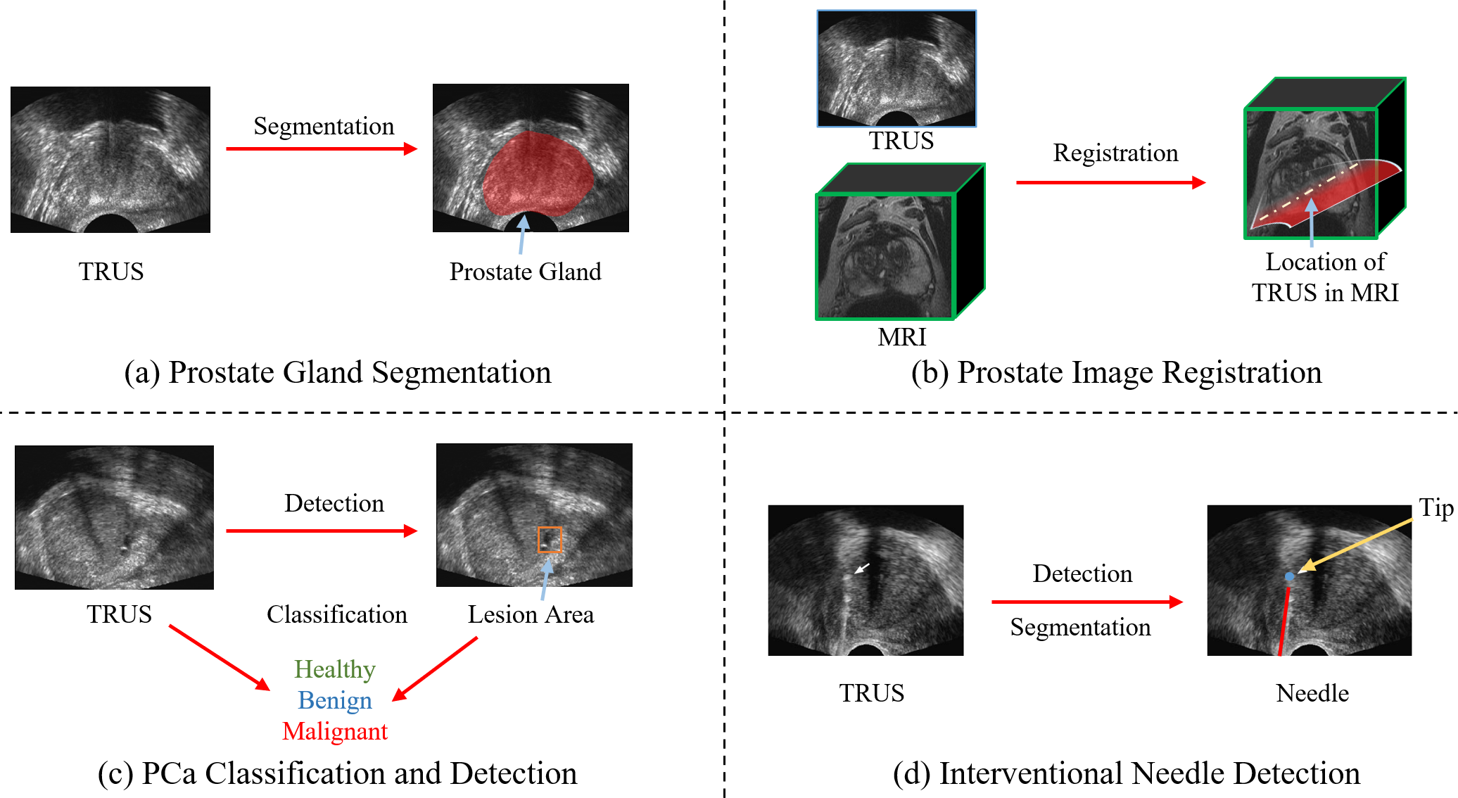
The development of image processing technology has greatly promoted the progress of the medical imaging field (Shen et al.,, 2017; Litjens et al.,, 2017; Duncan and Ayache,, 2000), especially playing an important role in computer assisted diagnosis (CAD) and computer assisted intervention (CAI). With the help of advanced image processing methods, it is beneficial for the physicians to make more accurate and efficient clinical decisions. In the field of TRUS image processing, lots of relevant methods have been developed and applied in different tasks, mainly including prostate gland segmentation, prostate image registration, PCa classification and detection, and interventional needle detection, as illustrated in Fig. 1.
This survey summarizes the related image processing methods in TRUS over the past 20 years, including gland segmentation, image registration, PCa classification and detection, and interventional needle detection. We classify and summarize these methods according to modality, dimension, and technical route, presenting the similarities and differences of these methods. We also list the primary evaluation metrics for different tasks, as well as the results of representative methods under their datasets and metrics, making a rough comparison of different methods. Finally, we also summarize these methods to propose current challenges faced by each task and possible future research directions.
The organization of this survey is as follows. In Section 2, we define each task of TRUS image processing, and list the definitions and formulas of their primary evaluation metrics. Section 3 is the main part of this survey, where we review and summarize the methods of prostate gland segmentation, prostate image registration, PCa classification and detection, and needle detection in Section 3.1, 3.2, 3.3 and 3.4, respectively. In Section 4, we discuss and analyze existing challenges and suggest future research directions for these four tasks. A summary of the entire survey is provided in Section 5.
2 Task Definition
2.1 Prostate Gland Segmentation
Image segmentation is one of the most common task in the field of medical image processing. The purpose of prostate segmentation is to accurately obtain the contour/region of the prostate gland, see Fig. 1(a). Prostate segmentation plays a crucial role in different stages of the clinical decision-making process. For example, prostate volume, which can be directly determined through prostate segmentation, aids in the diagnosis of BPH. Moreover, the boundary information of the prostate is important and useful in various treatment scenarios, such as prostate brachytherapy, high-intensity focused ultrasound (HIFU) therapy, cryotherapy, and transurethral microwave therapy. In addition, prostate segmentation also facilitates other image processing tasks, such as surface-based multi-modality image registration. To achieve prostate segmentation, lots of shape-/region-based methods, as well as conventional machine learning methods and advanced deep learning methods have been developed. We classify and summarize these segmentation methods in Section 3.1, according to TRUS modality, dimension, and technical route.
2.1.1 Evaluation Metrics
The evaluation of segmentation accuracy can be divided into qualitative and quantitative evaluations. For qualitative evaluation, the resulting contours are visually compared to the ground truth. For quantitative evaluation, the numerical calculation involves measuring the similarity between the obtained contours and the ground truth. The commonly used quantitative evaluation metrics consist of region-based metrics, boundary-based metrics, and classification-based metrics. Since traditional segmentation methods often rely on contour constraints, they always employ boundary-based metrics for the evaluation. In contrast, recent deep learning methods usually use region-based metrics.
Dice similarity coefficient (DSC) is currently the most popular region-based evaluation metric for the segmentation task, which measures the overlap between the segmented result and the ground truth:
| (1) |
where TP, FP, FN denotes true positive, false positive, and false negative regions, respectively.
Intersection over union (IoU) is another commonly used region-based metric, also known as the Jaccard coefficient. It is defined as follows:
| (2) |
Larger values of DSC and IoU indicate better segmentation accuracy.
As for the boundary-based metrics, mean surface distance (MSD), mean absolute distance (MAD), and average symmetric surface distance (ASD) are often employed to measure the similarity between the segmented boundary (in 2D)/surface (in 3D) and the ground truth:
| (3) |
| (4) |
| (5) |
where is the total number of points from the segmented boundary/surface. represents the shortest distance from the i-th point on the segmentation result to the ground truth, while represents the shortest distance from the i-th point on the ground truth to the segmentation result. It can be seen that MSD, MAD, and ASD measures the average distance, mean absolute distance, and average symmetric distance, respectively.
In contrast to MSD, MAD, and ASD, maximum distance (MAXD) and Hausdorff distance (HD) are used to represent the dissimilarity between the segmented boundary/surface and the ground truth:
| (6) |
| (7) |
MAXD measures the maximum distance between each point from the segmentation result and its corresponding point from the ground truth. HD measures the maximum distance between the segmentation result and ground truth, and with 95HD denoting the 95 percentile of HD. For all aforementioned boundary-based metrics, smaller values denotes better segmentation accuracy.
In addition to region-/boundary-based metrics, point-wise classification-based metrics can also be applied to measure segmentation accuracy. Accuracy (ACC) represents the proportion of correctly predicted pixels to the total number of pixels in the segmentation result:
| (8) |
where TN denotes true negative. Sensitivity and Recall indicate the proportion of all positive pixels that are correctly segmented as true positive:
| (9) |
Precision indicates the proportion of all predicted positive pixels that are TP pixels:
| (10) |
There is usually a trade-off between precision and sensitivity. Specificity is the proportion of all negative pixels that are correctly segmented as true negative:
| (11) |
Larger values of these classification-based metrics indicate better segmentation.
2.2 Prostate Image Registration
Image registration is the process of aligning two or more images acquired at varying times, viewpoints, or by different sensors. Taking registering a pair of images as an example, one is denoted as fixed image and the other as moving image. The objective is to estimate the optimal deformation field between the fixed and moving images, thus the warped moving image can be matched with the fixed image, enabling the alignment of the regions of interest. Prostate image registration plays a crucial role in assisting surgeons during preoperative planning and intraoperative surgery, with TRUS being a frequently utilized modality in this context. Its purpose is to provide surgeons with more complementary and valuable information. This task often involves analyzing multi-modality images (e.g., MR to TRUS, see Fig. 1(b)), thus remaining challenging in clinical practice. We classify and summarize relevant registration methods in Section 3.2, according to modality, dimension, deformation type and technical route.
2.2.1 Evaluation Metrics
Considering the purpose of image registration, the numerical evaluation metrics are mainly aimed at quantifying the similarity of corresponding regions between the fixed image and the warped moving image. Therefore the metrics like DSC, HD, MAD, and MAXD can also be used to evaluate the registration performance, by calculating the overlap of corresponding regions or the distance of corresponding boundaries/surfaces.
Target registration error (TRE) is one of the most commonly used metrics in medical image registration. It involves measuring the Euclidean distance of the manually identified corresponding landmarks in the fixed and moving images:
| (12) |
where denotes the landmark set, and is the total number of corresponding landmark pair in . and are the fixed and moving images, respectively. is the estimated deformation field, and denotes the warping operation. is the L2 norm.
Surface registration error (SRE) has the similar computing method with TRE, but the difference is SRE measures the similarity of surface points between fixed and moving images:
| (13) |
where denotes the prostate surface point set, and is the total number of corresponding point pair in . Smaller values of TRE and SRE indicate better registration accuracy.
In addition to assess registration accuracy using above metrics, some studies also evaluate the quality of the estimated deformation field . The Jacobian matrix evaluates the regularity of the deformation field by capturing the local gradient of around pixel . Smaller value of the percentage of pixels with non-positive Jacobian determinant (%) indicates smoother .
2.3 PCa Classification and Detection
Early and accurate diagnosis, along with precise staging, effectively improves the success rate of treatment. The standard method for diagnosing and grading PCa is the histopathological analysis of prostate tissue samples, typically obtained via TRUS-guided biopsy. Consequently, the accurate identification of target lesions during the biopsy procedure has been a long-standing and active research task. To better support TRUS-guided targeted biopsies, automated detection methods have been developed to predict PCa based on TRUS images, see Fig. 1(c). Most of these methods approach cancer detection as a classification problem, where small regions of interest or whole images are categorized as benign or malignant. We classify and introduce these methods in Section 3.3, according to TRUS modality and technical route.
2.3.1 Evaluation Metrics
The evaluation metrics for classification task mainly include area under the receiver operating characteristic curve (AUC), accuracy (8), sensitivity (9), specificity (11), precision (10), and F-score.
The AUC is a measure of the diagnostic ability of a binary classifier, which computes the area under the receiver operating characteristic (ROC) curve. The ROC curve is a curve drawn with the true positive rate (TPR, namely sensitivity) as the Y-axis and the false positive rate (FPR, namely specificity) as the X-axis. The area under ROC curve can be calculated as:
| (14) |
It represents the probability that a randomly chosen positive instance is ranked higher than a randomly chosen negative instance.
The F-score is a weighted harmonic mean of precision and recall:
| (15) |
2.4 Needle Detection
Prostate needles are primarily used during surgical procedures for various purposes, including biopsy, brachytherapy, HIFU, cryotherapy, and so on. Automated detection and segmentation of interventional needles are crucial in surgical settings because this ensures precise localization and safe interventions, as illustrated in Fig. 1(d). We summarize needle detection methods in Section 3.4, according to clinical scenario and technical route.
2.4.1 Evaluation Metrics
The numerical metrics for evaluating the needle detection/segmentation mainly include shaft error and tip error. Shaft error measures the deviation between the predicted and actual axes of the needle, usually expressed in terms of distance or angle:
| (16) |
| (17) |
where is the number of points in a needle, represents the predicted position for the i-th point, and is the ground truth position for the of the i-th point. and represent the actual and predicted axis direction vectors.
Tip error measures the difference between the predicted and actual tip positions of the needle:
| (18) |
where and represent coordinates of the actual and predicted needle tip.
3 Method Overview

In this section, we provide an overview of the key technologies involved in the four tasks of transrectal ultrasound (TRUS) prostate imaging analysis. To visually demonstrate the key terms and methods involved in these tasks, we present a comprehensive word cloud at the beginning of this section (see Fig. 3). The word cloud visually highlights the terms that frequently appear and are of significant research importance in each task, with the font size of the terms reflecting their prominence and widespread application in the literature.
3.1 Prostate Gland Segmentation
3.1.1 Modalities
Multi-modality TRUS images including B-mode, color Doppler, elastography, and micro-ultrasound have been utilized for the diagnosis of PCa. Each modality offers unique insights into the physiological and pathological states of the diseases. Among the TRUS modalities, the B-mode ultrasound is predominantly employed for prostate segmentation. The B-mode TRUS affords a morphological representation of the prostate, thereby facilitating the accurate delineation of the prostate’s contour.
Almost all segmentation studies mentioned in this survey used B-mode TRUS for the prostate segmentation. Few studies employed multi-modality TRUS or high-resolution micro-ultrasound. Mahdavi et al., (2012) boosted the performance of B-mode TRUS segmentation by offering supplementary information from the vibro-elastography images. Jiang et al., (2024) and Zhang et al., (2024) segmented prostate gland from the micro-ultrasound images. Their research has underscored the potential of this high-resolution modality in the precise segmentation of the prostate.
3.1.2 Dimensions
We categorize prostate segmentation approaches as 2D, 2.5D, 3D methods, according to the TRUS dimension they processed. As depicted in Fig. 2, the majority of researches have concentrated on 2D and 3D techniques, with a comparatively modest exploration of the 2.5D methodology.
The 2D segmentation is executed on an individual slice of the prostate image. This approach is frequently employed for intraoperative real-time navigation, serving to assist clinicians in the precise identification of the prostate gland’s location and boundary. The merits of 2D segmentation include its simplicity and high computational efficiency. Nonetheless, it falls short in capturing the 3D morphology of the prostate, potentially leading to inaccuracies in the volumetric assessment of the prostate, particularly concerning the apex and base regions.
In contrast to 2D segmentation, three-dimensional segmentation is conducted on the entire volumetric TRUS data. This technique is predominantly utilized in preoperative planning, aiding physicians in delineating the extent and trajectory for surgical excision or in radiation therapy planning by enabling radiation oncologists to pinpoint the target region for irradiation. 3D segmentation affords an exhaustive and accurate quantitative analysis of the prostate’s shape, volume, and boundaries. However, it is encumbered by high computational demands and necessitates the processing of a vast array of voxels and intricate 3D structures.
Beyond the 2D and 3D approaches, the 2.5D segmentation technique operates on multiple contiguous slices of the prostate image. By integrating information from multiple slices, 2.5D segmentation can partially capture the volumetric information. It offers an advantage over 2D segmentation in better handling the 3D morphological variations of the prostate, albeit with limitations imposed by the selection and thickness of the slices. Compared with 3D segmentation, 2.5D segmentation can alleviate the computational load, presenting a more efficient compromise between computational demands and segmentation accuracy.
3.1.3 Segmentation Methods
We categorize prostate segmentation methods into two ways: traditional methods and deep learning (DL) methods. Fig. 4 shows the categorization in detail. It can be seen that before 2016 almost all of the studies were about traditional methods, and after that DL methods dominated this area.
Traditional Methods.
We present the traditional methods for prostate segmentation in four categories: contour/shape-based methods, region-based methods, conventional machine learning methods, and hybrid methods. Such categorization is determined on the underlying theoretical computational approaches employed to address the segmentation task. Table 1 gives a comprehensive summary of the representative studies using traditional methods for TRUS segmentation.
(a) Contour/Shape-based Methods. These methods use prostate contour/edge information to guide the segmentation. Contour-based methods typically initiate with edge detection. Pathak et al., (2000) introduced a Sticks algorithm to enhance contrast and reduce speckle noise in TRUS, thus facilitating the rendering of prostate edges as a visual guide for subsequent edge detection and manual refinement. Shen et al., (2003) expanded on this by employing a Gabor filter bank to characterize prostate boundary features across multiple scales and orientations, followed by a hierarchical deformation strategy to adaptively focus on feature similarity. This approach, while more time-consuming, captured a broader range of boundary directions. Wu et al., (2015) diverged from the traditional view by leveraging speckles’ intrinsic properties to aid segmentation, arguing that speckle characteristics provide useful cues for prostate boundary delineation.
Active shape models (ASM), as pioneered by Cootes et al., (1995), offer a statistical shape-based method that has been further developed by researchers such as Cosío, (2008), who presented a robust automatic initialization of the ASM for prostate. This method used pixel classification for the initialization of prostate region and a multi-population genetic algorithm for ASM pose adjustment. Mahdavi et al., (2012) and Bi et al., (2020) extended the ASM by incorporating high-contrast vibro-elastography images and clustering techniques using a Rayleigh mixture model, respectively, to enhance the initialization and deformation guidance of the ASM model.
Beyond ASM, other shape-prior-based segmentation techniques have been explored. Badiei et al., (2006) proposed an image warping technique to impose an elliptical shape on the prostate, with subsequent optimization of the elliptical fitting using boundary points from an edge detector. Mahdavi et al., (2011) further advanced this by suggesting twisted and tapered ellipsoids as suitable a priori 3D shapes, transforming the shape fitting into a convex problem for efficient solution. Yan et al., 2010a proposed adaptive learning of local neighborhood shape statistics to capture patient-specific variations. Qiu et al., (2012) introduced a semi-automatic method integrating shape constraints with a modified level set formulation for 3D segmentation. These methods emphasized the importance of integrating shape priors with computational strategies to improve accuracy and efficiency.
(b) Region-based Methods. These methods harness local intensity values or statistical measures within an energy minimization framework to achieve segmentation. Fan et al., (2002) initially developed a fast discrimination technique for extracting the prostate region and subsequently addressed the “boundary leaking” issue by integrating regional information, rather than gradient information, into the level set method. Yuan et al., (2013) employed rotationally re-sliced images around a specified axis to delineate 3D prostate boundaries, a technique that effectively capitalized on the inherent rotational symmetry of prostate shapes to adjust a series of 2D slice segmentation in a unified 3D context. The work (Yuan et al.,, 2013) was further extended to (Qiu et al., 2014b, ), with more validation data. Qiu et al., 2014a introduced a convex optimization strategy for extracting the prostate surface, while maintaining the global volume size prior. Moreover, they implemented an efficient numerical solver on GPUs. Additionally, some methods accomplish the extraction of the prostate region through template matching. Nouranian et al., (2013) designed a multi-atlas fusion framework to segment TRUS images using pairwise atlas shape similarity. Nouranian et al., (2014) further introduced the atlas agreement factor to boost the robustness of the previous work (Nouranian et al.,, 2013). This factor was integrated into an atlas selection algorithm to refine the dataset prior to merging the atlas contours, yielding consistent segmentation.
| Traditional Methods | References | Dimension | Auto/Semi-auto | Methods | Samples | Results | |
| Metrics | Values | ||||||
| Contour and Shape | Pathak et al., (2000) | 2D | Semi-auto | Probabilistic filters, edge detection, manual editing | 125 | MAD HD | mm mm |
| Shen et al., (2003) | 2D | Automatic | Gabor filter bank, statistical shape model | 8 | 1-DSC 1-IoU MAD | pixels | |
| Badiei et al., (2006) | 2D | Semi-auto | Image warping, edge detection, ellipse fitting | 17 | MAD MAXD | mm mm | |
| Cosío, (2008) | 2D | Automatic | Pixel classification, active shape model | 22 | MAD MAXD | mm mm | |
| Mahdavi et al., (2011) | 3D | Semi-auto | Shape fitting, convex optimization | 40 | 1-DSC | ||
| Qiu et al., (2012) | 3D | Semi-auto | Shape constraint, level set | 20 | DSC MAD | mm | |
| Mahdavi et al., (2012) | 3D | Automatic | Elastography, active shape model | 11 | DSC | ||
| Wu et al., (2015) | 2D | Automatic | Rotation-invariant texture feature, graph cut, level set | 598 | MAD | mm | |
| Bi et al., (2020) | 2D | Automatic | Rayleigh mixture model, active shape model | 30 | DSC MAD | pixels | |
| Region | Fan et al., (2002) | 3D | Semi-auto | Region information, level set | 8 | Visual Comparison | Visual Comparison |
| Yuan et al., (2013) | 3D | Automatic | Rotational symmetry, convex optimization | 20 | DSC MAD MAXD | mm mm | |
| Nouranian et al., (2013) | 3D | Automatic | Multi-atlas fusion, pairwise shape similarity | 50 | DSC MAD MAXD | mm mm | |
| Qiu et al., 2014a | 3D | Automatic | Volume-preserving prior, convex optimization | 12 | DSC MAD MAXD | mm mm | |
| Qiu et al., 2014b | 3D | Automatic | Axial symmetry, convex optimization | 25 | DSC | ||
| Nouranian et al., (2014) | 3D | Automatic | Multi-atlas fusion, atlas agreement factor | 280 | DSC MSD | mm | |
| Machine Learning | Ghose et al., (2013) | 2D | Automatic | Statistical shape and appearance model, random forest | 126 | DSC MAD HD | mm mm |
| Nouranian et al., (2015) | 2D | Automatic | Sparse dictionary learning | 590 | MSD HD | mm mm | |
| Yang et al., (2016) | 3D | Automatic | Kernel support vector machine | 10 | DSC | ||
| Peng et al., 2022b | 2D | Semi-auto | Improved closed principal curve | 50 | DSC Jaccard ACC | ||
| Peng et al., 2024b | 2D | Semi-auto | Adaptive polygon tracking model, principal curve | 1320 | DSC Jaccard ACC | ||
| Hybrid | Zhan and Shen, (2003) | 3D | Automatic | Kernel support vector machine, statistical shape model | 3 | 1-IoU MAD | voxel |
| Gong et al., (2004) | 2D | Automatic | Deformable superellipses, Bayesian model | 125 | MAD HD | mm mm | |
| Tutar et al., (2006) | 3D | Semi-auto | Spherical harmonics, statistical analysis | 30 | MAD MAXD | mm mm | |
| Zhan and Shen, (2006) | 3D | Automatic | Gabor-support vector machine, statistical texture matching | 5 | 1-IoU MAD | voxels | |
| Yan et al., 2010b | 2D | Automatic | Partial active shape model, region model | 301 | MAD | mm | |
| Garnier et al., (2010) | 3D | Semi-auto | Discrete dynamic contour, optimal surface detection | 28 | ASD | mm | |
| Jaouen et al., (2018) | 3D | Automatic | Hybrid edge/region-based parametric deformable model | 36 | DSC MAD HD | mm mm | |
(c) Conventional Machine Learning Methods. Early prostate segmentation methods also encompass machine learning approaches. These techniques leverage features such as intensity or higher-dimensional attributes like filter responses to cluster and/or classify the image into prostate and background regions. Ghose et al., (2013) introduced a method for prostate segmentation that constructed a multiple average parameter model derived from the principal component analysis of shape and a posterior probabilities within a multi-resolution framework. The model parameters were subsequently refined using a priori knowledge of the optimization space to achieve optimal prostate segmentation. Nouranian et al., (2015) proposed a learning-based multi-label segmentation algorithm. A sparse representation method was integrated into the algorithm to learn a dictionary encoding information of images as well as clinical and planning target volume segmentation. Yang et al., (2016) developed a block-based feature learning framework, where patient-specific anatomical features extracted from aligned training images served as signatures for each voxel. A feature selection process identified the most robust and informative features to train a kernel support vector machine (KSVM).
Additionally, some methods have employed the principal curve, which provided a means of representing low-dimensional data manifolds in high-dimensional spaces, thereby effectively capturing the complexity of prostate shapes. Peng et al., 2022b proposed an improved closed principal curve approach, utilizing only a few seed points defined by radiologists as an approximate initialization. Peng et al., 2024b further developed an adaptive polygon tracking model to alleviate the issue of determining the segment number of the principal curve automatically. In addition, an interpretable mathematical function was formulated to constrain a smooth boundary.
(d) Hybrid Methods. The objective of hybrid methods is to leverage advantages from contour, shape, region, and conventional machine learning to achieve better segmentation results. Zhan and Shen, (2003) combined kernel support vector machine and statistical shape model (SSM) to segment 3D TRUS. The KSVM was employed to classify the prostate from surrounding tissues, and then the SSM was used to refine the contour. Later, Zhan and Shen, (2006) improved (Zhan and Shen,, 2003) by using a set of Gabor-support vector machines (G-SVMs) and the statistical texture matching model. Gong et al., (2004) emphasized that the prior knowledge of prostate shapes played an important role for reliable segmentation. They used deformable superellipses to model the shape of prostate and further developed a robust Bayesian segmentation approach. Tutar et al., (2006) employed the eighth spherical harmonic function to depict the shape of the prostate, and statistically analyzed the shape parameters. Yan et al., 2010b employed partial active shape model and region model to address the issue of missing boundaries in shadow areas. Garnier et al., (2010) sequentially combined discrete dynamic contour and optimal surface detection for semi-automatic 3D TRUS segmentation in HIFU Therapy. Jaouen et al., (2018) proposed a hybrid edge and region-based parametric deformable model. The edge-based model was derived from the radial bas-relief approach, while the region-based model was regarded as a parametric active surface driven by the Bhattacharyya gradient flow.
Deep Learning Methods.
We categorize deep learning methods for TRUS segmentation into three classes: convolutional neural network (CNN), recurrent neural network (RNN), and attention enhanced networks, according to their network structure. Due to limited studies on Transformer currently in this field, relevant researches are included in the attention enhanced networks section. Table 2 summarizes representative studies related to the DL-based segmentation in TRUS.
(a) Convolutional Neural Network. CNNs are among the most successful and widely used architectures in DL, particularly for computer vision tasks. Following the advent of U-net Ronneberger et al., (2015), a plethora of CNNs with encoder-decoder structures have been employed for medical image segmentation. Several methods have sought to enhance segmentation performance by improving loss functions. For instance, Lei et al., 2019a developed a multi-directional DL method to segment TRUS for US-guided radiation therapy. They combined binary cross-entropy (BCE) loss and batch-based Dice loss into a staged hybrid loss function for deeply-supervised training. Karimi et al., (2019) improved the uncertain segmentation by incorporating prior shape information in the form of a statistical shape model. Karimi and Salcudean, (2019) presented a loss function for training the segmentation network with the goal of directly reducing Hausdorff distance.
Additionally, other methods have focused on the improvement of the network structures. Lei et al., 2021b developed an anchor-free mask CNN that consisted of three subnetworks: a backbone, a fully convolutional one-state object detector, and a mask head. Xu et al., (2022) proposed a novel polar transform network to address the issue of difficult segmentation in the challenging base and apex regions. The proposed network represented the prostate shape in the polar coordinate space. This representation provided much denser samples of the prostate volume, thus facilitating the learning of discriminative features for accurate prostate segmentation.
Furthermore, some studies have focused on fully utilizing data information. Xu et al., (2021) proposed a shadow-consistent semi-supervised segmentation method that included shadow augmentation (Shadow-AUG) and shadow dropout (Shadow-DROP). Shadow-AUG enlarged training samples by including simulated shadow artifacts to the images, while Shadow-DROP enhanced the network to leverage neighboring high-quality pixels to infer boundaries. Orlando et al., (2022) developed an automatic 3D TRUS segmentation network, trained on a large, clinically diverse dataset with variable image quality.
(b) Recurrent Neural Network. RNNs are commonly utilized for processing sequential data, such as speech, text, videos, and time-series data. Unlike unidirectional feedforward neural networks, RNNs are bidirectional neural networks, allowing the outputs of certain nodes to influence subsequent inputs from the same nodes. Their capability to process arbitrary input sequences using internal state, or memory, makes them suitable for tasks such as unsegmented, connected handwriting recognition or speech recognition.
In the context of TRUS segmentation, Yang et al., (2017) first serialized static 2D TRUS images into dynamic sequences and then predicted prostate shape by sequentially exploring the shape prior. Subsequently, to mitigate the bias caused by different serialization approaches, they employed a multi-view fusion strategy to merge shape predictions obtained from various perspectives. Furthermore, they integrated the RNN core into a multi-scale automatic context scheme to continuously refine the details of the shape prediction graph. Similarly, Anas et al., 2018a not only utilized CNNs to extract spatial features but also employed RNNs to leverage the temporal information among a series of TRUS images. Additionally, they utilized cyclic connections within and between different layers of the network to maximize the utilization of temporal information.
| Deep Learning Methods | References | Dimension | Methods | Samples | Results | |
| Metrics | Values | |||||
| CNN | Lei et al., 2019a | 3D | 1) Multi-directional deeply supervised V-Net 2) A hybrid loss with binary cross-entropy and batch-based Dice | 44 | DSC MSD HD | mm mm |
| Karimi et al., (2019) | 2D | 1) An adaptive sampling strategy to address difficult cases 2) A CNN ensemble to identify uncertain segmentation | 675 | DSC HD | mm | |
| Karimi and Salcudean, (2019) | 2D | 1) A loss function to reduce the Hausdorff distance (HD) 2) Three methods to estimate HD | 2625 | DSC ASD HD | mm mm | |
| Lei et al., 2021b | 3D | 1) An anchor-free mask CNN | 83 | DSC 95HD | mm | |
| Xu et al., (2022) | 3D | 1) Represent prostate shape in the polar coordinate space 2) A centroid perturbed test-time augmentation strategy | 315 | DSC ASD HD | mm mm | |
| Xu et al., (2021) | 3D | 1) Shadow augmentation and shadow dropout 2) Semi-supervised Learning | 2423 | DSC ASD HD | mm mm | |
| Orlando et al., (2022) | 2D | 1) Modified U-Net and U-Net | 6721 | DSC MSD HD | mm mm | |
| RNN | Yang et al., (2017) | 2D | 1) Serialize static TRUS images into dynamic sequences 2) A multi-view shape fusion strategy 3) A multiscale auto-context scheme | 530 | DSC Recall Precision | |
| Anas et al., 2018a | 2D | 1) Residual convolution in recurrent networks 2) Recurrent connections for deep network layers | 3892 | DSC HD | mm | |
| Attention | Wang et al., 2018a | 2D | 1) Select useful complementary information from multi-level features to refine the features at each individual layer | 530 | DSC Recall Precision | |
| Wang et al., (2019) | 3D | 1) Selectively leverage multi-level features from different layers | 40 | DSC Recall Precision | ||
| Girum et al., (2020) | 2D | 1) A channel-wise attention strategy 2) Learning-based prior shape knowledge | 145 | DSC 95HD | mm | |
| Vesal et al., (2022) | 2.5D | 1) Feature positioning information 2) A training scheme that utilizes knowledge distillation | 2892 | DSC 95HD | mm | |
| Peng et al., 2024a | 2D | 1) Attention gate modules with squeeze-and-excitation U-net 2) An improved polygon searching model 3) A storage-based quantum evolution model | 945 | DSC ACC | ||
(c) Attention Enhanced Network. In DL methods, an attention block can selectively modify inputs or assign different weights to input variables according to their varying importance. Attention mechanisms in DL are fundamentally analogous to the selective visual attention mechanisms in humans, with the core objective being to select the most critical information from a vast amount of data for the current task’s goal.
In the context of TRUS segmentation, Wang et al., 2018a firstly developed a network equipped with deep attentional feature (DAF) modules to enhance segmentation by fully exploiting the complementary information encoded in different layers of the CNN. The DAF module selectively utilized multi-level features integrated across different layers to refine each individual layer, suppressing non-prostatic noise in shallow layers of the CNN, and enriching deeper features with more prostatic detail. Further, Wang et al., (2019) extended (Wang et al., 2018a, ) to 3D TRUS segmentation, by using more powerful backbone architecture. Girum et al., (2020) presented a multi-task DL method for boundary detection in intraoperative TRUS, by leveraging both low-level and high-level information. The channel-wise attention strategy was employed for low-level feature refinement, and the learning-based prior knowledge was utilized for high-level feature modeling. Vesal et al., (2022) addressed the limitations of transfer learning and fine-tuning methods by introducing a 2.5D deep neural network, which further relied on an attention module that considered feature positioning information to improve segmentation accuracy. Peng et al., 2024a developed a coarse-to-fine architecture for TRUS segmentation. They employed the attention gate module with squeeze-and-excitation U-net to conduct initial segmentation, and then jointly used a modified polygon-searching model and a storage-based quantum evolution model to refine the segmentation.
3.2 Prostate Image Registration
3.2.1 Modalities
Prostate image registration is useful for assisting surgeons during preoperative planning and intraoperative surgery. It can provide surgeons with more complementary information, especially the multi-modality registration. Fig. 5 lists the categorization of registration approaches according to the image modality. The majority of studies focused on MRI-TRUS registration, which registered the preoperative MRI scans to the TRUS images, providing better resolution and soft tissue contrast to assist targeted biopsy. In addition, CT-TRUS registration methods have also been investigated, mainly in the scenarios of treatment planning and prognosis evaluation. Other studies payed attention to the registration of preoperative and intraoperative TRUS images.
3.2.2 Dimensions
We classify registration methods into three categories based on dimensionality: 2D-2D, 2D-3D, and 3D-3D. Images acquired intraoperatively are predominantly 2D TRUS, whereas those acquired preoperatively are mostly 3D (MRI, CT, 3D TRUS). Ultimately, the registration methods intended for prostate biopsy would necessitate the registration between intraoperative 2D TRUS and preoperative 3D images.
The process of registering 2D TRUS to 3D images from different modalities presents two main challenges: differing dimensions and distinct modalities. There was a scarcity of literature that addressed both of these issues simultaneously. Consequently, most 2D-3D studies concentrated on registration between TRUS images to bridge the dimensional gap, while 3D-3D studies primarily focused on registration between TRUS and other modalities to overcome the challenge posed by different imaging modalities.
3.2.3 Deformation Types
According to different deformation types, we categorize the registration methods into linear and non-linear registration, as shown in Fig. 7. Linear transformation, also referred to as isometric transformation, involves only translation and rotation. Affine transformation includes scaling and shear in addition to translation and rotation, making linear transformation a special case of affine transformation. In this survey, both affine and linear transformation are uniformly classified as the category of linear registration. In contrast, non-linear registration refers to methods that go beyond linear registration, primarily involving deformable transformation. Unlike linear registration, deformable registration methods employ non-linear dense transformation to align images. Most of prostate image registration methods focused on non-linear registration to address the issue of large deformation in TRUS images.
3.2.4 Registration Methods
We categorize registration methods into two main types: traditional methods and DL methods, as shown in Fig. 8. Traditional methods, while often time-consuming due to the iterative estimation of the deformation field, are known for their stability and reliability across various scenarios. In contrast, DL methods have gained popularity due to advancements in GPU computing power, enabling them to achieve more efficient registration than traditional methods.
Traditional Methods.
Traditional registration methods mainly investigated the following two aspects: matching criteria and deformation models. The matching criterion defines the similarity of the image pair, with intensity-based methods relying on the similarity of intensities or intensity-related features of images, and shape-based methods focusing on the similarity of prostate’s region/boundary. The deformation model is also critical, as the registration process involves optimizing the parameters of the deformation fields to warp the moving image to match with the fixed image. We summarize the traditional registration methods on TRUS images according to different matching criteria.
(a) Intensity-based Methods. Table 3 summarizes some representative traditional registration methods, including intensity-based methods and shape-based methods. Xu et al., (2007) employed spatial tracking information to initialize the registration between intraoperative 2D TRUS and preoperative 3D TRUS. They then optimized the registration by calculating the grayscale similarity to eliminate spatial tracking errors. Additionally, they manually registered 3D TRUS and 3D MRI preoperatively, achieving a 2D TRUS-3D TRUS-3D MRI registration process. Hungr et al., (2012) used the correlation coefficient as a similarity metric to register 3D TRUS, while Baumann et al., (2012) employed the shift correlation model combined with the inverse consistency constraint, performing linear registration separately from deformable registration. Sun et al., (2014) used the modality independent neighborhood descriptor (MIND) as the local similarity feature for MRI-TRUS registration. They also developed a duality-based convex optimization algorithm to compute MIND similarity. De Silva et al., (2013) used Powell’s method to optimize the normalized cross-correlation (NCC) metric between images, completing the linear registration of 2D TRUS to 3D TRUS. Later, Gillies et al., (2017) also employed Powell’s method to optimize the NCC metric and realized real-time registration of 2D and 3D TRUS.
| Traditional Methods | References | Dimensions Modalities | Deformation | Methods | Samples | Results | |
| Metrics | Values: final (initial) | ||||||
| Intensity | Baumann et al., (2012) | 2D TRUS 3D MRI | Non-linear | 1) Shift correlation and inverse consistency constraint 2) Local rigid and linear elastic deformation | 40 | TRE | () mm |
| De Silva et al., (2013) | 2D TRUS 3D TRUS | Linear | 1) Optimize the normalized cross-correlation (NCC) metric using Powell’s method | 8 | TRE | () mm | |
| Sun et al., (2014) | 3D TRUS 3D MRI | Non-linear | 1) Modality independent neighborhood descriptor 2) Duality-based convex optimization algorithm | 20 | TRE DSC MAD MAXD | () mm (n/a) (n/a) mm (n/a) mm | |
| Gillies et al., (2017) | 2D TRUS 3D TRUS | Linear | 1) Optimize NCC using Powell’s method 2) Golden section search technique | 14 | TRE | () mm | |
| Shape | Mitra et al., (2012) | 2D TRUS 2D MRI | Non-linear | 1) Point correspondences obtained from a statistical measure of shape-contexts 2) The radial-basis function of thin-plate splines | 20 | TRE DSC 95HD | (n/a) mm (n/a) (n/a) mm |
| Hu et al., (2012) | 3D TRUS 3D MRI | Non-linear | 1) Statistical motion model 2) Expectation maximization algorithm for model-to-image registration | 8 | TRE | () mm | |
| Zettinig et al., (2015) | 3D TRUS 3D MRI | Non-linear | 1) Coherent point drift algorithm 2) Thin-plate splines | 13 | TRE | () mm | |
| Hu et al., (2015) | 3D TRUS 3D MRI | Non-linear | 1) Statistical motion model 2) Kernel regression analysis | 44 | TRE | () mm | |
| Khallaghi et al., 2015c | 3D TRUS 3D MRI | Non-linear | 1) Finite element model with biomechanical constraint 2) Gaussian mixture model | 19 | TRE | () mm | |
| Khallaghi et al., 2015b | 3D TRUS 3D MRI | Non-linear | 1) Statistical shape model 2) Finite element model | 19 | TRE | () mm | |
| Wang et al., (2016) | 3D TRUS 3D MRI | Non-linear | 1) Personalized statistical deformable model 2) Hybrid point matching | 18 | TRE | () mm | |
| Wang et al., 2018b | 3D TRUS 3D MRI | Non-linear | 1) Robust projective dictionary learning 2) Personalized statistical deformable model | 18 | TRE | () mm | |
| Samei et al., (2018) | 2.5D TRUS 3D MRI | Non-linear | 1) Linear stress-strain quasi-static FEM 2) 2.5D-3D non-rigid registration | 11 | TRE | () mm | |
(b) Shape-based Methods. It remains challenging to use intensity-based metrics to evaluate the similarity of multi-modality images. Therefore, most of MRI-TRUS and CT-TRUS registration approaches sought the shape-based similarities as the solution. Although the shape-based methods required additional steps to obtain the prostate region or corresponding landmarks, they offered more reliable and robust registration for multi-modality tasks. Mitra et al., (2012) introduced a non-linear thin-plate spline (TPS) regularized 2D MRI-TRUS registration method. The boundary point correspondences were estimated based on a statistical measure of shape-contexts. Zettinig et al., (2015) applied the coherent point drift (CPD) algorithm to elastically align surfaces extracted from TRUS and MRI. They then propagated the surface deformation to the whole gland using the TPS interpolation. Lee and Song, (2018) proposed a deformable CT-TRUS registration method based on the structural descriptor map (SDM), calculated by solving the Laplace’s equation using the segmented prostate surface. The SDM represented the surface distribution and could be employed for deformable registration. Samei et al., (2018) proposed an efficient algorithm to extract arbitrary slices in real-time from deformed 3D images represented by a discrete grid. They simulated tissue deformation using a patient-specific finite element model (FEM) with boundary conditions for image-guided, robot-assisted laparoscopic radical prostatectomy (RALRP). Later, Samei et al., (2020) incorporated this technology into a virtual reality-based RALRP system.
Certain methods (Hu et al.,, 2009, 2012) employed both statistical shape modeling (SSM) and biomechanical modeling to capture the motion of the prostate, which has been proved as a robust solution for MRI-TRUS registration. leveraging the SSM and biomechanical modeling, Hu et al., (2012) further calculated the normal vector field of the prostate surface in TRUS and used the expectation maximization (EM) algorithm to maximize the log-likelihood function for model-to-image registration. Hu et al., (2015) later used kernel regression analysis to express the multivariate subject-specific probability density function for the construction of subject-specific statistical motion model. Khallaghi et al., 2015c used a probabilistic framework based on Gaussian mixture model (GMM) to combine FEM with biomechanical prior knowledge for extrapolating and constraining deformation fields during MRI-TRUS registration. Khallaghi et al., 2015b employed probabilistic SSM-FEM to map two surfaces and their interiors to a common intermediate SSM instance, aligning MRI to 3D TRUS through this intermediate shape. Wang et al., (2016) introduced a hybrid point matching method to establish reliable surface point correspondences. A series of FEM were conducted using personalized anatomical and biomedical information. And a personalized statistical deformable model was constructed by the principal component analysis (PCA) of FEMs to estimate the deformation. To enhance the efficacy of statistical shape modeling in (Wang et al.,, 2016), Wang et al., 2018b developed a robust projective dictionary learning algorithm to integrate dimensionality reduction and dictionary learning into a unified shape prior modeling framework, and applied it for MRI-TRUS registration.
Deep Learning Methods.
With the rapid development of GPUs, using DL methods to solve non-linear registration with high computational complexity has become mainstream. These DL methods for TRUS image registration can be categorized into image-based networks, label-driven networks, and point set networks. The first two types often take images as input, while the last one uses surface point clouds. Image-based networks mainly rely on the similarity between moving and fixed images as the loss function to update network parameters. In contrast, label-driven networks directly use prostate annotations or landmarks to aid registration, either by incorporating them into the loss function or by directly registering them. Point set networks transform the image registration problem in the form of point cloud matching and train the network using the point-based loss function. Table 4 introduces the representative methods in each category.
(a) Image-based Network. Some methods have focused on the development of novel network architectures. Yan et al., (2018) proposed an adversarial MRI-TRUS registration framework, which trained two CNNs concurrently - a generator for image registration and a discriminator to assess the registration quality. This method only considered linear transformation and required ground truth transformation for the supervised learning. Guo et al., (2021) proposed a 2D-3D TRUS registration network. This network used a dual-branch encoding module to extract 2D and 3D features for the estimation of linear transformation parameters. And a 2D plane sampling module was designed to enable efficient training and inference. This approach was subsequently extended to (Guo et al.,, 2023), by newly designing a frame-to-slice correction network to refine the transformation estimation and a similarity filtering mechanism to assess the registration quality. Song et al., (2021) introduced a cross-modal attention module to explicitly utilize spatial correspondence for a linear transformation in 3D MRI-TRUS registration, and further extended this approach to other applications (Song et al., 2022b, ).
Other methods have explored various training strategies and loss functions. Haskins et al., (2019) employed a CNN to learn similarity metrics for the representation of MRI-TRUS, and then used the learned similarity metric to optimize the registration. Guo et al., (2020) utilized data generation methods to create diverse training data distributions, allowing the network to perform well for MRI-TRUS registration. Song et al., 2022a introduced the transformed grid distance loss, which addressed the issue of rotation representation and bridged the gap between translation and rotation.
(b) Label-driven Network. These networks leveraged prostate annotations or landmarks to evaluate the similarity of multi-modality prostate images. Hu et al., 2018a optimized a discriminator network to identify the deformation field predicted by registration network from FE-simulated motion data. The MRI-TRUS registration network simultaneously aimed to maximize similarity between anatomical labels to drive image alignment and minimize an adversarial generator loss that measured divergence between the predicted and simulated deformation. Hu et al., 2018b also investigated using various types of identifiable corresponding anatomical structures to train the network for the prediction of dense voxel correspondence. These anatomical structures could be prostate region, vessels, point landmarks or other ad hoc structures. Later, Hu et al., 2018c extended the work (Hu et al., 2018b, ), by designing a new multiscale Dice for the network training and an improved memory-efficient network architecture. Hu et al., (2019) applied segmentation concepts to registration, predicting the transformed region-of-interest (ROI) directly without predicting the deformation field. Zeng et al., 2020b employed a CNN for segmentation and then supervised the training of registration network using a surface loss. Chen et al., (2021) also used segmentation-based learning to train the MRI-TRUS registration network for the application of prostate brachytherapy. Song et al., (2023) trained a CNN to extract landmarks and then leveraged these landmarks for affine registration.
(c) Point Set Network. Baum et al., (2020) described a point set registration algorithm based on a free point transformer network for points extracted from segmented MRI-TRUS surfaces. Based on the preliminary work in (Baum et al.,, 2020), Baum et al., (2021) developed a partial point-set registration network, which allowed non-linear matching without known correspondence or heuristic constraints. Fu et al., (2021) utilized tetrahedron meshing to generate volumetric prostate point clouds from segmented masks, and trained a point cloud matching network using deformation fields generated by FE analysis. Min et al., (2023) developed a physically-informed neural network to impose elastic constraints on the estimated displacements, and formulated an end-to-end registration network training algorithm by minimizing surface distances as estimated boundary conditions in partial differential equations.
| Deep Learning Methods | References | Dimensions Modalities | Deformation | Methods | Samples | Results | |
| Metrics | Values: final (initial) | ||||||
| Image-based Network | Yan et al., (2018) | 3D TRUS 3D MRI | Linear | 1) Adversarial learning 2) Supervised-learning using ground truth transformation | 763 | TRE | () mm |
| Haskins et al., (2019) | 3D TRUS 3D MRI | Linear | 1) Use CNNs to learn similarity metrics for MRI–TRUS 2) Composite optimization strategy | 679 | TRE | () mm | |
| Guo et al., (2020) | 3D TRUS 3D MRI | Linear | 1) Coarse-to-fine multi-stage framework 2) Generate different training data distributions | 679 | TRE | () mm | |
| Song et al., (2021) | 3D TRUS 3D MRI | Linear | 1) Cross-modal attention mechanism | 662 | TRE | () mm | |
| Guo et al., (2021) | 2D TRUS 3D TRUS | Linear | 1) Dual-branch feature extraction module 2) Differentiable 2D slice sampling module | 619 | TRE | () mm | |
| Guo et al., (2023) | 2D TRUS 3D TRUS | Linear | 1) Frame-to-slice correction network 2) Similarity filtering mechanism | 618 | TRE | (n/a) mm | |
| Label-driven Network | Hu et al., 2018a | 3D TRUS 3D MRI | Non-linear | 1) Weakly-supervised anatomical-label-driven network 2) Adversarial deformation regularization | 108 | TRE DSC | (n/a) mm (n/a) |
| Hu et al., 2018b | 3D TRUS 3D MRI | Non-linear | 1) Label-driven weakly-supervised learning | 111 | TRE DSC | (n/a) mm (n/a) | |
| Hu et al., 2018c | 3D TRUS 3D MRI | Non-linear | 1) Multiscale Dice for weakly-supervised training 2) Memory-efficient network architecture | 108 | TRE DSC | (n/a) mm (n/a) | |
| Hu et al., (2019) | 3D TRUS 3D MRI | Non-linear | 1) Conditional image segmentation paradigm | 115 | TRE DSC | (n/a) mm (n/a) | |
| Point Set Network | Baum et al., (2020) | 3D TRUS 3D MRI | Non-linear | 1) Free point transformer network | 108 | TRE HD | () mm () mm |
| Baum et al., (2021) | 3D TRUS 3D MRI | Non-linear | 1) Data-driven learning without heuristic constraints 2) Variable number of points in training and inference | 108 | TRE HD | () mm () mm | |
| Fu et al., (2021) | 3D TRUS 3D MRI | Non-linear | 1) Biomechanical constraints via finite element analysis 2) Surface Chamber distance loss | 50 | TRE DSC HD | (n/a) mm (n/a) (n/a) mm | |
| Min et al., (2023) | 3D TRUS 3D MRI | Non-linear | 1) Biomechanical constraints exerted from soft-tissue-modeling partial differential equations | 77 | TRE | (n/a) mm | |
3.3 Prostate Cancer Classification and Detection
3.3.1 Modalities
As depicted in Fig. 9, the predominant modalities for TRUS-based PCa classification and detection methods are B-mode TRUS, as well as contrast-enhanced ultrasound (CEUS) and micro-ultrasound (Micro-US). With the advent of multi-modality imaging techniques, such as shear wave elastography (SWE), there has been a growing interest in multi-modality TRUS-based PCa diagnosis in recent years.
3.3.2 Classification and Detection Methods
We categorize the TRUS-based PCa diagnosis methods into two classes: traditional methods and deep learning methods. Some representative studies are summarized in Table 5.
Traditional Methods.
These methods have payed attention to analyzing the hand-crafted features extracted from images to conduct diagnosis. Mohamed and Salama, (2005) extracted texture features from TRUS, including the gray level dependence matrix and gray level difference vector. These features were then ranked and selected using a mutual information based selection algorithm to ensure that only relevant features were used for classification using SVMs. Han et al., (2008) presented a method that combined multi-resolution auto-correlation texture features with clinical features, such as tumor location and shape, using a SVM for classification. Moradi et al., (2008) searched for the best-performing feature subsets from texture features, radio frequency (RF) time series features, and Lizzi-Feleppa (LF) features, and combined these optimal subsets to construct a SVM classifier. Maggio et al., (2009) designed a multi-feature kernel classification model based on generalized discriminant analysis. They further employed the predictive deconvolution to reduce system-dependent effects. Glotsos et al., (2010) developed a multi-classifier system for discriminating between normal, infectious, and cancerous prostate based on the texture analysis of TRUS. A two-level hierarchical decision tree was designed to first distinguish between normal and abnormal cases, and then to further classify the abnormal cases into infectious and cancerous categories. Classification at each level was conducted using three classifiers - cubic least square mapping probabilistic neural network, quadratic Bayesian, and SVM. Wildeboer et al., (2020) constructed a classification model that utilized TRUS, SWE, and CEUS. Initially, features concerning contrast perfusion and dispersion were extracted from CEUS videos. Subsequently, radiomics features were collected from all imaging modalities, and a random forest model was employed for classification. Sun et al., (2024) collected clinical risk factors, including age, PSA density, serum total PSA, rise time, as well as radiomics features from target lesions identified in B-mode and CEUS. These features were then used to develop a risk factors-radiomics combined model through multivariable logistic regression analysis.
| Types | References | Methods | Samples | Results | |||
| Acc (%) | Sen (%) | Spe (%) | AUC (%) | ||||
| Traditional Methods | Mohamed and Salama, (2005) | Texture features, support vector machine (SVM) | 33 | 87.75 | 83.33 | 90.00 | - |
| Han et al., (2008) | Texture features, clinical features, SVM | 51 | 96.40 | 92.00 | 95.90 | - | |
| Moradi et al., (2008) | Texture features, radio frequency (RF) features, SVM | 35 | - | - | - | 95.00 | |
| Maggio et al., (2009) | Multi-feature kernel classification, predictive deconvolution | 37 | 89.00 | 79.00 | 89.00 | 93.00 | |
| Glotsos et al., (2010) | Texture features, multi-classifier system, hierarchical decision tree | 165 | 88.70 | 86.70 | 89.80 | - | |
| Wildeboer et al., (2020) | Contrast enhanced features, radiomics features, random forest | 50 | - | - | - | 75.00 | |
| Sun et al., (2024) | Clinical risk factors, radiomics, multivariable logistic regression | 166 | 82.00 | 80.00 | 83.00 | 89.00 | |
| Deep Learning Methods | Shi et al., (2016) | Stacked deep polynomial network, representation learning | 70 | 86.11 | 76.40 | 91.45 | - |
| Azizi et al., 2017a | Deep belief network, tissue mimicking, Gaussian mixture model | 197 | 68.00 | 63.00 | 67.00 | 72.00 | |
| Feng et al., (2018) | 3D convolutional neural network (CNN), spatial-temporal features | 27841 | 90.18 | 82.83 | 91.45 | - | |
| Shao et al., (2020) | Generative adversarial network, modified U-net | 6607 | - | 95.10 | 87.70 | 93.40 | |
| Javadi et al., (2020) | Multiple instance learning, conditional variational auto encoders | 339 | 66.00 | 77.00 | 55.00 | 68.00 | |
| Gilany et al., (2022) | Co-teaching paradigm, evidential deep learning | 472 | - | 67.38 | 88.20 | 87.76 | |
| Gilany et al., (2023) | Transformer, CNN, self-supervised learning (SSL) | 1398 | - | 88.00 | 51.00 | 80.00 | |
| Sun et al., (2023) | 3D CNN, prostate segmentation | 832 | - | 81.00 | 78.00 | 85.00 | |
| Wilson et al., (2023) | SSL, pretrain and fine-tune | 1028 | - | - | - | 90.99 | |
| Wu et al., 2024a | 3D CNN, adaptive spatial fusion module, orthogonal regularization | 512 | 79.00 | - | - | 84.00 | |
| Wu et al., 2024b | 3D CNN, hybrid attention module, prototype correction module | 512 | 81.00 | 86.00 | 73.00 | 86.00 | |
Deep Learning Methods.
The DL methods for TRUS-based PCa diagnosis were primarily aimed at achieving a more powerful feature representation that enhanced the classification performance. Shi et al., (2016) employed a deep polynomial network (DPN) to improve the representation performance of the initially extracted texture features for small TRUS dataset, and further proposed a stacked DPN algorithm to enhance the original DPN. Azizi et al., 2017a employed deep belief networks to automatically learn high-level features from CEUS data for PCa detection and grading. Additionally, they utilized GMMs to model the distribution of these features across different Gleason grades. Feng et al., (2018) proposed a 3D CNN to extract the spatial-temporal features uniformly from the CEUS videos. The video frames were split into small image tensors, and each tensor was treated as a sample. Shao et al., (2020) utilized a generative adversarial network (GAN)-based three-player minimax game framework to address data source heterogeneity and enhance classification performance for PCa. A modified U-net was proposed to encode multi-scale PCa-related information, thereby facilitating more effective data analysis and classification. Javadi et al., (2020) employed multiple instance learning (MIL) networks to learn from TRUS regions corresponding to biopsy core pathology, which represented a statistical distribution of PCa. Independent conditional variational auto encoders were trained to extract label-invariant features from RF data, facilitating the generation of synthetic data that enhanced MIL network training. Gilany et al., (2022) proposed a deep learning model for the detection of PCa using micro-US. They employed a co-teaching paradigm to handle label noise and incorporated evidential DL to estimate predictive uncertainty. This combined approach addressed challenges such as weak labels and out-of-distribution data. Gilany et al., (2023) proposed a multi-scale and context-aware DL method for PCa detection from micro-US. Self-supervised learning (SSL) was used to train a CNN for extracting features from ROIs, while a Transformer model with attention mechanisms was employed for feature aggregation. Sun et al., (2023) proposed a mask-guided hierarchical 3D CNN for identifying clinically significant prostate cancer (csPCa) from TRUS videos. Wilson et al., (2023) employed a SSL approach to leverage abundant unlabeled micro-US data to train the PCa detection model. By pretraining models with SSL and fine-tuning them on limited labeled data, the approach outperformed traditional supervised learning methods. Wu et al., 2024a proposed a framework for the classification of csPCa from multi-modality TRUS videos. The framework utilized two encoders to extract features from B-mode and SWE. An adaptive spatial fusion module was introduced to aggregate two modalities’ features. An orthogonal regularized loss was further used to mitigate feature redundancy. Wu et al., 2024b further designed a hybrid attention module to aggregate features from B-mode and SWE, then employed a prototype correction module to refine these features for better classification of csPCa from multi-modality TRUS videos.
3.4 Needle Detection
3.4.1 Biopsy and Brachytherapy
As shown in Fig. 10, most detection/segmentation approaches for interventional needles from TRUS have been focused on the clinical scenarios of biopsy and brachytherapy procedures. In the biopsy, needles are inserted to obtain targeted tissue samples, which are analyzed to identify and diagnose PCa. In the brachytherapy, needles are used to implant radioactive seeds directly into cancerous tissues, maximizing treatment efficacy while minimizing damage to healthy areas.
| References | Methods | Samples | Results | ||
| Shaft Error (mm) | Shaft Error (∘) | Tip Error (mm) | |||
| Wei et al., (2005) | Grey-level change detection, thresholding, linear regression | Phantom | - | 0.520.16 | 0.820.11 |
| Cool et al., (2010) | Hough transform, temporal information | 108 | - | 2.302.00 | 2.104.00 |
| Qiu et al., (2013) | 3D Hough transform | 35 | - | 0.800.21 | 0.900.32 |
| Qiu et al., 2014c | 3D phase grouping technique, 3D Hough transform | 50 | - | 0.950.15 | 1.150.15 |
| Waine et al., 2015b | Random sample consensus (RANSAC) | Phantom | 0.370.15 | - | - |
| Zhang et al., 2020a | Bidirectional convolutional sparse coding, RANSAC | 10 | 0.110.09 | - | 0.320.35 |
| Zhang et al., 2020e | Large margin mask R-CNN | 23 | 0.090.04 | - | 0.320.30 |
| Zhang et al., 2020c | Unsupervised order-graph regularized sparse dictionary learning | 91 | 0.190.13 | - | 1.011.74 |
3.4.2 Needle Detection Methods
We categorize these methods into traditional methods and deep learning methods. Some representative studies are given in Table 6.
Traditional Methods.
Wei et al., (2005) used grey-level change detection by comparing 3D TRUS captured before and after needle insertion. The needle was segmented from the difference map using a series of steps including thresholding, removal of spurious signals, and linear regression. Waine et al., 2015b employed random sample consensus (RANSAC) to reconstruct the 3D needle shape during TRUS-guided prostate brachytherapy. The Hough transform, traditionally employed in image processing for the identification of geometrical shapes like lines and circles, has been adapted for the segmentation of interventional needles in TRUS. Cool et al., (2010) introduced a temporal-based algorithm that harnessed the Hough transform to detect and track needle edges across multiple 2D TRUS frames. Qiu et al., (2013) further employed the 3D Hough transform, leveraging edge detection techniques in conjunction with assembly into coherent structures. This method facilitated the estimation of needle deflection angles with enhanced precision. Qiu et al., 2014c grouped voxel gradient vectors into line support regions (LSRs). Each LSR was processed using least-squares fitting and a 3D randomized Hough transform to estimate the needle axis.
Deep Learning Methods.
In recent years, as DL has rapidly advanced, it has also gained increasing prominence in the task of needle detection. Wang et al., (2020) developed a modified U-net for segmenting pixels belonging to brachytherapy needles from TRUS, supplemented by an additional VGG-16-based network that predicted the locations of the needle tips. Zhang et al., 2020a adopted a weakly supervised framework that referenced corresponding CT images due to their higher signal-to-noise ratio. The approach was centered around the bidirectional convolutional sparse coding (BiCSC) model, which concurrently learned dictionaries for both US and CT images to enhance needle visualization and reduce noise. The whole process included image preprocessing, needle reconstruction using the BiCSC model, and needle modeling through iterative RANSAC algorithms. Zhang et al., 2020e utilized a large margin mask R-CNN model to localize needle shafts in TRUS, and further employed a needle-based density-based spatial clustering of applications with noise (DBSCAN) algorithm to refine shaft localization and detect needle tips. Zhang et al., 2020c integrated sparse dictionary learning with order-graph regularization to enhance spatial continuity in TRUS. The key idea lied in a label-free approach that leveraged auxiliary images without needles to learn intrinsic features for reconstructing target images.
4 Discussion and Future Research Directions
In this section, we discuss the current challenges and suggest possible research directions for each of the main tasks in TRUS image processing.
4.1 Prostate Gland Segmentation
The evolution of TRUS segmentation techniques is pivotal for enhancing the efficiency of PCa diagnosis and treatment. The intrinsic challenge in TRUS segmentation arises from its imaging characteristics and the physiological properties of the prostate, which collectively make the delineation of prostate boundaries in TRUS ambiguously. Section 3.1 provides a systematic review of the research progress in the field of TRUS segmentation, analyzing it from the perspectives of imaging modalities, dimensions, and segmentation methods. Diverse imaging modalities such as B-mode, elastography, and micro-US offer various structural representations, aiding in the identification of the prostate’s morphology and boundaries. Moreover, 2D, 2.5D, and 3D segmentation methods each have their advantages, ranging from efficient segmentation of single slices to comprehensive analysis of the entire volume, offering a variety of technical options for accurate prostate segmentation. Traditional segmentation methods and DL-based approaches each have their characteristics, with DL showing great potential in improving segmentation accuracy.
Despite significant advancements in TRUS segmentation techniques, multiple challenges remain. The ambiguity and noise in TRUS images pose a particular difficulty for the automatic segmentation of prostate boundaries. Moreover, while 3D segmentation methods offer more comprehensive structural information of the prostate, their high computational costs and demands on computational resources limit their application in clinical practice. The scarcity and inconsistency of data quality also challenge the generalizability of models, especially in surgical navigation and treatment planning, where high-precision segmentation is required. The demand for real-time performance further complicates the design of segmentation algorithms, particularly in surgical environments where there are stringent requirements for the response speed of the algorithms.
Future research should focus on enhancing the automation, accuracy, and real-time performance of TRUS segmentation techniques. Automated and intelligent algorithms will reduce user interaction, enhancing the efficiency and repeatability of segmentation. Continuous optimization of DL models, including improvements in network architecture and training strategies, will better adapt to the specific requirements of prostate segmentation. Few-shot learning, semi-supervised and weakly-supervised learning methods will address the scarcity of high-quality annotated data, improving model performance with limited data. Moreover, cross-modality and multi-task learning methods will utilize the correlation between different imaging modalities and related clinical tasks to enhance the overall effectiveness of segmentation. Finally, improving the interpretability and generalizability of models, as well as optimizing models to meet the demands of real-time processing, will be key to advancing the development of prostate segmentation technology.
4.2 Prostate Image Registration
In the domain of PCa diagnosis and therapy, the registration of TRUS images constitutes a critical technological component. By aligning preoperative imaging (could be 3D MRI, CT, TRUS) with real-time 2D TRUS obtained during surgery, it facilitates the precise targeting and treatment by clinicians. Conventional approaches mainly include intensity-based methods and shape-based methods, where the shape-based statistical model combined with biomechanical modeling methods provide a robust set of solutions. Advancing with the evolution of DL, registration methods driven by point clouds or annotated labels have exhibited enhanced automation and precision by discerning intricate mappings between sets of images.
Most of the articles we surveyed involve multi-modality registration, predominantly focusing on 3D-3D registration. Consequently, shape-based methods or label-driven networks are often employed in these tasks. These methods are generally unaffected by different modalities and have demonstrated promising results in preoperative applications. However, shape-based methods, which register according to the prostate contours, tend to produce sparse deformation fields, leading to sub-optimal deformation effects in internal areas. Additionally, both shape-based and label-driven methods require segmentation and annotation, which not only suffer from the segmentation challenges discussed in Section 4.1 but also increase time complexity, rendering them unsuitable for intraoperative scenarios. Intraoperative registration involves the challenging leap from 3D to 2D dimensions, making it difficult to ensure both accuracy and real-time performance simultaneously, which is why an end-to-end method has yet to be developed. While image-based methods can quickly generate dense deformation fields, their accuracy needs improvement. Finding an effective similarity metric to measure different modalities may be key to solving this problem.
Looking ahead to the future development of TRUS image registration, a suite of innovative research directions is anticipated to propel the advancement of this field. At the forefront, researchers are committed to crafting novel algorithms that integrate detailed internal structural information of the prostate, alongside the design of loss functions capable of surmounting the disparities between various imaging modalities. Achieving real-time, end-to-end registration from 2D TRUS to 3D MRI stands as a significant milestone, promising to greatly enhance the precision and efficiency of surgical navigation. Additionally, the integration of SWE into the registration process to leverage the biomechanical properties of tissues for improved registration accuracy is an important avenue for future exploration. Collectively, these research directions aim to foster progress in TRUS image registration, equipping the clinical realm with more accurate diagnostic and therapeutic tools.
4.3 Prostate Cancer Classification and Detection
In the realm of PCa classification and detection, B-mode TRUS has emerged as a predominant imaging modality, complemented by CEUS, Micro-US, and SWE to enhance the accuracy of diagnosis. Traditional methods are primarily relied on texture features, or combined with RF features for analysis through classifiers like SVM. Moreover, radiomic analysis offers a comprehensive feature assessment, extracting a vast array of quantitative features from images. The application of DL has revolutionized PCa classification, with various network structures and training strategies significantly improving classification performance.
However, several challenges persist in this field. Data annotation remains a difficult and time-consuming task due to the intricate nature of prostate lesions in TRUS. Additionally, the heterogeneity of data sources and the limited availability of high-quality labeled data complicate the development of robust models. These issues underscore the need for innovative solutions to enhance data quality and model performance.
Future research should explore the integration of multi-modality data and advanced learning strategies to overcome these challenges. By combining various imaging modalities and employing techniques like weakly-supervised and semi-supervised learning, researchers can better utilize the vast amounts of available but unlabeled data. Such approach can help mitigate the difficulties in data annotation and improve the generalization capabilities of their models. Furthermore, while most current studies focus on classification tasks, there is a crucial need to develop methods for lesion-scale detection. Advancing research in this direction will enable more precise localization and characterization of cancerous regions, enhancing the overall efficacy of PCa diagnostics in clinical practice.
4.4 Needle Detection
Recent advancements in interventional needle detection have leveraged both traditional and DL methods. Traditional techniques, such as the Hough transform and random sample consensus (RANSAC), have been used to detect and track needle edges in TRUS images. These methods rely on identifying geometrical shapes and managing noise and artifacts effectively. Meanwhile, DL approaches have revolutionized needle detection by automatically learning complex patterns from large datasets, improving the accuracy and efficiency of needle detection.
Despite these advancements, several challenges remain in this field. The poor imaging quality of TRUS often complicates the detection process. Furthermore, there is often a discrepancy in annotating the gold standard, leading to errors in needle segmentation. These issues highlight the need for more robust techniques to enhance image quality and reduce annotation errors.
Future research should consider the inherent errors in the gold standard annotations, weakly-supervised and semi-supervised learning methods should be explored to improve model training. Further advancements could include developing innovative algorithms that can better handle the inherent noise and artifacts in TRUS images, paving the way for more precise and reliable prostate needle segmentation methods. The integration of these techniques into clinical workflows will further facilitate the delivery of personalized and targeted therapies, contributing to the overall improvement in patient care and outcomes.
5 Summary
This paper conducts a comprehensive review of the advancements in transrectal ultrasound image processing. The paper meticulously reviews a plethora of studies spanning two decades, delineating the paradigm shift from conventional methods to deep learning approaches. For the segmentation, the paper summarizes the traditional shape/contour-based, region-based, machine learning methods, as well as the DL models. For 3D TRUS segmentation, the exigency for high accuracy in preoperative planning, image registration, and volume measurement is paramount, necessitating solutions to low image resolution and large shape variations. Conversely, 2D TRUS segmentation confronts the challenge of achieving real-time speed, especially in intraoperative navigation scenarios. For the registration, the paper deliberates on the efficacy of traditional intensity-based and shape-based methods, as well as the burgeoning DL approaches, including image-based, label-driven, and point set methods. It notes the predominance of multi-modality 3D-3D registration tasks and the challenges associated with real-time property and accuracy in intraoperative settings. For the PCa diagnosis, the paper highlights the advancements made by traditional texture feature analysis and deep learning’s capacity to discern complex patterns from large datasets. It acknowledges the enduring challenges of data annotation and the heterogeneity of data sources, advocating for innovative solutions to enhance data quality and model performance. For the needle detection, traditional geometrical detection methods and DL’s automatic pattern recognition have improved accuracy and efficiency. This paper points out the necessity for more robust techniques to address poor imaging quality and reduce annotation errors, suggesting the exploration of weakly-supervised and semi-supervised learning methods as future directions. In conclusion, this survey provides an exhaustive overview of the current methodologies and challenges in prostate TRUS image processing and suggests potential research directions to advance the field further.
References
- Aarnink et al., (1998) Aarnink, R., Pathak, S. D., De La Rosette, J. J., Debruyne, F. M., Kim, Y., and Wijkstra, H. (1998). Edge detection in prostatic ultrasound images using integrated edge maps. Ultrasonics, 36(1-5):635–642.
- Abolmaesumi and Sirouspour, (2004) Abolmaesumi, R. and Sirouspour, M. R. (2004). Segmentation of prostate contours from ultrasound images. In IEEE International Conference on Acoustics, Speech, and Signal Processing, volume 3, pages iii–517. IEEE.
- Ahmed et al., (2017) Ahmed, H. U., Bosaily, A. E.-S., Brown, L. C., Gabe, R., Kaplan, R., Parmar, M. K., Collaco-Moraes, Y., Ward, K., Hindley, R. G., Freeman, A., et al. (2017). Diagnostic accuracy of multi-parametric MRI and TRUS biopsy in prostate cancer (PROMIS): a paired validating confirmatory study. The Lancet, 389(10071):815–822.
- Akatsuka et al., (2022) Akatsuka, J., Numata, Y., Morikawa, H., Sekine, T., Kayama, S., Mikami, H., Yanagi, M., Endo, Y., Takeda, H., Toyama, Y., et al. (2022). A data-driven ultrasound approach discriminates pathological high grade prostate cancer. Scientific Reports, 12(1):860.
- Akbari et al., (2011) Akbari, H., Yang, X., Halig, L. V., and Fei, B. (2011). 3d segmentation of prostate ultrasound images using wavelet transform. In Medical Imaging 2011: Image Processing, volume 7962, pages 812–817. SPIE.
- (6) Anas, E. M. A., Mousavi, P., and Abolmaesumi, P. (2018a). A deep learning approach for real time prostate segmentation in freehand ultrasound guided biopsy. Medical Image Analysis, 48:107–116.
- (7) Anas, E. M. A., Mousavi, P., and Abolmaesumi, P. (2018b). A deep learning approach for real time prostate segmentation in freehand ultrasound guided biopsy. Medical image analysis, 48:107–116.
- Ayvaci et al., (2011) Ayvaci, A., Yan, P., Xu, S., Soatto, S., and Kruecker, J. (2011). Biopsy needle detection in transrectal ultrasound. Computerized Medical Imaging and Graphics, 35(7-8):653–659.
- (9) Azizi, S., Bayat, S., Yan, P., Tahmasebi, A., Nir, G., Kwak, J. T., Xu, S., Wilson, S., Iczkowski, K. A., Lucia, M. S., et al. (2017a). Detection and grading of prostate cancer using temporal enhanced ultrasound: combining deep neural networks and tissue mimicking simulations. International Journal of Computer Assisted Radiology and Surgery, 12:1293–1305.
- (10) Azizi, S., Mousavi, P., Yan, P., Tahmasebi, A., Kwak, J. T., Xu, S., Turkbey, B., Choyke, P., Pinto, P., Wood, B., et al. (2017b). Transfer learning from RF to B-mode temporal enhanced ultrasound features for prostate cancer detection. International Journal of Computer Assisted Radiology and Surgery, 12:1111–1121.
- Badiei et al., (2006) Badiei, S., Salcudean, S. E., Varah, J., and Morris, W. J. (2006). Prostate segmentation in 2D ultrasound images using image warping and ellipse fitting. In Medical Image Computing and Computer Assisted Intervention, pages 17–24. Springer.
- Bashkanov et al., (2021) Bashkanov, O., Meyer, A., Schindele, D., Schostak, M., Tönnies, K.-D., Hansen, C., and Rak, M. (2021). Learning multi-modal volumetric prostate registration with weak inter-subject spatial correspondence. In 2021 IEEE 18th International Symposium on Biomedical Imaging, pages 1817–1821. IEEE.
- Baum et al., (2020) Baum, Z. M., Hu, Y., and Barratt, D. C. (2020). Multimodality biomedical image registration using free point transformer networks. In Medical Ultrasound, and Preterm, Perinatal and Paediatric Image Analysis: First International Workshop, ASMUS 2020, and 5th International Workshop, PIPPI 2020, Held in Conjunction with MICCAI 2020, pages 116–125. Springer.
- Baum et al., (2021) Baum, Z. M., Hu, Y., and Barratt, D. C. (2021). Real-time multimodal image registration with partial intraoperative point-set data. Medical Image Analysis, 74:102231.
- Baum et al., (2022) Baum, Z. M., Hu, Y., and Barratt, D. C. (2022). Meta-learning initializations for interactive medical image registration. IEEE Transactions on Medical Imaging, 42(3):823–833.
- Baumann et al., (2012) Baumann, M., Mozer, P., Daanen, V., and Troccaz, J. (2012). Prostate biopsy tracking with deformation estimation. Medical Image Analysis, 16(3):562–576.
- Beerlage et al., (2001) Beerlage, H. P., Aarnink, R. G., Ruijter, E., Witjes, J. A., Wijkstra, H., van de Kaa, C. A., Debruyne, F., and De La Rosette, J. (2001). Correlation of transrectal ultrasound, computer analysis of transrectal ultrasound and histopathology of radical prostatectomy specimen. Prostate Cancer and Prostatic Diseases, 4(1):56–62.
- Beitone and Troccaz, (2022) Beitone, C. and Troccaz, J. (2022). Multi-expert fusion: An ensemble learning framework to segment 3D TRUS prostate images. Medical Physics, 49(8):5138–5148.
- Betrouni et al., (2005) Betrouni, N., Vermandel, M., Pasquier, D., Maouche, S., and Rousseau, J. (2005). Segmentation of abdominal ultrasound images of the prostate using a priori information and an adapted noise filter. Computerized Medical Imaging and Graphics, 29(1):43–51.
- Bhardwaj et al., (2020) Bhardwaj, A., Park, J.-S., Mukhopadhyay, S., Sharda, S., Son, Y., Ajani, B., and Kudavelly, S. R. (2020). Rigid and deformable corrections in real-time using deep learning for prostate fusion biopsy. In Medical Imaging 2020: Image-Guided Procedures, Robotic Interventions, and Modeling, volume 11315, pages 486–499. SPIE.
- Bi et al., (2020) Bi, H., Jiang, Y., Tang, H., Yang, G., Shu, H., and Dillenseger, J.-L. (2020). Fast and accurate segmentation method of active shape model with rayleigh mixture model clustering for prostate ultrasound images. Computer Methods and Programs in Biomedicine, 184:105097.
- Bi et al., (2022) Bi, H., Sun, J., Jiang, Y., Ni, X., and Shu, H. (2022). Structure boundary-preserving u-net for prostate ultrasound image segmentation. Frontiers in Oncology, 12:900340.
- Borghesi et al., (2017) Borghesi, M., Ahmed, H., Nam, R., Schaeffer, E., Schiavina, R., Taneja, S., Weidner, W., and Loeb, S. (2017). Complications after systematic, random, and image-guided prostate biopsy. European Urology, 71(3):353–365.
- Brown et al., (2015) Brown, A. M., Elbuluk, O., Mertan, F., Sankineni, S., Margolis, D. J., Wood, B. J., Pinto, P. A., Choyke, P. L., and Turkbey, B. (2015). Recent advances in image-guided targeted prostate biopsy. Abdominal Imaging, 40:1788–1799.
- Buzurovic et al., (2012) Buzurovic, I., Misic, V., and Yu, Y. (2012). Needle identification in high-dose-rate prostate brachytherapy using ultrasound imaging modality. In 2012 Annual International Conference of the IEEE Engineering in Medicine and Biology Society, pages 476–479. IEEE.
- Carriere et al., (2022) Carriere, J., Sloboda, R., Usmani, N., and Tavakoli, M. (2022). Autonomous prostate segmentation in 2D B-mode ultrasound images. Applied Sciences, 12(6):2994.
- Castro-Pareja et al., (2005) Castro-Pareja, C. R., Zagrodsky, V., Bouchet, L., and Shekhar, R. (2005). Automated prostate localization in external-beam radiotherapy using mutual information-based registration of treatment planning CT and daily 3D ultrasound images. In International Congress Series, volume 1281, pages 435–440. Elsevier.
- Chen et al., (2021) Chen, Y., Xing, L., Yu, L., Liu, W., Pooya Fahimian, B., Niedermayr, T., Bagshaw, H. P., Buyyounouski, M., and Han, B. (2021). MR to ultrasound image registration with segmentation-based learning for HDR prostate brachytherapy. Medical Physics, 48(6):3074–3083.
- Choi et al., (2023) Choi, M., Zhou, S., Kornberg, Z., Sommer, E., Fan, R., Brooks, J., Sonn, G., and Rusu, M. (2023). MIC-CUSP: Multimodal image correlations for ultrasound-based prostate cancer detection. In Simplifying Medical Ultrasound: 4th International Workshop, ASMUS 2023, Held in Conjunction with MICCAI 2023, volume 14337, page 121. Springer Nature.
- Cool et al., (2010) Cool, D. W., Gardi, L., Romagnoli, C., Saikaly, M., Izawa, J. I., and Fenster, A. (2010). Temporal-based needle segmentation algorithm for transrectal ultrasound prostate biopsy procedures. Medical Physics, 37(4):1660–1673.
- Cootes et al., (1995) Cootes, T. F., Taylor, C. J., Cooper, D. H., and Graham, J. (1995). Active shape models-their training and application. Computer Vision and Image Understanding, 61(1):38–59.
- Cosío, (2008) Cosío, F. A. (2008). Automatic initialization of an active shape model of the prostate. Medical Image Analysis, 12(4):469–483.
- De Silva et al., (2017) De Silva, T., Cool, D. W., Yuan, J., Romagnoli, C., Samarabandu, J., Fenster, A., and Ward, A. D. (2017). Robust 2-D–3-D registration optimization for motion compensation during 3-D TRUS-guided biopsy using learned prostate motion data. IEEE Transactions on Medical Imaging, 36(10):2010–2020.
- De Silva et al., (2013) De Silva, T., Fenster, A., Cool, D. W., Gardi, L., Romagnoli, C., Samarabandu, J., and Ward, A. D. (2013). 2D-3D rigid registration to compensate for prostate motion during 3D TRUS-guided biopsy. Medical Physics, 40(2):022904.
- Diaz and Castaneda, (2008) Diaz, K. and Castaneda, B. (2008). Semi-automated segmentation of the prostate gland boundary in ultrasound images using a machine learning approach. In Medical Imaging 2008: Image Processing, volume 6914, pages 1364–1371. SPIE.
- Ding et al., (2003) Ding, M., Chen, C., Wang, Y., Gyacskov, I., and Fenster, A. (2003). Prostate segmentation in 3D US images using the cardinal-spline-based discrete dynamic contour. In Medical Imaging 2003: Visualization, Image-Guided Procedures, and Display, volume 5029, pages 69–76. SPIE.
- Ding and Fenster, (2004) Ding, M. and Fenster, A. (2004). Projection-based needle segmentation in 3D ultrasound images. Computer Aided Surgery, 9(5):193–201.
- (38) Ding, M., Gyacskov, I., Yuan, X., Drangova, M., Fenster, A., and Downey, D. (2006a). Slice-based prostate segmentation in 3D US images using continuity constraint. In 2005 IEEE Engineering in Medicine and Biology, pages 662–665. IEEE.
- (39) Ding, M., Wei, Z., Gardi, L., Downey, D. B., and Fenster, A. (2006b). Needle and seed segmentation in intra-operative 3D ultrasound-guided prostate brachytherapy. Ultrasonics, 44:e331–e336.
- Duncan and Ayache, (2000) Duncan, J. and Ayache, N. (2000). Medical image analysis: progress over two decades and the challenges ahead. IEEE Transactions on Pattern Analysis and Machine Intelligence, 22(1):85–106.
- Dupuy et al., (2021) Dupuy, T., Beitone, C., Troccaz, J., and Voros, S. (2021). 2D/3D deep registration for real-time prostate biopsy navigation. In Medical Imaging 2021: Image-Guided Procedures, Robotic Interventions, and Modeling, volume 11598, pages 463–471. SPIE.
- Eskandari et al., (2012) Eskandari, H., Talebpour, A., Tabrizi, S. H., and Nowroozi, M. R. (2012). Development of a fast algorithm for automatic delineation of prostate gland on 2D ultrasound images. In 2012 19th Iranian Conference of Biomedical Engineering, pages 313–317. IEEE.
- Fan et al., (2002) Fan, S., Voon, L. K., and Sing, N. W. (2002). 3D prostate surface detection from ultrasound images based on level set method. In Medical Image Computing and Computer Assisted Intervention—MICCAI 2002, pages 389–396. Springer.
- Fang et al., (2023) Fang, Z., Delingette, H., and Ayache, N. (2023). Anatomical landmark detection for initializing US and MR image registration. In International Workshop on Advances in Simplifying Medical Ultrasound, pages 165–174. Springer.
- Fedorov et al., (2015) Fedorov, A., Khallaghi, S., Sánchez, C. A., Lasso, A., Fels, S., Tuncali, K., Sugar, E. N., Kapur, T., Zhang, C., Wells, W., et al. (2015). Open-source image registration for MRI–TRUS fusion-guided prostate interventions. International Journal of Computer Assisted Radiology and Surgery, 10:925–934.
- Fei et al., (2011) Fei, B., Master, V., Nieh, P., Akbari, H., Yang, X., Fenster, A., and Schuster, D. (2011). A PET/CT directed, 3D ultrasound-guided biopsy system for prostate cancer. In Prostate Cancer Imaging. Image Analysis and Image-Guided Interventions: International Workshop, Held in Conjunction with MICCAI 2011, pages 100–108. Springer.
- Fei et al., (2012) Fei, B., Schuster, D. M., Master, V., Akbari, H., Fenster, A., and Nieh, P. (2012). A molecular image-directed, 3D ultrasound-guided biopsy system for the prostate. In Medical Imaging 2012: Image-Guided Procedures, Robotic Interventions, and Modeling, volume 8316, pages 345–352. SPIE.
- Feleppa et al., (2004) Feleppa, E. J., Ketterling, J. A., Porter, C. R., Gillespie, J., Wuu, C.-S., Urban, S., Kalisz, A., Ennis, R. D., and Schiff, P. B. (2004). Ultrasonic tissue-type imaging (TTI) for planning treatment of prostate cancer. In Medical Imaging 2004: Ultrasonic Imaging and Signal Processing, volume 5373, pages 223–230. SPIE.
- Feng et al., (2023) Feng, Y., Atabansi, C. C., Nie, J., Liu, H., Zhou, H., Zhao, H., Hong, R., Li, F., and Zhou, X. (2023). Multi-stage fully convolutional network for precise prostate segmentation in ultrasound images. Biocybernetics and Biomedical Engineering, 43(3):586–602.
- Feng et al., (2018) Feng, Y., Yang, F., Zhou, X., Guo, Y., Tang, F., Ren, F., Guo, J., and Ji, S. (2018). A deep learning approach for targeted contrast-enhanced ultrasound based prostate cancer detection. IEEE/ACM Transactions on Computational Biology and Bioinformatics, 16(6):1794–1801.
- Figl et al., (2019) Figl, M., Hoffmann, R., Kaar, M., and Hummel, J. (2019). Deformable registration of 3D ultrasound volumes using automatic landmark generation. PloS one, 14(3):e0213004.
- Fooladgar et al., (2022) Fooladgar, F., To, M. N. N., Javadi, G., Samadi, S., Bayat, S., Sojoudi, S., Eshumani, W., Hurtado, A., Chang, S., Black, P., et al. (2022). Uncertainty-aware deep ensemble model for targeted ultrasound-guided prostate biopsy. In 2022 IEEE 19th International Symposium on Biomedical Imaging, pages 1–5. IEEE.
- Fu et al., (2021) Fu, Y., Lei, Y., Wang, T., Patel, P., Jani, A. B., Mao, H., Curran, W. J., Liu, T., and Yang, X. (2021). Biomechanically constrained non-rigid MR-TRUS prostate registration using deep learning based 3D point cloud matching. Medical Image Analysis, 67:101845.
- Garnier et al., (2010) Garnier, C., Bellanger, J.-J., Wu, K., Shu, H., Costet, N., Mathieu, R., De Crevoisier, R., and Coatrieux, J.-L. (2010). Prostate segmentation in HIFU therapy. IEEE Transactions on Medical Imaging, 30(3):792–803.
- Geng et al., (2020) Geng, L., Li, S., Xiao, Z., and Zhang, F. (2020). Multi-channel feature pyramid networks for prostate segmentation, based on transrectal ultrasound imaging. Applied Sciences, 10(11):3834.
- Georgieva et al., (2018) Georgieva, V., Mihaylova, A., and Petrov, P. (2018). Prostate segmentation in ultrasound images using hybrid method. In 2018 International Conference on High Technology for Sustainable Development, pages 1–4. IEEE.
- Ghanei et al., (2001) Ghanei, A., Soltanian-Zadeh, H., Ratkewicz, A., and Yin, F.-F. (2001). A three-dimensional deformable model for segmentation of human prostate from ultrasound images. Medical Physics, 28(10):2147–2153.
- Ghavami et al., (2019) Ghavami, N., Hu, Y., Bonmati, E., Rodell, R., Gibson, E., Moore, C., and Barratt, D. (2019). Integration of spatial information in convolutional neural networks for automatic segmentation of intraoperative transrectal ultrasound images. Journal of Medical Imaging, 6(1):011003–011003.
- (59) Ghose, S., Mitra, J., Oliver, A., Marti, R., Lladó, X., Freixenet, J., Vilanova, J. C., Comet, J., Sidibé, D., and Meriaudeau, F. (2012a). A supervised learning framework for automatic prostate segmentation in trans rectal ultrasound images. In Advanced Concepts for Intelligent Vision Systems: 14th International Conference, ACIVS 2012, pages 190–200. Springer.
- (60) Ghose, S., Oliver, A., Martí, R., Lladó, X., Freixenet, J., Mitra, J., Vilanova, J. C., Comet, J., and Meriaudeau, F. (2011a). Multiple mean models of statistical shape and probability priors for automatic prostate segmentation. In Prostate Cancer Imaging. Image Analysis and Image-Guided Interventions: International Workshop, Held in Conjunction with MICCAI 2011, pages 35–46. Springer.
- (61) Ghose, S., Oliver, A., Martí, R., Lladó, X., Freixenet, J., Mitra, J., Vilanova, J. C., Comet-Batlle, J., and Meriaudeau, F. (2012b). Statistical shape and texture model of quadrature phase information for prostate segmentation. International Journal of Computer Assisted Radiology and Surgery, 7:43–55.
- (62) Ghose, S., Oliver, A., Martí, R., Lladó, X., Freixenet, J., Mitra, J., Vilanova, J. C., Comet-Batlle, J., and Meriaudeau, F. (2012c). Statistical shape and texture model of quadrature phase information for prostate segmentation. International Journal of Computer Assisted Radiology and Surgery, 7:43–55.
- Ghose et al., (2010) Ghose, S., Oliver, A., Martí, R., Lladó, X., Freixenet, J., Vilanova, J. C., and Meriaudeau, F. (2010). Texture guided active appearance model propagation for prostate segmentation. In Prostate Cancer Imaging. Computer-Aided Diagnosis, Prognosis, and Intervention: International Workshop, Held in Conjunction with MICCAI 2010, pages 111–120. Springer.
- (64) Ghose, S., Oliver, A., Marti, R., Lladó, X., Freixenet, J., Vilanova, J. C., and Meriaudeau, F. (2011b). A probabilistic framework for automatic prostate segmentation with a statistical model of shape and appearance. In 2011 18th IEEE International Conference on Image Processing, pages 713–716. IEEE.
- (65) Ghose, S., Oliver, A., Marti, R., Lladó, X., Freixenet, J., Vilanova, J. C., and Meriaudeau, F. (2011c). Prostate segmentation with local binary patterns guided active appearance models. In Medical Imaging 2011: Image Processing, volume 7962, pages 389–396. SPIE.
- Ghose et al., (2013) Ghose, S., Oliver, A., Mitra, J., Martí, R., Lladó, X., Freixenet, J., Sidibé, D., Vilanova, J. C., Comet, J., and Meriaudeau, F. (2013). A supervised learning framework of statistical shape and probability priors for automatic prostate segmentation in ultrasound images. Medical Image Analysis, 17(6):587–600.
- Gilany et al., (2022) Gilany, M., Wilson, P., Jamzad, A., Fooladgar, F., To, M. N. N., Wodlinger, B., Abolmaesumi, P., and Mousavi, P. (2022). Towards confident detection of prostate cancer using high resolution micro-ultrasound. In Medical Image Computing and Computer Assisted Intervention, pages 411–420. Springer.
- Gilany et al., (2023) Gilany, M., Wilson, P., Perera-Ortega, A., Jamzad, A., To, M. N. N., Fooladgar, F., Wodlinger, B., Abolmaesumi, P., and Mousavi, P. (2023). TRUSformer: improving prostate cancer detection from micro-ultrasound using attention and self-supervision. International Journal of Computer Assisted Radiology and Surgery, 18(7):1193–1200.
- Gillies et al., (2017) Gillies, D. J., Gardi, L., De Silva, T., Zhao, S.-R., and Fenster, A. (2017). Real-time registration of 3D to 2D ultrasound images for image-guided prostate biopsy. Medical Physics, 44(9):4708–4723.
- Girum et al., (2020) Girum, K. B., Lalande, A., Hussain, R., and Créhange, G. (2020). A deep learning method for real-time intraoperative US image segmentation in prostate brachytherapy. International Journal of Computer Assisted Radiology and Surgery, 15(9):1467–1476.
- Glotsos et al., (2010) Glotsos, D., Kalatzis, I., Theocharakis, P., Georgiadis, P., Daskalakis, A., Ninos, K., Zoumboulis, P., Filippidou, A., and Cavouras, D. (2010). A multi-classifier system for the characterization of normal, infectious, and cancerous prostate tissues employing transrectal ultrasound images. Computer Methods and Programs in Biomedicine, 97(1):53–61.
- Gong et al., (2004) Gong, L., Pathak, S. D., Haynor, D. R., Cho, P. S., and Kim, Y. (2004). Parametric shape modeling using deformable superellipses for prostate segmentation. IEEE Transactions on Medical Imaging, 23(3):340–349.
- Guo et al., (2020) Guo, H., Kruger, M., Xu, S., Wood, B. J., and Yan, P. (2020). Deep adaptive registration of multi-modal prostate images. Computerized Medical Imaging and Graphics, 84:101769.
- Guo et al., (2023) Guo, H., Xu, X., Song, X., Xu, S., Chao, H., Myers, J., Turkbey, B., Pinto, P. A., Wood, B. J., and Yan, P. (2023). Ultrasound frame-to-volume registration via deep learning for interventional guidance. IEEE Transactions on Ultrasonics, Ferroelectrics, and Frequency Control, 70(9):1016–1025.
- Guo et al., (2021) Guo, H., Xu, X., Xu, S., Wood, B. J., and Yan, P. (2021). End-to-end ultrasound frame to volume registration. In Medical Image Computing and Computer Assisted Intervention–MICCAI 2021, pages 56–65. Springer.
- Han et al., (2008) Han, S. M., Lee, H. J., and Choi, J. Y. (2008). Computer-aided prostate cancer detection using texture features and clinical features in ultrasound image. Journal of Digital Imaging, 21:121–133.
- Haskins et al., (2019) Haskins, G., Kruecker, J., Kruger, U., Xu, S., Pinto, P. A., Wood, B. J., and Yan, P. (2019). Learning deep similarity metric for 3d mr–trus image registration. International Journal of Computer Assisted Radiology and Surgery, 14:417–425.
- Hassan et al., (2022) Hassan, M. R., Islam, M. F., Uddin, M. Z., Ghoshal, G., Hassan, M. M., Huda, S., and Fortino, G. (2022). Prostate cancer classification from ultrasound and MRI images using deep learning based explainable artificial intelligence. Future Generation Computer Systems, 127:462–472.
- Hassanien et al., (2011) Hassanien, A. E., Al-Qaheri, H., and El-Dahshan, E.-S. A. (2011). Prostate boundary detection in ultrasound images using biologically-inspired spiking neural network. Applied Soft Computing, 11(2):2035–2041.
- Hodge et al., (2006) Hodge, A. C., Fenster, A., Downey, D. B., and Ladak, H. M. (2006). Prostate boundary segmentation from ultrasound images using 2D active shape models: Optimisation and extension to 3D. Computer Methods and Programs in Biomedicine, 84(2-3):99–113.
- Hricak et al., (2007) Hricak, H., Choyke, P. L., Eberhardt, S. C., Leibel, S. A., and Scardino, P. T. (2007). Imaging prostate cancer: a multidisciplinary perspective. Radiology, 243(1):28–53.
- Hrinivich et al., (2017) Hrinivich, W. T., Hoover, D. A., Surry, K., Edirisinghe, C., Montreuil, J., D’Souza, D., Fenster, A., and Wong, E. (2017). Simultaneous automatic segmentation of multiple needles using 3D ultrasound for high-dose-rate prostate brachytherapy. Medical Physics, 44(4):1234–1245.
- Hu et al., (2002) Hu, N., Downey, D. B., Fenster, A., and Ladak, H. M. (2002). Prostate surface segmentation from 3D ultrasound images. In Proceedings IEEE International Symposium on Biomedical Imaging, pages 613–616. IEEE.
- Hu et al., (2009) Hu, Y., Ahmed, H. U., Allen, C., Pendsé, D., Sahu, M., Emberton, M., Hawkes, D., and Barratt, D. (2009). MR to ultrasound image registration for guiding prostate biopsy and interventions. In Medical Image Computing and Computer Assisted Intervention–MICCAI 2009, pages 787–794. Springer.
- Hu et al., (2012) Hu, Y., Ahmed, H. U., Taylor, Z., Allen, C., Emberton, M., Hawkes, D., and Barratt, D. (2012). MR to ultrasound registration for image-guided prostate interventions. Medical Image Analysis, 16(3):687–703.
- Hu et al., (2015) Hu, Y., Gibson, E., Ahmed, H. U., Moore, C. M., Emberton, M., and Barratt, D. C. (2015). Population-based prediction of subject-specific prostate deformation for MR-to-ultrasound image registration. Medical Image Analysis, 26(1):332–344.
- Hu et al., (2019) Hu, Y., Gibson, E., Barratt, D. C., Emberton, M., Noble, J. A., and Vercauteren, T. (2019). Conditional segmentation in lieu of image registration. In Medical Image Computing and Computer Assisted Intervention–MICCAI 2019, pages 401–409. Springer.
- (88) Hu, Y., Gibson, E., Ghavami, N., Bonmati, E., Moore, C. M., Emberton, M., Vercauteren, T., Noble, J. A., and Barratt, D. C. (2018a). Adversarial deformation regularization for training image registration neural networks. In Medical Image Computing and Computer Assisted Intervention–MICCAI 2018, pages 774–782. Springer.
- (89) Hu, Y., Modat, M., Gibson, E., Ghavami, N., Bonmati, E., Moore, C. M., Emberton, M., Noble, J. A., Barratt, D. C., and Vercauteren, T. (2018b). Label-driven weakly-supervised learning for multimodal deformable image registration. In 2018 IEEE 15th International Symposium on Biomedical Imaging, pages 1070–1074. IEEE.
- (90) Hu, Y., Modat, M., Gibson, E., Li, W., Ghavami, N., Bonmati, E., Wang, G., Bandula, S., Moore, C. M., Emberton, M., et al. (2018c). Weakly-supervised convolutional neural networks for multimodal image registration. Medical Image Analysis, 49:1–13.
- Huang et al., (2023) Huang, T.-L., Lu, N.-H., Huang, Y.-H., Twan, W.-H., Yeh, L.-R., Liu, K.-Y., and Chen, T.-B. (2023). Transfer learning with cnns for efficient prostate cancer and bph detection in transrectal ultrasound images. Scientific Reports, 13(1):21849.
- Huang et al., (2020) Huang, X., Chen, M., Liu, P., Du, Y., et al. (2020). Texture feature-based classification on transrectal ultrasound image for prostatic cancer detection. Computational and Mathematical methods in Medicine, 2020:7359375.
- Hungr et al., (2012) Hungr, N., Baumann, M., Long, J.-A., and Troccaz, J. (2012). A 3-D ultrasound robotic prostate brachytherapy system with prostate motion tracking. IEEE Transactions on Robotics, 28(6):1382–1397.
- Igarasihi et al., (2020) Igarasihi, R., Koizumi, N., Nishiyama, Y., Tomita, K., Shigenari, Y., and Shoji, S. (2020). Sagittal alignment in an MR-TRUS fusion biopsy using only the prostate contour in the axial image. ROBOMECH Journal, 7:1–7.
- Jaouen et al., (2018) Jaouen, V., Bert, J., Mountris, K. A., Boussion, N., Schick, U., Pradier, O., Valeri, A., and Visvikis, D. (2018). Prostate volume segmentation in TRUS using hybrid edge-bhattacharyya active surfaces. IEEE Transactions on Biomedical Engineering, 66(4):920–933.
- Javadi et al., (2020) Javadi, G., Samadi, S., Bayat, S., Pesteie, M., Jafari, M. H., Sojoudi, S., Kesch, C., Hurtado, A., Chang, S., Mousavi, P., et al. (2020). Multiple instance learning combined with label invariant synthetic data for guiding systematic prostate biopsy: a feasibility study. International Journal of Computer Assisted Radiology and Surgery, 15:1023–1031.
- Jendoubi et al., (2004) Jendoubi, A., Zeng, J., and Chouikha, M. F. (2004). Segmentation of prostate ultrasound images using an improved snakes model. In Proceedings 7th International Conference on Signal Processing, 2004, volume 3, pages 2568–2571. IEEE.
- Jiang et al., (2024) Jiang, H., Imran, M., Muralidharan, P., Patel, A., Pensa, J., Liang, M., Benidir, T., Grajo, J. R., Joseph, J. P., Terry, R., et al. (2024). Microsegnet: a deep learning approach for prostate segmentation on micro-ultrasound images. Computerized Medical Imaging and Graphics, 112:102326.
- Kachouie and Fieguth, (2007) Kachouie, N. N. and Fieguth, P. (2007). A medical texture local binary pattern for TRUS prostate segmentation. In 2007 29th Annual International Conference of the IEEE Engineering in Medicine and Biology Society, pages 5605–5608. IEEE.
- Kachouie et al., (2006) Kachouie, N. N., Fieguth, P., and Rahnamayan, S. (2006). An elliptical level set method for automatic TRUS prostate image segmentation. In 2006 IEEE International Symposium on Signal Processing and Information Technology, pages 191–196. IEEE.
- Karimi and Salcudean, (2019) Karimi, D. and Salcudean, S. E. (2019). Reducing the hausdorff distance in medical image segmentation with convolutional neural networks. IEEE Transactions on Medical Imaging, 39(2):499–513.
- Karimi et al., (2019) Karimi, D., Zeng, Q., Mathur, P., Avinash, A., Mahdavi, S., Spadinger, I., Abolmaesumi, P., and Salcudean, S. E. (2019). Accurate and robust deep learning-based segmentation of the prostate clinical target volume in ultrasound images. Medical Image Analysis, 57:186–196.
- (103) Khallaghi, S., Sánchez, C. A., Nouranian, S., Sojoudi, S., Chang, S., Abdi, H., Machan, L., Harris, A., Black, P., Gleave, M., et al. (2015a). A 2D-3D registration framework for freehand TRUS-guided prostate biopsy. In Medical Image Computing and Computer Assisted Intervention–MICCAI 2015, pages 272–279. Springer.
- (104) Khallaghi, S., Sánchez, C. A., Rasoulian, A., Nouranian, S., Romagnoli, C., Abdi, H., Chang, S. D., Black, P. C., Goldenberg, L., Morris, W. J., et al. (2015b). Statistical biomechanical surface registration: application to MR-TRUS fusion for prostate interventions. IEEE Transactions on Medical Imaging, 34(12):2535–2549.
- (105) Khallaghi, S., Sánchez, C. A., Rasoulian, A., Sun, Y., Imani, F., Khojaste, A., Goksel, O., Romagnoli, C., Abdi, H., Chang, S., et al. (2015c). Biomechanically constrained surface registration: Application to MR-TRUS fusion for prostate interventions. IEEE Transactions on Medical Imaging, 34(11):2404–2414.
- Knoll et al., (1999) Knoll, C., Alcañiz, M., Grau, V., Monserrat, C., and Juan, M. C. (1999). Outlining of the prostate using snakes with shape restrictions based on the wavelet transform (doctoral thesis: Dissertation). Pattern Recognition, 32(10):1767–1781.
- Krefting et al., (2007) Krefting, D., Haupt, B., Tolxdorff, T., Kempkensteffen, C., and Miller, K. (2007). Segmentation of prostate biopsy needles in transrectal ultrasound images. In Medical Imaging 2007: Image Processing, volume 6512, pages 1001–1008. SPIE.
- Kwoh et al., (1998) Kwoh, C., Teo, M., Ng, W., Tan, S., and Jones, L. (1998). Outlining the prostate boundary using the harmonics method. Medical and Biological Engineering and Computing, 36:768–771.
- Ladak et al., (2000) Ladak, H. M., Mao, F., Wang, Y., Downey, D. B., Steinman, D. A., and Fenster, A. (2000). Prostate segmentation from 2d ultrasound images. In Proceedings of the 22nd Annual International Conference of the IEEE Engineering in Medicine and Biology Society (Cat. No. 00CH37143), volume 4, pages 3188–3191. IEEE.
- Lee and Song, (2018) Lee, J. and Song, D. Y. (2018). CT-ultrasound deformable registration for PET-determined prostate brachytherapy. In Medical Imaging 2018: Image-Guided Procedures, Robotic Interventions, and Modeling, volume 10576, pages 706–712. SPIE.
- (111) Lei, Y., Tian, S., He, X., Wang, T., Wang, B., Patel, P., Jani, A. B., Mao, H., Curran, W. J., Liu, T., et al. (2019a). Ultrasound prostate segmentation based on multidirectional deeply supervised V-net. Medical Physics, 46(7):3194–3206.
- (112) Lei, Y., Wang, T., Fu, Y., Roper, J., Jani, A. B., Liu, T., Patel, P., and Yang, X. (2021a). Catheter position prediction using deep-learning-based multi-atlas registration for high-dose rate prostate brachytherapy. Medical Physics, 48(11):7261–7270.
- (113) Lei, Y., Wang, T., Roper, J., Jani, A. B., Patel, S. A., Curran, W. J., Patel, P., Liu, T., and Yang, X. (2021b). Male pelvic multi-organ segmentation on transrectal ultrasound using anchor-free mask CNN. Medical Physics, 48(6):3055–3064.
- (114) Lei, Y., Wang, T., Wang, B., He, X., Tian, S., Jani, A. B., Mao, H., Curran, W. J., Patel, P., Liu, T., et al. (2019b). Ultrasound prostate segmentation based on 3D V-net with deep supervision. In Medical Imaging 2019: Ultrasonic Imaging and Tomography, volume 10955, pages 198–204. SPIE.
- Li et al., (2016) Li, X., Li, C., Fedorov, A., Kapur, T., and Yang, X. (2016). Segmentation of prostate from ultrasound images using level sets on active band and intensity variation across edges. Medical Physics, 43(6Part1):3090–3103.
- Lilja et al., (2008) Lilja, H., Ulmert, D., and Vickers, A. J. (2008). Prostate-specific antigen and prostate cancer: prediction, detection and monitoring. Nature Reviews Cancer, 8(4):268–278.
- Litjens et al., (2017) Litjens, G., Kooi, T., Bejnordi, B. E., Setio, A. A. A., Ciompi, F., Ghafoorian, M., Van Der Laak, J. A., Van Ginneken, B., and Sánchez, C. I. (2017). A survey on deep learning in medical image analysis. Medical Image Analysis, 42:60–88.
- Liu et al., (2019) Liu, C., Dong, G., Lin, M., Zou, Y., Liang, T., He, X., Chen, Z., Ni, D., Xiong, Y., and Zhu, L. (2019). ARS-Net: Adaptively rectified supervision network for automated 3D ultrasound image segmentation. In Medical Image Computing and Computer Assisted Intervention–MICCAI 2019, pages 375–383. Springer.
- Liu et al., (2002) Liu, H., Cheng, G., Rubens, D., Strang, J. G., Liao, L., Brasacchio, R., Messing, E., and Yu, Y. (2002). Automatic segmentation of prostate boundaries in transrectal ultrasound (TRUS) imaging. In Medical Imaging 2002: Image Processing, volume 4684, pages 412–423. SPIE.
- Liu et al., (2023) Liu, M., Wu, K., and Jiang, L. (2023). ADC-Net: adaptive detail compensation network for prostate segmentation in 3D transrectal ultrasound images. In Medical Imaging 2023: Ultrasonic Imaging and Tomography, volume 12470, pages 211–220. SPIE.
- Liu et al., (1997) Liu, Y., Ng, W., Teo, M., and Lim, H. (1997). Computerised prostate boundary estimation of ultrasound images using radial bas-relief method. Medical and Biological Engineering and Computing, 35:445–454.
- Liu et al., (2021) Liu, Z., Yang, C., Huang, J., Liu, S., Zhuo, Y., and Lu, X. (2021). Deep learning framework based on integration of S-Mask R-CNN and Inception-v3 for ultrasound image-aided diagnosis of prostate cancer. Future Generation Computer Systems, 114:358–367.
- Llobet et al., (2007) Llobet, R., Pérez-Cortés, J. C., Toselli, A. H., and Juan, A. (2007). Computer-aided detection of prostate cancer. International Journal of Medical Informatics, 76(7):547–556.
- Ma et al., (2017) Ma, L., Guo, R., Tian, Z., and Fei, B. (2017). A random walk-based segmentation framework for 3D ultrasound images of the prostate. Medical Physics, 44(10):5128–5142.
- Ma et al., (2016) Ma, L., Guo, R., Tian, Z., Venkataraman, R., Sarkar, S., Liu, X., Nieh, P. T., Master, V. V., Schuster, D. M., and Fei, B. (2016). Random walk based segmentation for the prostate on 3D transrectal ultrasound images. In Medical Imaging 2016: Image-Guided Procedures, Robotic Interventions, and Modeling, volume 9786, pages 75–82. SPIE.
- Maggio et al., (2009) Maggio, S., Palladini, A., De Marchi, L., Alessandrini, M., Speciale, N., and Masetti, G. (2009). Predictive deconvolution and hybrid feature selection for computer-aided detection of prostate cancer. IEEE Transactions on Medical Imaging, 29(2):455–464.
- Mahdavi et al., (2011) Mahdavi, S. S., Chng, N., Spadinger, I., Morris, W. J., and Salcudean, S. E. (2011). Semi-automatic segmentation for prostate interventions. Medical Image Analysis, 15(2):226–237.
- Mahdavi et al., (2012) Mahdavi, S. S., Moradi, M., Morris, W. J., Goldenberg, S. L., and Salcudean, S. E. (2012). Fusion of ultrasound b-mode and vibro-elastography images for automatic 3-D segmentation of the prostate. IEEE Transactions on Medical Imaging, 31(11):2073–2082.
- Manavalan and Thangavel, (2011) Manavalan, R. and Thangavel, K. (2011). TRUS image segmentation using morphological operators and DBSCAN clustering. In 2011 World Congress on information and communication technologies, pages 898–903. IEEE.
- Mathur et al., (2019) Mathur, P., Samei, G., Tsang, K., Lobo, J., and Salcudean, S. (2019). On the feasibility of transperineal 3D ultrasound image guidance for robotic radical prostatectomy. International Journal of Computer Assisted Radiology and Surgery, 14:923–931.
- Medina et al., (2006) Medina, R., Bravo, A., Windyga, P., Toro, J., Yan, P., and Onik, G. (2006). A 2-D active appearance model for prostate segmentation in ultrasound images. In 2005 IEEE Engineering in Medicine and Biology 27th Annual Conference, pages 3363–3366. IEEE.
- Min et al., (2023) Min, Z., Baum, Z. M., Saeed, S. U., Emberton, M., Barratt, D. C., Taylor, Z. A., and Hu, Y. (2023). Non-rigid medical image registration using physics-informed neural networks. In International Conference on Information Processing in Medical Imaging, pages 601–613. Springer.
- Mitra et al., (2012) Mitra, J., Kato, Z., Martí, R., Oliver, A., Lladó, X., Sidibé, D., Ghose, S., Vilanova, J. C., Comet, J., and Meriaudeau, F. (2012). A spline-based non-linear diffeomorphism for multimodal prostate registration. Medical Image Analysis, 16(6):1259–1279.
- Mizowaki et al., (2002) Mizowaki, T., Gilad, N. C., Fung, A. Y., and Zaider, M. (2002). Towards integrating functional imaging in the treatment of prostate cancer with radiation: the registration of the mr spectroscopy imaging to ultrasound/ct images and its implementation in treatment planning. International Journal of Radiation Oncology Biology Physics, 54(5):1558–1564.
- Mohamed et al., (2003) Mohamed, M., Abdel-Galil, T., El-Saadany, E., Fenster, A., Downey, D., Rizkalla, K., et al. (2003). Prostate cancer diagnosis based on gabor filter texture segmentation of ultrasound image. In CCECE 2003-Canadian Conference on Electrical and Computer Engineering. Toward a Caring and Humane Technology (Cat. No. 03CH37436), volume 3, pages 1485–1488. IEEE.
- Mohamed et al., (2006) Mohamed, S., Youssef, A. M., El-Saadany, E., and Salama, M. M. (2006). Prostate tissue characterization using TRUS image spectral features. In Image Analysis and Recognition: Third International Conference, ICIAR 2006, pages 589–601. Springer.
- Mohamed and Salama, (2005) Mohamed, S. S. and Salama, M. M. (2005). Computer-aided diagnosis for prostate cancer using support vector machine. In Medical Imaging 2005: Visualization, Image-Guided Procedures, and Display, volume 5744, pages 898–906. SPIE.
- Mohamed and Salama, (2008) Mohamed, S. S. and Salama, M. M. (2008). Prostate cancer spectral multifeature analysis using TRUS images. IEEE Transactions on Medical Imaging, 27(4):548–556.
- Moradi et al., (2008) Moradi, M., Abolmaesumi, P., Siemens, D. R., Sauerbrei, E. E., Boag, A. H., and Mousavi, P. (2008). Augmenting detection of prostate cancer in transrectal ultrasound images using SVM and RF time series. IEEE Transactions on Biomedical Engineering, 56(9):2214–2224.
- Mukhopadhyay et al., (2021) Mukhopadhyay, S., Mathur, P., Bharadwaj, A., Son, Y., Park, J.-S., Kudavelly, S. R., Song, S., and Kang, H. (2021). Deep learning based needle tracking in prostate fusion biopsy. In Medical Imaging 2021: Image-Guided Procedures, Robotic Interventions, and Modeling, volume 11598, pages 605–613. SPIE.
- Naji et al., (2018) Naji, L., Randhawa, H., Sohani, Z., Dennis, B., Lautenbach, D., Kavanagh, O., Bawor, M., Banfield, L., and Profetto, J. (2018). Digital rectal examination for prostate cancer screening in primary care: a systematic review and meta-analysis. The Annals of Family Medicine, 16(2):149–154.
- Nouranian et al., (2013) Nouranian, S., Mahdavi, S. S., Spadinger, I., Morris, W. J., Salcudean, S. E., and Abolmaesumi, P. (2013). An automatic multi-atlas segmentation of the prostate in transrectal ultrasound images using pairwise atlas shape similarity. In Medical Image Computing and Computer Assisted Intervention–MICCAI 2013, pages 173–180. Springer.
- Nouranian et al., (2014) Nouranian, S., Mahdavi, S. S., Spadinger, I., Morris, W. J., Salcudean, S. E., and Abolmaesumi, P. (2014). A multi-atlas-based segmentation framework for prostate brachytherapy. IEEE Transactions on Medical Imaging, 34(4):950–961.
- Nouranian et al., (2015) Nouranian, S., Ramezani, M., Spadinger, I., Morris, W. J., Salcudean, S. E., and Abolmaesumi, P. (2015). Learning-based multi-label segmentation of transrectal ultrasound images for prostate brachytherapy. IEEE Transactions on Medical Imaging, 35(3):921–932.
- Orlando et al., (2020) Orlando, N., Gillies, D. J., Gyacskov, I., Romagnoli, C., D’Souza, D., and Fenster, A. (2020). Automatic prostate segmentation using deep learning on clinically diverse 3D transrectal ultrasound images. Medical Physics, 47(6):2413–2426.
- Orlando et al., (2022) Orlando, N., Gyacskov, I., Gillies, D. J., Guo, F., Romagnoli, C., D’Souza, D., Cool, D. W., Hoover, D. A., and Fenster, A. (2022). Effect of dataset size, image quality, and image type on deep learning-based automatic prostate segmentation in 3D ultrasound. Physics in Medicine & Biology, 67(7):074002.
- Pareek et al., (2013) Pareek, G., Acharya, U. R., Sree, S. V., Swapna, G., Yantri, R., Martis, R. J., Saba, L., Krishnamurthi, G., Mallarini, G., El-Baz, A., et al. (2013). Prostate tissue characterization/classification in 144 patient population using wavelet and higher order spectra features from transrectal ultrasound images. Technology in Cancer Research & Treatment, 12(6):545–557.
- Pathak et al., (2000) Pathak, S. D., Haynor, D., and Kim, Y. (2000). Edge-guided boundary delineation in prostate ultrasound images. IEEE Transactions on Medical Imaging, 19(12):1211–1219.
- (149) Peng, T., Dong, Y., Di, G., Zhao, J., Li, T., Ren, G., Zhang, L., and Cai, J. (2023a). Boundary delineation in transrectal ultrasound images for region of interest of prostate. Physics in Medicine & Biology, 68(19):195008.
- (150) Peng, T., Tang, C., and Wang, J. (2022a). Prostate segmentation of ultrasound images based on interpretable-guided mathematical model. In International Conference on Multimedia Modeling, pages 166–177. Springer.
- (151) Peng, T., Tang, C., Wu, Y., and Cai, J. (2022b). H-SegMed: a hybrid method for prostate segmentation in trus images via improved closed principal curve and improved enhanced machine learning. International Journal of Computer Vision, 130(8):1896–1919.
- (152) Peng, T., Tang, C., Wu, Y., and Cai, J. (2022c). Semi-automatic prostate segmentation from ultrasound images using machine learning and principal curve based on interpretable mathematical model expression. Frontiers in Oncology, 12:878104.
- (153) Peng, T., Wang, C., Tang, C., Gu, Y., Zhao, J., Li, Q., and Cai, J. (2024a). A multi-center study of ultrasound images using a fully automated segmentation architecture. Pattern Recognition, 145:109925.
- (154) Peng, T., Wu, Y., and Cai, J. (2022d). Improving the detection of the prostrate in ultrasound images using machine learning based image processing. In 2022 IEEE 19th International Symposium on Biomedical Imaging, pages 1–4. IEEE.
- (155) Peng, T., Wu, Y., Gu, Y., Xu, D., Wang, C., Li, Q., and Cai, J. (2023b). Intelligent contour extraction approach for accurate segmentation of medical ultrasound images. Frontiers in Physiology, 14:1177351.
- (156) Peng, T., Wu, Y., Qin, J., Wu, Q. J., and Cai, J. (2022e). H-ProSeg: Hybrid ultrasound prostate segmentation based on explainability-guided mathematical model. Computer Methods and Programs in Biomedicine, 219:106752.
- (157) Peng, T., Wu, Y., Zhao, J., Wang, C., Wang, J., and Cai, J. (2023c). Ultrasound prostate segmentation using adaptive selection principal curve and smooth mathematical model. Journal of Digital Imaging, 36(3):947–963.
- (158) Peng, T., Wu, Y., Zhao, J., Wang, C., Wu, Q. J., and Cai, J. (2024b). Organ boundary delineation for automated diagnosis from multi-center using ultrasound images. Expert Systems with Applications, 238:122128.
- (159) Peng, T., Xu, D., Tang, C., Zhao, J., Shen, Y., Yang, C., and Cai, J. (2023d). Automatic coarse-to-refinement-based ultrasound prostate segmentation using optimal polyline segment tracking method and deep learning. Applied Intelligence, 53(18):21390–21406.
- (160) Peng, T., Xu, D., Wu, Y., Zhao, J., Mao, H., Cai, J., and Zhang, L. (2023e). Delineation of prostate boundary from medical images via a mathematical formula-based hybrid algorithm. In International Conference on Artificial Neural Networks, pages 160–171. Springer.
- (161) Peng, T., Zhang, Y., Dong, Y., Ruan, Y., Jin, R., Liu, Z., Wu, H., Shen, Y., and Zhang, L. (2023f). AI-based intelligent-annotation algorithm for medical segmentation from ultrasound data. In Pacific Rim International Conference on Artificial Intelligence, pages 28–40. Springer.
- (162) Peng, T., Zhao, J., Gu, Y., Wang, C., Wu, Y., Cheng, X., and Cai, J. (2022f). H-ProMed: Ultrasound image segmentation based on the evolutionary neural network and an improved principal curve. Pattern Recognition, 131:108890.
- Peng et al., (2021) Peng, T., Zhao, J., and Wang, J. (2021). Interpretable mathematical model-guided ultrasound prostate contour extraction using data mining techniques. In 2021 IEEE International Conference on Bioinformatics and Biomedicine, pages 1037–1044. IEEE.
- (164) Peng, T., Zhao, J., Xu, Y., and Cai, J. (2022g). Combining an optimized closed principal curve-based method and evolutionary neural network for ultrasound prostate segmentation. International Journal of Electronics and Communication Engineering, 16(11):131–136.
- (165) Qiu, W., Rajchl, M., Guo, F., Sun, Y., Ukwatta, E., Fenster, A., and Yuan, J. (2014a). 3D prostate TRUS segmentation using globally optimized volume-preserving prior. In Medical Image Computing and Computer Assisted Intervention–MICCAI 2014, pages 796–803. Springer.
- (166) Qiu, W., Yuan, J., Ukwatta, E., Sun, Y., Rajchl, M., and Fenster, A. (2014b). Prostate segmentation: an efficient convex optimization approach with axial symmetry using 3-D TRUS and MR images. IEEE Transactions on Medical Imaging, 33(4):947–960.
- Qiu et al., (2012) Qiu, W., Yuan, J., Ukwatta, E., Tessier, D., and Fenster, A. (2012). Rotational-slice-based prostate segmentation using level set with shape constraint for 3D end-firing TRUS guided biopsy. In Medical Image Computing and Computer Assisted Intervention–MICCAI 2012, pages 537–544. Springer.
- (168) Qiu, W., Yuchi, M., and Ding, M. (2014c). Phase grouping-based needle segmentation in 3-D trans-rectal ultrasound-guided prostate trans-perineal therapy. Ultrasound in Medicine & Biology, 40(4):804–816.
- Qiu et al., (2013) Qiu, W., Yuchi, M., Ding, M., Tessier, D., and Fenster, A. (2013). Needle segmentation using 3d hough transform in 3D TRUS guided prostate transperineal therapy. Medical Physics, 40(4):042902.
- Rawla, (2019) Rawla, P. (2019). Epidemiology of prostate cancer. World Journal of Oncology, 10(2):63.
- Richard and Keen, (1996) Richard, W. D. and Keen, C. G. (1996). Automated texture-based segmentation of ultrasound images of the prostate. Computerized Medical Imaging and Graphics, 20(3):131–140.
- Ronneberger et al., (2015) Ronneberger, O., Fischer, P., and Brox, T. (2015). U-net: Convolutional networks for biomedical image segmentation. In Medical Image Computing and Computer Assisted Intervention–MICCAI 2015, pages 234–241. Springer.
- Sahba et al., (2005) Sahba, F., Tizhoosh, H. R., and Salama, M. M. (2005). Segmentation of prostate boundaries using regional contrast enhancement. In IEEE International Conference on Image Processing 2005, volume 2, pages II–1266. IEEE.
- Samei et al., (2018) Samei, G., Goksel, O., Lobo, J., Mohareri, O., Black, P., Rohling, R., and Salcudean, S. (2018). Real-time FEM-based registration of 3-D to 2.5-D transrectal ultrasound images. IEEE Transactions on Medical Imaging, 37(8):1877–1886.
- Samei et al., (2020) Samei, G., Tsang, K., Kesch, C., Lobo, J., Hor, S., Mohareri, O., Chang, S., Goldenberg, S. L., Black, P. C., and Salcudean, S. (2020). A partial augmented reality system with live ultrasound and registered preoperative MRI for guiding robot-assisted radical prostatectomy. Medical Image Analysis, 60:101588.
- Sang et al., (2024) Sang, S., Jahanandish, H., Li, X., Vesal, S., Bhattacharya, I., Zhang, L., Fan, R. E., Sonn, G., and Rusu, M. (2024). Swin transformer-based affine registration of mri and ultrasound images of the prostate. In Medical Imaging 2024: Ultrasonic Imaging and Tomography, volume 12932, pages 39–44. SPIE.
- Saroul et al., (2008) Saroul, L., Bernard, O., Vray, D., and Friboulet, D. (2008). Prostate segmentation in echographic images: A variational approach using deformable super-ellipse and rayleigh distribution. In 2008 5th IEEE International Symposium on Biomedical Imaging: From Nano to Macro, pages 129–132. IEEE.
- Scheipers et al., (2003) Scheipers, U., Ermert, H., Sommerfeld, H.-J., Garcia-Schürmann, M., Senge, T., and Philippou, S. (2003). Ultrasonic multifeature tissue characterization for prostate diagnostics. Ultrasound in Medicine & Biology, 29(8):1137–1149.
- Sedghi et al., (2020) Sedghi, A., Mehrtash, A., Jamzad, A., Amalou, A., Wells, W. M., Kapur, T., Kwak, J. T., Turkbey, B., Choyke, P., Pinto, P., et al. (2020). Improving detection of prostate cancer foci via information fusion of MRI and temporal enhanced ultrasound. International Journal of Computer Assisted Radiology and Surgery, 15:1215–1223.
- Shakeri et al., (2019) Shakeri, S., Menard, C., Lopes, R., and Kadoury, S. (2019). Deformable MRI-TRUS surface registration from statistical deformation models of the prostate. In Medical Imaging 2019: Image-Guided Procedures, Robotic Interventions, and Modeling, volume 10951, pages 504–510. SPIE.
- Shao et al., (2020) Shao, Y., Wang, J., Wodlinger, B., and Salcudean, S. E. (2020). Improving prostate cancer (PCa) classification performance by using three-player minimax game to reduce data source heterogeneity. IEEE Transactions on Medical Imaging, 39(10):3148–3158.
- Shen et al., (2017) Shen, D., Wu, G., and Suk, H.-I. (2017). Deep learning in medical image analysis. Annual Review of Biomedical Engineering, 19:221–248.
- Shen et al., (2003) Shen, D., Zhan, Y., and Davatzikos, C. (2003). Segmentation of prostate boundaries from ultrasound images using statistical shape model. IEEE Transactions on Medical Imaging, 22(4):539–551.
- Shi et al., (2016) Shi, J., Zhou, S., Liu, X., Zhang, Q., Lu, M., and Wang, T. (2016). Stacked deep polynomial network based representation learning for tumor classification with small ultrasound image dataset. Neurocomputing, 194:87–94.
- Siegel et al., (2023) Siegel, R. L., Miller, K. D., Wagle, N. S., and Jemal, A. (2023). Cancer statistics, 2023. CA: A Cancer Journal for Clinicians, 73(1):17–48.
- (186) Song, X., Chao, H., Xu, S., Turkbey, B., Wood, B. J., Wang, G., and Yan, P. (2022a). Transformed grid distance loss for supervised image registration. In International Workshop on Biomedical Image Registration, pages 177–181. Springer.
- (187) Song, X., Chao, H., Xu, X., Guo, H., Xu, S., Turkbey, B., Wood, B. J., Sanford, T., Wang, G., and Yan, P. (2022b). Cross-modal attention for multi-modal image registration. Medical Image Analysis, 82:102612.
- Song et al., (2021) Song, X., Guo, H., Xu, X., Chao, H., Xu, S., Turkbey, B., Wood, B. J., Wang, G., and Yan, P. (2021). Cross-modal attention for MRI and ultrasound volume registration. In Medical Image Computing and Computer Assisted Intervention–MICCAI 2021, pages 66–75. Springer.
- Song et al., (2023) Song, X., Xu, X., Xu, S., Turkbey, B., Sanford, T., Wood, B. J., and Yan, P. (2023). Distance map supervised landmark localization for MR-TRUS registration. In Medical Imaging 2023: Image Processing, volume 12464, pages 708–713. SPIE.
- Sultana et al., (2019) Sultana, S., Song, D. Y., and Lee, J. (2019). Deformable registration of PET/CT and ultrasound for disease-targeted focal prostate brachytherapy. Journal of Medical Imaging, 6(3):035003–035003.
- Sun et al., (2024) Sun, Y., Fang, J., Shi, Y., Li, H., Wang, J., Xu, J., Zhang, B., and Liang, L. (2024). Machine learning based on radiomics features combing B-mode transrectal ultrasound and contrast-enhanced ultrasound to improve peripheral zone prostate cancer detection. Abdominal Radiology, 49(1):141–150.
- Sun et al., (2014) Sun, Y., Yuan, J., Qiu, W., Rajchl, M., Romagnoli, C., and Fenster, A. (2014). Three-dimensional nonrigid MR-TRUS registration using dual optimization. IEEE Transactions on Medical Imaging, 34(5):1085–1095.
- Sun et al., (2013) Sun, Y., Yuan, J., Rajchl, M., Qiu, W., Romagnoli, C., and Fenster, A. (2013). Efficient convex optimization approach to 3D non-rigid MR-TRUS registration. In Medical Image Computing and Computer Assisted Intervention–MICCAI 2013, pages 195–202. Springer.
- Sun et al., (2023) Sun, Y.-K., Zhou, B.-Y., Miao, Y., Shi, Y.-L., Xu, S.-H., Wu, D.-M., Zhang, L., Xu, G., Wu, T.-F., Wang, L.-F., et al. (2023). Three-dimensional convolutional neural network model to identify clinically significant prostate cancer in transrectal ultrasound videos: a prospective, multi-institutional, diagnostic study. Eclinicalmedicine, 60.
- Tang et al., (2019) Tang, C., Xie, G., Omisore, O. M., Xiong, J., and Xia, Z. (2019). A real-time needle tracking algorithm with first-frame linear structure removing in 2D ultrasound-guided prostate therapy. In 2019 IEEE International Conference on Robotics and Biomimetics, pages 1240–1245. IEEE.
- Tutar et al., (2006) Tutar, I. B., Pathak, S. D., Gong, L., Cho, P. S., Wallner, K., and Kim, Y. (2006). Semiautomatic 3-D prostate segmentation from TRUS images using spherical harmonics. IEEE Transactions on Medical Imaging, 25(12):1645–1654.
- (197) Vafaie, R., Alirezaie, J., and Babyn, P. (2012a). A fast model-based prostate boundary segmentation using normalized cross-correlation and representative patterns in ultrasound images. In 2012 IEEE-EMBS Conference on Biomedical Engineering and Sciences, pages 723–727. IEEE.
- (198) Vafaie, R., Alirezaie, J., and Babyn, P. (2012b). Fully automated model-based prostate boundary segmentation using markov random field in ultrasound images. In 2012 International Conference on Digital Image Computing Techniques and Applications, pages 1–8. IEEE.
- van de Ven et al., (2013) van de Ven, W. J., Hu, Y., Barentsz, J. O., Karssemeijer, N., Barratt, D., and Huisman, H. J. (2013). Surface-based prostate registration with biomechanical regularization. In Medical Imaging 2013: Image-Guided Procedures, Robotic Interventions, and Modeling, volume 8671, pages 447–452. SPIE.
- van de Ven et al., (2015) van de Ven, W. J., Hu, Y., Barentsz, J. O., Karssemeijer, N., Barratt, D., and Huisman, H. J. (2015). Biomechanical modeling constrained surface-based image registration for prostate mr guided trus biopsy. Medical Physics, 42(5):2470–2481.
- Van Sloun et al., (2018) Van Sloun, R., Wildeboer, R., Postema, A., Mannaerts, C., Gayer, M., Wijkstra, H., and Mischi, M. (2018). Zonal segmentation in transrectal ultrasound images of the prostate through deep learning. In 2018 IEEE International Ultrasonics Symposium, pages 1–4. IEEE.
- Vesal et al., (2022) Vesal, S., Gayo, I., Bhattacharya, I., Natarajan, S., Marks, L. S., Barratt, D. C., Fan, R. E., Hu, Y., Sonn, G. A., and Rusu, M. (2022). Domain generalization for prostate segmentation in transrectal ultrasound images: A multi-center study. Medical Image Analysis, 82:102620.
- (203) Waine, M., Rossa, C., Sloboda, R., Usmani, N., and Tavakoli, M. (2015a). 3D shape visualization of curved needles in tissue from 2D ultrasound images using RANSAC. In 2015 IEEE International Conference on Robotics and Automation, pages 4723–4728. IEEE.
- (204) Waine, M., Rossa, C., Sloboda, R., Usmani, N., and Tavakoli, M. (2015b). Three-dimensional needle shape estimation in TRUS-guided prostate brachytherapy using 2-D ultrasound images. IEEE Journal of Biomedical and Health Informatics, 20(6):1621–1631.
- Wang et al., (2020) Wang, F., Xing, L., Bagshaw, H., Buyyounouski, M., and Han, B. (2020). Deep learning applications in automatic needle segmentation in ultrasound-guided prostate brachytherapy. Medical Physics, 47(9):3797–3805.
- Wang et al., (2016) Wang, Y., Cheng, J.-Z., Ni, D., Lin, M., Qin, J., Luo, X., Xu, M., Xie, X., and Heng, P. A. (2016). Towards personalized statistical deformable model and hybrid point matching for robust MR-TRUS registration. IEEE Transactions on Medical Imaging, 35(2):589–604.
- (207) Wang, Y., Deng, Z., Hu, X., Zhu, L., Yang, X., Xu, X., Heng, P.-A., and Ni, D. (2018a). Deep attentional features for prostate segmentation in ultrasound. In Medical Image Computing and Computer Assisted Intervention–MICCAI 2018, pages 523–530. Springer.
- Wang et al., (2019) Wang, Y., Dou, H., Hu, X., Zhu, L., Yang, X., Xu, M., Qin, J., Heng, P.-A., Wang, T., and Ni, D. (2019). Deep attentive features for prostate segmentation in 3D transrectal ultrasound. IEEE Transactions on Medical Imaging, 38(12):2768–2778.
- (209) Wang, Y., Ni, D., Qin, J., Lin, M., Xie, X., Xu, M., and Heng, P. A. (2014a). Towards personalized biomechanical model and MIND-weighted point matching for robust deformable MR-TRUS registration. In Computer-Assisted and Robotic Endoscopy: First International Workshop, CARE 2014, Held in Conjunction with MICCAI 2014, pages 121–130. Springer.
- (210) Wang, Y., Ni, D., Qin, J., Yang, X., Xie, X., Lin, M., and Heng, P. A. (2014b). Personalized modeling of prostate deformation based on elastography for MRI-TRUS registration. In 2014 IEEE 11th International Symposium on Biomedical Imaging, pages 782–785. IEEE.
- (211) Wang, Y., Zheng, Q., and Heng, P. A. (2018b). Online robust projective dictionary learning: shape modeling for MR-TRUS registration. IEEE Transactions on Medical Imaging, 37(4):1067–1078.
- Wei et al., (2004) Wei, Z., Gardi, L., Downey, D. B., and Fenster, A. (2004). Oblique needle segmentation for 3D TRUS-guided robot-aided transperineal prostate brachytherapy. In 2004 2nd IEEE International Symposium on Biomedical Imaging: Nano to Macro, pages 960–963. IEEE.
- Wei et al., (2005) Wei, Z., Gardi, L., Downey, D. B., and Fenster, A. (2005). Oblique needle segmentation and tracking for 3D TRUS guided prostate brachytherapy. Medical Physics, 32(9):2928–2941.
- Wildeboer et al., (2019) Wildeboer, R., Mannaerts, C. K., van Sloun, R., Wijkstra, H., Salomon, G., and Mischi, M. (2019). Machine learning for multiparametric ultrasound classification of prostate cancer using B-mode, shear-wave elastography, and contrast-enhanced ultrasound radiomics. In 2019 IEEE International Ultrasonics Symposium, pages 1902–1905. IEEE.
- Wildeboer et al., (2016) Wildeboer, R., Postema, A., Demi, L., Kuenen, M., Wijkstra, H., and Mischi, M. (2016). Multiparametric dynamic contrast-enhanced ultrasound classification of prostate cancer. In 2016 IEEE International Ultrasonics Symposium, pages 1–4. IEEE.
- Wildeboer et al., (2020) Wildeboer, R. R., Mannaerts, C. K., van Sloun, R. J., Budäus, L., Tilki, D., Wijkstra, H., Salomon, G., and Mischi, M. (2020). Automated multiparametric localization of prostate cancer based on B-mode, shear-wave elastography, and contrast-enhanced ultrasound radiomics. European Radiology, 30:806–815.
- Wilson et al., (2023) Wilson, P. F., Gilany, M., Jamzad, A., Fooladgar, F., To, M. N. N., Wodlinger, B., Abolmaesumi, P., and Mousavi, P. (2023). Self-supervised learning with limited labeled data for prostate cancer detection in high frequency ultrasound. IEEE Transactions on Ultrasonics, Ferroelectrics, and Frequency Control, 70:1073–1083.
- (218) Wu, H., Fu, J., Ye, H., Zhong, Y., Zhou, X., Zhou, J., and Wang, Y. (2024a). Multi-modality transrectal ultrasound video classification for identification of clinically significant prostate cancer. In 2024 IEEE 21st International Symposium on Biomedical Imaging. IEEE.
- (219) Wu, H., Fu, J., Ye, H., Zhong, Y., Zhou, X., Zhou, J., and Wang, Y. (2024b). Towards multi-modality fusion and prototype-based feature refinement for clinically significant prostate cancer classification in transrectal ultrasound. In Medical Image Computing and Computer Assisted Intervention. Springer.
- Wu et al., (2015) Wu, P., Liu, Y., Li, Y., and Liu, B. (2015). Robust prostate segmentation using intrinsic properties of TRUS images. IEEE Transactions on Medical Imaging, 34(6):1321–1335.
- Xia et al., (2022) Xia, C., Dong, X., Li, H., Cao, M., Sun, D., He, S., Yang, F., Yan, X., Zhang, S., Li, N., et al. (2022). Cancer statistics in China and United States, 2022: profiles, trends, and determinants. Chinese Medical Journal, 135(05):584–590.
- Xu et al., (2010) Xu, R. S., Michailovich, O., and Salama, M. (2010). Information tracking approach to segmentation of ultrasound imagery of the prostate. IEEE Transactions on Ultrasonics, Ferroelectrics, and Frequency Control, 57(8):1748–1761.
- Xu et al., (2007) Xu, S., Kruecker, J., Guion, P., Glossop, N., Neeman, Z., Choyke, P., Singh, A. K., and Wood, B. J. (2007). Closed-loop control in fused MR-TRUS image-guided prostate biopsy. In Medical Image Computing and Computer Assisted Intervention–MICCAI 2007, pages 128–135. Springer.
- Xu et al., (2021) Xu, X., Sanford, T., Turkbey, B., Xu, S., Wood, B. J., and Yan, P. (2021). Shadow-consistent semi-supervised learning for prostate ultrasound segmentation. IEEE Transactions on Medical Imaging, 41(6):1331–1345.
- Xu et al., (2022) Xu, X., Sanford, T., Turkbey, B., Xu, S., Wood, B. J., and Yan, P. (2022). Polar transform network for prostate ultrasound segmentation with uncertainty estimation. Medical Image Analysis, 78:102418.
- Yan et al., (2012) Yan, P., Cheeseborough III, J. C., and Chao, K. C. (2012). Automatic shape-based level set segmentation for needle tracking in 3-D TRUS-guided prostate brachytherapy. Ultrasound in Medicine & Biology, 38(9):1626–1636.
- Yan et al., (2018) Yan, P., Xu, S., Rastinehad, A. R., and Wood, B. J. (2018). Adversarial image registration with application for MR and TRUS image fusion. In Machine Learning in Medical Imaging: 9th International Workshop, MLMI 2018, Held in Conjunction with MICCAI 2018, pages 197–204. Springer.
- (228) Yan, P., Xu, S., Turkbey, B., and Kruecker, J. (2010a). Adaptively learning local shape statistics for prostate segmentation in ultrasound. IEEE Transactions on Biomedical Engineering, 58(3):633–641.
- (229) Yan, P., Xu, S., Turkbey, B., and Kruecker, J. (2010b). Discrete deformable model guided by partial active shape model for TRUS image segmentation. IEEE Transactions on Biomedical Engineering, 57(5):1158–1166.
- Yang et al., (2006) Yang, F., Suri, J., and Fenster, A. (2006). Segmentation of prostate from 3-D ultrasound volumes using shape and intensity priors in level set framework. In 2006 International Conference of the IEEE Engineering in Medicine and Biology Society, pages 2341–2344. IEEE.
- (231) Yang, X., Akbari, H., Halig, L., and Fei, B. (2011a). 3D non-rigid registration using surface and local salient features for transrectal ultrasound image-guided prostate biopsy. In Medical Imaging 2011: Visualization, Image-Guided Procedures, and Modeling, volume 7964, pages 863–870. SPIE.
- Yang and Fei, (2012) Yang, X. and Fei, B. (2012). 3D prostate segmentation of ultrasound images combining longitudinal image registration and machine learning. In Medical Imaging 2012: Image-Guided Procedures, Robotic Interventions, and Modeling, volume 8316, pages 803–811. SPIE.
- Yang et al., (2015) Yang, X., Rossi, P., Mao, H., Jani, A. B., Ogunleye, T., Curran, W. J., and Liu, T. (2015). A MR-TRUS registration method for ultrasound-guided prostate interventions. In Medical Imaging 2015: Image-Guided Procedures, Robotic Interventions, and Modeling, volume 9415, pages 482–490. SPIE.
- Yang et al., (2016) Yang, X., Rossi, P. J., Jani, A. B., Mao, H., Curran, W. J., and Liu, T. (2016). 3D transrectal ultrasound (TRUS) prostate segmentation based on optimal feature learning framework. In Medical Imaging 2016: Image Processing, volume 9784, pages 654–660. SPIE.
- (235) Yang, X., Schuster, D., Master, V., Nieh, P., Fenster, A., and Fei, B. (2011b). Automatic 3D segmentation of ultrasound images using atlas registration and statistical texture prior. In Medical Imaging 2011: Visualization, Image-Guided Procedures, and Modeling, volume 7964, pages 912–918. SPIE.
- Yang et al., (2017) Yang, X., Yu, L., Wu, L., Wang, Y., Ni, D., Qin, J., and Heng, P.-A. (2017). Fine-grained recurrent neural networks for automatic prostate segmentation in ultrasound images. In Proceedings of the AAAI Conference on Artificial Intelligence, volume 31.
- Yi et al., (2023) Yi, W., Stavrinides, V., Baum, Z. M., Yang, Q., Barratt, D. C., Clarkson, M. J., Hu, Y., and Saeed, S. U. (2023). Boundary-RL: Reinforcement learning for weakly-supervised prostate segmentation in TRUS images. In International Workshop on Machine Learning in Medical Imaging, pages 277–288. Springer.
- Younes et al., (2018) Younes, H., Voros, S., and Troccaz, J. (2018). Automatic needle localization in 3D ultrasound images for brachytherapy. In 2018 IEEE 15th International Symposium on Biomedical Imaging, pages 1203–1207. IEEE.
- Yu et al., (2016) Yu, Y., Chen, Y., and Chiu, B. (2016). Fully automatic prostate segmentation from transrectal ultrasound images based on radial bas-relief initialization and slice-based propagation. Computers in Biology and Medicine, 74:74–90.
- Yu et al., (2014) Yu, Y., Cheng, J., Li, J., Chen, W., and Chiu, B. (2014). Automatic prostate segmentation from transrectal ultrasound images. In 2014 IEEE Biomedical Circuits and Systems Conference (BioCAS) Proceedings, pages 117–120. IEEE.
- Yuan et al., (2013) Yuan, J., Qiu, W., Ukwatta, E., Rajchl, M., Tai, X.-C., and Fenster, A. (2013). Efficient 3D endfiring TRUS prostate segmentation with globally optimized rotational symmetry. In Proceedings of the IEEE Conference on Computer Vision and Pattern Recognition, pages 2211–2218.
- Zaim, (2005) Zaim, A. (2005). Automatic segmentation of the prostate from ultrasound data using feature-based self organizing map. In Image Analysis: 14th Scandinavian Conference, SCIA 2005, pages 1259–1265. Springer.
- Zaim and Jankun, (2007) Zaim, A. and Jankun, J. (2007). An energy-based segmentation of prostate from ultrasouind images using dot-pattern select cells. In 2007 IEEE International Conference on Acoustics, Speech and Signal Processing-ICASSP’07, volume 1, pages I–297. IEEE.
- Zaim et al., (2007) Zaim, A., Yi, T., and Keck, R. (2007). Feature-based classification of prostate ultrasound images using multiwavelet and kernel support vector machines. In 2007 International Joint Conference on Neural Networks, pages 278–281. IEEE.
- (245) Zeng, Q., Fu, Y., Jeong, J., Lei, Y., Wang, T., Mao, H., Jani, A. B., Patel, P., Curran, W. J., Liu, T., et al. (2020a). Weakly non-rigid MR-TRUS prostate registration using fully convolutional and recurrent neural networks. In Medical Imaging 2020: Image Processing, volume 11313, pages 754–760. SPIE.
- (246) Zeng, Q., Fu, Y., Tian, Z., Lei, Y., Zhang, Y., Wang, T., Mao, H., Liu, T., Curran, W. J., Jani, A. B., et al. (2020b). Label-driven magnetic resonance imaging (MRI)-transrectal ultrasound (TRUS) registration using weakly supervised learning for MRI-guided prostate radiotherapy. Physics in Medicine & Biology, 65(13):135002.
- Zeng et al., (2018) Zeng, Q., Samei, G., Karimi, D., Kesch, C., Mahdavi, S. S., Abolmaesumi, P., and Salcudean, S. E. (2018). Prostate segmentation in transrectal ultrasound using magnetic resonance imaging priors. International Journal of Computer Assisted Radiology and Surgery, 13:749–757.
- Zettinig et al., (2015) Zettinig, O., Shah, A., Hennersperger, C., Eiber, M., Kroll, C., Kübler, H., Maurer, T., Milletarì, F., Rackerseder, J., Schulte zu Berge, C., et al. (2015). Multimodal image-guided prostate fusion biopsy based on automatic deformable registration. International Journal of Computer Assisted Radiology and Surgery, 10:1997–2007.
- Zhan and Shen, (2003) Zhan, Y. and Shen, D. (2003). Automated segmentation of 3D US prostate images using statistical texture-based matching method. In Medical Image Computing and Computer Assisted Intervention-MICCAI 2003, pages 688–696. Springer.
- Zhan and Shen, (2004) Zhan, Y. and Shen, D. (2004). An efficient method for deformable segmentation of 3D US prostate images. In International Workshop on Medical Imaging and Virtual Reality, pages 103–112. Springer.
- Zhan and Shen, (2005) Zhan, Y. and Shen, D. (2005). Increasing efficiency of SVM by adaptively penalizing outliers. In International Workshop on Energy Minimization Methods in Computer Vision and Pattern Recognition, pages 539–551. Springer.
- Zhan and Shen, (2006) Zhan, Y. and Shen, D. (2006). Deformable segmentation of 3-D ultrasound prostate images using statistical texture matching method. IEEE Transactions on Medical Imaging, 25(3):256–272.
- Zhang et al., (2024) Zhang, L., Zhou, S. R., Choi, M., Fan, R. E., Sang, S., Sonn, G. A., and Rusu, M. (2024). Deep learning for prostate and central gland segmentation on micro-ultrasound images. In Medical Imaging 2024: Ultrasonic Imaging and Tomography, volume 12932, pages 34–38. SPIE.
- (254) Zhang, Y., Harms, J., Lei, Y., Wang, T., Liu, T., Jani, A. B., Curran, W. J., Patel, P., and Yang, X. (2020a). Weakly supervised multi-needle detection in 3D ultrasound images with bidirectional convolutional sparse coding. In Medical Imaging 2020: Ultrasonic Imaging and Tomography, volume 11319, pages 229–236. SPIE.
- (255) Zhang, Y., He, X., Tian, Z., Jeong, J., Lei, Y., Wang, T., Zeng, Q., Jani, A. B., Curran, W., Patel, P., et al. (2020b). Multi-needle detection in 3d ultrasound images with sparse dictionary learning. In Medical Imaging 2020: Ultrasonic Imaging and Tomography, volume 11319, pages 97–104. SPIE.
- (256) Zhang, Y., He, X., Tian, Z., Jeong, J. J., Lei, Y., Wang, T., Zeng, Q., Jani, A. B., Curran, W. J., Patel, P., et al. (2020c). Multi-needle detection in 3D ultrasound images using unsupervised order-graph regularized sparse dictionary learning. IEEE Transactions on Medical Imaging, 39(7):2302–2315.
- (257) Zhang, Y., Lei, Y., He, X., Tian, Z., Jeong, J., Wang, T., Zeng, Q., Jani, A. B., Curran, W., Patel, P., et al. (2021a). Ultrasound multi-needle detection using deep attention U-net with TV regularizations. In Medical Imaging 2021: Image-Guided Procedures, Robotic Interventions, and Modeling, volume 11598, pages 599–604. SPIE.
- (258) Zhang, Y., Lei, Y., Qiu, R. L., Wang, T., Wang, H., Jani, A. B., Curran, W. J., Patel, P., Liu, T., and Yang, X. (2020d). Multi-needle localization with attention U-net in US-guided HDR prostate brachytherapy. Medical Physics, 47(7):2735–2745.
- (259) Zhang, Y., Tian, Z., Lei, Y., Wang, T., Patel, P., Jani, A. B., Curran, W. J., Liu, T., and Yang, X. (2020e). Automatic multi-needle localization in ultrasound images using large margin mask RCNN for ultrasound-guided prostate brachytherapy. Physics in Medicine & Biology, 65(20):205003.
- (260) Zhang, Y., Tian, Z., Lei, Y., Wang, T., Patel, P., Jani, A. B., Curran, W. J., Liu, T., and Yang, X. (2021b). Multi-needle detection in ultrasound image using max-margin mask R-CNN. In Medical Imaging 2021: Ultrasonic Imaging and Tomography, volume 11602, pages 264–269. SPIE.
- Zhu et al., (2019) Zhu, N., Najafi, M., Han, B., Hancock, S., and Hristov, D. (2019). Feasibility of image registration for ultrasound-guided prostate radiotherapy based on similarity measurement by a convolutional neural network. Technology in Cancer Research & Treatment, 18:1533033818821964.
- Zouqi and Samarabandu, (2008) Zouqi, M. and Samarabandu, J. (2008). Prostate segmentation from 2-D ultrasound images using graph cuts and domain knowledge. In 2008 Canadian Conference on Computer and Robot Vision, pages 359–362. IEEE.