A Low Duty Cycle Pulsed UV Technique for Spectroscopy of Aluminum Monochloride
††journal: opticajournal††articletype: Research Articlehttp://molecules.ucr.edu
We present a novel technique to minimize UV-induced damage in experiments that employ second-harmonic generation cavities. The principle of our approach is to reduce the duty cycle of the UV light as much as possible to prolong the lifetime of the used optics. The low duty cycle is achieved by ramping the cavity into resonance for a short time during the experimental cycle when the light is used and tuning it to an off-resonant state otherwise. The necessary fast ramp and length-stabilization control of the cavity is implemented with the FPGA-based STEMlab platform. We demonstrate the utility of this method by measuring the isotope shift of the electronic transition () in AlCl at 261.5 nm in a pulsed molecular beam experiment.
1 Introduction
The invention of the laser led to a myriad of applications in science and industry [1]. In fundamental research, the laser enabled applications ranging from precision spectroscopy and tests of fundamental theories [2], cooling and trapping of atoms and molecules [3, 4], to cold controlled chemistry [5, 6, 7]. This type of research often uses narrow-band continuous-wave (CW) lasers to provide a high frequency resolution. Many of these applications also require laser light in the deep ultraviolet (UV) to cover optical transitions in atoms and molecules for carrying out precision spectroscopy and laser cooling [3]. Specific examples include atomic cadmium with a laser cooling transition at 229 nm [8, 9] and atomic mercury at 254 nm [10, 11].
While solid-state, robust and tunable CW laser systems exist for a wide range of wavelengths in the visible and infrared, at the same time, developing such systems for the deep ultraviolet range is technically very challenging, partly because deep-UV radiation damages and degrades the involved materials. Nevertheless, recent efforts were able to produce UV emitting diodes at 271.8 nm [12] and ongoing research efforts keep on pushing the development of UV laser technology with a focus on AlGaN-based edge emitters [13].
In addition to a specific wavelength in the deep-UV, many of the interesting applications require the use of high UV laser power. For instance, driving dipole-forbidden transitions, as is done for spectroscopy of hydrogen [14, 15, 16, 17], muonium [18] and xenon [19], requires deep-UV laser light at the level of Watts. Another example involves the laser cooling of molecules [20, 4, 21, 22]. Two of the promising candidates, AlCl [23, 24] and AlF [25], have laser cooling transitions at 261 nm and 228 nm, respectively. These molecules potentially provide high capture velocities for magneto-optical traps of up to 30–40 m/s [24]. To achieve this level, though, high intensities are required to saturate the cooling transitions. At the same time, this requirement is in conflict with the desire to use laser beams with large beam cross sections. Here, the aim is to provide a large overlap with a molecular beam to render the capture process as efficient as possible. To address such experimental needs, tremendous progress has been made over the years and various laser systems at wavelengths in the deep-UV below 300 nm with output levels ranging from 50 mW to more than 1 Watt, have been developed [26, 27, 28, 29, 30, 31, 32, 33, 34, 35, 36, 37].
A major challenge when using deep-UV lasers is the laser-induced damage that can occur in any of the involved optical components. The intensity threshold, typically designated as laser-induced damage threshold (LIDT), at which significant damage occurs depends on the optical material itself and varies with the wavelength. The mechanisms of these effects have been studied for typical used optical materials, e.g. CaF2 [38, 39] and fused silica [40, 41, 42]. Among the detrimental effects causing damage is the absorption of moisture by materials that are hygroscopic, the breaking of chemical bonds, the depletion of oxygen and the contamination with hydrocarbons upon exposure [43, 44, 45]. Various methods have been explored to revert these effects that focus on keeping a oxygen near the surface of the optics through oxide coatings [45] or the submersion of the optics in an oxygen environment [32]. Moreover, fluoride-coated optics that avoid the need for the presence of oxygen have been shown to be beneficial [46]. Non-linear crystals, which are used for higher-harmonic generation to produce deep-UV light, are often hygroscopic and suffer from UV-induced damage. Extensive studies have been carried out to characterize these damages and to find mitigation techniques for non-linear crystals, such as BaB2O4 (BBO) [47, 48, 49] and CsLiB6O10 (CLBO) [50, 51, 52, 53, 49], which are relevant for this work. Optical fibers, another essential component in today’s experiments involving lasers, have been shown to be able to withstand larger amounts of UV intensities if they are pretreated with hydrogen and irradiate with UV light [54].
While the choice of materials and the developed specific treatments have shown promising results to prepare optical components for operating in the deep-UV regime, a material-independent method to reduce UV-degradation in the first place is to minimize high intensity UV-exposure of an optical material. In some optical setups this is possible by avoiding laser beam foci near or inside optical materials, which consequently reduces the local UV intensity [55]. This technique, however, cannot be used in setups where optical foci are a requirement. This is the case for resonant enhancement cavities that are typically used to efficiently implement the higher harmonic generation of CW laser light by focusing the cavity mode into a non-linear conversion crystal to maximize the electric field inside the crystal [56]. Here, an alternative approach is to reduce the duty cycle of the UV light to a minimum instead. This technique is most naturally applied in pulsed experiments that often exhibit times between experimental repeats where the laser is only idling. However, it can also be used to bridge dead times in continuous experiments.
In this work, we present a method to reduce the duty cycle of a second-harmonic generation (SHG) cavity for UV light by using an FPGA-based fast control to length-stabilize the cavity for a short period during the experiment only and move it off-resonant otherwise. We demonstrate an application of our technique by carrying out detailed spectroscopy on the isotope shift of the diatomic molecule AlCl, as described in Section 2.2. We note that recently a similar technique that reduces the duty cycle where the circulating power inside a SHG cavity was quickly switched (s) with an acousto-optic modulator has been developed [28]. Both of these methods are particularly suitable for experiments geared towards fundamental research since the lack of readily available solid-state UV technology often requires the use of SHG cavities to produce UV light.
2 Experimental Method
Our method can be summarized as follows. We produce UV light at 261.5 nm in a second-harmonic generation cavity by ramping the cavity into resonance for only 50 ms during each experimental cycle, which repeats every s. The molecules traverse and interact with the laser beam during this time window and the cavity is off-resonant till the next experimental cycle, effectively providing a duty cycle of the UV light of .
The experiment starts with a cryogenic helium buffer-gas beam source [57] to produce a beam of aluminum monochloride (AlCl) molecules. AlCl molecules are brought into the gas phase via short-pulsed laser ablation of a solid precursor made of a mixture of Al and KCl [58] at 532 nm with a Nd:Yag laser. The molecular beam travels through our vacuum system and is subject to laser-induced fluorescence spectroscopy m downstream from the source. The time-resolved fluorescence is collected with a photomultiplier tube (Hamamatsu, H10722-04) and analyzed. In the following, we focus the discussion on the spectroscopy laser setup, while further details on the cryogenic buffer-gas beam part of our apparatus can be found in Refs. [23, 24].
2.1 Resonance-Triggered Stabilization Technique

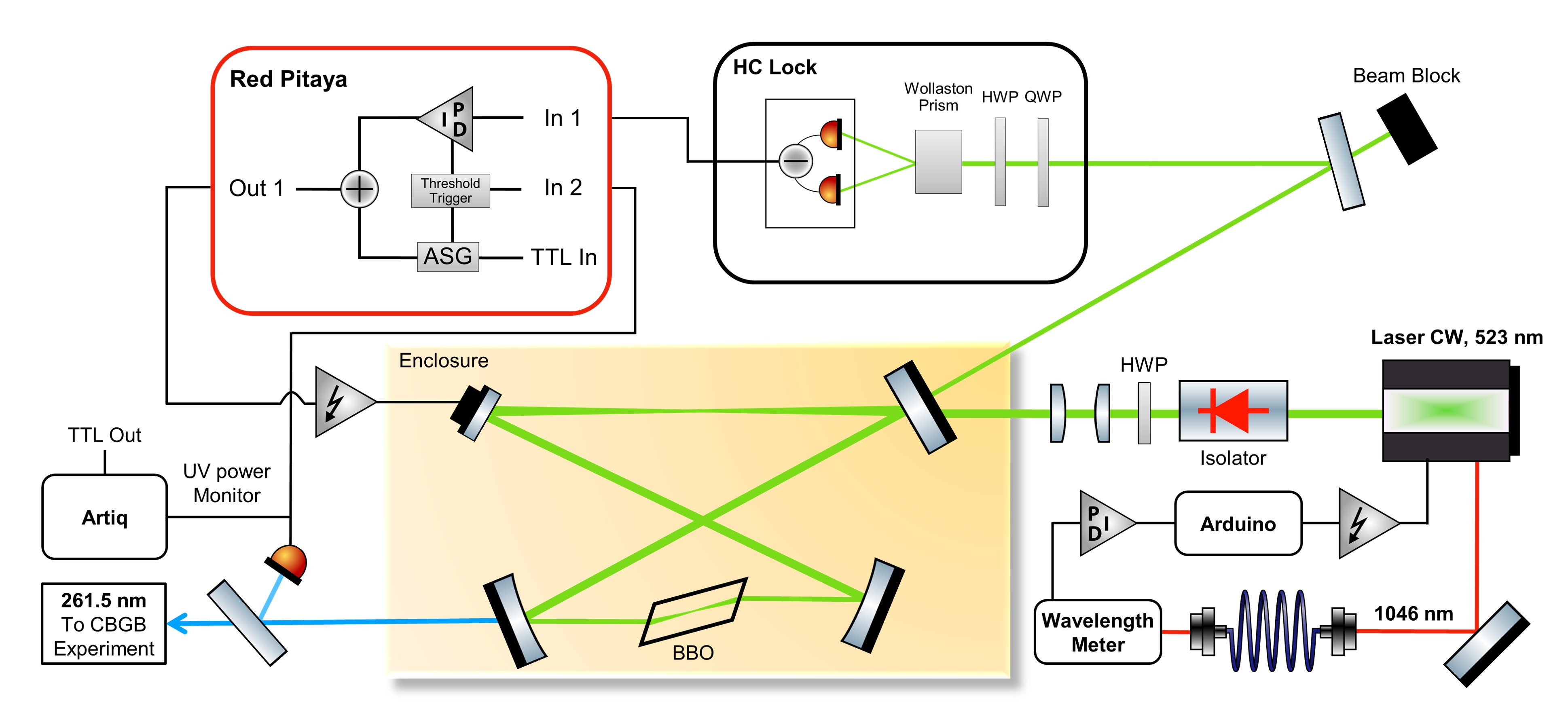
The spectroscopy light at nm is produced by frequency-doubling CW laser light at 523 nm (Vexlum, VALO SHG SF) in a homebuilt second-harmonic generation (SHG) bow-tie cavity [59] with a non-linear beta-barium-borate (BBO) crystal (Newlight Photonics), as shown in Fig. 1. Frequency drifts of the Vexlum laser are compensated by comparing it to a wavelength meter (High Finesse, WS-7). Specifically, a part of the light from a monitoring output of the laser at 1046 nm is continuously measured by the wavelength meter and compared to a set frequency to generate an error signal. This signal is then fed into a software-based proportional-integral controller (PI) to produce a control signal, which is sent to the internal frequency-tuning piezo of the laser via the analog voltage output of a microcontroller (Arduino Due).
The absolute frequency calibration of the wavelength meter is done by regularly comparing a Doppler-free saturated absorption spectrum of rubidium to known literature values [60]. This calibration step uses a separate CW Ti:Saph (Coherent 899-21) laser. The shot-to-shot variations of the measured laser frequency are a convolution of the limited resolution of the wavelength meter (specified to MHz), the intrinsic frequency noise of the laser, the limited bandwidth of the feedback loop due to the software-based PI control and the time it takes to read out the laser frequency ( ms). Overall, these effects lead to a conservative upper limit of the frequency stability of MHz in the UV.
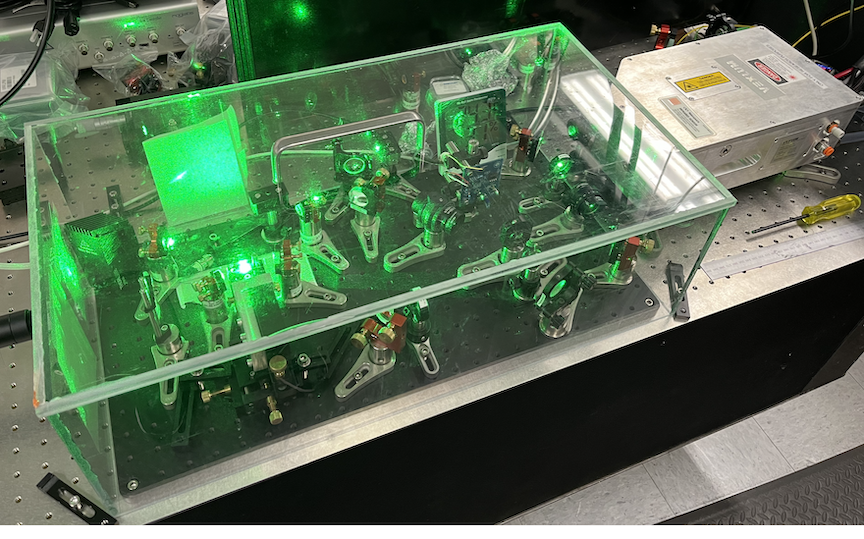
The output of the 523 nm laser is sent through an optical isolator (Thorlabs, IO-5-532-HP) and focused into the SHG cavity using mode-shaping lenses to hit the optimal target beam waist of m inside the crystal, which is determined by the Boyd Kleinman theory [61, 56]. For the SHG process in the BBO at this wavelength, critical type I phase matching at room temperature is achieved by using a crystal that is cut at an angle of deg. The crystal facets are cut at the Brewster angle for the fundamental wavelength ( deg) to minimize reflection losses at each round trip in the cavity. The cavity is enclosed in a box made of acrylic, as shown in Fig. 5, and the box is continuously flushed with compressed air from the building supply lines. To minimize contamination, an air filter that blocks particulates larger than 0.01 µm (Parker, 9933-11-BQ) is installed inline. After flushing the enclosure for min, a humidity level of and a clean environment is achieved around the cavity. The measured finesse of the cavity is approximately and the incoupling efficiency is . For the measurements presented in this work, we typically operated with a few milliwatt UV power. We didn’t optimize these cavity parameters further due to the degradation issues of the crystal. Moreover, the measurements presented here only needed low UV power but required us to still use our resonance-triggered stabilization technique. For future experiments that require higher UV power, we will use a CLBO crystal for the SHG cavity, as described in Section 2.3.
Resonant enhancement of the non-linear conversion process is achieved by length-stabilizing the SHG cavity to the fundamental wavelength using the Hänsch-Couillaud (HC) technique [62]. The error signal created by measuring the difference of the two quadratures in the HC setup is fed into the fast 14-bit ADC input of a field-programmable gate array (FPGA)-based platform (Red Pitaya, STEMLab 125-14, clocked at 125 Msps) for further processing. The 125-14 board has found wide applications in experimental physics and has been used and characterized in various experiments to stabilize cavities and lock lasers [59, 63, 64, 65, 66]. To program the FPGA, we built on and customized the modules from the open source software package Python Red Pitaya Lockbox (PyRPL) [67].
As shown in Fig. 1, the error signal is sent via the ADC input (In 1) to the PI module, which produces a control signal at the fast DAC output (Out 1) of the FPGA board. This signal is then amplified by an HV amplifier (TEM Messtechnik, miniPIA) to drive a piezoactuated cavity mirror. The control signal has added the output of an arbitrary signal generator module (ASG), which is used to apply a voltage ramp to the piezo. The second ADC input (In 2) monitors the UV intensity output of the cavity by reading a photodiode that picks up a small fraction of the output beam. When the UV intensity reaches a predefined threshold, an internal threshold trigger is latched that switches on the PI control module and switches the ASG output to a hold state, maintaining its current voltage output. This step switches off the voltage ramp and switches on the feedback loop for a predefined time interval.
The complete experimental sequence, shown in Fig. 2, is as follows: First, the main laser wavelength is tuned and stabilized to a given set point by the computer. Then, our data acquisition system Artiq [68] sends a TTL trigger (T) to the FPGA board to initiate the voltage ramp (Ramp). The FPGA switches on the cavity stabilization (PID on) as soon as it reaches the resonance condition. After a short delay, which is chosen such that the cavity resonance condition is always met by ramping over at least one free spectral range, Artiq triggers an ablation laser shot (Yag) to produce a beam of molecules. The SHG cavity is then held on resonance for a time interval that is sufficient to let the molecules traverse the experimental apparatus ( ms) and be detected on a photomultiplier (PMT detection). After this time, the PI control module is switched off and the piezo voltage ramp output is set to zero to intentionally tune the cavity out of resonance and eliminate the UV output. This sequence is repeated for a preset number of averages after which the laser wavelength is tuned to a new set point. An example of the voltage ramp (solid blue curve) and the UV output intensity (solid orange curve) of a single experimental cycle is shown in Fig. 2.
2.2 Measurement and Ab Initio Calculations of the Isotope Shift in AlCl
Using the method described in Section 2.1, we carried out detailed spectroscopy on the isotope shift of the -transitions at 261.5 nm in AlCl. Fig. 3 shows the laser-induced fluorescence spectrum of both isotopologues, Al35Cl and Al37Cl, as a function of the laser detuning. The blue dots are the measured data and each point corresponds to 30 averages, and the red solid curve corresponds to a Gaussian multi-peak fit to identify the center of each line. This data was taken using the calibrated wavelength meter, as described above. The difference in the amplitudes of two isotope manifolds comes from the natural abundances of 75.8% for 35Cl and 24.2% for 37Cl. The individual peaks of each manifold are a combination of the -transitions and the hyperfine structure splittings of the involved states that originates from the nuclear spins of Al () and 35/37Cl () [23, 24]. The short lifetime of ns of the electronic state results in a broad natural linewidth ( MHz) that does not resolve the chlorine hyperfine splitting which spreads over MHz [69]. The similarity of the rotational constants of the - and -states [23] results in an overlap of the first few transitions. Therefore, we chose to compare the frequency of the fitted combined peaks, labeled and , to extract an average isotope shift. The result of this comparison is shown in the inset of Fig. 3 for all seven peaks with an average of MHz. This measurement is consistent with the isotope shift extracted from the lower resolution absorption spectroscopy in our previous work, which yields MHz [23] and is the first report of the electronic isotope shift in AlCl to the best of our knowledge.
The theoretical calculations for AlCl are discussed in detail in our prior work[23], so only a brief summary will be given here. Accurate ab initio electronic structure calculations were performed using MOLPRO [70] to compute the potential energy curves for the ground singlet , excited singlet and triplet states. These potential energy curves were then used to perform a numerically exact solution of the one-dimensional diatomic rovibrational Schrödinger equation for the rovibrational wave functions and energies. The relevant transition frequencies for the , and branches for the transition were then calculated. Overall, excellent agreement was obtained between the theoretical and experimental frequencies [23]. In the present work, the rotational structure within the branch is computed for both Al35Cl and Al37Cl for the transition. The isotope shift between the two branches is also calculated enabling a direct comparison with the experimental spectra and isotope shift presented in Fig. 3. Table 1 lists the experimental frequencies for the various features (fitted peaks) labeled by to for Al35Cl and by to for Al37Cl in Fig. 3. The isotope shift for each feature is also tabulated. The corresponding theoretical frequencies for each transition (i.e., , , ) are also listed for each isotopologue as well as the associated isotope shift.
The average theoretical isotope shift is MHz which is within of the experimental value. This level of agreement is considered excellent. The small differences are attributed to the residual numerical errors in the ab initio calculations, potential energy curve fitting errors, and numerical errors in the rovibrational calculations. We note that the and features observed in the experimental spectra are most likely associated with hyperfine structure within the overlapping transitions and is not included in the current level of theory. The theoretical frequencies listed in Table 1 are used to simulate the experimental spectra by adding up the appropriately weighted contributions from each transition with a Lorentzian line-width function [71] centered at each transition frequency. The line widths were chosen to increase linearly with the rotational quantum number via MHz to qualitatively reproduce the experimentally observed widths of the peaks in Fig. 3. The weights of each transition were chosen to match the relative intensities of the experimental spectra for Al35Cl in Fig. 3. The weights for the Al37Cl transitions were set by scaling the Al35Cl weights by the relative isotope abundance ratio: .
Overall, the theoretically simulated spectra presented in Fig. 3 reproduces the experimental one quite well confirming the assignments of the primary overlapping branch features. The theoretical analysis also confirms that the experimentally measured isotope shift is due to an increase in the relative difference between the vibrational zero point energies (ZPE) for the and -states upon isotopic substitution. That is, the vibrational ZPEs for the and states of Al35Cl are and , respectively. Whereas for Al37Cl the ZPEs for the and states are and . The corresponding isotope shifts in the vibrational ZPEs for the and states are therefore and . Thus, we see that the ZPE for the state shifts lower in energy than that for the state and the relative difference increases by (or MHz). The increased relative difference in vibrational ZPE accounts for of the observed isotope shift (). The remaining is due to shifts in the rotational energies due to changes in the rotational constants upon isotopic substitution. We note that the difference in the vibrational ZPE shifts between the and states is due to the difference in the potential energy curves (e.g., force constants). Also, the electronic transition energy is not affected by isotopic substitution and is the same for both isotopologues.
| Experimental | Theoretical | ||||||
|---|---|---|---|---|---|---|---|
| Label | Q(i) | Al35Cl | Al37Cl | Isotope Shift | Al35Cl | Al37Cl | Isotope Shift |
| a,a’ | 1 | -3417 | 2717 | 6134 | -3113 | 2769 | 5882 |
| b,b’ | 2 | -3212 | 2915 | 6127 | -3057 | 2825 | 5882 |
| c,c’ | -3112 | 3001 | 6114 | ||||
| d,d’ | 3 | -2976 | 3144 | 6120 | -2973 | 2909 | 5882 |
| e,e’ | -2807 | 3323 | 6130 | ||||
| f,f’ | 4 | -2632 | 3501 | 6133 | -2861 | 3020 | 5881 |
| g,g’ | 5 | -2383 | 3729 | 6112 | -2721 | 3159 | 5880 |
2.3 Characterization of Long-Term Behaviour and Further Improvements
Using our technique, the measurements in Fig. 3 were conducted over a two-day period during which no degradation of the BBO crystal was observed. The primary reason for the long duration of the experimental run is the low repetition rate of Hz and the high number of averages per data point. The experiment is run at such a slow rate to keep the heat load from the ablation laser on the cryogenic cell at a minimum, as is typically done in CBGB experiments. High-number averaging is necessary to compensate for variations in the molecular yield when the ablation laser rasters over the target.
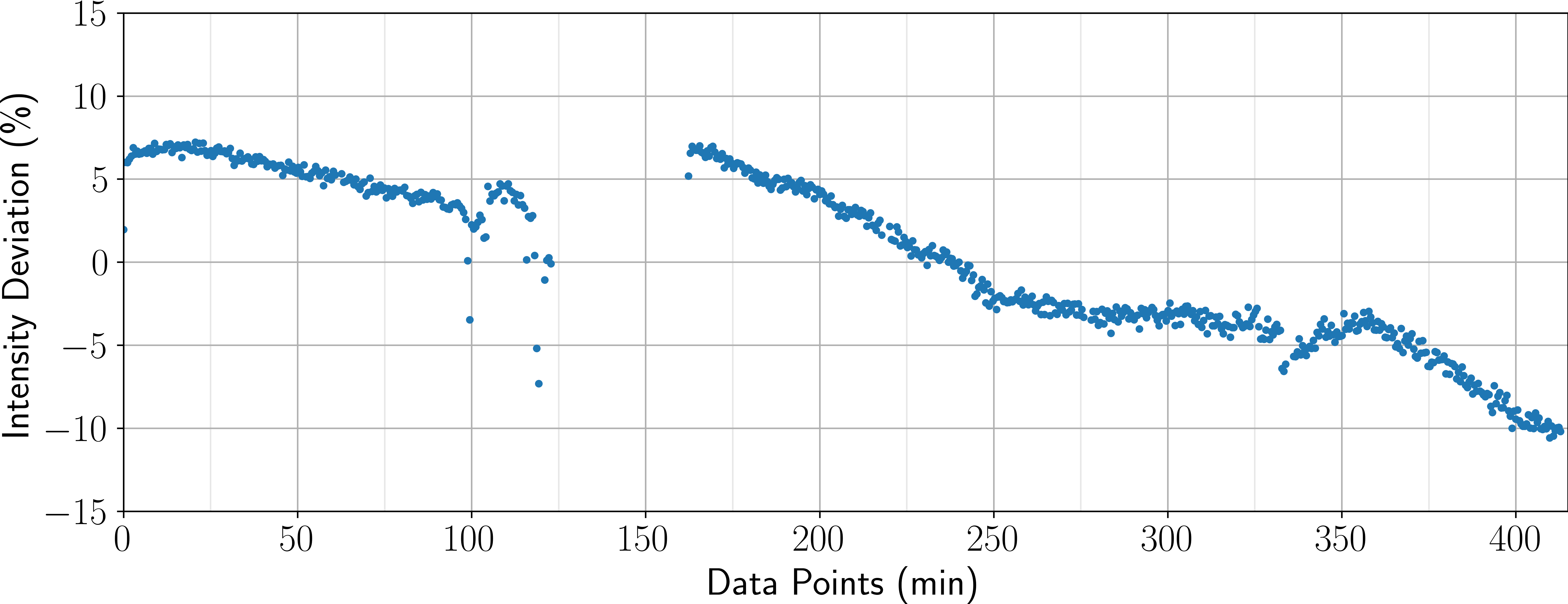
To characterize the long-term behaviour of our method, we monitored the output intensity over a long period of time, as shown in Fig. 4. The average intensity variations between frequency points in the scans were mostly below for at least 6 hours without any cavity alignment optimization. We attribute these fluctuations primarily to set point drifts in the SHG cavity stabilization circuitry. While these fluctuations introduce no systematic frequency offsets in the experiment, improved power stability could be achieved by incorporating an acousto-optic modulator at the output of the cavity for intensity stabilization [72].
We note that the SHG cavity can drift in and out of resonance outside the experimental sequence window when the piezo is held at a constant voltage. This effect leads to unwanted threshold trigger events that switch on the cavity feedback loop. A simple way to avoid this issue is to only arm the threshold trigger at the beginning of the applied voltage ramp. Moreover, if it is desirable to avoid that the cavity drifts into resonance at any time, an easy mitigation technique would be to monitor the output intensity outside the sequence window and use another threshold trigger in combination with an inverted PI control to deliberately push the cavity out of resonance. For the data presented in this work, we didn’t restrict the trigger window and relied on occasional manual adjustments of the offset voltage of the ramp to circumvent the issue since we did not observe any degradation due to the sporadically resonant cavity.
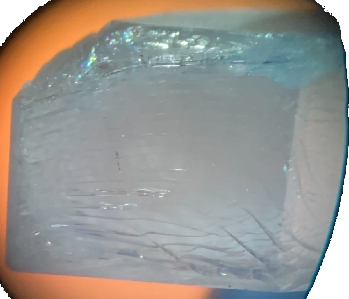
To increase the UV output power, e.g. for laser cooling purposes, in our home built system, we also explored using a CLBO crystal (Conex Optics Inc.) for the SHG conversion. This choice of crystal is motivated by the fact that CLBO has an overall higher conversion efficiency than BBO at our fundamental wavelength of 523 nm, which is mainly attributed to the reduced walk-off angle of CLBO [27, 73]. Overall, the cavity UV output increased by an order of magnitude using the CLBO. However, when we used the cavity in continuous mode, i.e. before the development of the pulsed technique, we observed a severe degradation of the crystal performance over the course of minutes. The cavity output power could be brought back to a maximum by translating the crystal, which in turn moves the focus and the entry and exit points of the fundamental cavity mode to a different spot. Translating the focus back to the original point resulted in the low output power again. This behaviour was observed both with BBO and CLBO crystals, both being at room temperature, and prompted the development of the low-duty cycle technique. In addition, in the case of CLBO, we observed fractures in the crystal that developed over the course of several days of operation, as shown in Fig. 5. We attribute these damages to the high sensitivity of CLBO to the presence of residual moisture and dust in our cavity enclosure, which is not perfectly sealed. The building of cracks in CLBO due to water is a known fact and has been studied previously [74, 75]. However, the operation of the cavity in continuous mode seemingly accelerated this effect. On the other hand, taking advantage of the pulsed technique, we were able to operate the SHG cavity with a new CLBO crystal for several weeks without any notable crystal damage.
Finally, we note that any technique that runs an experimental setup with a reduced duty cycle exhibits pulsed heat loads, resulting in a non-equilibrated state of the setup. In this case of an SHG cavity, the absorption of the light in the crystal leads to what is known as self-heating, which has been observed with both BBO and CLBO crystals, especially in high-power UV systems [32, 11, 9]. As a consequence, the phase matching condition is slightly altered due to different net heat loads between the configuration when the user aligns the cavity setup for optimal output power and when the setup is used for an experimental run. This change leads to a reduction of the conversion efficiency and can be mitigated in different ways. In one approach, users align and optimize the SHG cavities in a configuration that mimics the experimental run in terms of, for instance, repetition rates and used laser powers as close as possible to operate under the same heat loads. In a second approach, users intentionally lower the temperature of their crystals, slightly deviating from the optimal phase matching condition, as has been shown in previous works [11, 9, 29]. Yet another way is to maintain the crystal at an elevated temperature such that the relative temperature increase due to self-heating is small compared to the crystal temperature. The latter approach is best applied to crystals that use angle, instead of temperature, crystal phase-matching, which is the case for BBO and CLBO around 500 nm. Heating the crystal is desirable in any case since the detrimental effects of moisture can be reduced. CLBO, for instance, has been successfully operated without degradation at an elevated temperature of 150 deg C and produced stable Watt-level UV output power, as described in Ref. [27]. We note that a major difference of that experiment is the use of commercial cavities made of stainless steel enclosures, which are well sealed and can readily be kept clean and dry. Nevertheless, especially at such high UV output powers, most optics to guide or manipulate the laser light after the cavity is susceptible to UV-induced damage. Our pulsed approach will minimize the impact of the UV light and prolong the lifetime of the SHG cavity and the following optics as well.
3 Conclusion
In this work, we have developed and tested a technique to use a deep-UV SHG laser system while reducing UV-induced damage on optics to a bare minimum. Our approach to reduce the duty cycle of the used UV light is ideal for any pulsed experiments but can also be applied in CW experiments to bridge, often unavoidable, dead times and reduce the UV operation time as much as possible. The frequency accuracy of the laser system should not be affected when using this method since the ramping of the piezo-actuated mirror only affects the output intensity and the frequency stability remains with the fundamental laser. The long-term stability of our setup is sufficient for typical time scales of atomic and molecular experiments. While our technique is particularly well-suited for protecting non-linear crystals in resonant-enhancement cavities where the circulating intensity of the fundamental laser light is large, any subsequent optical components in the experiment, such as mirrors and optical fibers, experience less degradation as well due to the reduced average power of the used UV light. We applied this technique to measure the isotope shift of the electronic - transition in AlCl and found excellent agreement with ab initio calculations.
Funding L. L. and B. H. acknowledge funding from the National Science Foundation under Grant No. 2145147. This material is based upon work supported by the Air Force Office of Scientific Research under award number FA9550-21-1-0263. B. K. K. acknowledges that part of this work was done under the auspices of the U.S. Department of Energy under Project No. 20240256ER of the Laboratory Directed Research and Development Program at Los Alamos National Laboratory. Los Alamos National Laboratory is operated by Triad National Security, LLC, for the National Nuclear Security Administration of the U.S. Department of Energy (Contract No. 89233218CNA000001).
Acknowledgments We would like to thank Grady Kestler and Julio T. Barreiro for helpful discussions on the FPGA implementation and the PyRPL code. We would also like to thank Daniel J. McCarron for useful discussions and feedback on this manuscript.
Disclosures The authors declare no conflicts of interest.
Data availability Data underlying the results presented in this paper are available upon request.
References
- [1] F. Duarte, Tunable Laser Applications, ISSN (CRC Press, 2016).
- [2] M. Safronova, D. Budker, D. DeMille, et al., “Search for new physics with atoms and molecules,” \JournalTitleReviews of Modern Physics 90, 025008 (2018).
- [3] M. H. J and van der Straten P, Laser Cooling and Trapping (New York: Springer, 1999).
- [4] D. McCarron, “Laser cooling and trapping molecules,” \JournalTitleJournal of Physics B: Atomic, Molecular and Optical Physics 51, 212001 (2018).
- [5] R. V. Krems, “Cold controlled chemistry,” \JournalTitlePhysical Chemistry Chemical Physics 10, 4079 (2008).
- [6] L. D. Carr, D. DeMille, R. V. Krems, and J. Ye, “Cold and ultracold molecules: science, technology and applications,” \JournalTitleNew Journal of Physics 11, 055049 (2009).
- [7] R. V. Krems, B. Friedrich, and W. C. Stwalley, Cold molecules: theory, experiment, applications. (CRC PRESS, 2019).
- [8] K.-A. Brickman, M.-S. Chang, M. Acton, et al., “Magneto-optical trapping of cadmium,” \JournalTitlePhysical Review A 76, 043411 (2007).
- [9] Y. Kaneda, J. M. Yarborough, Y. Merzlyak, et al., “Continuous-wave, single-frequency 229 nm laser source for laser cooling of cadmium atoms,” \JournalTitleOptics Letters 41, 705 (2016).
- [10] P. Villwock, S. Siol, and T. Walther, “Magneto-optical trapping of neutral mercury,” \JournalTitleThe European Physical Journal D 65, 251–255 (2011).
- [11] Y. Zhang, Q. Liu, X. Fu, et al., “A stable deep-ultraviolet laser for laser cooling of mercury atoms,” \JournalTitleOptics & Laser Technology 139, 106956 (2021).
- [12] Z. Zhang, M. Kushimoto, T. Sakai, et al., “A 271.8 nm deep-ultraviolet laser diode for room temperature operation,” \JournalTitleApplied Physics Express 12, 124003 (2019).
- [13] H. Amano, R. Collazo, C. D. Santi, et al., “The 2020 uv emitter roadmap,” \JournalTitleJournal of Physics D: Applied Physics 53, 503001 (2020).
- [14] C. G. Parthey, A. Matveev, J. Alnis, et al., “Improved Measurement of the Hydrogen 1s–2s Transition Frequency,” \JournalTitlePhysical Review Letters 107, 203001 (2011).
- [15] M. Ahmadi, B. X. R. Alves, C. J. Baker, et al., “Characterization of the 1S–2S transition in antihydrogen,” \JournalTitleNature 557, 71–75 (2018).
- [16] H. Fleurbaey, S. Galtier, S. Thomas, et al., “New Measurement of the 1s-3s Transition Frequency of Hydrogen: Contribution to the Proton Charge Radius Puzzle,” \JournalTitlePhysical Review Letters 120, 183001 (2018).
- [17] A. Grinin, A. Matveev, D. C. Yost, et al., “Two-photon frequency comb spectroscopy of atomic hydrogen,” \JournalTitleScience 370, 1061–1066 (2020).
- [18] P. Crivelli, “The Mu-MASS (muonium laser spectroscopy) experiment,” \JournalTitleHyperfine Interactions 239, 49 (2018).
- [19] E. Altiere, E. R. Miller, T. Hayamizu, et al., “High-resolution two-photon spectroscopy of a transition of xenon,” \JournalTitlePhysical Review A 97, 012507 (2018).
- [20] M. D. D. Rosa, “Laser-cooling molecules,” \JournalTitleThe European Physical Journal D 31, 395–402 (2004).
- [21] M. R. Tarbutt, “Laser cooling of molecules,” \JournalTitleContemporary Physics 59, 356–376 (2018).
- [22] E. Chae, “Laser cooling of molecules,” \JournalTitleJournal of the Korean Physical Society 82, 851–863 (2023).
- [23] J. R. Daniel, C. Wang, K. Rodriguez, et al., “Spectroscopy on the x – a transition of buffer-gas cooled alcl,” \JournalTitlePhysical Review A 104, 012801 (2021).
- [24] J. R. Daniel, J. C. Shaw, C. Wang, et al., “Hyperfine structure of the a state of alcl and its relevance to laser cooling and trapping,” \JournalTitlePhysical Review A 108, 062821 (2023).
- [25] S. Hofsäss, M. Doppelbauer, S. C. Wright, et al., “Optical cycling of AlF molecules,” \JournalTitleNew Journal of Physics 23, 075001 (2021).
- [26] J. Mes, E. J. van Duijn, R. Zinkstok, et al., “Third-harmonic generation of a continuous-wave ti:sapphire laser in external resonant cavities,” \JournalTitleApplied Physics Letters 82, 4423–4425 (2003).
- [27] J. C. Shaw, S. Hannig, and D. J. McCarron, “Stable 2 w continuous-wave 261.5 nm laser for cooling and trapping aluminum monochloride,” \JournalTitleOptics Express 29, 37140–37149 (2021).
- [28] N. Zhadnov, A. Golovizin, I. Cortinovis, et al., “Pulsed CW laser for long-term spectroscopic measurements at high power in deep-UV,” \JournalTitleOptics Express 31, 28470–28479 (2023).
- [29] Z. Burkley, A. D. Brandt, C. Rasor, et al., “Highly coherent, watt-level deep-uv radiation via a frequency-quadrupled yb-fiber laser system,” \JournalTitleApplied Optics pp. 1657–1661 (2019).
- [30] J. Hu, L. Zhang, H. Liu, et al., “High power room temperature 1014.8 nm Yb fiber amplifier and frequency quadrupling to 253.7 nm for laser cooling of mercury atoms,” \JournalTitleOptics Express 21, 30958–30963 (2013).
- [31] S. Sayama and M. Ohtsu, “Tunable uv cw generation by frequency tripling of a ti:sapphire laser,” \JournalTitleOptics Communications 137, 295–298 (1997).
- [32] S. F. Cooper, Z. Burkley, A. D. Brandt, et al., “Cavity-enhanced deep ultraviolet laser for two-photon cooling of atomic hydrogen,” \JournalTitleOptics Letters 43, 1375 (2018).
- [33] S. Imai, H. Inoue, T. Nomura, and T. Tojo, “Cw 198.5-nm light generation in clbo,” \JournalTitleAdvanced Solid-State Photonics (2003).
- [34] Y. Asakawa, J. Sakuma, H. Sekita, and M. Obara, “High-power cw duv coherent light source around 200 nm,” \JournalTitleAdvanced Solid-State Photonics (TOPS) (2004).
- [35] S. Huang, T. Ando, Y. Orii, and T. Sumiyoshi, “A cw 266 nm coherent light source pumped by the shg beam of the single frequency fiber amplifier radiation,” \JournalTitleAdvanced Solid-State Photonics (2008).
- [36] M. Oka, L. Liu, W. Wiechmann, et al., “1 w continuous-wave 266 nm radiation from an all solid-state frequency quadrupled nd:yag laser,” \JournalTitleAdvanced Solid State Lasers (2004).
- [37] H. Masuda, K. Kimura, N. Eguchi, et al., “All-solid-state, continuous-wave, 195 nm light generation in -bab2o4,” \JournalTitleAdvanced Solid-State Lasers (2001).
- [38] M. Bauer, M. Bischoff, S. Jukresch, et al., “Exterior surface damage of calcium fluoride outcoupling mirrors for duv lasers,” \JournalTitleOptics Express 17, 8253 (2009).
- [39] M. Bauer, M. Bischoff, T. Hülsenbusch, et al., “Onset of the optical damage in caf optics caused by deep-uv lasers,” \JournalTitleOptics Letters 34, 3815 (2009).
- [40] R. Schenker, P. Schermerhorn, and W. G. Oldham, “Deep-ultraviolet damage to fused silica,” \JournalTitleJournal of Vacuum Science & Technology B: Microelectronics and Nanometer Structures Processing, Measurement, and Phenomena 12, 3275–3279 (1994).
- [41] R. E. Schenker, L. Eichner, H. Vaidya, et al., “Ultraviolet damage properties of various fused silica materials,” \JournalTitleLaser-Induced Damage in Optical Materials: 1994 2428, 458–468 (1995).
- [42] R. A. Negres, M. A. Norton, D. A. Cross, and C. W. Carr, “Growth behavior of laser-induced damage on fused silica optics under UV, ns laser irradiation,” \JournalTitleOptics Express 18, 19966–19976 (2010).
- [43] R. R. Kunz, V. Liberman, and D. K. Downs, “Experimentation and modeling of organic photocontamination on lithographic optics,” \JournalTitleJournal of Vacuum Science & Technology B: Microelectronics and Nanometer Structures Processing, Measurement, and Phenomena 18, 1306–1313 (2000).
- [44] S. Heinbuch, F. Dong, J. J. Rocca, and E. R. Bernstein, “Gas-phase study of the reactivity of optical coating materials with hydrocarbons by use of a desktop-size extreme-ultraviolet laser,” \JournalTitleJournal of the Optical Society of America B pp. B85–B91 (2008).
- [45] D. Gangloff, M. Shi, T. Wu, et al., “Preventing and reversing vacuum-induced optical losses in high-finesse tantalum (V) oxide mirror coatings,” \JournalTitleOptics Express 23, 18014–18028 (2015).
- [46] Z. Burkley, L. d. S. Borges, B. Ohayon, et al., “Stable high power deep-uv enhancement cavity in ultra-high vacuum with fluoride coatings,” \JournalTitleOptics Express 29, 27450–27459 (2021).
- [47] M. Watanabe, K. Hayasaka, H. Imajo, et al., “Generation of continuous-wave coherent radiation tunable down to 190.8nm in -BaB2O4,” \JournalTitleApplied Physics B 53, 11–13 (1991).
- [48] M. Takahashi, A. Osada, A. Dergachev, et al., “Effects of Pulse Rate and Temperature on Nonlinear Absorption of Pulsed 262-nm Laser Light in -BaB2O4,” \JournalTitleJapanese Journal of Applied Physics 49, 080211 (2010).
- [49] H. Turcicova, O. Novak, J. Muzik, et al., “Laser induced damage threshold (LIDT) of -barium borate (BBO) and cesium lithium borate (CLBO) – Overview,” \JournalTitleOptics & Laser Technology 149, 107876 (2022).
- [50] M. Nishioka, A. Kanoh, M. Yoshimura, et al., “Improvement in UV Optical Properties of CsLiB6O10 by Reducing Water Molecules in the Crystal,” \JournalTitleJapanese Journal of Applied Physics 44, L699 (2005).
- [51] T. Kawamura, M. Yoshimura, Y. Honda, et al., “Effect of water impurity in cslib6i10 crystals on bulk laser-induced damage threshold and transmittance in the ultraviolet region,” \JournalTitleApplied Optics pp. 1658–1662 (2009).
- [52] K. Takachiho, M. Yoshimura, Y. Takahashi, et al., “Ultraviolet laser-induced degradation of CsLiBO and -BaBO,” \JournalTitleOptical Materials Express 4, 559–567 (2014).
- [53] M. Yoshimura, Y. Oeki, Y. Takahashi, et al., “Ultraviolet laser-induced degradation of cslib6o10,” \JournalTitleAdvanced Solid State Lasers p. AM5A.11 (2015).
- [54] Y. Colombe, D. H. Slichter, A. C. Wilson, et al., “Single-mode optical fiber for high-power, low-loss uv transmission,” \JournalTitleOptics Express 22, 19783–19793 (2014).
- [55] B. Hemmerling, F. Gebert, Y. Wan, et al., “A single laser system for ground-state cooling of 25mg+,” \JournalTitleApplied Physics B 104, 583–590 (2011).
- [56] G. D. Boyd and D. A. Kleinman, “Parametric interaction of focused gaussian light beams,” \JournalTitleJournal of Applied Physics 39, 3597–3639 (2021).
- [57] N. R. Hutzler, H.-I. I. Lu, and J. M. Doyle, “The buffer gas beam: An intense, cold, and slow source for atoms and molecules,” \JournalTitleChemical Reviews 112, 4803–4827 (2012).
- [58] T. N. Lewis, C. Wang, J. R. Daniel, et al., “Optimizing pulsed-laser ablation production of alcl molecules for laser cooling,” \JournalTitlePhysical Chemistry Chemical Physics 23, 22785 (2021).
- [59] S. Hannig, J. Mielke, J. A. Fenske, et al., “A highly stable monolithic enhancement cavity for second harmonic generation in the ultraviolet,” \JournalTitleReview of Scientific Instruments 89, 013106 (2018).
- [60] D. A. Steck, “Rubidium 85 d line data,” \JournalTitlehttp://steck.us/alkalidata (2021).
- [61] D. A. Kleinman, A. Ashkin, and G. D. Boyd, “Second-harmonic generation of light by focused laser beams,” \JournalTitlePhysical Review 145, 338–379 (2021).
- [62] T. W. Hansch and B. Couillaud, “Laser frequency stabilization by polarization spectroscopy of a reflecting reference cavity,” \JournalTitleOptics Communications 35, 441–444 (2021).
- [63] T. Preuschoff, M. Schlosser, and G. Birkl, “Digital laser frequency and intensity stabilization based on the STEMlab platform (originally Red Pitaya),” \JournalTitleReview of Scientific Instruments 91, 083001 (2020).
- [64] E. Pultinevicius, M. Rockenhäuser, F. Kogel, et al., “A scalable scanning transfer cavity laser stabilization scheme based on the Red Pitaya STEMlab platform,” \JournalTitleArXiv (2023). ArXiv:2307.10217 [physics] type: article.
- [65] P. Lauria, W.-T. Kuo, N. R. Cooper, and J. T. Barreiro, “Experimental Realization of a Fermionic Spin-Momentum Lattice,” \JournalTitlePhysical Review Letters 128, 245301 (2022).
- [66] G. Kestler, K. Ton, D. Filin, et al., “State-Insensitive Trapping of Alkaline-Earth Atoms in a Nanofiber-Based Optical Dipole Trap,” \JournalTitlePRX Quantum 4, 040308 (2023).
- [67] L. Neuhaus, M. Croquette, R. Metzdorff, et al., “Python red pitaya lockbox (pyrpl): An open source software package for digital feedback control in quantum optics experiments,” \JournalTitleReview of Scientific Instruments 95, 033003 (2024).
- [68] S. Bourdeauducq, R. Jördens, P. Zotov, et al., “Artiq,” \JournalTitleZenodo (2021).
- [69] K. D. Hensel, C. Styger, W. Jäger, et al., “Microwave spectra of metal chlorides produced using laser ablation,” \JournalTitleThe Journal of Chemical Physics 99, 3320–3328 (1993).
- [70] H.-J. Werner, P. J. Knowles, G. Knizia, et al., “MOLPRO, version 2015.1, a package of ab initio programs,” Http://www.molpro.net.
- [71] P. F. Bernath, Spectra of Atoms and Molecules (Oxford University Press, 2005).
- [72] F. Tricot, D. H. Phung, M. Lours, et al., “Power stabilization of a diode laser with an acousto-optic modulator,” \JournalTitleReview of Scientific Instruments 89, 113112 (2018).
- [73] W. L. Zhou, Y. Mori, T. Sasaki, and S. Nakai, “High-efficiency intracavity continuous-wave ultraviolet generation using crystals CsLiB6O10, -BaB2O4 and LiB3O5,” \JournalTitleOptics Communications 123, 583–586 (1996).
- [74] Y. V. Seryotkin, E. A. Fomina, and L. I. Isaenko, “Humidity effect on hydration of CsLiB6O10 nonlinear optical crystal: X-ray diffraction study,” \JournalTitleOptical Materials 35, 1646–1651 (2013).
- [75] F. Pan, X. Wang, G. Shen, and D. Shen, “Cracking mechanism in CLBO crystals at room temperature,” \JournalTitleJournal of Crystal Growth 241, 129–134 (2002).