Uncertainty-Aware PPG-2-ECG for Enhanced Cardiovascular Diagnosis using Diffusion Models
Abstract
Analyzing the cardiovascular system condition via Electrocardiography (ECG) is a common and highly effective approach, and it has been practiced and perfected over many decades. ECG sensing is non-invasive and relatively easy to acquire, and yet it is still cumbersome for holter monitoring tests that may span over hours and even days. A possible alternative in this context is Photoplethysmography (PPG): An optically-based signal that measures blood volume fluctuations, as typically sensed by conventional “wearable devices”. While PPG presents clear advantages in acquisition, convenience, and cost-effectiveness, ECG provides more comprehensive information, allowing for a more precise detection of heart conditions. This implies that a conversion from PPG to ECG, as recently discussed in the literature, inherently involves an unavoidable level of uncertainty. In this paper we introduce a novel methodology for addressing the PPG-2-ECG conversion, and offer an enhanced classification of cardiovascular conditions using the given PPG, all while taking into account the uncertainties arising from the conversion process. We provide a mathematical justification for our proposed computational approach, and present empirical studies demonstrating its superior performance compared to state-of-the-art baseline methods.
1 Introduction
Cardiovascular diseases are a significant public health problem, affecting millions of people worldwide and remaining a leading cause of mortality [1]. The analysis of cardiovascular conditions using Electrocardiography (ECG) signals has emerged as one of the most widely utilized diagnostic tool [2], and its practice and efficiency have been continuously refined over many decades.
While ECG sensing is non-invasive and relatively easy to acquire, it is time-consuming and requires the expertise of trained professionals with specialized skills in order to ensure accurate diagnosis. Consequently, there has been a surge in the development of numerous contemporary wearable ECG systems in recent decades for digital diagnosis. However, the materials used to deliver a high-quality signal via electrodes can often lead to skin irritation and discomfort during extended usage, thereby limiting the long-term viability of these devices [3].
A possible alternative to ECG sensing and diagnosis is the use of Photoplethysmography (PPG), which is an optically-based non-invasive signal associated with rhythmic changes in blood volume within tissues [4]. Unlike ECG, PPG signals are easier to obtain, convenient, and cost-effective, as they are widely available in clinics and hospitals and can be sensed through finger/toe clips. Moreover, the popularity of PPG is increasing with the emergence of wearable devices like smart watches, enabling continuous long-term monitoring without causing skin irritations.
Naturally, the PPG and ECG signals are inter-related, as the timing, amplitude, and shape characteristics of the PPG waveform contain information about the interaction between the heart and the blood vessels. These features of the PPG have been leveraged to measure heart rate, heart rate variability, respiration rate [5], blood oxygen saturation [6], blood pressure [7], and to assess vascular function [8, 9]. Despite these capabilities, using PPG-based monitoring during daily activities and light physical exercises poses unavoidable uncertainty challenges due to signal inaccuracies and inherent noise, stemming from their indirect nature, compared to ECG signals sourced directly from the heart.
As the use of wearable devices capturing PPG signals is becoming widespread, there is an emerging interest in accurately converting PPG sources into ECG signals [10, 11, 12, 13, 14, 15, 16, 17, 18, 19, 20, 21, 22]. However, such a mapping can be regarded as an ill-posed inverse problem. This implies that multiple ECG solutions may correspond to the same input PPG signal. Nevertheless, previous work in this domain has tended to focus on providing a single ECG solution to the conversion task, ignoring the inherent uncertainty in the conversion and thus failing to capture the complete information contained in the PPG signal. Such studies suggest optimizing neural models either via regularized regression with an / loss [11, 10, 12, 15, 16, 17, 19, 20, 21] or relying on a generative approach that produces a single solution that approximates a sample from the posterior distribution of ECG given the PPG signal [13, 14, 18, 22]. Here, we argue that both these strategies are lacking, and the conversion in question calls for a different approach.
In this paper, we introduce Uncertainty-Aware PPG-2-ECG (UA-P2E), a novel PPG-2-ECG conversion methodology, aimed at enhancing the classification of cardiovascular conditions using ECG signals derived from PPG data. Unlike prior work, our approach aims to address both the uncertainty arising from the conversion task, regarding the spread and variability of the possible ECG solutions given the PPG [23, 24], and also the uncertainty in the classification task, enabling the selection of PPG samples with low classification risk and high confidence [25].
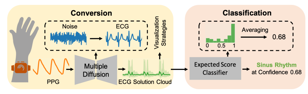
To achieve the above, we leverage a conditional diffusion-based methodology [26, 27, 28]. These generative models have recently emerged as the leading synthesis approach in various tasks [29, 30], and have been shown to enable high-fidelity posterior modeling for inverse problems [30, 31, 32, 33, 34]. We utilize this capability to produce a multitude of posterior sampled ECG results that correspond to the input PPG, and provide classification decisions based on this cluster of outcomes. Additionally, we introduce several methods for displaying the resulting ECG candidates for better human interpretability. Figure 1 provides an illustration of our approach to the conversion-classification process.
Our computational methodology is grounded on a mathematical proof showcasing the optimality of the expected classification score derived from our proposed approach for classifying cardiovascular conditions from source PPG signals. This optimality relies on the assumption that we have access to a perfect posterior sampler and ECG classification models. This stands in contrast to the single-solution approach used by prior work, as described above, which necessarily performs worse.
Our work presents a thorough empirical study demonstrating superior performance in classification and uncertainty estimation compared to baseline methods. Our main results are demonstrated on the “Computing in Cardiology” (CinC) dataset [35], which provides the most challenging classification data in cardiology.
In summary, our contributions are the following: (1) We introduce a state-of-the-art diffusion-based methodology for PPG-2-ECG conversion-classification that accounts for the uncertainty in the conversion task, thereby enhancing performance; (2) We provide a mathematical proof that justifies our proposed computational process for the classification task; (3) We offer effective methods for displaying the ECG solutions obtained, enhancing interpretability while taking into account the uncertainty perspective; and (4) We present an empirical study that supports our mathematical proof and demonstrates the superiority of our methodology over baseline strategies.
2 Related Work
Naturally, our work is not the first one to consider the conversion of PPG signals to ECG ones. Below we outline the main trends in this line of work, and add a discussion on uncertainty quantification, as we aim to practice in this paper.
Regression: Earlier approaches to the PPG-2-ECG conversion problem primarily focused on approximating either the Minimum Mean Absolute Error (MMAE) estimator using loss [11, 10, 16, 19] or the Minimum Mean Squared Error (MMSE) estimator with loss [12, 15, 17, 20, 21]. These works commonly employed regularization techniques to enhance perceptual outcomes. However, the work reported in [36] which discovered the distortion-perception tradeoff has shown that when optimizing for distortion (of any kind), perceptual quality is necessarily compromised, thus pushing the solutions in the above methods to signals that are likely to drift out of the valid ECG manifold, thus weakening their classification performance.
Generation: The first attempt at PPG-2-ECG conversion using generative models was introduced by the authors of CardioGAN [13]. They employed an adversarial training scheme with an encoder-decoder architecture. Similarly, P2E-WGAN [14] utilized conditional Wasserstein GANs for this task. [18] introduced a variational inference methodology to approximate the posterior distribution with a Gaussian prior assumption. The authors of RDDM [22] have first adapted a diffusion-based model that accelerates a DDPM [27] diffusion process for this conversion task. However, despite the success of the generative approach, all the above works have chosen to use a single (possibly random) ECG solution from the posterior distribution, thus missing the complete picture of the possibilities of the different ECGs given the PPG signal.
Uncertainty Quantification: In the framework of the conversion-classification explored in this paper, uncertainty quantification serves a dual purpose. Firstly, it seeks to characterize the range, spread, and variability of potential solutions for a given input PPG signal [23, 24]. Secondly, it also aims at assessing the confidence in the correctness of classification predictions, e.g. via a selective classification [25] scheme, which we adopt in this paper. Building upon principles from these two threads of work, we characterize the diverse solutions that can potentially describe the input, leading to an improved methodology within the conversion-classification framework.
3 Problem Formulation and Goals
Let be a random vector representing an ECG signal that follows the prior distribution . We assume access only to its observation , where is an unknown, possibly stochastic, and non-invertible function. The observation represents a PPG signal that follows the distribution . In this paper, our first and primary objective is to provide a comprehensive and interpretable estimation of the ECG signal(s) that corresponds to the given , focusing on the approximation of the posterior distribution . Prior work in this field (see Section 2) adopted a single-solution approach for the conversion task, focusing on estimating a single reconstructed ECG solution from , typically achieved through regression or generation models.
Here, we propose a generalization of this conversion approach using a conditional stochastic generative model, denoted as s.t. , where denotes a stochastic component that typically follows a normal distribution , and represents a potential ECG solution derived as a sample from the approximate posterior distribution . In this context, a single ECG solution may be derived by fixing in , or by marginalizing over the random seed, i.e., . In contrast, allowing to be random may provide a more comprehensive set of solutions to the inverse problem, exposing the inherent uncertainty within this conversion task. Our prime goal in this work is therefore to train the stochastic conversion model using a training data consisting of many (ECG, PPG) signal pairs, denoted by .
A second objective in this work is classification, enabling diagnosis of various cardiovascular conditions via the incoming PPG signals. Consider a binary classification problem where the input is an observation with a corresponding binary label that follows a conditional distribution . In this classification setting, we aim to model a classifier for approximating the posterior distribution, denoted as .
Addressing this classification task, one might be tempted to adopt a straightforward and naive strategy, of training directly on data that consists of PPG signals and their associated labels. Nevertheless, appropriate cardiovascular labels linked with PPG signals are relatively rare and hard to acquire. An alternative is to use labels that were produced for ECG signals, , and tie these to their matched PPG, . However, as the mapping between PPG and ECG is not injective, this implies that other ECG signals (possibly with a different label) could have led to the same PPG. As a consequence, these labels are necessarily noisy, impairing the learning task.
A presumably better alternative is to operate in the reconstructed ECG domain, hoping for improved performance. Indeed, the classification proposed in prior work operates on the single estimated ECG , approximating this way the posterior . This is achieved using a classifier , trained over data of the form , where represents a grounded ECG signal and denotes its associating label. As we rely now on labels that do match their corresponding signals, this may seem like a better approach. However, the same flaw as above applies here – the single-produced ECG, , captures a partial truth about the given PPG, thus weakening the classification. Here is a formal definition of this classification strategy, as practiced in prior work:
Definition 3.1 (Single Score Classifier).
We define a stochastic classifier as a Single Score Classifier (SSC) if it is given by
When choosing , the term becomes the MMSE estimate of the ECG, and when for , we get a single sample from the approximate posterior distribution, .
Following the data processing inequality [37], any transformation satisfies the inequality
| (1) |
where denotes the mutual information between the random variables and . The above inequality implies that the classification information contained within a single reconstructed ECG signal , as utilized by prior work, is always weakly lower than the classification information contained within the original PPG signal . This suggests that despite the transformation from PPG to ECG, there may be some loss or reduction in the discriminative power for classification tasks due to information processing constraints.
To address this challenge, we adopt a multi-solution formulation approach that takes into account all possible ECG solutions that can be derived from the PPG signal . Below is the formal definition of our classification strategy.
Definition 3.2 (Expected Score Classifier).
We define a classifier as an Expected Score Classifier (ESC) if it is given by
Intuitively, this strategy averages the classification scores of posterior samples that emerge from the posterior distribution , rather than averaging the samples themselves. In Appendix A we prove Theorem A.1, implying that our approach utilizes the full extent of the information within the observation , and is therefore optimal.
4 UA-P2E: Uncertainty-Aware PPG-2-ECG
We now turn to introduce Uncertainty-Aware PPG-2-ECG (UA-P2E), our methodology for PPG-2-ECG conversion, aiming to provide a comprehensive understanding of the various ECG solutions derived from a given PPG signal. UA-P2E may assist medical professionals in analyzing diverse ECG signal variations resulting from the conversion process. Additionally, our method can be leveraged to enhance cardiovascular diagnostic accuracy and confidence assessments, facilitating more informed clinical decisions.
While our proposed approach is applicable using any conditional stochastic generative estimator, denoted as , we focus in this work on conditional diffusion-based models [26, 27, 28], which have emerged as the leading synthesis approach in various domains [29, 30]. Our diffusion process is trained and evaluated on raw signal data of fixed-length PPG and ECG signal pairs, sampled at Hz, thus covering seconds.111in our experiments and thus the signals cover seconds. The central part of the diffusion process involves a conditional denoiser, denoted as . This denoiser is trained to predict the noise in , which is a combination of (representing a clean instance) and white additive Gaussian noise at a noise level denoted by . In this conditional process, the denoiser leverages the PPG signal and the noise level as supplementary information. Training is based on pairs of signals - a clean ECG and its corresponding PPG. After training, we apply multiple diffusion operations on a given PPG signal to draw potential ECG solutions , where each is a sample from the approximated posterior distribution of the ECG given the PPG signal.
In Section 4.1 we describe our enhanced classification procedure that relies on the above posterior sampler, denoted by . Additionally, in Section 4.2 we discuss how to improve the quantification of uncertainty in classification, aiming to enhance confidence in predictions. Lastly, in Section 4.3, we offer methods to visualize the cloud of ECG solutions . This visualization aims at enhancing interpretability for medical professionals, allowing for a better understanding of the various ECG signals that emerge from a given PPG signal.
4.1 UA-P2E: Enhanced Classification
The methodology for UA-P2E’s enhanced classification consists of three phases. The first involves training the probabilistic classification model, denoted as , using ECG signals and their associated labels. can be optimized through the commonly used Cross-Entropy loss function, producing an output that has a probabilistic interpretation. In the second phase, each potential posterior sample from the set (derived by applying to a given ) is evaluated by this classifier, extracting an output score , being an estimated probability of belonging to the positive class (). In the final phase, the average classification score, , is computed, and compared to a decision threshold . The complete workflow of our proposed method to enhance classification is detailed in Algorithm 1.
4.2 UA-P2E: Uncertainty Quantification
The proposed PPG-2-ECG conversion has two layers of uncertainty awareness, as described herein. The first takes into account the inherent uncertainty that emerges from the ill-posed conversion itself, quantifying the spread and variability of the possible ECG candidates that derive from the PPG inputs. In Appendix C, we present empirical evidence illustrating the uncertainty from this perspective. The second uncertainty layer leverages the above property for providing classification predictions with a better confidence in their correctness. This serves a selective classification [25] scheme, in which a carefully chosen subset of the test PPG data is selected by their calculated confidence, exhibiting improved accuracy.
Selective classification [25] enables handling cases in which classifying every PPG signal within a test set is challenging due to uncertainty or data corruption. This technique allows for abstaining from making predictions for signals with low confidence, thus reducing classification errors on the remaining data. A confidence function, defined as , serves as the engine for this task by predicting the correctness of predictions. One common choice of this confidence function is to use the maximal softmax score of a classifier. In a similar spirit, in this work we set to take the value , where approximates the ESC (Definition 3.2), as summarized in Algorithm 1.
In addition to the above, we may consider applying a calibration scheme on a heldout set, denoted as , to determine a threshold for that defines the reliable test PPG signals whose classification error is statistically guaranteed to be lower than a user-defined risk level . Such a guarantee has the following form:
| (2) |
where is a user-defined calibration error. Intuitively, by setting , the above implies that the classification error among selected PPG signals, whose confidence scores are greater than , will be lower than () with probability of (). In Appendix B, we describe how to calibrate to provide the guarantee stated in Equation (2).
4.3 Visualizing UA-P2E’s ECG Output(s)
A primary objective of UA-P2E is the PPG-2-ECG conversion, and a natural question to pose is what ECG signal to present to medical professionals after such a conversion. Due to the stochastic nature of our estimation approach, this question becomes even more intricate, as we have multitude of candidate ECG signals, all valid from a probabilistic point of view. We emphasize, however, that there is no “correct” answer to the question of the ECG result to present, and thus the options discussed hereafter can be considered as possibilities to be chosen by the human observer.
Note that although a single ECG solution represents only a partial truth that refers to the given PPG signal (see Section 3), here we aim to present such a single ECG that encapsulates the main information conveyed by the given PPG signal. In terms of interpretability, we assume that professionals might prefer receiving a single solution that closely aligns with the underlying medical information within the PPG signal.
To address this challenge, while acknowledging the inherent uncertainty in converting PPG-2-ECG from a classification perspective, we first apply our diagnostic approach as described in Section 4.1, which involves analyzing multiple ECG solutions. Consequently, we propose presenting a single ECG that corresponds to the diagnostic findings, specifically tied to the histogram of ECG solutions whose classification aligns with the classification obtained using the ESC (Definition 3.2) approximation. Formally, these ECG solutions are derived from , where and , with representing the decision threshold. We may offer to use one of the following strategies:
Most Likely Score ECG: Present an ECG sample that matches the most likely score. i.e.,
| (3) |
where denotes the bin intervals of the KDE function of .
Expected Score ECG: Present an ECG sample that best matches the average score, i.e.,
| (4) |
Min/Max Score ECG: Present an ECG sample that best represents the classification, i.e.,
| (5) |
5 Empirical Study
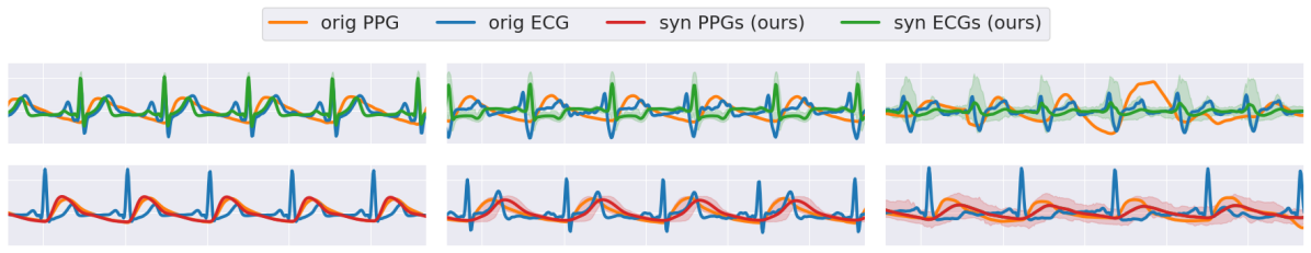
(a) Low Uncertainty (b) Medium Uncertainty (c) High Uncertainty
This section presents a comprehensive empirical study of our proposed method, UA-P2E, for converting PPG signals to ECG ones (lead II), as detailed in Section 4. All experiments described hereafter are conducted using three random seeds, and the results shown are the average of their outcomes. We employ this conversion technique with two prime objectives in mind: (1) Providing an interpretable estimation of the ECG signals, denoted as , that correspond to the given PPG , while targeting the approximation of samples from the posterior distribution ; and (2) Providing accurate classification and confidence assessment for a series of cardiovascular conditions. In both objectives, the presented results showcase the superiority of the proposed paradigm across all conducted experiments.
Unlike prior work, this paper aims to address the uncertainty in the conversion of PPG to ECG signals, and this is obtained by leveraging a diffusion-based conversion methodology. More specifically, we adopt the DDIM framework [28], aiming to reduce the number of iterations (in our experiments we applied iterations) required for the sampling procedure, and thus increasing the efficiency of our conversion algorithm. The diffusion denoising model is trained using the MIMIC-III matched waveform database [38], which is one of the most extensive dataset for paired PPG and ECG signals.
Our strategy relies on the core ability of generative models to obtain an effective sampling from the posterior distribution , where stands for the given PPG and for its corresponding ECG. Figure 2 (top row) illustrates typical sampling outcomes of our diffusion-based conversion model, showcasing the diverse ECG signals generated from different PPG inputs, each exhibiting varying levels of uncertainty, as expressed by the variability of the samples obtained.
As for the classification objective, we train and evaluate the classification model, denoted as , using the ‘Computing in Cardiology” (CinC) [35] dataset, containing multi-labeled ECG signals of 11 cardiovascular conditions 222The following cardiovascular conditions are considered: (1) Ventricular premature beats, (2) Atrial flutter, (3) Sinus rhythm, (4) Atrial fibrillation, (5) Supraventricular premature beats, (6) Bradycardia, (7) Pacing rhythm, (8) Sinus tachycardia, (9) 1st degree AV block, (10) Sinus bradycardia, and (11) Sinus arrhythmia. that are detectable by lead II ECG signals. Interestingly, our experiments rely in part on a reversed diffusion-based conversion model, designed to sample from the likelihood distribution , i.e. converting ECG (lead II) to PPG. This reversed sampling is utilized in order to complete the CinC [35] dataset used in our tests, enabling an analysis of challenging cardiac classification tasks. Figure 2 (bottom row) presents typical sampling outcomes of the reversed conversion model.
For further details on experimental details, including the datasets used for training and evaluating the conversion and classification models, along with details on data pre-processing and training schemes, we refer the reader to Appendix E.
5.1 Diffusion-Based Conversion: Quality Assessment
| RMSE ↓ | 1-FD ↓ | 100-FD ↓ | ||
| Baseline Methods | CardioGAN [13] | 0.54 | 99.0 | - |
| RDDM [22] (T=50) | 0.22 | 6.71 | - | |
| Our Method: UA-P2E | MMSE-Approx | 0.2222 0.0000 | 11.527 0.0179 | - |
| DDIM (T=25) | 0.3031 0.0000 | 2.8671 0.0034 | 2.7873 0.0007 | |
| DDIM (T=50) | 0.2956 0.0000 | 0.5194 0.0015 | 0.4418 0.0007 | |
| DDIM (T=100) | 0.2939 0.0001 | 0.3198 0.0020 | 0.2379 0.0005 |
We begin by assessing the quality of our diffusion-based PPG-2-ECG conversion model using the MIMIC-III [38] dataset, showcasing its superiority over state-of-the-art baseline methods. Here, we examine the quality of with , sampled from .
Table 1 presents the conversion performance results, evaluating the quality of the obtained ECG signals. we provide a comparison with two state-of-the-art methods, CardioGAN [13] and RDDM [22], using their reported metrics. More specifically, we assess the conversion quality through RMSE calculations between the generated ECG(s) and the ground truth signals. Additionally, we employ the Fréchet Distance (FD) to evaluate the perceptual quality of the produced signals, considering either a single ECG sample from each PPG (1-FD) or 100 samples from each (100-FD). The results in Table 1 demonstrate the superiority of our conversion model. Observe that by averaging the samples, we obtain an approximation of the MMSE estimator, yielding a comparable performance to the reported result in [22]. When considering the individual ECG solutions for each PPG, the RMSE values increases, as expected [36]. In terms of perceptual quality, the single-solution and multi-solution approaches show pronounced improvement in FD compared to the baselines. Note that computing the FD on multiple solutions helps in improving further the signals’ quality.
5.2 Classifying Cardiovascular Conditions
To demonstrate the benefits of our diffusion-based synthesized ECGs, we present classification results on two datasets, MIMIC-III [38] and CinC [35], analyzing the classification performance across a series of 11 cardiovascular conditions mentioned earlier. For both datasets, we compare our approach to classification strategies utilized by prior work, varied by the choice of in Definition 3.1, which refers to as the SSC. Specifically, we examine the classification performance of the MMSE estimate and a random single sample from the approximated posterior distribution, . For more details on the classification strategies considered, we refer the reader to Appendix F.
Note that MIMIC-III lacks cardiovascular labels, and thus cannot be utilized as-is for classification. As a remedy, we apply our experiments using the CinC dataset. However, while CinC contains ECG signals (all leads) and their corresponding labels, it does not include PPG signals, and as such it cannot serve our overall PPG classification goals. We resolve this difficulty by generating synthetic PPG signals from the given ECG ones, using a reversed diffusion-based methodology. The idea is to sample valid and random PPG signals that correspond to their ECG (lead II) counterparts, this way augmenting CinC to form a complete suite. In Appendix G we provide empirical evidence for the validity of our reversal conversion model of ECG-2-PPG in comparison with the original PPG signal.
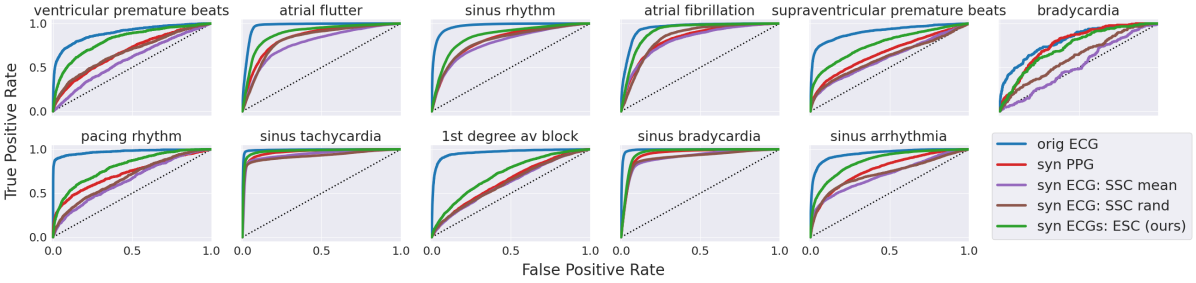
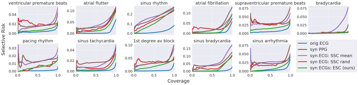
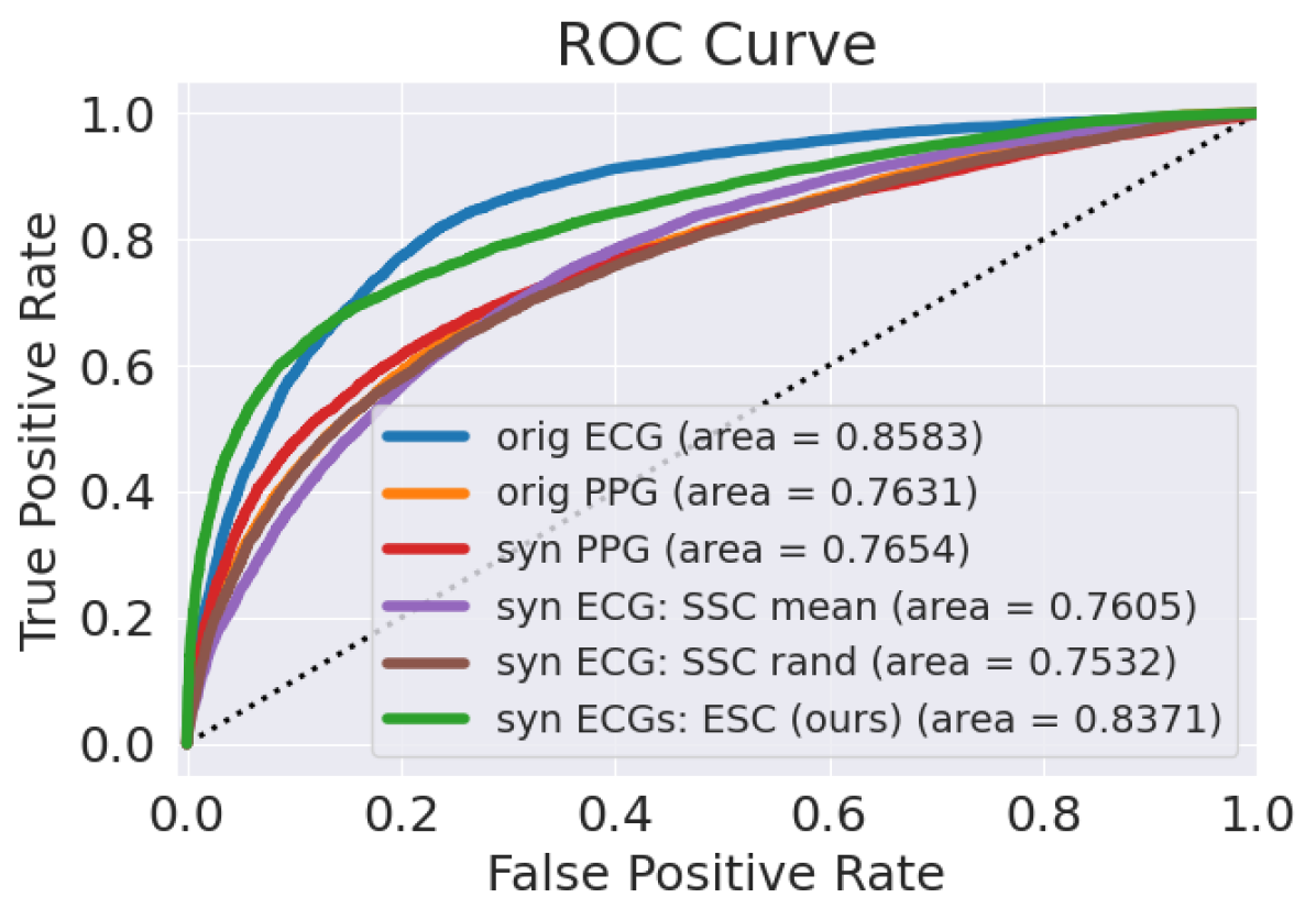
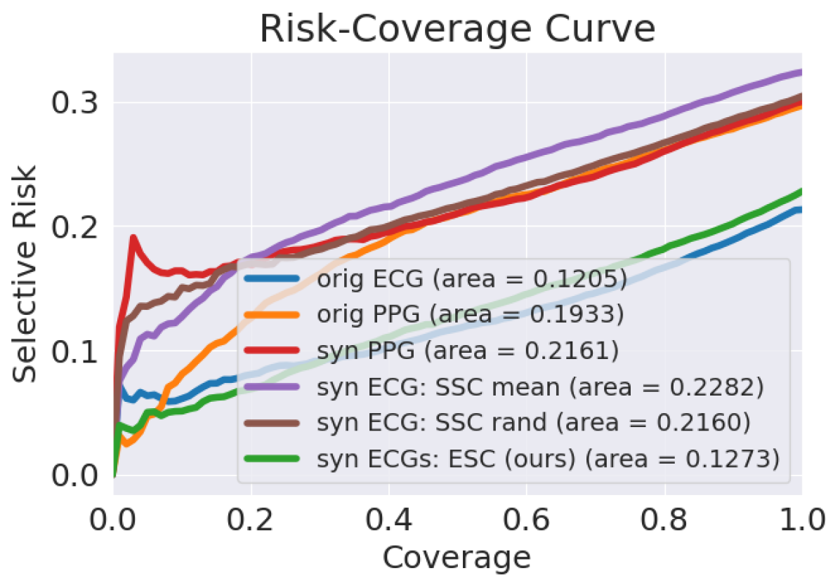
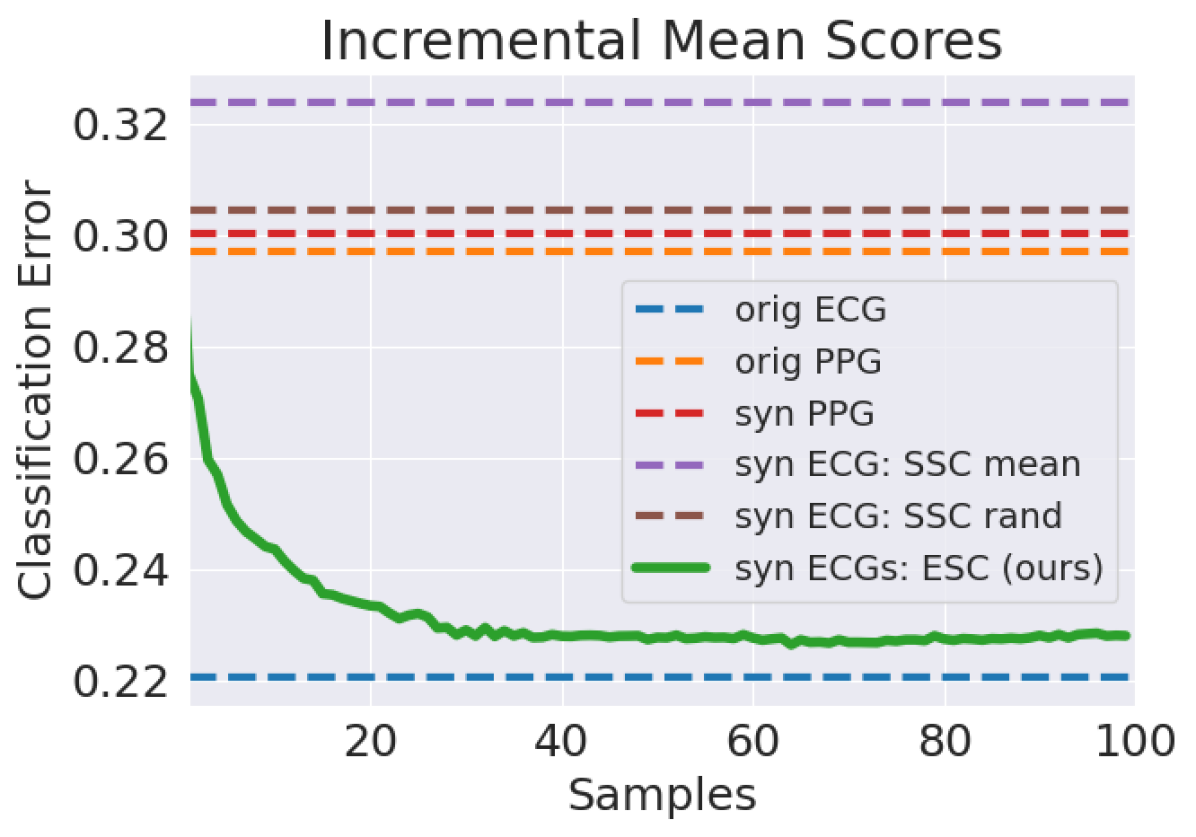
We now turn to describe the main classification results of cardiovascular conditions, referring to the augmented CinC dataset, and using the synthesized PPG signals. Figure 3 presents ROC curves for the classification strategies considered, and Figure 4 completes this description by presenting risk-coverage curves [25], which evaluate the performance of different confidence functions, denoted as , for PPG signals in regard to uncertainty quantification. For detailed numerical results, including standard errors, refer to Appendix H.
As can be seen from these two figures, our approach (GREEN) demonstrates superior performance over all the alternatives, apart from the direct ECG (BLUE) classification, which serves as an upper-bound for the achievable performance.
Theorem A.1 may be interpreted as claiming that our approach is expected to align with that of classifying the original PPG signals directly (ORANGE), under optimal conditions. However, we observe that our approach achieves better performance. We argue that this gap is due to the erroneous labels used in the direct classification: Note that these labels refer to the original ECG signals, and do not necessarily match the corresponding PPG, which may correspond to other valid ECG signals.
Still referring to the results in these two figures, it is evident that prior work which takes advantage of either the classification strategies of the mean estimate (PURPLE) or a random ECG sample (BROWN), exhibits a weaker performance compared to our approach. These findings strengthen our claim that ignoring the uncertainty that emerges from the conversion task might impair performance.
6 Concluding Remarks
This paper presents Uncertainty-Aware PPG-2-ECG (UA-P2E), a novel approach that addresses the inherent uncertainty in the PPG-2-ECG task. By incorporating the potential spread and variability of derived ECG signals from PPG inputs through sampling from a diffusion-based model, we achieve state-of-the-art performance in the conversion task. In addition, we show that our approach enhances cardiovascular classification, through the approximation of our proposed Expected Score Classifier (ESC), which averages classification scores of the ECG solution cloud for each input PPG signal. This classification strategy is proven to be optimal, and demonstrated to perform better than baseline methods in practice. Finally, we present ECG visualization methods, designed to enhance interpretability for medical professionals while considering the multitude ECG solutions associated with each PPG signal.
Our work presents rich empirical results showcasing the superior classification performance of our approach, compared to direct classification methods operating on given PPG signals. Nevertheless, these results are partially reliant on synthetic PPG signals. While we provide empirical validation for these synthetic signals in this paper, further investigation should be dedicated for the study of our approach with original PPG signals, along with further exploration of additional cardiovascular conditions. Future studies in these areas will require additional data collection.
References
- [1] Connie W Tsao, Aaron W Aday, Zaid I Almarzooq, Alvaro Alonso, Andrea Z Beaton, Marcio S Bittencourt, Amelia K Boehme, Alfred E Buxton, April P Carson, Yvonne Commodore-Mensah, et al. Heart disease and stroke statistics—2022 update: a report from the american heart association. Circulation, 145(8):e153–e639, 2022.
- [2] Paul Kligfield, Leonard S Gettes, James J Bailey, Rory Childers, Barbara J Deal, E William Hancock, Gerard Van Herpen, Jan A Kors, Peter Macfarlane, David M Mirvis, et al. Recommendations for the standardization and interpretation of the electrocardiogram: part i: the electrocardiogram and its technology: a scientific statement from the american heart association electrocardiography and arrhythmias committee, council on clinical cardiology; the american college of cardiology foundation; and the heart rhythm society endorsed by the international society for computerized electrocardiology. Circulation, 115(10):1306–1324, 2007.
- [3] Qiang Zhu, Xin Tian, Chau-Wai Wong, and Min Wu. Ecg reconstruction via ppg: A pilot study. In 2019 IEEE EMBS international conference on biomedical & health informatics (BHI), pages 1–4. IEEE, 2019.
- [4] Andrew Reisner, Phillip A Shaltis, Devin McCombie, H Harry Asada, David S Warner, and Mark A Warner. Utility of the photoplethysmogram in circulatory monitoring. The Journal of the American Society of Anesthesiologists, 108(5):950–958, 2008.
- [5] Walter Karlen, Srinivas Raman, J Mark Ansermino, and Guy A Dumont. Multiparameter respiratory rate estimation from the photoplethysmogram. IEEE Transactions on Biomedical Engineering, 60(7):1946–1953, 2013.
- [6] Takuo Aoyagi and Katsuyuki Miyasaka. Pulse oximetry: its invention, contribution to medicine, and future tasks. Anesthesia and analgesia, 94(1 Suppl):S1–3, 2002.
- [7] RA Payne, CN Symeonides, DJ Webb, and SRJ Maxwell. Pulse transit time measured from the ecg: an unreliable marker of beat-to-beat blood pressure. Journal of Applied Physiology, 100(1):136–141, 2006.
- [8] William A Marston. Ppg, apg, duplex: which noninvasive tests are most appropriate for the management of patients with chronic venous insufficiency? In Seminars in vascular surgery, volume 15, pages 13–20. Elsevier, 2002.
- [9] J Allen and A Murray. Development of a neural network screening aid for diagnosing lower limb peripheral vascular disease from photoelectric plethysmography pulse waveforms. Physiological Measurement, 14(1):13, 1993.
- [10] Hong-Yu Chiu, Hong-Han Shuai, and Paul C-P Chao. Reconstructing qrs complex from ppg by transformed attentional neural networks. IEEE Sensors Journal, 20(20):12374–12383, 2020.
- [11] Yuenan Li, Xin Tian, Qiang Zhu, and Min Wu. Inferring ecg from ppg for continuous cardiac monitoring using lightweight neural network. arXiv preprint arXiv:2012.04949, 2020.
- [12] Qiang Zhu, Xin Tian, Chau-Wai Wong, and Min Wu. Learning your heart actions from pulse: Ecg waveform reconstruction from ppg. IEEE Internet of Things Journal, 8(23):16734–16748, 2021.
- [13] Pritam Sarkar and Ali Etemad. Cardiogan: Attentive generative adversarial network with dual discriminators for synthesis of ecg from ppg. In Proceedings of the AAAI Conference on Artificial Intelligence, volume 35, pages 488–496, 2021.
- [14] Khuong Vo, Emad Kasaeyan Naeini, Amir Naderi, Daniel Jilani, Amir M Rahmani, Nikil Dutt, and Hung Cao. P2e-wgan: Ecg waveform synthesis from ppg with conditional wasserstein generative adversarial networks. In Proceedings of the 36th Annual ACM Symposium on Applied Computing, pages 1030–1036, 2021.
- [15] Qunfeng Tang, Zhencheng Chen, Yanke Guo, Yongbo Liang, Rabab Ward, Carlo Menon, and Mohamed Elgendi. Robust reconstruction of electrocardiogram using photoplethysmography: A subject-based model. Frontiers in Physiology, 13:859763, 2022.
- [16] Wen-Hsien Ho, Chia-Te Liao, Yenming J Chen, Kao-Shing Hwang, and Yanyun Tao. Quickly convert photoplethysmography to electrocardiogram signals by a banded kernel ensemble learning method for heart diseases detection. IEEE Access, 10:51079–51092, 2022.
- [17] Xin Tian, Qiang Zhu, Yuenan Li, and Min Wu. Cross-domain joint dictionary learning for ecg inference from ppg. IEEE Internet of Things Journal, 10(9):8140–8154, 2022.
- [18] Khuong Vo, Mostafa El-Khamy, and Yoojin Choi. Ppg to ecg signal translation for continuous atrial fibrillation detection via attention-based deep state-space modeling. arXiv preprint arXiv:2309.15375, 2023.
- [19] Ella Lan. Performer: A novel ppg-to-ecg reconstruction transformer for a digital biomarker of cardiovascular disease detection. In Proceedings of the IEEE/CVF Winter Conference on Applications of Computer Vision, pages 1991–1999, 2023.
- [20] Khaled M Abdelgaber, Mostafa Salah, Osama A Omer, Ahmed EA Farghal, and Ahmed S Mubarak. Subject-independent per beat ppg to single-lead ecg mapping. Information, 14(7):377, 2023.
- [21] Shun Hinatsu, Masamichi Tanji, Hiroki Ishizuka, Sei Ikeda, and Osamu Oshiro. Heart side-channel: Estimation of cardiovascular signal waveforms through skin vibration sensing. IEEE Sensors Letters, 2023.
- [22] Debaditya Shome, Pritam Sarkar, and Ali Etemad. Region-disentangled diffusion model for high-fidelity ppg-to-ecg translation. arXiv preprint arXiv:2308.13568, 2023.
- [23] Anastasios N Angelopoulos, Amit Pal Kohli, Stephen Bates, Michael Jordan, Jitendra Malik, Thayer Alshaabi, Srigokul Upadhyayula, and Yaniv Romano. Image-to-image regression with distribution-free uncertainty quantification and applications in imaging. In International Conference on Machine Learning, pages 717–730. PMLR, 2022.
- [24] Omer Belhasin, Yaniv Romano, Daniel Freedman, Ehud Rivlin, and Michael Elad. Principal uncertainty quantification with spatial correlation for image restoration problems. IEEE Transactions on Pattern Analysis and Machine Intelligence, 2023.
- [25] Yonatan Geifman and Ran El-Yaniv. Selective classification for deep neural networks. Advances in neural information processing systems, 30, 2017.
- [26] Jascha Sohl-Dickstein, Eric Weiss, Niru Maheswaranathan, and Surya Ganguli. Deep unsupervised learning using nonequilibrium thermodynamics. In International conference on machine learning, pages 2256–2265. PMLR, 2015.
- [27] Jonathan Ho, Ajay Jain, and Pieter Abbeel. Denoising diffusion probabilistic models. Advances in neural information processing systems, 33:6840–6851, 2020.
- [28] Jiaming Song, Chenlin Meng, and Stefano Ermon. Denoising diffusion implicit models. arXiv preprint arXiv:2010.02502, 2020.
- [29] Prafulla Dhariwal and Alexander Nichol. Diffusion models beat gans on image synthesis. Advances in neural information processing systems, 34:8780–8794, 2021.
- [30] Ling Yang, Zhilong Zhang, Yang Song, Shenda Hong, Runsheng Xu, Yue Zhao, Wentao Zhang, Bin Cui, and Ming-Hsuan Yang. Diffusion models: A comprehensive survey of methods and applications. ACM Computing Surveys, 56(4):1–39, 2023.
- [31] Bahjat Kawar, Gregory Vaksman, and Michael Elad. Snips: Solving noisy inverse problems stochastically. Advances in Neural Information Processing Systems, 34:21757–21769, 2021.
- [32] Bahjat Kawar, Michael Elad, Stefano Ermon, and Jiaming Song. Denoising diffusion restoration models. Advances in Neural Information Processing Systems, 35:23593–23606, 2022.
- [33] Jiaming Song, Arash Vahdat, Morteza Mardani, and Jan Kautz. Pseudoinverse-guided diffusion models for inverse problems. In International Conference on Learning Representations, 2022.
- [34] Hyungjin Chung, Jeongsol Kim, Michael T Mccann, Marc L Klasky, and Jong Chul Ye. Diffusion posterior sampling for general noisy inverse problems. arXiv preprint arXiv:2209.14687, 2022.
- [35] Ary L Goldberger, Luis AN Amaral, Leon Glass, Jeffrey M Hausdorff, Plamen Ch Ivanov, Roger G Mark, Joseph E Mietus, George B Moody, Chung-Kang Peng, and H Eugene Stanley. Physiobank, physiotoolkit, and physionet: components of a new research resource for complex physiologic signals. circulation, 101(23):e215–e220, 2000.
- [36] Yochai Blau and Tomer Michaeli. The perception-distortion tradeoff. In Proceedings of the IEEE conference on computer vision and pattern recognition, pages 6228–6237, 2018.
- [37] Normand J Beaudry and Renato Renner. An intuitive proof of the data processing inequality. arXiv preprint arXiv:1107.0740, 2011.
- [38] Alistair EW Johnson, Tom J Pollard, Lu Shen, Li-wei H Lehman, Mengling Feng, Mohammad Ghassemi, Benjamin Moody, Peter Szolovits, Leo Anthony Celi, and Roger G Mark. Mimic-iii, a freely accessible critical care database. Scientific data, 3(1):1–9, 2016.
- [39] Vladimir Vovk, Alexander Gammerman, and Glenn Shafer. Algorithmic learning in a random world, volume 29. Springer, 2005.
- [40] Harris Papadopoulos, Kostas Proedrou, Volodya Vovk, and Alex Gammerman. Inductive confidence machines for regression. In Machine learning: ECML 2002: 13th European conference on machine learning Helsinki, Finland, August 19–23, 2002 proceedings 13, pages 345–356. Springer, 2002.
- [41] Jing Lei and Larry Wasserman. Distribution-free prediction bands for non-parametric regression. Journal of the Royal Statistical Society Series B: Statistical Methodology, 76(1):71–96, 2014.
- [42] Anastasios N Angelopoulos, Stephen Bates, Emmanuel J Candès, Michael I Jordan, and Lihua Lei. Learn then test: Calibrating predictive algorithms to achieve risk control. arXiv preprint arXiv:2110.01052, 2021.
- [43] Anastasios N Angelopoulos and Stephen Bates. A gentle introduction to conformal prediction and distribution-free uncertainty quantification. arXiv preprint arXiv:2107.07511, 2021.
- [44] Wassily Hoeffding. Probability inequalities for sums of bounded random variables. The collected works of Wassily Hoeffding, pages 409–426, 1994.
- [45] Olaf Ronneberger, Philipp Fischer, and Thomas Brox. U-net: Convolutional networks for biomedical image segmentation. In Medical image computing and computer-assisted intervention–MICCAI 2015: 18th international conference, Munich, Germany, October 5-9, 2015, proceedings, part III 18, pages 234–241. Springer, 2015.
- [46] Karen Simonyan and Andrew Zisserman. Very deep convolutional networks for large-scale image recognition. arXiv preprint arXiv:1409.1556, 2014.
Appendix A Optimality of the Expected Score Classifier
This section provides theoretical results proving the optimality of the Expected Score Classifier (ESC), defined in Definition 3.2.
A.1 Optimal Settings
We begin by showing the optimality of the ESC in optimal settings:
Theorem A.1 (Optimality of the Expected Score Classifier).
Consider the Markovian dependency chain , implying that , and assume the following:
-
1.
The conversion model is a posterior sampler, i.e., ; and
-
2.
The classification model is optimal by satisfying ,
Then the obtained classification is optimal, satisfying .
The following is the proof:
Consider the Markovian dependency chain , implying that , and recalling the assumptions of Theorem A.1:
-
1.
The conversion model is a posterior sampler, i.e., ; and
-
2.
The classification model is optimal by satisfying .
By definition, the ESC is written as
Next, we make a change of variables . This gives us
where we rely on the assumption that is a posterior sampler, namely, . Therefore, for any , it follows from the optimality of that
where the second-to-last transition holds due to the Markov chain assumption. Thus, we obtain , completing the proof.
A.2 Optimality for General Classifiers
Here, we show that the ESC (Definition 3.2), denoted as , is an optimal estimator of , regardless of the optimality of .
Theorem.
Let be the source ECG signal whose observation is the PPG signal . Additionally, assume the conversion model is a posterior sampler such that and let denote the ESC, as defined by Definition 3.2. Then, constitutes the minimum mean square error (MMSE) estimator of . Namely,
where the expectation above is taken over given .
The proof follows a straightforward approach. We first expand the squared error term
Differentiating the above expression with respect to and setting it to zero yields
Recognizing that , we conclude that
Therefore, is the MMSE estimator of .
Appendix B Assessing Reliable PPG Signals via Calibration
Recalling the desired selective guarantee specified in Equation (2),
To ensure the selection of valid PPG signals meeting the above guarantee, a calibration scheme is necessary. This scheme’s goal is to identify a calibrated parameter that maximizes coverage of selected PPG signals whose classification error is below a user-specified bound, denoted by . Then, a new unseen PPG signal, , will be considered reliable if its confidence score surpasses .
The calibration algorithm is based on a conformal prediction scheme [39, 40, 41], a general method for distribution-free risk control. Specifically, we employ the Learn Then Test (LTT) [42] procedure in this paper. The conformal prediction’s objective is to bound a user-defined risk function, denoted as , by adjusting a calibration parameter based on empirical data, aiming for the tightest upper bound feasible [43]. Through this calibration process, our aim is to identify that ensures the following guarantee:
| (6) |
where is an upper bound for the risk function, and is the calibration error. These parameters are user-defined and should approach zero for an effective calibration.
We now turn to describe the steps for calibration to achieve the guarantee outlined in Equation (2). To simplify Equation (2) into the form posed in Equation (6), we define the selective risk as:
| (7) |
Intuitively, this measure quantifies the classification error among selected PPG signals with confidence scores surpassing .
In practical scenarios, computing the above risk function, , is not feasible due to the unavailability of the joint distribution . However, we do have samples from this distribution within our calibration data, . Hence, we utilize the empirical selective risk, denoted as , and defined as:
| (8) |
Note that , where represents the decision threshold.
For each , where represents a set of calibration parameter values defined by the user as , we utilize the empirical risk function mentioned earlier to establish an upper-confidence bound, denoted as , and defined as:
| (9) |
where serves as a concentration bound, such as Hoeffding’s bound, which is calculated as . It is shown in [44] that .
Finally, to achieve the desired guarantee outlined in Equation (2), we seek calibration parameters that satisfy Equation (2) while using the upper confidence bound defined in Equation (9). The goal is to maximize the coverage of selected PPG signals, thus the smallest that meets Equation (2) is selected as follows:
| (10) |
The algorithm below summarizes the steps outlined in this section for applying calibration.
Appendix C Conversion-Classification Uncertainty Evidence
Conversion Uncertainty Classification Uncertainty
Here, we present empirical evidence of the inherent uncertainty in the proposed PPG-2-ECG conversion, along with the classification uncertainty implied. Figure 5 summarizes the results that quantify these uncertainty layers, referring to the CinC [35] dataset.
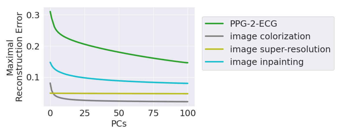
In Figure 5(a), we illustrate the uncertainty as proposed by [24], showing the maximal reconstruction error of the projected ground truth ECG signals among of the temporal signal values using a varying number of Principal Components (PCs) of the ECG solutions’ cloud. Intuitively, a high reconstruction error using many PCs indicates high uncertainty in terms of the spread and variability of possible solutions to the inverse problem tackled. For additional information and details on this metric, we refer the reader to previous work [24].
Figure 5(a) compares the reconstruction error for our PPG-2-ECG conversion with three similar graphs that correspond to image restoration tasks (colorization, super-resolution and inpainting). This comparison exposes the tendency of our task to have a much wider uncertainty, as far more than PCs are required in order to reconstruct the ground truth ECG signals with a small error. This exposes the fact that PPG-2-ECG conversion is highly ill-posed. Put bluntly, this means that given a PPG signal, the ECG corresponding to it could be one of a wide variety of possibilities. This fact undermines our task, as it suggests that too much information has been lost in migrating from ECG to PPG, to enable a “safe” come-back. However, we argue that this is a misleading observation, and, in fact, the true uncertainty is much better behaved. More specifically, within the variability that our posterior sampler exposes, many changes manifested in the obtained ECG signals are simply meaningless and medically transparent. Indeed, if we migrate from the signal domain to classification, this uncertainty shrinks to a meaningful and crisp decisions.
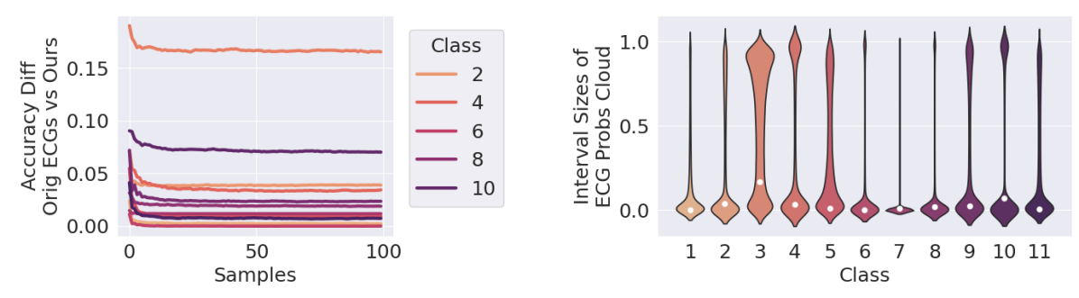
The above takes us to the results shown in Figure 5(b), depicting the uncertainty in classification, as arises from potential variability in the conversion process. Inspired by [23], in Figure 5(b) (right) we present histograms of interval sizes that contain 90% of the probability scores extracted using the ECG cloud that corresponds to each PPG signal, denoted as . These histograms refer to the series of 11 cardiovascular conditions. Small intervals in this context correspond to a high certainty in classification, and the results reveal that for most of the conditions assessed this is indeed the case. We also see that a small fraction of PPG signals may exhibit a wide range of possible probabilities among ECG candidates, a fact that may motivate our later selective classification approach.
Figure 5(b) (left) presents the classification accuracy gaps for the 11 cardiac conditions mentioned. These gaps correspond to the difference between the accuracy of original ECG classification, which serves as an upper bound for our performance, and the accuracy of the average of ECG classification scores, as summarized in Algorithm 1. As can be seen, these graphs show a rapid stabilization, suggesting that using samples is definitely sufficient for modeling the uncertainty that arises from the conversion process in the classification domain.
Appendix D Quality Assessment of ECG Visualization Approaches
| Signal Domain | Embedding Domain | |||
|---|---|---|---|---|
| Visualization Strategy | RMSE ↓ | 1-FD ↓ | RMSE ↓ | 1-FD ↓ |
| Min/Max Score ECG (5) | 0.3130 0.0004 | 1.3113 0.0302 | 0.3079 0.0037 | 16.676 0.7135 |
| Expected Score ECG (4) | 0.3119 0.0004 | 1.2820 0.0144 | 0.3091 0.0044 | 13.172 0.2228 |
| Most Likely Score ECG (3) | 0.3105 0.0004 | 1.2726 0.0101 | 0.3040 0.0043 | 10.491 0.1879 |
In this section, we examine the ECG visualization strategies, as discussed in Section 4.3, over the CinC dataset. By comparing our proposed visualization strategies, we aim to provide empirical insights on how to choose between them. As already mentioned in Section 4.3, there is no “correct” choice for the visualization strategy, and it should be considered by the medical user. Table 2 presents the comparative results of the three proposed strategies: Min/Max Score ECG, Expected Score ECG, and Most Likely Score ECG. This table brings RMSE results for the signals chosen and FD values for their distributions, all compared with their ECG origins. The three strategies are evaluated across two domains, first, in the signal domain, and second, in the embedding domain using our classification model, denoted as . As can be seen from the table, while all three considered approaches are good performing in these measures, the “Most Likely Score ECG” seem to be the best, both in RMSE and FD.
Appendix E Experimental Details
This section provides a thorough description of the experimental methodology utilized in our paper. It encompasses the datasets employed for both training and evaluating the conversion and classification models, along with details regarding data pre-processing and training schemes.
E.1 Datasets and Preprocessing
In this work we rely on two datasets: the MIMIC-III matched waveform database [38], which consists of pairs of measured ECG signals, denoted as , and corresponding PPG signals, denoted as ; and the “Computing in Cardiology” dataset (CinC) [35], which contains pairs of measured ECG signals along with associated cardiovascular condition labels, referred to as .
MIMIC-III is one of the most extensive datasets for paired PPG and ECG signals. It comprises 22,317 records from 10,282 distinct patients, with each record spanning approximately 45 hours.
The MIMIC-III dataset includes a substantial portion of noisy signals, which are unsuitable for training models due to various distortions, artifacts, synchronization issues, and other anomalies. Consequently, a comprehensive preprocessing protocol is employed for preparing the data for subsequent analysis. The preprocessing of each record in MIMIC-III is conducted through the following steps:
-
1.
Resampling: Each record is resampled to a uniform sampling rate of 125 Hz.
-
2.
Signal Smoothing: A Butterworth bandpass filter is applied to all signals to reduce noise.
-
3.
Signal Alignment: Ensures that all signals are temporally aligned.
-
4.
Artifact Removal: Signal blocks with unacceptable heart rates, as extracted from the electrocardiogram (ECG), are removed.
-
5.
Consistency Check: Removes signal blocks where the heart rates derived from the photoplethysmogram (PPG) differ by more than 30% from those extracted from the ECG.
-
6.
Trend Removal: The global trend (DC-drift) in the signal is removed to prevent bias.
-
7.
Normalization: All signals are normalized to have zero mean and unit variance.
Moving to CinC, which to the best of our knowledge, is the largest and most challenging classification dataset in cardiology, containing 94,630 of 10-second multi-labeled ECG recordings from distinct patients. Each recording is labeled with the presence or absence (binary label) of 11 cardiovascular conditions: (1) Ventricular premature beats, (2) Atrial flutter, (3) Sinus rhythm, (4) Atrial fibrillation, (5) Supraventricular premature beats, (6) Bradycardia, (7) Pacing rhythm, (8) Sinus tachycardia, (9) 1st degree AV block, (10) Sinus bradycardia, and (11) Sinus arrhythmia. We note that the labels considered in CinC exhibit significant imbalance.
The CinC database, noted for its cleaner and more structured data, lacks the pairings and complexities found in the MIMIC-III dataset and only includes ECG samples. For this dataset, the preprocessing steps are slightly modified and include:
-
1.
Resampling: Each record is resampled to 125 Hz to standardize the data input.
-
2.
Signal Smoothing: A Butterworth bandpass filter is employed to smooth the ECG signals.
-
3.
Trend Removal: Global trends are removed.
-
4.
Normalization: Signals are normalized to have zero mean and unit variance.
E.2 Training Details for the Conversion Model
Our study leverages a diffusion-based conversion methodology. Specifically, we adopt the DDIM framework [28]. All the training details for the reversed conversion model, designed for generating PPG signals given ECG ones, are similar to the training details for the ECG-2-PPG conversion, as described hereafter.
The denoising model employed in our experiments is based on a U-Net [45] architecture with an attention mechanism, as proposed in [27, 29]. Originally introduced in the field of image processing, this architecture comprises a series of residual layers and downsampling convolutions, followed by a subsequent series of residual layers with upsampling convolutions. Additionally, a global attention layer with a single head is incorporated, along with a projection of the timestep embedding into each residual block. To adapt the architecture for our purposes, we replace the convolutions and attention layers with 1D counterparts, enabling training and evaluation directly in the raw 1D signal domain. In our study we use PPG and ECG signal length.
Regarding hyperparameters for the U-Net architecture, we set 256 dimensions for time embedding. The downsampling and upsampling is applied by factors of [1, 1, 2, 2, 4, 4, 8], with each factor corresponding to a stack of 2 residual blocks. Attention is applied after the downsampling/upsampling matching to the factor of 4. To address overfitting, we incorporate dropout with a ratio of 0.2.
Concerning training, the model architecture is trained on MIMIC-III. We randomly split the data into 70% for training and 30% for validation after preprocessing. The model architecture is trained for 10,000 epochs, employing a batch size of 2,048 PPG and ECG recordings across 8V100 GPUs. The noise schedule progresses linearly over 1,000 time steps, starting at 1e-6 and ending at 1e-2. For optimization, we employed the standard Adam optimizer with a fixed learning rate of 1e-4. An loss is utilized to estimate the noise in the input instance.
For evaluation, we sample ECG candidates for each PPG signal using the diffusion-based framework. We adopt a non-stochastic diffusion process as proposed in [28], with skip jumps of 10 iterations, resulting in a total of diffusion iterations.
E.3 Training Details for the Classification Model
Our paper adopts a classification methodology over the CinC dataset, referred to as , which classifies ECG signals, whether original or synthesized, into the 11 cardiovascular conditions in CinC. Additionally, we report the classification performance of a direct approach, denoted as , which directly gets PPG signals instead of ECG ones. The training scheme outlined hereafter applies to both classifiers.
As discussed earlier, the presence of each cardiovascular condition in the CinC dataset is imbalanced, and thus CinC poses significant challenges for developing a robust classifier. In Figure 6 (red) we report the frequency of each cardiovascular condition in the dataset. To address this issue, we implement a novel class-balance sampling technique. Given the multi-label nature of the classification, where class dependencies exist, our strategy not only ensures a uniform class distribution for positive samples but also carefully selects negative samples from a balanced pool reflecting common class co-occurrences. This approach significantly aids in achieving a more uniform label distribution, the effectiveness of which is demonstrated in Figure 6 (blue).
The classification architecture utilized in our studies is based on a simplified VGG [46] model with 8 layers, incorporating batch normalization modules to promote stability, along with dropout at ratio of 0.2 to address overfitting. Similar to the conversion model, we modify the VGG architecture for 1D input signals by reconfiguring the convolutional layers for 1D operation.
Optimization is performed on 1V100 GPU using the Adam optimizer and an weight decay of 1e-4. The model is trained over 20 epochs with a batch size of 128, employing a learning rate of 1e-3 with a linear decay and using binary cross-entropy loss as the loss function.
Appendix F Classification Strategies
In our experiments we assess several classification strategies, all relying on paired ECG and PPG signals and their corresponding labels, as obtained using MIMIC-III [38] and CinC [35]. We assume the availability of pretrained classifiers, and , for input ECG and PPG signals, respectively. These classifiers output classification likelihoods considering cardiovascular conditions.
We emphasize that the baseline approaches, as utilized in prior work, ignore the uncertainty in the conversion of PPG to ECG by maintaining only a single ECG solution given the PPG, whereas our approach maintains and manipulates an aggregation of multiple ECG candidates for the classification. With this in mind, here are the classification strategies considered in this work:
Original ECG: Classifier performance when tested on original ECG signals, i.e., the performance of , where . Since we do not have access to the original ECG signal in our PPG-2-ECG setting, this measure serves as an upper bound for the achievable performance.
Original PPG: Classifier performance when tested on the original PPG signals, i.e., the performance of , where .
Synthesized PPG: Classifier performance when tested on diffusion-based random PPG samples, i.e., the performance of , where are outcomes of the reversed diffusion-based conversion model. See Section 5.2 for detailed explanation on the need for these signals.
Synthesized ECG: SSC Mean Classifier performance when tested on single-solution (Definition 3.1) ECG signals, as practiced by prior work, referring to the mean ECG signal from the diffusion-based ECG samples, i.e., the performance of , where .
Synthesized ECG: SSC Random: Classifier performance when tested on single-solution (Definition 3.1) ECG signals, as practiced by prior work, referring to randomly chosen diffusion-based ECG samples, i.e., the performance of , where .
Appendix G Exploring the Validity of Synthesized PPG Signals within MIMIC-III
The main question arising from the results depicted in Section 5.2 is whether using synthesized PPG signals for drawing the various conclusions is a fair and trust-worthy strategy. One potential medical rationale for this approach is that PPG signals inherently encompass noise. Consequently, employing synthesized PPG signals, even if noisy themselves, can accurately model real-world scenarios. Still, in order to address this question in a more direct form, we return to the MIMIC-III [38] dataset, this time using a much smaller held-out subset of it that contains binary labels indicating the presence of atrial fibrillation (AFib). Thus, for this heart condition, we can evaluate classification performance for both true PPG signals and synthesized ones.
As previously stated in Section 5.2, the MIMIC-III matched waveform database lacks cardiovascular labels. However, textual cardiac reports are available for a subset of patients within MIMIC-III, referred to as the MIMIC-III clinical database [38]. In order to extract AFib labels from this subset, we filtered 400 patients, with 200 reported cases of AFib and 200 with normal sinus rhythm. The determination of AFib or sinus rhythm presence was made by searching for the terms "atrial fibrillation" and "sinus rhythm" within the cardiac reports. We note that extracting labels using this searching approach may provide erroneous labels.
In Figure 7 we bring the classification results in an experiment that follows the ones performed on CinC, with one distinct difference – we assess both real and synthesized PPG signals. Broadly speaking, all the conclusions from Section 5.2 generally hold, namely (i) The best performance is obtained by classifying ECG directly; (ii) Our approach is superior to all other alternatives, including a direct classification of PPG (be it real or synthesized).
Focusing on the main theme of this experiment, we see that the performance of classifying the original PPG (ORANGE) is comparable to the alternative of operating on the synthetic signals (RED). In terms of AUROC, the performances are nearly identical. When evaluating the area under the Risk-Coverage curve [25], we observe a similar trend; the uncertainty performance of both approaches is almost identical when considering around half of the test samples. These findings offer empirical evidence supporting the validity of our reversal conversion model.
Appendix H CinC Numerical Results
| Classification Strategy | Macro-AUROC ↑ | Macro-AURC ↓ |
|---|---|---|
| Original ECG | 0.9435 0.0020 | 0.0048 0.0000 |
| Synthesized PPG | 0.7857 0.0021 | 0.0313 0.0001 |
| Synthesized ECG: SSC Mean | 0.7286 0.0068 | 0.0405 0.0017 |
| Synthesized ECG: SSC Random | 0.7458 0.0007 | 0.0392 0.0002 |
| Synthesized ECG: ESC (Ours) | 0.8496 0.0010 | 0.0211 0.0001 |
| Cardiovascular Condition | Classification Strategy | AUROC ↑ | AURC ↓ |
|---|---|---|---|
| 1) Ventricular premature beat | Original ECG | 0.8953 0.0081 | 0.0042 0.0005 |
| Synthesized PPG | 0.6724 0.0033 | 0.0216 0.0009 | |
| Synthesized ECG: SSC Mean | 0.6290 0.0194 | 0.0171 0.0006 | |
| Synthesized ECG: SSC Random | 0.6344 0.0087 | 0.0178 0.0014 | |
| Synthesized ECG: ESC (Ours) | 0.8006 0.0072 | 0.0077 0.0006 | |
| 2) Atrial flutter | Original ECG | 0.9710 0.0004 | 0.0056 0.0001 |
| Synthesized PPG | 0.8457 0.0018 | 0.0289 0.0006 | |
| Synthesized ECG: SSC Mean | 0.7648 0.0170 | 0.0424 0.0040 | |
| Synthesized ECG: SSC Random | 0.8018 0.0035 | 0.0380 0.0009 | |
| Synthesized ECG: ESC (Ours) | 0.8816 0.0009 | 0.0220 0.0003 | |
| 3) Sinus rhythm | Original ECG | 0.9728 0.0004 | 0.0175 0.0003 |
| Synthesized PPG | 0.8111 0.0010 | 0.1309 0.0005 | |
| Synthesized ECG: SSC Mean | 0.7638 0.0018 | 0.1700 0.0012 | |
| Synthesized ECG: SSC Random | 0.7773 0.0009 | 0.1618 0.0010 | |
| Synthesized ECG: ESC (Ours) | 0.8547 0.0004 | 0.1114 0.0013 | |
| 4) Atrial fibrillation | Original ECG | 0.9489 0.0008 | 0.0059 0.0001 |
| Synthesized PPG | 0.8263 0.0007 | 0.0283 0.0005 | |
| Synthesized ECG: SSC Mean | 0.8320 0.0144 | 0.0262 0.0004 | |
| Synthesized ECG: SSC Random | 0.8582 0.0044 | 0.0317 0.0010 | |
| Synthesized ECG: ESC (Ours) | 0.9196 0.0016 | 0.0147 0.0003 | |
| 5) Supraventricular premature beats | Original ECG | 0.9080 0.0017 | 0.0068 0.0002 |
| Synthesized PPG | 0.6673 0.0033 | 0.0302 0.0011 | |
| Synthesized ECG: SSC Mean | 0.6176 0.0223 | 0.0313 0.0027 | |
| Synthesized ECG: SSC Random | 0.6404 0.0054 | 0.0351 0.0006 | |
| Synthesized ECG: ESC (Ours) | 0.7739 0.0069 | 0.0163 0.0006 | |
| 6) Bradycardia | Original ECG | 0.8072 0.0224 | 0.0010 0.0002 |
| Synthesized PPG | 0.7734 0.0154 | 0.0023 0.0001 | |
| Synthesized ECG: SSC Mean | 0.6405 0.0284 | 0.0143 0.0057 | |
| Synthesized ECG: SSC Random | 0.6927 0.0098 | 0.0028 0.0005 | |
| Synthesized ECG: ESC (Ours) | 0.7853 0.0113 | 0.0011 0.0001 | |
| 7) Pacing rhythm | Original ECG | 0.9734 0.0048 | 0.0009 0.0002 |
| Synthesized PPG | 0.7258 0.0119 | 0.0054 0.0001 | |
| Synthesized ECG: SSC Mean | 0.6148 0.0212 | 0.0139 0.0010 | |
| Synthesized ECG: SSC Random | 0.6193 0.0181 | 0.0137 0.0008 | |
| Synthesized ECG: ESC (Ours) | 0.7966 0.0061 | 0.0055 0.0002 | |
| 8) Sinus tachycardia | Original ECG | 0.9932 0.0005 | 0.0015 0.0001 |
| Synthesized PPG | 0.9674 0.0003 | 0.0094 0.0002 | |
| Synthesized ECG: SSC Mean | 0.9451 0.0030 | 0.0136 0.0010 | |
| Synthesized ECG: SSC Random | 0.9263 0.0030 | 0.0184 0.0009 | |
| Synthesized ECG: ESC (Ours) | 0.9792 0.0018 | 0.0050 0.0005 | |
| 9) 1st degree AV block | Original ECG | 0.9634 0.0011 | 0.0029 0.0001 |
| Synthesized PPG | 0.6350 0.0043 | 0.0365 0.0006 | |
| Synthesized ECG: SSC Mean | 0.6139 0.0092 | 0.0332 0.0009 | |
| Synthesized ECG: SSC Random | 0.6184 0.0050 | 0.0347 0.0011 | |
| Synthesized ECG: ESC (Ours) | 0.7193 0.0095 | 0.0203 0.0010 | |
| 10) Sinus bradycardia | Original ECG | 0.9935 0.0002 | 0.0025 0.0001 |
| Synthesized PPG | 0.9438 0.0008 | 0.0304 0.0005 | |
| Synthesized ECG: SSC Mean | 0.8932 0.0028 | 0.0565 0.0028 | |
| Synthesized ECG: SSC Random | 0.9005 0.0010 | 0.0502 0.0011 | |
| Synthesized ECG: ESC (Ours) | 0.9580 0.0003 | 0.0199 0.0011 | |
| 11) Sinus arrhythmia | Original ECG | 0.9512 0.0003 | 0.0039 0.0001 |
| Synthesized PPG | 0.7741 0.0032 | 0.0207 0.0004 | |
| Synthesized ECG: SSC Mean | 0.6999 0.0039 | 0.0272 0.0019 | |
| Synthesized ECG: SSC Random | 0.7344 0.0037 | 0.0268 0.0006 | |
| Synthesized ECG: ESC (Ours) | 0.8771 0.0006 | 0.0086 0.0001 |
This section extends the empirical classification results referring to CinC [35], that were outlined in Section 5.2. We provide here the numerical results of the mean metric values along with their corresponding standard errors.
Tables 3 and 4 display the AUROC (Area Under the ROC curve) and AURC (Area Under the Risk-Coverage curve [25]) metrics. Specifically, Table 4 outlines AUROC/AURC mean and standard error values associated with each classification strategy per cardiovascular condition, whereas Table 3 provides macro-level metrics derived by averaging over all examined cardiovascular conditions.