The Developing Human Connectome Project: A Fast Deep Learning-based Pipeline for Neonatal Cortical Surface Reconstruction
Abstract
The Developing Human Connectome Project (dHCP) aims to explore developmental patterns of the human brain during the perinatal period. An automated processing pipeline has been developed to extract high-quality cortical surfaces from structural brain magnetic resonance (MR) images for the dHCP neonatal dataset. However, the current implementation of the pipeline requires more than 6.5 hours to process a single MRI scan, making it expensive for large-scale neuroimaging studies. In this paper, we propose a fast deep learning (DL) based pipeline for dHCP neonatal cortical surface reconstruction, incorporating DL-based brain extraction, cortical surface reconstruction and spherical projection, as well as GPU-accelerated cortical surface inflation and cortical feature estimation. We introduce a multiscale deformation network to learn diffeomorphic cortical surface reconstruction end-to-end from T2-weighted brain MRI. A fast unsupervised spherical mapping approach is integrated to minimize metric distortions between cortical surfaces and projected spheres. The entire workflow of our DL-based dHCP pipeline completes within only 24 seconds on a modern GPU, which is nearly 1000 times faster than the original dHCP pipeline. Manual quality control demonstrates that for 82.5% of the test samples, our DL-based pipeline produces superior (54.2%) or equal quality (28.3%) cortical surfaces compared to the original dHCP pipeline.
keywords:
MSC:
34A12, 68T07, 68U10, 92C50 \KWDThe developing human connectome project , Neuroimage pipeline , Deep learning , Neonatal brain MRI , Cortical surface reconstruction1 Introduction
1.1 Background
During the perinatal phase, the human brain undergoes significant developmental changes, characterized by the formation of anatomical and functional connectivity. The Developing Human Connectome Project (dHCP) plays a pivotal role in this context. It aims at compiling an extensive dataset of brain magnetic resonance imaging (MRI) for both fetuses and neonates [30, 44, 17]. This ambitious project is instrumental in facilitating an in-depth understanding of early brain development and in constructing a four-dimensional connectome for young brains. The dHCP dataset is a valuable asset for investigating the normal and abnormal patterns of brain structural development and their respective connectomes. Notably, the dHCP has publicly released its neonatal dataset, comprising MRI scans of 783 newborn infants, both pre-term and full-term, resulting in a total of 887 images [17].
Cortical surfaces refer to the inner and outer surfaces of the cerebral cortex. The inner surface, also called white matter surface, is the boundary between the white matter (WM) and cortical grey matter (cGM). The outer surface, also called pial surface, is the boundary between the cGM and cerebrospinal fluid (CSF). Cortical surfaces are often represented by 3D polygon meshes and should have genus-0 topology, i.e., topologically equivalent to a 2-sphere without any ”holes”. The cortical folding and brain development of newborn infants can be modeled and quantified by the morphological features of cortical surfaces such as cortical thickness, mean curvature and sulcal depth [45, 24]. The notable achievements of FreeSurfer [15, 22, 21] and the Human Connectome Project (HCP) [62, 25], recognized for their automated neuroimaging processing pipelines for adults, underscore the importance of implementing cortical surface-based structural brain MRI processing and analysis for the dHCP fetal and neonatal datasets.
However, the pronounced differences between adult and neonatal brains present significant challenges in adapting existing adult MRI processing pipelines for use in newborn infants [21, 25, 58, 40, 32]. One primary challenge lies in the limited resolution of neonatal brain MRI, a consequence of the necessarily brief data acquisition times [30]. This limitation is further compounded by the smaller head size in neonates, which results in a reduced region of interest (ROI) [46, 42]. Additionally, the rapid developmental changes in the neonatal brain contribute to a significant variability in the geometric shape and size of the brain across different ages. Another notable aspect is the reversed contrast in neonatal brain MRI, attributed to the ongoing myelination process in the white matter, which is not yet complete [49]. As a result of these factors, T2-weighted (T2w) images are predominantly used in the structural MRI processing of neonatal brains.
1.2 Related Work
To address the challenges of neonatal cortical surface extraction, [44] proposed an automated structural brain MRI processing pipeline for dHCP neonatal dataset. The pipeline consists of brain extraction, bias field correction, brain segmentation, cortical surface reconstruction, inflation and parcellation, cortical feature estimation, and spherical projection. The dHCP neonatal structural pipeline [44] employs the FMRIB software library (FSL) brain extraction tool (BET) [59, 31] for skull stripping and the N4 algorithm [61] for bias correction. The Draw-EM approach [43] is adopted for brain regional and tissue segmentation. A deformable model-based approach [56] is introduced for cortical surface reconstruction. Such an approach iteratively deforms an initial convex hull surface to fit the zero level set of an implicit surface defined by the WM segmentation. Based on the T2w MRI intensity, the surface is then deformed towards the WM/cGM and cGM/CSF interface to generate white matter and pial surfaces. The spherical topology is guaranteed and no topology correction [57, 6, 7] is required. For spherical projection, the dHCP pipeline adopts the Spherical Multi-Dimensional Scaling (MDS) approach [18] to minimize the geodesic distance distortions between the sphere and the white matter surface. The remaining parts of the pipeline follow the design of FreeSurfer [21] and the HCP pipeline [25].
The dHCP structural pipeline has shown its advantages on cortical surface-based neonatal MRI analysis [44] and provides crucial support for downstream applications [55, 11, 20]. However, the current dHCP pipeline, as well as other conventional infant neuroimage processing pipelines such as infant FreeSurfer [73] and iBEAT [14], still have limitations. First of all, these pipelines require several hours to process a single brain MRI scan, which are computationally expensive for large-scale neuroimage studies. Besides, the quality of the cortical surfaces extracted by the pipelines heavily relies on the brain tissue segmentations. Therefore, inaccurate segmentations can cause subsequent corruptions in the cortical surfaces, especially in the parietal and occipital lobes where the T2w MRI have low image contrast [43].
To overcome the limitations in the traditional neuroimage pipelines, one alternative solution is deep learning (DL), which trains a deep neural network on a large dataset and makes end-to-end predictions during inference. DL-based approaches are supported by mature frameworks such as TensorFlow [1] and PyTorch [48], and accelerated by modern graphics processing units (GPUs) with fast parallel computation. Recently, several DL-based brain MRI processing pipelines have been proposed such as FastSurfer [28], DeeepCSR [13] and iBEAT V2.0 [65]. These pipelines introduce learning-based modules to predict implicit surfaces from brain MRI for cortical surface reconstruction. The implicit surfaces are represented as a segmentation or a signed distance function (SDF), which is a level set indicating the signed distance to the surface [47]. An explicit 3D mesh can be extracted from the implicit surface using the Marching Cubes algorithm [36]. Subsequently, topology correction algorithms [57, 6, 7] are usually applied to fix topological errors in the extracted surface such that it is topologically equivalent to a sphere. However, such topology correction algorithms are usually time-consuming as they either repeatedly project the surface onto a sphere to detect topological defects [57], or iterate over all voxels to detect critical points [6, 7]. Hence, current DL-based pipelines [28, 65, 9] still require more than half an hour to process a single subject, although they can extract accurate cortical surfaces.
In contrast to learning implicit surfaces, explicit learning-based approaches [67, 39, 29, 35, 54, 10, 38, 37] predict a sequence of deformations from brain MRI to deform an initial template surface explicitly into cortical surfaces. Since explicit deformations do not change the surface topology, time-consuming topology correction is not necessary if the initial surface has genus-0 topology. Therefore, explicit approaches, being both fast and capable of end-to-end learning, require only a few seconds to extract cortical surfaces, effectively circumventing the corruptions caused by imprecise segmentations. Recent explicit approaches such as CortexODE [38], CorticalFlow [35], CorticalFlow++ [54] and CoTAN [37] focus on learning diffeomorphic deformations, which have been widely used for medical image registration [8, 3, 63, 5]. Such diffeomorphic surface deformations preserve the surface topology and effectively prevent surface self-intersections, which will affect the estimation of cortical geometric features such as cortical volume, surface area and cortical thickness [38].
After surface reconstruction, spherical projection or spherical mapping, is usually performed to project the cortical surface onto a spherical mesh while minimizing their metric distortions [22], e.g., the distortions of edge length, face area, or geodesic distance. The spherical projection allows the cortical surface to be represented in a spherical coordinate system for downstream tasks such as cortical surface registration and atlas construction [23, 52, 51, 11]. Conventional spherical projection approaches [22, 18] are computationally intensive due to the iterative inflation of cortical surfaces and optimization of metric distortions. Recently, [70] proposed an end-to-end unsupervised learning framework for spherical mapping. Multiscale Spherical U-Net models [71, 72] are employed to learn coarse-to-fine diffeomorphic deformation fields to deform an initial projected sphere. The Spherical U-Nets are trained by minimizing the unsupervised metric distortions between the deformed sphere and resampled cortical surfaces for each resolution.
1.3 Contributions
Based on the original dHCP structural pipeline [44], we propose a new DL-based pipeline for dHCP neonatal cortical surface reconstruction. Such a pipeline comprises brain extraction, bias field correction, cortical surface reconstruction, cortical surface inflation, cortical feature estimation, and spherical projection. A brain mask is learned by a 3D U-Net [53] for skull stripping. A multiscale diffeomorphic deformation network is proposed to extract cortical surfaces from the T2w MRI end-to-end without the requirement of tissue segmentations. We refine the original unsupervised spherical mapping framework [70] such that the runtime is accelerated from 2.1 to 0.3 seconds, while the edge and area distortions are reduced significantly. Following the procedure of the original dHCP pipeline, the remaining steps of the pipeline are re-implemented with GPU acceleration.
Our DL-based dHCP neonatal pipeline is primarily implemented in Python. Most of the processing steps in the pipeline are implemented with PyTorch [48], a well-known deep learning library enabling GPU acceleration. Our DL-based pipeline is fast and memory-efficient, which only needs 24 seconds to complete all procedures on a modern GPU with 12GB memory (or less than 3 minutes on CPUs only). This is nearly 1000 faster than the original dHCP pipeline, which requires more than 6.5 hours of runtime. We perform manual quality control of the cortical surfaces in the test set, which demonstrates that our dHCP DL-based pipeline produces superior cortical surface quality than the original dHCP pipeline [44] while being orders of magnitude faster. The code for our DL-based dHCP neonatal pipeline is publicly available111https://github.com/m-qiang/dhcp-dl-neonatal/.
2 Deep Learning-based dHCP Neonatal Pipeline
2.1 Data Acquisition
In this paper, we use the neonatal dataset from the third public dHCP data release222https://biomedia.github.io/dHCP-release-notes/ [17]. The dHCP neonatal dataset includes 783 neonatal subjects with 887 scans. A small portion of the subjects were scanned more than once for the purpose of longitudinal study. The neonates were scanned at post-menstrual age (PMA) between 27 and 45 weeks and the distribution of their ages at birth and scan is shown in Figure 1.
For MRI acquisition, the infants were imaged in natural sleep on a 3T Philips scanner with a dedicated neonatal brain imaging system [30]. The T2w images are acquired in two stacks of 2D slices in axial and sagittal planes at resolution and overlapping slices. The acquisition uses the parameters repetition/echo time TR/TE=12s/156ms and SENSE factor of 2.11 (axial) and 2.60 (sagittal). A motion correction technique [12] is adopted to compensate for head motion during imaging. The acquired images are reconstructed to high-quality 3D volumes at isotropic resolution [34]. The detailed MRI acquisition protocol is described in [17].


2.2 Pipeline Overview
The workflow of our DL-based dHCP neonatal pipeline is illustrated in Figure 2. Our DL-based pipeline accelerates the original dHCP structural pipeline by integrating several DL-based modules and re-implemented algorithms with GPU acceleration. The pipeline consists of five main steps: preprocessing, cortical surface reconstruction, cortical surface inflation, cortical feature estimation, and spherical projection.
Following the original dHCP structural pipeline [44], we use T2w structural brain MRI as the primary inputs. Given a T2w image, the pipeline uses a 3D U-Net to learn a brain mask to extract the brain ROI. Then, N4 bias field correction [61] is applied to normalize the intensity of the T2w image. The processed images are affinely aligned to a 40-week dHCP atlas [55]. A multiscale deformation network, which learns a sequence of diffeomorphic surface deformations, is introduced to reconstruct cortical white matter and pial surfaces end-to-end from the aligned T2w image. The predicted white matter surface is iteratively inflated and smoothed to generate inflated and very inflated surfaces. Next, the morphological features of the cortex, including cortical thickness, curvature and sulcal depth, are computed from the extracted cortical surfaces. If the T1w MRI is provided, the pipeline estimates the myelin map based on the T1/T2 ratio. Finally, the pipeline employs a Spherical U-Net [71, 70] to project the white matter surface onto a sphere while minimizing the metric distortions.
2.3 Dataset Preparation

To train and evaluate each component of the proposed DL-based pipeline, we use 879 images in the dHCP neonatal dataset, in which 8 samples are excluded during data cleaning. Each data sample includes T2w structural brain MRI as well as pseudo ground truth (GT) segmentations and cortical surfaces generated by the original dHCP structural pipeline [44]. The original T2w image has the size of . The dataset is randomly split into training/validation/testing sets with the ratio of .
We generate the initial template image, surface and sphere for training and running the pipeline. The generated templates are shared for all subjects. As depicted in Figure 3, we create an image template from a 40-week dHCP atlas [55]. To reduce the GPU memory cost, we downsample the original atlas to isotropic resolution with the size of . The downsampled atlas is used as the target image template for affine registration. Next, we run the original dHCP pipeline to extract the midthickness surfaces from the dHCP atlas. For each brain hemisphere, the midthickness surface is iteratively smoothed and remeshed to create an inflated initial surface, which is in the same space as the image template. Such an initial surface mesh has 150k vertices and is used as the input template surface for cortical surface reconstruction. Finally, we use FreeSurfer mris_inflate and mris_sphere [22, 21] to project the initial surface onto an initial sphere, which has the same mesh connectivity as the initial surface with minimized metric distortions. The initial sphere is used as the input for spherical projection.
2.4 Preprocessing

Brain Extraction
The preprocessing steps consist of brain extraction, bias field correction and affine registration. For brain extraction, as shown in Figure 4, we train a 3D U-Net [53] to predict a brain mask from the input original T2w image. To reduce the GPU memory usage, we downsample the input T2w images to the size of . The predicted brain masks are upsampled to the original size to increase accuracy. We compute the Binary Cross Entropy (BCE) loss between the predicted and GT brain masks. The GT brain masks are generated by FSL BET [59], which is integrated into the original dHCP structural pipeline [44]. The 3D U-Net model is trained using the Adam optimizer [33] with learning rate for 100 epochs.
Bias Field Correction and Affine Registration
We use N4 bias field correction [61] to normalize the intensity of the brain-extracted T2w images. Then, we affinely align the bias-corrected image to the dHCP 40-week image template as described in Section 2.3. To increase accuracy, we first apply rigid registration to obtain an initial transformation, and then apply the affine registration. The affine registration transforms all MRI into a template space with the size of , which reduces the GPU memory cost for the following process. The proposed pipeline integrates Advanced Normalization Tools in Python (ANTsPy) [4], which provides CPU-based implementation for both N4 algorithm and affine registration.
2.5 Cortical Surface Reconstruction
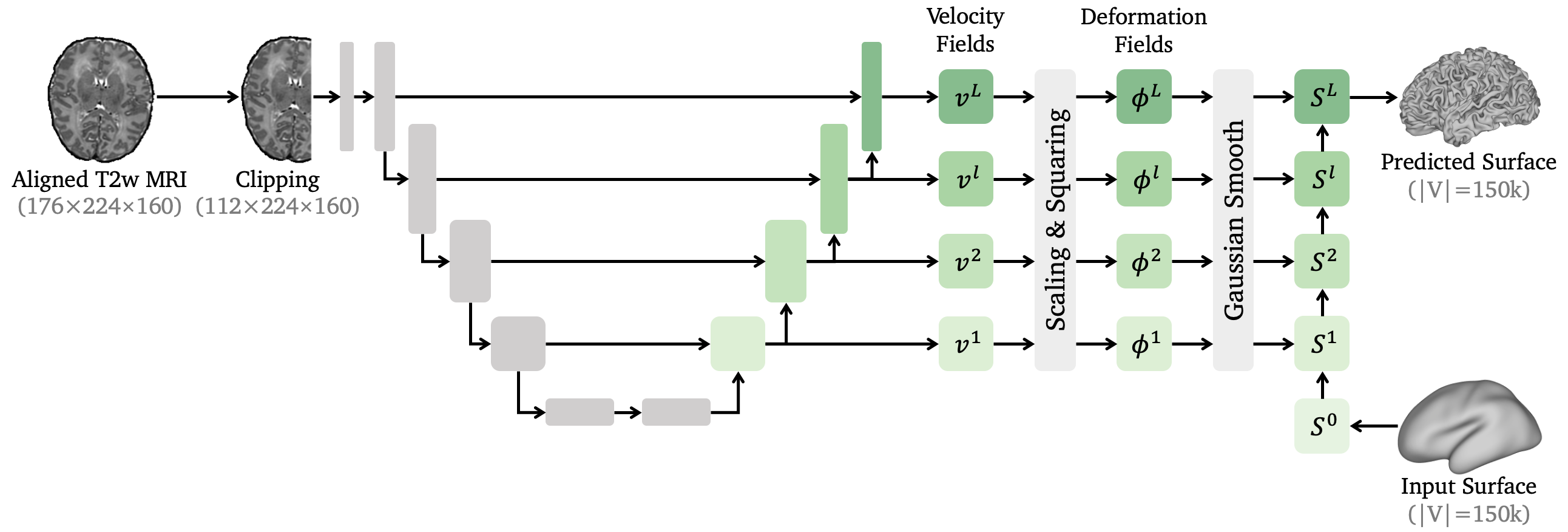
We propose an end-to-end learning-based approach for cortical surface reconstruction. To tackle the large deformation required by neonatal cortical surfaces, we introduce multiscale deformation networks, which learn a sequence of diffeomorphic transformations to deform an initial surface to white matter and pial surfaces. The architecture of the proposed framework is shown in Figure 5.
2.5.1 Diffeomorphic Surface Deformation
Diffeomorphic transformation has been extensively applied to medical image registration [8, 3, 63, 5] and been introduced for learning-based cortical surface reconstruction [35, 54, 38, 37]. For any domain , the diffeomorphic deformations can be modeled by the following flow ordinary differential equation (ODE):
| (1) |
where is a stationary velocity field (SVF), is a deformation field for , and is an identity map. If the SVF is sufficiently smooth, then represents an one-parameter subgroup of diffeomorphisms generated by and satisfies the group law [3, 5]. Therefore, we can solve the ODE (1), of which the solution is a diffeomorphism , using the scaling and squaring method [2, 3, 5] to iteratively compute . For surface deformation, the deformation field evolves an initial surface to a target surface while preserving the topology of the surface. Since is a differentiable bijection, any two distinct points on the initial surface will not be mapped to the same location, which provides theoretical guarantee for preventing surface self-intersections.
2.5.2 Multiscale Deformation Network
Since a single SVF has limited representation ability, in this work, we propose multiscale deformation networks to learn a series of diffeomorphic deformation fields for cortical surface reconstruction. As shown in Figure 5, given an affinely aligned T2w image, we clip it to the size of for the left or right brain hemispheres. The multiscale deformation network predicts cortical surfaces end-to-end from the clipped brain MRI. Note that the brain segmentations are not required as the input, which would cause subsequent corruptions on the surfaces in cases where segmentations are inaccurate.
We use a 3D U-Net to extract multiscale feature maps from the input clipped T2w image. Then, a sequence of multiscale volumetric SVFs are predicted from the extracted feature maps. Previous work [35, 38] adopted the forward Euler method to solve the ODE (1), which is fast but may introduce large numerical errors. Instead, we use the scaling and squaring method [2] for integration, which is more accurate and widely used for medical image registration tasks [3, 5]. For each scale , we set the initial transformation as , and then the deformation field can be updated recurrently by , where is the steps of the scaling and squaring. This is equivalent to steps of forward Euler.
By integration, the network predicts multi-resolution diffeomorphic deformation fields , each of which downsamples the original volume by a factor of for . This allow the network to model coarse-to-fine surface deformations. The last deformation field has the same size as the input MRI volume to refine surface details. To improve the smoothness of the deformation fields, we further employ Gaussian smoothing with standard deviation . Finally, we apply the deformation fields iteratively to an input surface . The displacements of the surface mesh vertices are sampled by linear interpolation. The surfaces are updated by for as shown in Figure 6, where is the final predicted white matter or pial surface. For white matter surface extraction, we use an initial template surface described in Figure 3 as the input surface for all subjects. The predicted white matter surfaces are further refined by Taubin smoothing [60] to improve the mesh quality. For pial surface reconstruction, we use the predicted white matter surface as the input surface. Since the initial surface has genus-0 topology and the diffeomorphic deformations are topology-preserving, all reconstructed cortical surfaces have the same topology as a 2-sphere.

2.5.3 Model Training
We train four multiscale deformation networks for white matter and pial surface reconstruction for both brain hemispheres. We consider the following loss function:
| (2) |
where is the Chamfer distance [19, 67], is the edge length loss [66, 67], is the normal consistency loss [66, 67, 37], and are weights for regularization terms. The Chamfer distance is defined as the bidirectional distance between two point sets [19]:
| (3) |
We generate pseudo GT cortical surfaces using the original dHCP pipeline, and compute the Chamfer distance between the mesh vertices of the predicted and pseudo GT surfaces. All pseudo GT surfaces are remeshed to have 150k vertices to reduce the errors introduced by point matching during the computation of the Chamfer distance. We adopt PyTorch3D [50] which provides fast computation and gradient backpropagation for the Chamfer distance.
Edge length and normal consistency constraints are introduced to encourage the smoothness and improve the mesh quality of the predicted cortical surfaces. For a surface mesh , where are the sets for vertices, edges and faces respectively, the edge length loss is defined as:
| (4) |
where are two vertices connected by the edge . The normal consistency loss is defined as:
| (5) |
Here is the unit normal vector of the face , and is the edge where the adjacent faces and intersect. The normal consistency loss penalizes the dot product of the normal vectors between any two adjacent faces.
For the architecture of the multiscale deformation network, we set =4 as the number of scales. The numbers of channels for hidden layers are set to for white matter surface reconstruction and for pial surface to avoid overfitting. We use the same hyperparameters for both white matter and pial surface reconstruction. The regularization weights are set to =0.3 and =3.0. The number of scaling and squaring steps is set to =7. We use the Adam optimizer with learning rate to train the multiscale deformation networks for 200 epochs. After training, we select the best models with minimum validation error, i.e., minimum Chamfer distances between predicted and pseudo GT cortical surfaces.
2.6 Cortical Surface Inflation
Following the HCP [25] and original dHCP pipelines [44], our dHCP DL pipeline incorporates two types of cortical surface inflation. Firstly, the original pipeline employs Connectome Workbench [25] to iteratively inflate and smooth the midthickness surface to generate inflated and very inflated surfaces. These surfaces are primarily used for visualization. Our pipeline re-implements these surface inflation approaches using PyTorch to facilitate GPU acceleration.
Secondly, the original dHCP pipeline adopts FreeSurfer mris_inflate command [22, 21], which is reproduced in the Medical Image Registration ToolKit (MIRTK) [56, 44], to inflate the white matter surface while minimizing the metric distortion between white matter and inflated surfaces. During the inflation, the cortical sulcal depth, or average convexity for each mesh vertex is computed by integrating the displacement of the vertex along its normal direction. Our DL-based pipeline also accelerates such an inflation process by PyTorch with parallel computation on a GPU.
2.7 Cortical Feature Estimation
Based on the extracted cortical surfaces, our DL-based dHCP pipeline provides the estimation of cortical morphological features such as cortical thickness, curvature and sulcal depth, as well as functional features such as a myelin map. The estimation of cortical features is consistent with previous pipelines [25, 44]. The cortical thickness is computed by averaging the bidirectional distances between white matter and pial surfaces. This is implemented with the SciPy package [64] in Python. Furthermore, the mean curvature of the white matter surface is computed via PyTorch-based re-implementation [41, 25]. The sulcal depth is measured during cortical surface inflation as described in Section 2.6.
For the myelin map estimation, if the T1 image is provided and aligned to the T2 image, we divide the T1 image by the original T2 image before intensity normalization to compute the T1/T2 ratio. Then, the myelin map is estimated by projecting the T1/T2 ratio onto the midthickness surface using volume-to-surface mapping [26, 25, 44], which is implemented with the Connectome Workbench. For each vertex of the midthickness surface mesh, we find its corresponding location in the T1/T2 ratio volume, and compute the Gaussian average of its neighboring voxels as the myelin map value, where the size of the neighbor depends on the cortical thickness. Only voxels within the cortical ribbon ROI are considered. To accelerate the volume-to-surface mapping, we train a 3D U-Net to learn the binary cortical ribbon mask, i.e., the cortical GM segmentation. The learning procedure is similar to brain extraction as shown in Figure 4. The GT segmentation is generated by the Draw-EM approach integrated in the original dHCP pipeline.
2.8 Spherical Projection
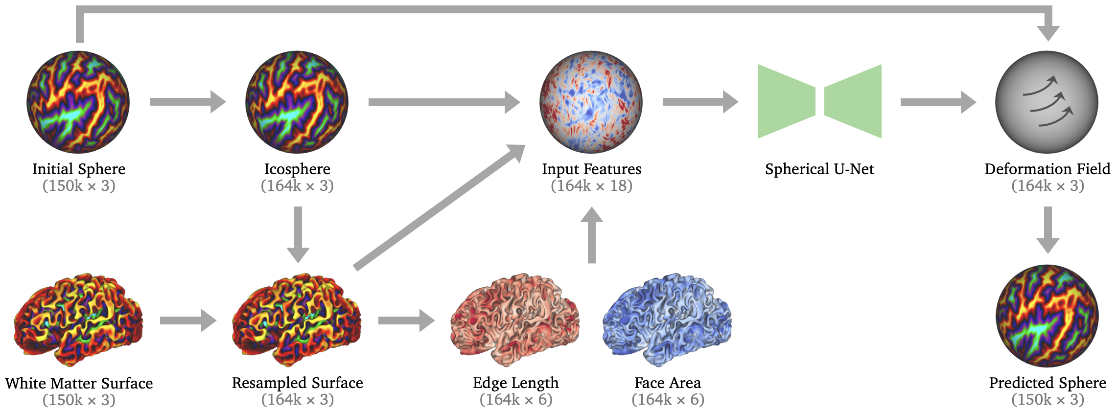
The purpose of spherical projection or spherical mapping is to project the white matter surface onto a sphere while minimizing metric distortions. Motivated by [70], we propose an unsupervised learning framework for spherical projection and integrate it into our DL-based dHCP pipeline. As illustrated in Figure 7, the inputs of our spherical projection framework include a white matter surface and a fixed initial sphere for all subjects. Note that since all white matter surfaces are deformed from the same initial template surface, the white matter surface has the same mesh connectivity as the initial sphere, which is generated from the initial surface by FreeSurfer (see Figure 3). Therefore, instead of inflating the white matter surface and projecting the inflated surface to create a sphere for each subject [22, 18, 70], our approach learns to deform the initial sphere directly while minimizing metric distortions between the deformed sphere and the white matter surface. In this work, we primarily consider edge and area distortions.
We resample the input white matter surface mesh such that it has the same connectivity as a standard icosphere with 163,842 vertices. This is achieved by barycentric interpolation [52, 51] between the initial sphere and the icosphere, of which the barycentric coordinates are pre-computed to accelerate the resampling. Then, we compute the metrics of the resampled white matter surface including edge length and face area. Each vertex contains the metrics of its adjacent edges and faces. We concatenate the coordinates of the resampled white matter surface and the icosphere as well as computed metrics as input features of a Spherical U-Net [71], which has been widely used in cortical surface analysis tasks such as cortical surface registration [68], parcellation [72] and development prediction [69]. We train the Spherical U-Net to learn a diffeomorphic deformation field in the spherical domain by integrating a SVF using scaling and squaring [2]. Rather than adding regularization terms to the loss function [70], we apply Laplacian mesh smoothing to the deformation field to encourage smoothness. The predicted sphere is derived by deforming the initial sphere, where the displacement of each vertex is determined by sampling from the deformation field using barycentric interpolation.
[70] employed multi-resolution Spherical U-Nets and computed the losses between the deformed icosphere and the resampled white matter surface with 164k vertices. In contrast, our approach minimizes the metric distortions end-to-end between the predicted sphere and the original input white matter surface. Hence, a single-scale Spherical U-Net is sufficient to achieve accurate results. Inspired by [70], we train the Spherical U-Net by minimizing an unsupervised metric distortion loss, which is defined as the minimum root mean square deviation (RMSD) between the metrics of predicted sphere and the original white matter surface:
| (6) |
where are the metrics and is a scaling coefficient. The loss (6) achieves minimum when
| (7) |
The metrics in Eq. (6) can be replaced by the mesh edge length or face area. The overall loss function is defined as , where are the edge and area distortions, and are weights for distortions. For model training, we use the Adam optimizer with learning rate to train for 200 epochs. The weights of losses are set to and . After training, we select the model with lowest metric distortions on the validation set.
| Original dHCP Pipeline | DL-based dHCP Pipeline (Ours) | |
|---|---|---|
| Brain Extraction | FSL BET [59] | Supervised Learning + U-Net |
| Tissue Segmentation | Draw-EM [43] | — |
| Cortical Surface Reconstruction | Deformable Model [56] | Multiscale Deformation Networks |
| Spherical Projection | Spherical MDS [18] | Unsupervised Learning + Spherical U-Net |
3 Comparison to Existing Neuroimage Pipelines
3.1 Comparison to Original dHCP Pipeline
We compare our DL-based dHCP neonatal pipeline to the original dHCP pipeline [44] with respect to the approach, implementation and runtime. The comparison of surface and sphere quality are provided in the Section 4.
| Processing Steps | Implementation | GPU | CPU only |
|---|---|---|---|
| Brain Extraction | PyTorch | 0.25s | 1.63s |
| Bias Field Correction | ANTsPy | 2.62s | 2.64s |
| Affine Registration | ANTsPy | 2.76s | 2.77s |
| Surface Reconstruction | PyTorch3D | 2.37s | 37.01s |
| Surface Inflation | PyTorch | 4.99s | 21.28s |
| Spherical Projection | PyTorch | 0.68s | 1.35s |
| Cortical Thickness | SciPy | 8.73s | 102.15s |
| Mean Curvature | PyTorch | ||
| Sulcal Depth | PyTorch | ||
| Myelin Map | WB & PyTorch | ||
| Total Runtime | 23.98s | 170.29s |
3.1.1 Approach and Implementation
We compare the different approaches of the main processing steps between the original and our DL-based dHCP pipelines in Table 1. Our DL-based pipeline integrates novel learning-based approaches for the crucial processing steps, and predicts cortical surfaces end-to-end without the need of tissue segmentation.
The original dHCP pipeline is implemented with MIRTK [44], Connectome Workbench [25], and FSL [31]. Since the installation and configuration of the MIRTK package are complicated, it is difficult to reproduce or modify the original dHCP pipeline. For our DL-based dHCP pipeline, the implementation of each processing step is described in Table 2. Our DL-based dHCP pipeline is primarily implemented in Python and PyTorch [48], which simplify deployment, enhance reproducibility, and allow for more straightforward modifications and adaptations. The PyTorch library provides seamless support for parallel computation and acceleration on GPUs. The PyTorch3D package [50] is required for the training of cortical surface reconstruction and provides utilities for mesh-based processing. ANTsPy [4] is used for N4 bias correction and affine registration. The Connectome Workbench (WB) is used for visualization and myelin map estimation. Our DL-based pipeline is GPU memory-efficient, which only uses 12GB for training and 7.6GB GPU memory for inference. Therefore, it is simple to adapt the pipeline to a new dataset by fine-tuning the neural network models.
3.1.2 Runtime
To measure the runtime, we run both original and DL-based dHCP pipeline to process the brain MRI of 10 dHCP neonatal subjects, which are randomly selected from the test set. The runtime is measured on an 8-core Intel Core i7-11700K CPUs and a NVIDIA GeForce RTX 3080 GPU with 12GB memory. The original dHCP pipeline [44] runs exclusively on CPUs and requires 6h38min to process a single subject. The volume-based and surface-based processing requires 2h9min and 4h29min respectively. For our DL-based pipeline, we report the runtime with and without GPU acceleration in Table 2. With GPU acceleration, it only takes 24 seconds to run the entire DL-based pipeline, which is 995 faster than the original dHCP pipeline. The cortical surface reconstruction and spherical projection only needs 3 seconds in total owing to the powerful end-to-end deep learning approaches. Even without GPU acceleration, our DL-based pipeline only requires 170 seconds to complete on the CPU, which is still 140 faster compared to the original dHCP pipeline.
| Processing Steps | FastSurfer | iBEAT V2.0 | Ours |
|---|---|---|---|
| Preprocessing | ✓ | ✓ | |
| Brain Extraction | ✓ | ✓ | |
| Brain Segmentation | ✓ | ✓ | |
| Surface Reconstruction | ✓ | ✓ | ✓ |
| Surface Parcellation | ✓ | ✓ | |
| Spherical Projection | ✓ | ✓ | |
| Feature Estimation | ✓ | ✓ | ✓ |
| Approx Runtime | 1h | 4h | 24s |


3.2 Comparison to DL-based Neuroimage Pipelines
We compare our dHCP DL pipeline to existing DL-based neuroimage processing pipelines, including FastSurfer [28] and iBEAT V2.0 [65]. The comparisons regarding functionality and approximate runtime on a GPU are reported in Table 3. Table 3 shows that previous DL-based pipelines focus on learning-based approaches for brain regional and tissue segmentations. However, theses pipelines still adopt traditional approaches for cortical surface reconstruction including computationally intensive topology correction [6, 7, 57], and thus require more than one hour to process a single brain MRI scan. Our DL-based dHCP pipeline introduces fast learning-based methods to accelerate the most time-consuming cortical surface reconstruction and spherical projection. Therefore, our pipeline is 150 and 600 faster than FastSurfer and iBEAT V2.0 respectively.
Our DL-based dHCP pipeline extracts cortical surfaces end-to-end from the T2w brain MRI without the need of intermediate tissue segmentations. This prevents the propagation of errors from segmentations to cortical surfaces. Therefore, as shown in Table 3, currently our pipeline does not provide brain tissue segmentations and cortical surface parcellation. We will develop corresponding DL-based approaches and integrate them into our pipeline in future work.
4 Quality Control
4.1 Surface Quality
4.1.1 Visual Inspection
We conduct manual quality control (QC) via visual inspection to assess the quality of the cortical surfaces generated by our DL-based dHCP pipeline. More specifically, our QC aims to examine if our DL-based dHCP pipeline produces better cortical surfaces compared to the baseline of the original dHCP pipeline [44].
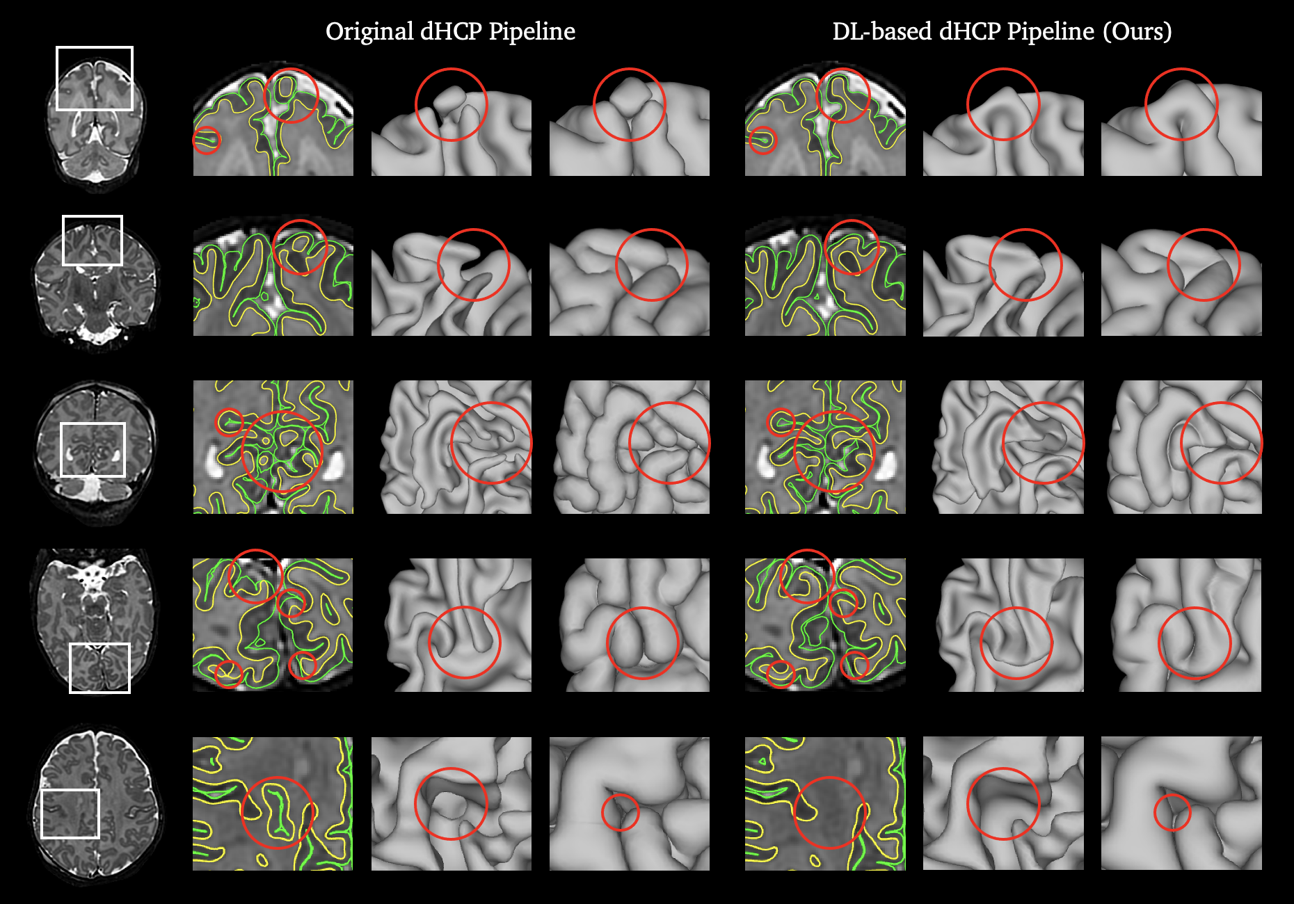
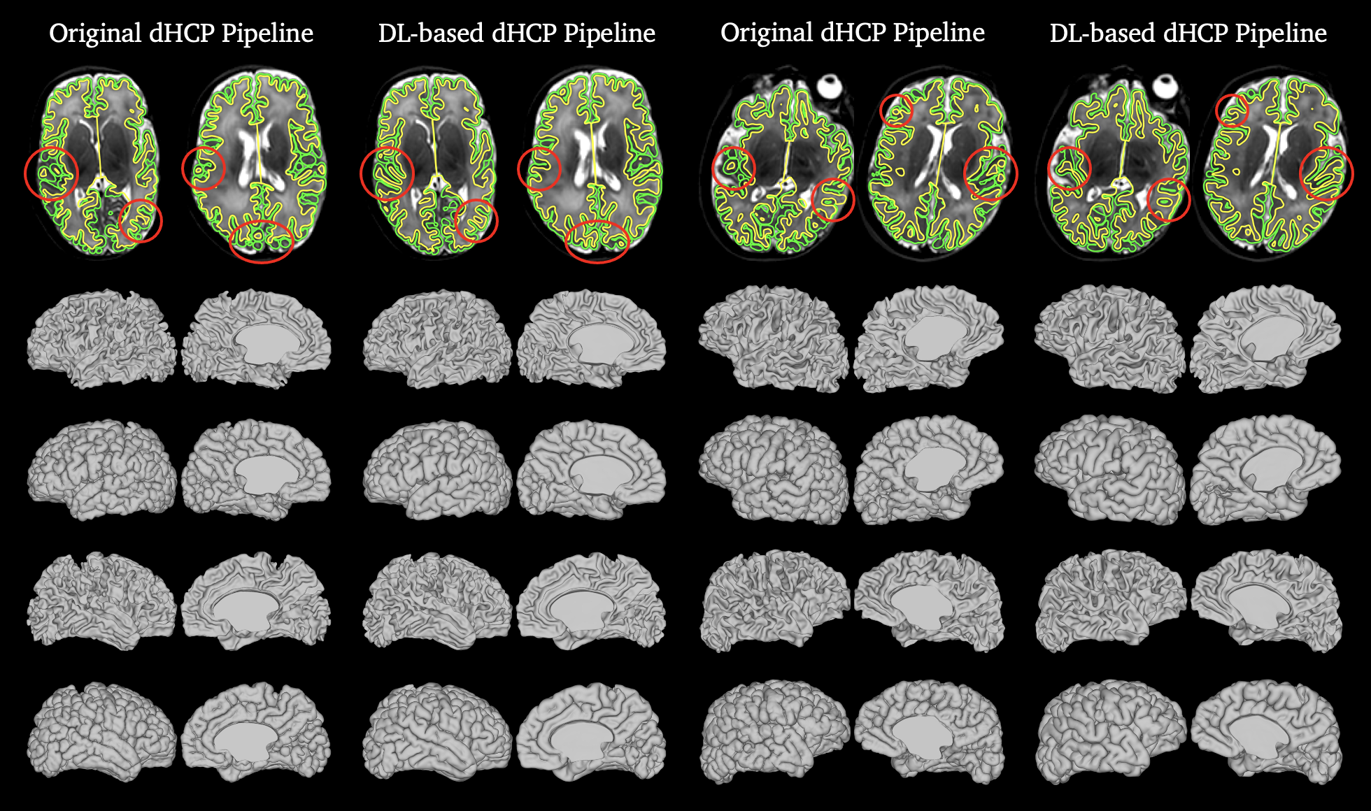
We randomly select 30 T2w brain MRI samples from 265 test cases and extract two groups of cortical surfaces for each sample by the original dHCP pipeline and our DL-based dHCP pipeline respectively. QC is performed by four expert assessors with clinical background or extensive experience in neuroimage analysis. To ensure fairness, all assessors are requested to visually evaluate two groups of surfaces independently, and select the group with better quality for each test sample without knowing the corresponding reconstruction approaches (original or DL-based pipeline). Equal quality is also an option when both groups of surfaces are anatomically accurate or corrupted to the same degree. The average and individual QC results are reported in Figure 8. The results show that for 82.5% of all test samples, our DL-based dHCP pipeline achieves superior (54.2%) or equal (28.3%) surface quality compared to the original dHCP pipeline. The individual QC results in Figure 8 also indicate that our DL-based dHCP pipeline achieves better performance in the evaluation of all assessors. These demonstrate that our DL-based dHCP pipeline reconstructs higher quality cortical surfaces, while being 995 faster than the original dHCP pipeline.
We further provide visual comparison between cortical surfaces reconstructed by original and DL-based dHCP pipeline. Figure 10 indicates that the original dHCP pipeline is prone to produce corruptions on the top and back (parietal and occipital lobes) of the cortical surfaces, due to the low image contrast in the corresponding regions of T2w brain MRI, whereas our DL-based pipeline achieves relatively high anatomical accuracy in these regions. In addition, the surfaces generated by the original dHCP pipeline are affected by the imprecise Draw-EM segmentations [43, 44], and thus occasionally produce unexpected ”caves” or ”holes” as shown in Figure 10. Our DL-based pipeline leverages diffeomorphic surface deformations and learns cortical surfaces end-to-end from brain MRI, which can effectively avoid such issues.
4.1.2 Brain Development
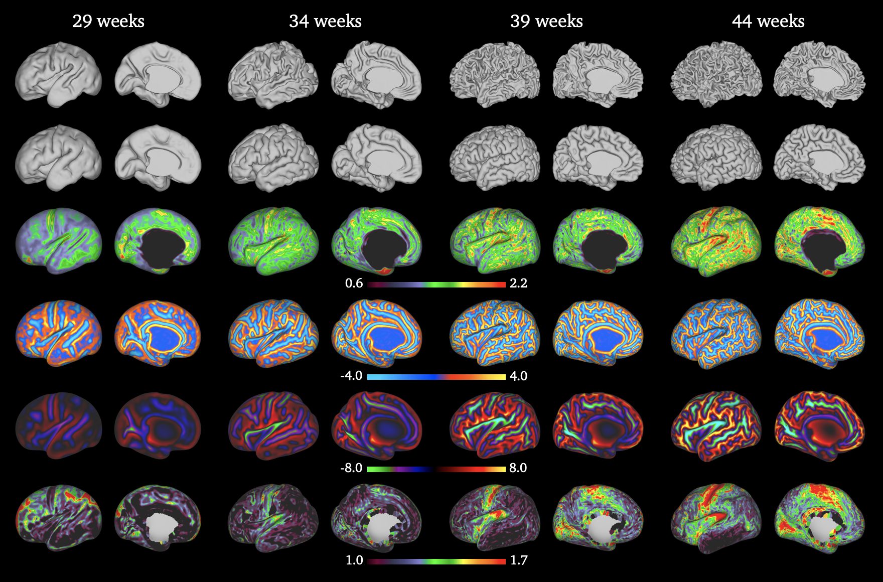
In addition to manual quality control, we provide qualitative and quantitative evaluation to validate that the cortical surfaces extracted by our dHCP DL pipeline can capture the brain development of neonates. We visualize the cortical surfaces as well as the corresponding cortical features of different neonatal subjects scanned at the PMA of 29,34,39,44 weeks in Figure 11. The cortical folding is observed in Figure 11 with the increasing of the PMA. The cortical thickness and sulcal depth are increasing as well with brain growth.
We further measure and plot cortical morphological features including cortical volume, surface area, cortical thickness and sulcal depth across all 265 test neonatal subjects in Figure 9. The cortical volume is equal to the volumes of pial surfaces minus those of the white matter surfaces for both brain hemispheres. The surface area refers to the mesh area of the pial surfaces. The cortical thickness and sulcal depth for each subject is measured by averaging the cortical thickness and the absolute value of the sulcal depth over all vertices on the surface meshes. Figure 9 shows the positive trends of the morphological features with increasing PMA, which reflect the growth and folding of the neonatal cerebral cortex. Moreover, we compute the Pearson correlation coefficients (PCC) to measure the correlation between the morphological features and the PMA. The PCCs are provided in Figure 9, which indicate that all cortical morphological features have strong positive correlation with the age of the neonates. This verifies that the cortical surfaces extracted by our DL-based pipeline are able to capture the brain development of neonates.
| Method | White Matter Surface | Pial Surface |
|---|---|---|
| Original dHCP pipeline | 0.4577.409 | 0.5177.418 |
| Forward Euler | 0.8645.712 | 4.92517.71 |
| w/o Gaussian smoothing | 0.6134.304 | 3.7159.824 |
| Ours | 0.1793.171 | 1.5008.875 |
4.1.3 Surface Topology
Our DL-based dHCP neonatal pipeline is able to guarantee the spherical topology of cortical surfaces. The proposed pipeline uses multiscale deformation networks to learn diffeomorphic deformations to deform an initial surface to cortical surfaces. As the diffeomorphic deformations preserve the topology of the surfaces and the initial surface has the same topology as a sphere, the cortical surfaces are topologically equivalent to a sphere as well. The Euler characteristic number is 2 for all extracted surface meshes as expected.
We further examine surface self-intersections, which usually appear in the narrow sulci of pial surfaces and will affect the estimation of cortical morphological features [38]. We detect and count the number of self-intersecting faces (SIFs) in white matter and pial surface meshes generated by both original and DL-based dHCP pipelines. To verify that the proposed multiscale deformation networks can effectively reduce SIFs, we conduct ablation experiments by removing the Gaussian smoothing layer or replacing the scaling and squaring method [2] with the forward Euler method for integration. The number of SIFs are reported in Table 4. It shows that our DL-based dHCP pipeline produces only 0.18 SIFs on average in white matter surfaces. The original dHCP pipeline produces marginally fewer SIFs in pial surfaces than ours since it performs collision detection during the iterative surface deformation. Table 4 also validates that SIFs can be reduced effectively by adding the Gaussian smoothing layer and using the scaling and squaring method.
4.1.4 Generalization ability
In addition to the dHCP neonatal dataset [17], we evaluate the generalization ability of our DL-based dHCP pipeline on an unseen dataset. We consider the Evaluation of Preterm Imaging (ePrime) dataset333https://www.npeu.ox.ac.uk/research/projects/22-eprime-mr-imaging. [16, 27], comprising 486 T2w MRI scans of neonates with gestational age between 23 and 33 weeks, in which 444 subjects are scanned at term-equivalent age between 38 and 45 weeks. The MR images are acquired with a Philips Intera 3T scanner, using a T2w turbo echo sequence (TES) with parameters repetition/echo time TR/TE=8670/160ms and TSE factor of 16. The ePrime dataset has relatively low image resolution with the voxel size of , and thus it is challenging to extract high-quality cortical surfaces.
To test the capacity of generalization, we run both original and our DL-based dHCP pipelines on unseen T2w MRI scans of the ePrime dataset without fine-tuning. The qualitative comparison in Figure 12 shows that there are severe corruptions and discontinuity on the gyri of cortical surfaces generated by the original dHCP pipeline, while our DL-based pipeline produces cortical surfaces with acceptable quality. This demonstrates that our DL-based pipeline has better generalize ability on the unseen and low-resolution dataset.
| Method | Edge () | Area () | Runtime |
| Spherical MDS | 0.1860.039 | 0.0640.020 | 74min |
| FastSurfer | 0.2840.064 | 0.1060.033 | 2.73s |
| FreeSurfer | 0.1230.025 | 0.0330.010 | 4min12s |
| Initial Sphere | 0.1650.033 | 0.0550.017 | – |
| [70] | 0.1390.026 | 0.0350.010 | 2.11s |
| Ours | 0.1190.023∗ | 0.0230.006∗ | 0.33s∗ |
4.2 Sphere Quality
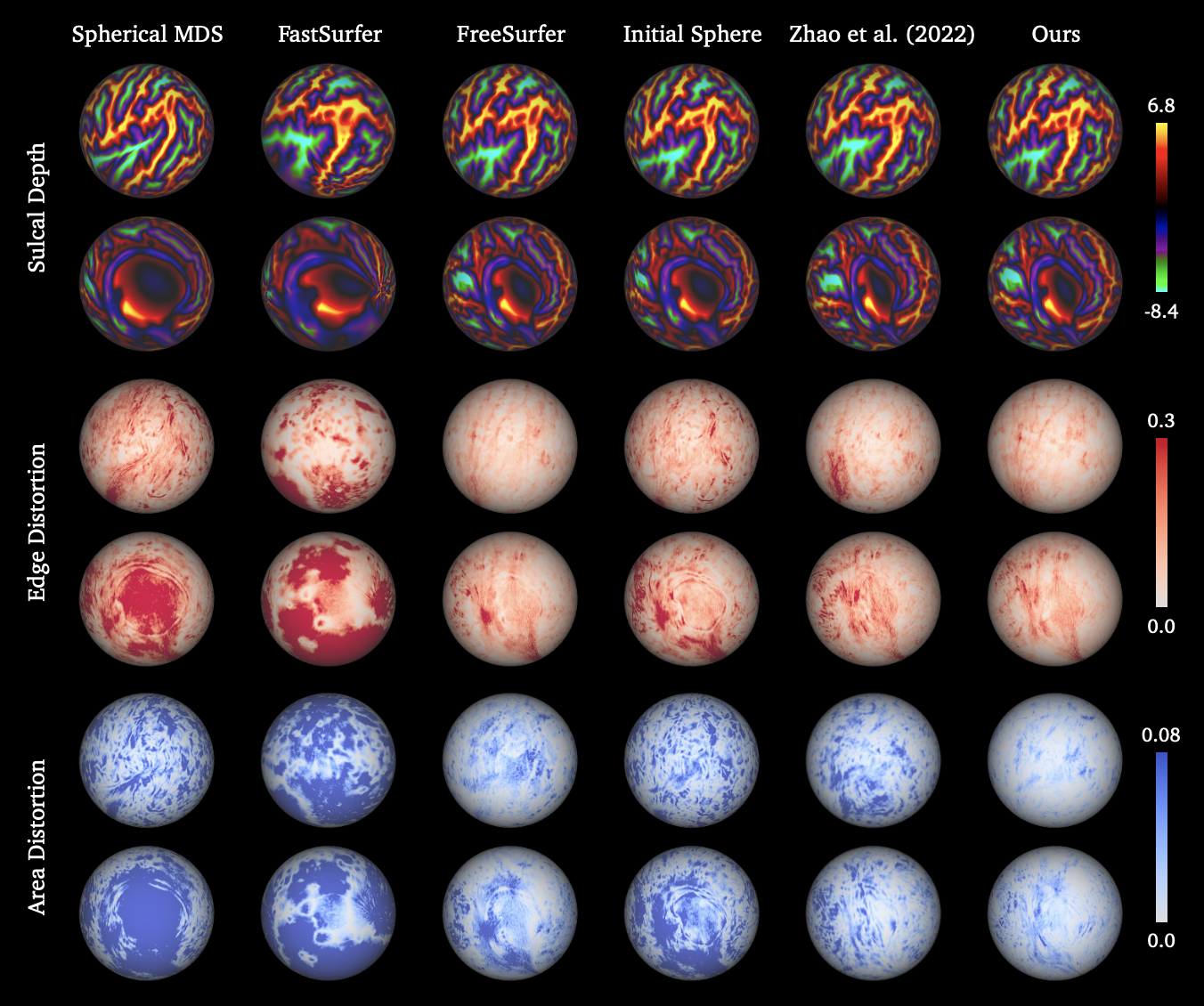
We evaluate the performance of the spherical projection by the metric distortions defined in Eq. (6) on 265 test samples in the dHCP neonatal dataset. Similar to the original dHCP pipeline [44], the quality of the sphere is measured by the distortions of the edge length and face area between the white matter surface and the projected sphere. We compare our unsupervised learning-based approach with existing spherical mapping methods including Spherical MDS [18], FreeSurfer [21], FastSurfer [28], and [70]. We also provide the metric distortions between the white matter surface and the initial sphere as a baseline. For fair comparison, we only use a single-scale Spherical U-Net in [70] like our approach. The edge and area distortions, as well as the average runtime for each brain hemisphere are reported in Table 5. We conduct paired t-tests to examine if our unsupervised learning approach performs significantly better than other baseline methods.
Table 5 shows that our approach achieves significantly better performance () compared to all baseline methods and only requires 0.33 seconds of runtime. As reported in Table 5, the runtime of Spherical MDS [18] is more than one hour due to the time-consuming pairwise geodesic distance computation. Despite the efficiency of the spherical embedding approach in FastSurfer [28], it does not optimize the metric distortions. FreeSurfer [21] achieves satisfactory performance but it takes 4 minutes to process each hemisphere. The initial sphere, which is generated by FreeSurfer as a good starting point, effectively reduces the baseline metric distortions without additional deformations.
Compared to [70], our approach has several advantages. First, [70] needs to inflate and project the white matter surfaces to create initial spheres, which requires 1.79 seconds for each brain hemisphere with GPU acceleration in our experiments. Instead, our approach uses an fixed initial sphere for all subjects without extra processing. The initial sphere is generated by FreeSurfer with minimized metric distortions as reported in Table 5. This provides a strong prior with small initial distortions for the training. Such a global initial surface also saves the runtime of interpolation and resampling, since the barycentric coordinates can be pre-computed. Hence, our approach is capable of projecting the cortical surface to a sphere in 0.33 seconds, which is 6 faster than [70]. Furthermore, [70] computes the metric distortions between the resampled white matter surface and the icosphere, and thus requires multiscale networks to improve the performance. As reported in Table 5, [70] does not outperform FreeSurfer in the case of single-scale architecture. Our end-to-end metric distortion loss is more effective so that a single-scale Spherical U-Net is sufficient to produce significantly smaller distortions than FreeSurfer.
We also provide qualitative comparison as shown in Figure 13, which visualizes the projected spheres overlaid with cortical sulcal depth, edge and area distortions for all approaches. Figure 13 shows that Spherical MDS and FastSurfer produce large distortions for edge length and face area. Compared to FreeSurfer, which is the best baseline method as reported in Table 5, our unsupervised learning approach achieves similar visualization of sulcal depth, similar edge distortions, much smaller area distortions, and faster runtime.
5 Discussion
In this work, we presented a fast DL-based cortical surface reconstruction pipeline for structural brain MRI processing and analysis on the dHCP neonatal dataset. The proposed DL-based dHCP pipeline, which is primarily implemented in Python and PyTorch, consists of learning-based brain extraction, cortical surface reconstruction and spherical projection, as well as GPU-accelerated cortical surface inflation and feature estimation. The entire pipeline executes within only 24 seconds accelerated by GPU and takes less than 3 minutes even without GPU support. This is orders of magnitude faster than traditional neuroimage pipelines [21, 25, 44] that require at least 6 hours of runtime, as well as existing DL-based cortical surface reconstruction pipelines [28, 65] that need more than one hour.
The quality control results validate that for more than 80% of test samples, our DL-based dHCP pipeline produces more anatomically accurate (54.2%) or equal quality (28.3%) cortical surfaces compared to the original dHCP pipeline [44]. Particularly, the visual inspection shows that our learning-based cortical surface reconstruction approach can effectively reduces the artifacts on the top and occipital lobe of the cortical surfaces produced by original dHCP pipeline. We also verify that the proposed DL-based dHCP pipeline is capable of generalizing well on unseen and low-resolution ePrime neonatal brain MRI data [16, 27]. In addition, our unsupervised learning-based spherical projection framework significantly reduces both edge and area distortions compared to previous work while only requires 0.33 seconds of runtime for each brain hemisphere.
However, despite the high efficacy and efficiency of the proposed dHCP DL pipeline, the anatomical accuracy of predicted cortical surfaces is inevitably restricted by the quality of pseudo GT cortical surfaces generated by the original dHCP pipeline, due to the limitations of supervised learning. Besides, the proposed multiscale deformation network predicts cortical surfaces from the input T2w brain MRI directly without the need of segmentations. Such an end-to-end framework facilitates the surface reconstruction procedure and avoids the potential errors in cortical surfaces introduced by imprecise segmentations, while this also means that our dHCP DL pipeline does not provide brain tissue segmentations and cortical surface parcellation.
In future work, we will consider semi-supervised learning technique to further enhance the anatomical accuracy of the cortical surfaces. In addition to pseudo GT cortical surfaces, the unsupervised brain MRI intensity will be utilized to deform the cortical surfaces towards the WM/cGM or cGM/CSF boundary where the MRI achieves maximum image contrast. Furthermore, we plan to enrich the functionality and incorporate more GPU-accelerated processing steps into our DL-based dHCP pipeline, e.g., brain regional and tissue segmentations, cortical parcellation, and local gyrification index estimation.
Acknowledgments
Qiang Ma is funded by the President’s PhD Scholarship at Imperial College London. Kaili Liang is supported by National Institute for Health Research (NIHR) Maudsley Biomedical Research Centre (BRC) PhD studentship. Liu Li is supported by Lee Family Scholarship. Support was also received from the ERC project MIA-NORMAL 101083647 and ERC project Deep4MI 884622. The dHCP neonatal dataset was provided by the developing Human Connectome Project, KCL-Imperial-Oxford Consortium funded by the ERC under the European Union Seventh Framework Programme (FP/2007-2013) / ERC Grant Agreement no. [319456]. We are grateful to the families who generously supported this trial.
References
- Abadi et al. [2016] Abadi, M., Agarwal, A., Barham, P., Brevdo, E., Chen, Z., Citro, C., Corrado, G.S., Davis, A., Dean, J., Devin, M., et al., 2016. Tensorflow: Large-scale machine learning on heterogeneous distributed systems. arXiv preprint arXiv:1603.04467 .
- Arsigny et al. [2006] Arsigny, V., Commowick, O., Pennec, X., Ayache, N., 2006. A log-euclidean framework for statistics on diffeomorphisms, in: Medical Image Computing and Computer-Assisted Intervention–MICCAI 2006: 9th International Conference, Copenhagen, Denmark, October 1-6, 2006. Proceedings, Part I 9, Springer. pp. 924–931.
- Ashburner [2007] Ashburner, J., 2007. A fast diffeomorphic image registration algorithm. Neuroimage 38, 95–113.
- Avants et al. [2009] Avants, B.B., Tustison, N., Song, G., et al., 2009. Advanced normalization tools (ANTS). Insight j 2, 1–35.
- Balakrishnan et al. [2019] Balakrishnan, G., Zhao, A., Sabuncu, M.R., Guttag, J., Dalca, A.V., 2019. VoxelMorph: a learning framework for deformable medical image registration. IEEE transactions on medical imaging 38, 1788–1800.
- Bazin and Pham [2005] Bazin, P.L., Pham, D.L., 2005. Topology correction using fast marching methods and its application to brain segmentation, in: International Conference on Medical Image Computing and Computer-Assisted Intervention, Springer. pp. 484–491.
- Bazin and Pham [2007] Bazin, P.L., Pham, D.L., 2007. Topology correction of segmented medical images using a fast marching algorithm. Computer methods and programs in biomedicine 88, 182–190.
- Beg et al. [2005] Beg, M.F., Miller, M.I., Trouvé, A., Younes, L., 2005. Computing large deformation metric mappings via geodesic flows of diffeomorphisms. International journal of computer vision 61, 139–157.
- Billot et al. [2023] Billot, B., Magdamo, C., Cheng, Y., Arnold, S.E., Das, S., Iglesias, J.E., 2023. Robust machine learning segmentation for large-scale analysis of heterogeneous clinical brain MRI datasets. Proceedings of the National Academy of Sciences 120, e2216399120.
- Bongratz et al. [2022] Bongratz, F., Rickmann, A.M., Pölsterl, S., Wachinger, C., 2022. Vox2Cortex: Fast explicit reconstruction of cortical surfaces from 3D MRI scans with geometric deep neural networks, in: Proceedings of the IEEE/CVF Conference on Computer Vision and Pattern Recognition, pp. 20773–20783.
- Bozek et al. [2018] Bozek, J., Makropoulos, A., Schuh, A., Fitzgibbon, S., Wright, R., Glasser, M.F., Coalson, T.S., O’Muircheartaigh, J., Hutter, J., Price, A.N., et al., 2018. Construction of a neonatal cortical surface atlas using multimodal surface matching in the developing human connectome project. NeuroImage 179, 11–29.
- Cordero-Grande et al. [2018] Cordero-Grande, L., Hughes, E.J., Hutter, J., Price, A.N., Hajnal, J.V., 2018. Three-dimensional motion corrected sensitivity encoding reconstruction for multi-shot multi-slice MRI: application to neonatal brain imaging. Magnetic resonance in medicine 79, 1365–1376.
- Cruz et al. [2021] Cruz, R.S., Lebrat, L., Bourgeat, P., Fookes, C., Fripp, J., Salvado, O., 2021. DeepCSR: A 3D deep learning approach for cortical surface reconstruction, in: Proceedings of the IEEE/CVF Winter Conference on Applications of Computer Vision, pp. 806–815.
- Dai et al. [2013] Dai, Y., Shi, F., Wang, L., Wu, G., Shen, D., 2013. iBEAT: a toolbox for infant brain magnetic resonance image processing. Neuroinformatics 11, 211–225.
- Dale et al. [1999] Dale, A.M., Fischl, B., Sereno, M.I., 1999. Cortical surface-based analysis I: Segmentation and surface reconstruction. Neuroimage 9, 179–194.
- Edwards et al. [2018] Edwards, A.D., Redshaw, M.E., Kennea, N., Rivero-Arias, O., Gonzales-Cinca, N., Nongena, P., Ederies, M., Falconer, S., Chew, A., Omar, O., et al., 2018. Effect of MRI on preterm infants and their families: a randomised trial with nested diagnostic and economic evaluation. Archives of Disease in Childhood-Fetal and Neonatal Edition 103, F15–F21.
- Edwards et al. [2022] Edwards, A.D., Rueckert, D., Smith, S.M., Seada, S.A., Alansary, A., Almalbis, J., Allsop, J., Andersson, J., Arichi, T., Arulkumaran, S., et al., 2022. The developing human connectome project neonatal data release. Frontiers in Neuroscience 16.
- Elad et al. [2005] Elad, A., Keller, Y., Kimmel, R., 2005. Texture mapping via spherical multi-dimensional scaling, in: International Conference on Scale-Space Theories in Computer Vision, Springer. pp. 443–455.
- Fan et al. [2017] Fan, H., Su, H., Guibas, L.J., 2017. A point set generation network for 3D object reconstruction from a single image, in: Proceedings of the IEEE conference on computer vision and pattern recognition, pp. 605–613.
- Fetit et al. [2020] Fetit, A.E., Alansary, A., Cordero-Grande, L., Cupitt, J., Davidson, A.B., Edwards, A.D., Hajnal, J.V., Hughes, E., Kamnitsas, K., Kyriakopoulou, V., et al., 2020. A deep learning approach to segmentation of the developing cortex in fetal brain MRI with minimal manual labeling, in: Medical Imaging with Deep Learning, PMLR. pp. 241–261.
- Fischl [2012] Fischl, B., 2012. Freesurfer. Neuroimage 62, 774–781.
- Fischl et al. [1999a] Fischl, B., Sereno, M.I., Dale, A.M., 1999a. Cortical surface-based analysis II: Inflation, flattening, and a surface-based coordinate system. Neuroimage 9, 195–207.
- Fischl et al. [1999b] Fischl, B., Sereno, M.I., Tootell, R.B., Dale, A.M., 1999b. High-resolution intersubject averaging and a coordinate system for the cortical surface. Human brain mapping 8, 272–284.
- Garcia et al. [2018] Garcia, K.E., Robinson, E.C., Alexopoulos, D., Dierker, D.L., Glasser, M.F., Coalson, T.S., Ortinau, C.M., Rueckert, D., Taber, L.A., Van Essen, D.C., et al., 2018. Dynamic patterns of cortical expansion during folding of the preterm human brain. Proceedings of the National Academy of Sciences 115, 3156–3161.
- Glasser et al. [2013] Glasser, M.F., Sotiropoulos, S.N., Wilson, J.A., Coalson, T.S., Fischl, B., Andersson, J.L., Xu, J., Jbabdi, S., Webster, M., Polimeni, J.R., et al., 2013. The minimal preprocessing pipelines for the Human Connectome Project. Neuroimage 80, 105–124.
- Glasser and Van Essen [2011] Glasser, M.F., Van Essen, D.C., 2011. Mapping human cortical areas in vivo based on myelin content as revealed by T1-and T2-weighted MRI. Journal of neuroscience 31, 11597–11616.
- Grigorescu et al. [2021] Grigorescu, I., Vanes, L., Uus, A., Batalle, D., Cordero-Grande, L., Nosarti, C., Edwards, A.D., Hajnal, J.V., Modat, M., Deprez, M., 2021. Harmonized segmentation of neonatal brain MRI. Frontiers in Neuroscience 15, 662005.
- Henschel et al. [2020] Henschel, L., Conjeti, S., Estrada, S., Diers, K., Fischl, B., Reuter, M., 2020. Fastsurfer-a fast and accurate deep learning based neuroimaging pipeline. NeuroImage 219, 117012.
- Hoopes et al. [2021] Hoopes, A., Iglesias, J.E., Fischl, B., Greve, D., Dalca, A.V., 2021. TopoFit: Rapid reconstruction of topologically-correct cortical surfaces, in: Medical Imaging with Deep Learning.
- Hughes et al. [2017] Hughes, E.J., Winchman, T., Padormo, F., Teixeira, R., Wurie, J., Sharma, M., Fox, M., Hutter, J., Cordero-Grande, L., Price, A.N., et al., 2017. A dedicated neonatal brain imaging system. Magnetic resonance in medicine 78, 794–804.
- Jenkinson et al. [2012] Jenkinson, M., Beckmann, C.F., Behrens, T.E., Woolrich, M.W., Smith, S.M., 2012. FSL. Neuroimage 62, 782–790.
- Kim et al. [2005] Kim, J.S., Singh, V., Lee, J.K., Lerch, J., Ad-Dab’bagh, Y., MacDonald, D., Lee, J.M., Kim, S.I., Evans, A.C., 2005. Automated 3-D extraction and evaluation of the inner and outer cortical surfaces using a Laplacian map and partial volume effect classification. Neuroimage 27, 210–221.
- Kingma and Ba [2014] Kingma, D.P., Ba, J., 2014. Adam: A method for stochastic optimization. arXiv preprint arXiv:1412.6980 .
- Kuklisova-Murgasova et al. [2012] Kuklisova-Murgasova, M., Quaghebeur, G., Rutherford, M.A., Hajnal, J.V., Schnabel, J.A., 2012. Reconstruction of fetal brain MRI with intensity matching and complete outlier removal. Medical image analysis 16, 1550–1564.
- Lebrat et al. [2021] Lebrat, L., Santa Cruz, R., de Gournay, F., Fu, D., Bourgeat, P., Fripp, J., Fookes, C., Salvado, O., 2021. CorticalFlow: A diffeomorphic mesh transformer network for cortical surface reconstruction. Advances in Neural Information Processing Systems 34, 29491–29505.
- Lorensen and Cline [1998] Lorensen, W.E., Cline, H.E., 1998. Marching cubes: A high resolution 3d surface construction algorithm, in: Seminal graphics: pioneering efforts that shaped the field, pp. 347–353.
- Ma et al. [2023] Ma, Q., Li, L., Kyriakopoulou, V., Hajnal, J.V., Robinson, E.C., Kainz, B., Rueckert, D., 2023. Conditional temporal attention networks for neonatal cortical surface reconstruction, in: International Conference on Medical Image Computing and Computer-Assisted Intervention, Springer. pp. 312–322.
- Ma et al. [2022] Ma, Q., Li, L., Robinson, E.C., Kainz, B., Rueckert, D., Alansary, A., 2022. CortexODE: Learning cortical surface reconstruction by neural ODEs. IEEE Transactions on Medical Imaging 42, 430–443.
- Ma et al. [2021] Ma, Q., Robinson, E.C., Kainz, B., Rueckert, D., Alansary, A., 2021. PialNN: A fast deep learning framework for cortical pial surface reconstruction, in: International Workshop on Machine Learning in Clinical Neuroimaging, Springer. pp. 73–81.
- MacDonald et al. [2000] MacDonald, D., Kabani, N., Avis, D., Evans, A.C., 2000. Automated 3-D extraction of inner and outer surfaces of cerebral cortex from MRI. NeuroImage 12, 340–356.
- Maillot et al. [1993] Maillot, J., Yahia, H., Verroust, A., 1993. Interactive texture mapping, in: Proceedings of the 20th annual conference on Computer graphics and interactive techniques, pp. 27–34.
- Makropoulos et al. [2016] Makropoulos, A., Aljabar, P., Wright, R., Hüning, B., Merchant, N., Arichi, T., Tusor, N., Hajnal, J.V., Edwards, A.D., Counsell, S.J., et al., 2016. Regional growth and atlasing of the developing human brain. Neuroimage 125, 456–478.
- Makropoulos et al. [2014] Makropoulos, A., Gousias, I.S., Ledig, C., Aljabar, P., Serag, A., Hajnal, J.V., Edwards, A.D., Counsell, S.J., Rueckert, D., 2014. Automatic whole brain MRI segmentation of the developing neonatal brain. IEEE transactions on medical imaging 33, 1818–1831.
- Makropoulos et al. [2018] Makropoulos, A., Robinson, E.C., Schuh, A., Wright, R., Fitzgibbon, S., Bozek, J., Counsell, S.J., Steinweg, J., Vecchiato, K., Passerat-Palmbach, J., et al., 2018. The developing human connectome project: A minimal processing pipeline for neonatal cortical surface reconstruction. Neuroimage 173, 88–112.
- Nie et al. [2012] Nie, J., Li, G., Wang, L., Gilmore, J.H., Lin, W., Shen, D., 2012. A computational growth model for measuring dynamic cortical development in the first year of life. Cerebral cortex 22, 2272–2284.
- Orasanu et al. [2014] Orasanu, E., Melbourne, A., Cardoso, M.J., Modat, M., Taylor, A.M., Thayyil, S., Ourselin, S., 2014. Brain volume estimation from post-mortem newborn and fetal MRI. NeuroImage: Clinical 6, 438–444.
- Park et al. [2019] Park, J.J., Florence, P., Straub, J., Newcombe, R., Lovegrove, S., 2019. Deepsdf: Learning continuous signed distance functions for shape representation, in: Proceedings of the IEEE/CVF conference on computer vision and pattern recognition, pp. 165–174.
- Paszke et al. [2019] Paszke, A., Gross, S., Massa, F., Lerer, A., Bradbury, J., Chanan, G., Killeen, T., Lin, Z., Gimelshein, N., Antiga, L., et al., 2019. Pytorch: An imperative style, high-performance deep learning library. Advances in neural information processing systems 32.
- Prastawa et al. [2005] Prastawa, M., Gilmore, J.H., Lin, W., Gerig, G., 2005. Automatic segmentation of mr images of the developing newborn brain. Medical image analysis 9, 457–466.
- Ravi et al. [2020] Ravi, N., Reizenstein, J., Novotny, D., Gordon, T., Lo, W.Y., Johnson, J., Gkioxari, G., 2020. Accelerating 3D deep learning with PyTorch3D. arXiv preprint arXiv:2007.08501 .
- Robinson et al. [2018] Robinson, E.C., Garcia, K., Glasser, M.F., Chen, Z., Coalson, T.S., Makropoulos, A., Bozek, J., Wright, R., Schuh, A., Webster, M., et al., 2018. Multimodal surface matching with higher-order smoothness constraints. Neuroimage 167, 453–465.
- Robinson et al. [2014] Robinson, E.C., Jbabdi, S., Glasser, M.F., Andersson, J., Burgess, G.C., Harms, M.P., Smith, S.M., Van Essen, D.C., Jenkinson, M., 2014. Msm: a new flexible framework for multimodal surface matching. Neuroimage 100, 414–426.
- Ronneberger et al. [2015] Ronneberger, O., Fischer, P., Brox, T., 2015. U-net: Convolutional networks for biomedical image segmentation, in: International Conference on Medical image computing and computer-assisted intervention, Springer. pp. 234–241.
- Santa Cruz et al. [2022] Santa Cruz, R., Lebrat, L., Fu, D., Bourgeat, P., Fripp, J., Fookes, C., Salvado, O., 2022. CorticalFlow++: Boosting cortical surface reconstruction accuracy, regularity, and interoperability, in: International Conference on Medical Image Computing and Computer-Assisted Intervention, Springer. pp. 496–505.
- Schuh et al. [2018] Schuh, A., Makropoulos, A., Robinson, E.C., Cordero-Grande, L., Hughes, E., Hutter, J., Price, A.N., Murgasova, M., Teixeira, R.P.A., Tusor, N., et al., 2018. Unbiased construction of a temporally consistent morphological atlas of neonatal brain development. BioRxiv , 251512.
- Schuh et al. [2017] Schuh, A., Makropoulos, A., Wright, R., Robinson, E.C., Tusor, N., Steinweg, J., Hughes, E., Grande, L.C., Price, A., Hutter, J., et al., 2017. A deformable model for the reconstruction of the neonatal cortex, in: IEEE 14th International Symposium on Biomedical Imaging, IEEE. pp. 800–803.
- Ségonne et al. [2007] Ségonne, F., Pacheco, J., Fischl, B., 2007. Geometrically accurate topology-correction of cortical surfaces using nonseparating loops. IEEE transactions on medical imaging 26, 518–529.
- Shattuck and Leahy [2002] Shattuck, D.W., Leahy, R.M., 2002. BrainSuite: an automated cortical surface identification tool. Medical image analysis 6, 129–142.
- Smith [2002] Smith, S.M., 2002. Fast robust automated brain extraction. Human brain mapping 17, 143–155.
- Taubin [1995] Taubin, G., 1995. A signal processing approach to fair surface design, in: Proceedings of the 22nd annual conference on Computer graphics and interactive techniques, pp. 351–358.
- Tustison et al. [2010] Tustison, N.J., Avants, B.B., Cook, P.A., Zheng, Y., Egan, A., Yushkevich, P.A., Gee, J.C., 2010. N4ITK: improved N3 bias correction. IEEE transactions on medical imaging 29, 1310–1320.
- Van Essen et al. [2013] Van Essen, D.C., Smith, S.M., Barch, D.M., Behrens, T.E., Yacoub, E., Ugurbil, K., Consortium, W.M.H., et al., 2013. The WU-Minn human connectome project: an overview. Neuroimage 80, 62–79.
- Vercauteren et al. [2009] Vercauteren, T., Pennec, X., Perchant, A., Ayache, N., 2009. Diffeomorphic demons: Efficient non-parametric image registration. NeuroImage 45, S61–S72.
- Virtanen et al. [2020] Virtanen, P., Gommers, R., Oliphant, T.E., Haberland, M., Reddy, T., Cournapeau, D., Burovski, E., Peterson, P., Weckesser, W., Bright, J., et al., 2020. SciPy 1.0: fundamental algorithms for scientific computing in Python. Nature methods 17, 261–272.
- Wang et al. [2023] Wang, L., Wu, Z., Chen, L., Sun, Y., Lin, W., Li, G., 2023. ibeat v2. 0: a multisite-applicable, deep learning-based pipeline for infant cerebral cortical surface reconstruction. Nature protocols 18, 1488–1509.
- Wang et al. [2018] Wang, N., Zhang, Y., Li, Z., Fu, Y., Liu, W., Jiang, Y.G., 2018. Pixel2Mesh: Generating 3D mesh models from single rgb images, in: Proceedings of the European conference on computer vision (ECCV), pp. 52–67.
- Wickramasinghe et al. [2020] Wickramasinghe, U., Remelli, E., Knott, G., Fua, P., 2020. Voxel2Mesh: 3D mesh model generation from volumetric data, in: Medical Image Computing and Computer Assisted Intervention–MICCAI 2020: 23rd International Conference, Lima, Peru, October 4–8, 2020, Proceedings, Part IV 23, Springer. pp. 299–308.
- Zhao et al. [2021a] Zhao, F., Wu, Z., Wang, F., Lin, W., Xia, S., Shen, D., Wang, L., Li, G., 2021a. S3reg: superfast spherical surface registration based on deep learning. IEEE transactions on medical imaging 40, 1964–1976.
- Zhao et al. [2021b] Zhao, F., Wu, Z., Wang, L., Lin, W., Gilmore, J.H., Xia, S., Shen, D., Li, G., 2021b. Spherical deformable u-net: Application to cortical surface parcellation and development prediction. IEEE transactions on medical imaging 40, 1217–1228.
- Zhao et al. [2022] Zhao, F., Wu, Z., Wang, L., Lin, W., Li, G., 2022. Fast spherical mapping of cortical surface meshes using deep unsupervised learning, in: International Conference on Medical Image Computing and Computer-Assisted Intervention, Springer. pp. 163–173.
- Zhao et al. [2019a] Zhao, F., Xia, S., Wu, Z., Duan, D., Wang, L., Lin, W., Gilmore, J.H., Shen, D., Li, G., 2019a. Spherical U-Net on cortical surfaces: methods and applications, in: International Conference on Information Processing in Medical Imaging, Springer. pp. 855–866.
- Zhao et al. [2019b] Zhao, F., Xia, S., Wu, Z., Wang, L., Chen, Z., Lin, W., Gilmore, J.H., Shen, D., Li, G., 2019b. Spherical U-Net for infant cortical surface parcellation, in: 2019 IEEE 16th International Symposium on Biomedical Imaging (ISBI 2019), IEEE. pp. 1882–1886.
- Zöllei et al. [2020] Zöllei, L., Iglesias, J.E., Ou, Y., Grant, P.E., Fischl, B., 2020. Infant FreeSurfer: An automated segmentation and surface extraction pipeline for T1-weighted neuroimaging data of infants 0–2 years. Neuroimage 218, 116946.