Infrared absorbers inspired by nature
Abstract
Efficient energy harvesting, conversion, and recycling technologies are crucial for addressing the challenges faced by modern societies and the global economy. The potential of harnessing mid-infrared (mid-IR) thermal radiation as a pervasive and readily available energy source has so far no been fully exploited, particularly through bioinspiration. In this article, by reviewing existing photon-based strategies and the efficiency of natural systems in harnessing light and thermal radiation, I highlight the promising role of bioinspiration in enhancing energy capture, conversion, and recycling. Natural photonic structures found in various organisms, including insects, birds, and plants, exhibit sophisticated optical properties that can be leveraged for energy-efficient applications. These developments pave the way for future research and innovation in bioinspired energy solutions. Ultimately, they contribute to the pursuit of a sustainable and environmentally conscious future by harnessing the beauty of nature’s designs to meet humankind’s energy needs.
Keywords: Light absorption; Infrared absorber; Solar energy; Clean energy; Energy efficiency; Renewable energy; Sustainable energy; Bioinspiration; Photonics
1 Introduction
Addressing the significant challenges faced by modern global society and the world economy, the advancement of efficient energy harvesting and recycling technologies [1, 2, 3] stands as a prominent area of research on a global scale. Mid-infrared (mid-IR) thermal radiation represents a pervasive and readily available energy source. This is not only due to the long illumination of some parts of the Earth by the Sun but also because many machinery, engines, and industrial processes dissipate energy in the form of heat radiation, distinct from thermal conduction or convection mechanisms.
While the primary energy source may vary in its environmental impact, the recycling of this "wasted" energy presents a sustainable approach to converting radiative heat losses into diverse forms of energy. Numerous mechanical components found in machinery, engines, industrial processes, and even household systems generate mid-IR thermal emissions at moderately elevated temperatures, typically ranging from 150°C to 950°C. These emissions are an intrinsic byproduct of these components’ regular functioning and constitute an unavoidable energy loss. The prospect of harnessing this radiative heat loss is compelling, as it offers the opportunity to transform it into electrical power, effectively enabling devices to utilise their own recycled radiative heat loss for enhanced functionality.
Photon-based strategies have already played a crucial role in harnessing solar energy, enhancing the performance of energy conversion devices [4, 5, 6, 7, 8, 9, 10, 11, 12]. For instance, devices designed for solar light trapping have effectively increased the efficiency of photovoltaic (PV) cells and thermal photovoltaic (TPV) cells. Similar photonic devices are instrumental in augmenting the efficiency of solar thermal panels, or in energy harvesting for thermoelectric generators (TEG), artificial photosynthesis, and photocatalysis.
In nature, numerous biological organisms have developed highly efficient mechanisms to harness thermal radiation, a crucial adaptation for their survival. Over millions of years of evolution, these natural systems have honed specialised characteristics to maximise their radiation harvesting abilities [13, 14]. Consequently, certain structures within their integuments have become increasingly inspiring for the development, design, and production of energy-efficient materials [15, 16, 17, 14]. Bioinspiration emerges as a powerful and promising strategy in this context.
Natural photonic structures found in various animals, including insects, birds, and fish, are example of effective thermal radiation collectors [15, 17, 18]. In addition, this type of structures exhibit a diverse array of properties, such as structural colours (resulting from light interference in nanostructures) [19, 20, 13, 14], antireflection features [18, 21, 16], thermoregulation mechanisms [22, 23, 24, 25, 26], light-trapping capabilities [27, 28, 29, 30], and enhanced light-extraction methods [31]. These properties emerge from the interaction between radiation and structures composed of biopolymers like chitin, keratin, collagen, or cellulose, sometimes in combination with pores.
The existence of these naturally occurring radiation management systems challenges human imagination. While human beings have access to a wide range of materials, human designs sometimes fall short in complexity compared to these remarkable natural structures. Identifying and comprehending these natural photonic devices not only expand human understanding but also empowers engineers and materials scientists to conceptualise new ideas and explore potential technological applications through bioinspired principles [15, 16, 17, 14]. These exciting possibilities have captivated the attention of researchers worldwide. Despite the development of artificial intelligence, bioinspiration remains a guiding force in the quest for novel technological applications. In fact, the convergence of both approaches holds promise for unprecedented advancements in this field.
In this article, I first review previously investigated cases of photonic structures enhancing electromagnetic-wave absorption (also known as structural absorption) in natural organisms across the UV, visible and infrared (IR) range. This is because the dimensions of a visible light absorber occurring in nature may be adjusted to another range such as IR through a bioinspiration approach due to the scalability of Maxwell’s equations. Finally, I review examples of bioinspired IR absorbers from the literature.
2 Light absorption enhanced by photonic structures in natural organisms
The management of electromagnetic radiation and thermoregulation are pivotal functions essential for the survival or benefice of various natural organisms, including plants, insects, and birds [23, 32, 33, 34, 35, 24, 36, 25, 26, 37], whether endotherms (organisms able to maintain their body temperature through their metabolisms), mesotherms (organisms with some metabolic strategies of heat production without any proper metabolic heat control), or ectotherms (organisms requiring external heat sources) [38]. For instance, photosynthesis implies absorbing visible radiation from the Sun whereas thermoregulation of ectothermic animals involves a subtle trade-off between radiation absorption and thermal emission in the near-infrared (near-IR) part of the electromagnetic spectrum. Photonic structures may play roles in the management of such thermal radiation. For instance, iridescent butterflies were reported to exhibit in general an absorptance higher than the one of non-iridescent species [39]. Other striking illustrations are the photonic structures occurring in the super-black feathers of the bird of paradise (as depicted in figure 7) [36], as well as in the scales covering of the black wings of insects like the Magellan birdwing and the Meander prepona butterflies [23, 27]. These feathers and wings exhibit remarkably high energy absorption properties within the spectral range of solar irradiance, encompassing the mid-IR spectrum in some instances. Often, in such natural integument, incident light is absorbed by pigments including melanin [40, 41, 42, 43]. Nanostructures, operating at micro- and nanometer length-scales, yield fascinating opportunities for both light and thermal radiation harvesting.
2.1 Wings of lepidopterans
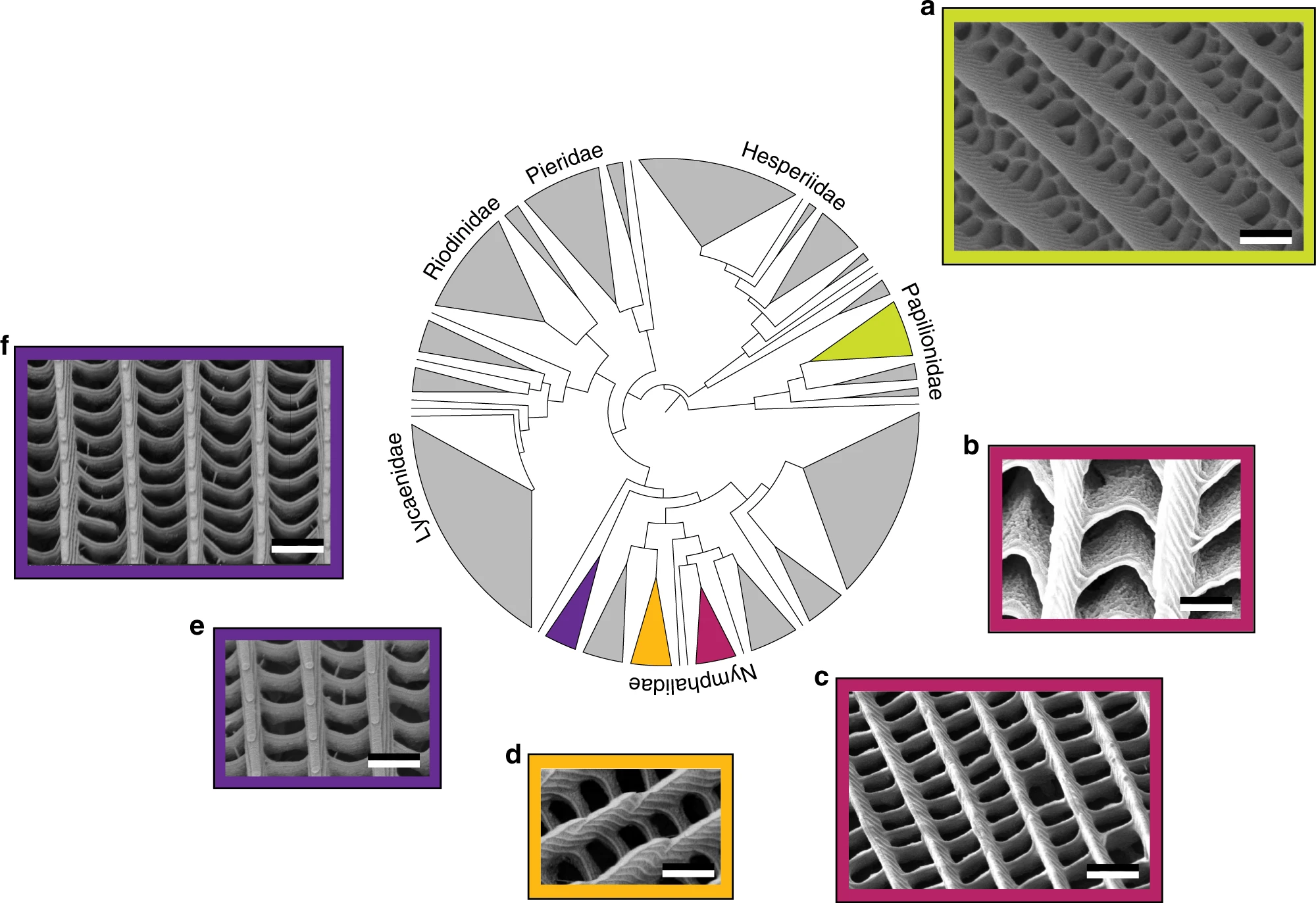
Insects such as lepidopterans, the taxonomic order encompassing the ethereal beauty of butterflies and the enigmatic allure of moths, are ectotherms, which means that their metabolisms rely on environmental heat sources. Harvesting incident energy appears crucial for these species. The phylogenetic diversity of structures giving rise to ultra-black colouration occurring in the order Lepidopetra was recently analysed in detail (figure 1) [44]. All these structures present some longitudinal ridges connected by crossribs in the upper lamina of the scales, forming 2D networks of quasi-periodic holes. The resulting high-surface area was described as increasing light absorption by underlying cuticular melanin and reducing reflection [45, 44]: whatever the size and shape of the holes -honeycomb, chevrons, or rectangles- scales giving rise to ultra-black visual appearances exhibit steeper ridges as well as deeper and wider trabeculae (namely, pillars connecting the upper and basal laminae of a scale) than scales with some regular black or brown colour. Through numerical modelling, these features were shown to play a significant role in reducing light reflection [45, 44]. Furthermore, coating these structures with gold does not lead to an increase in light reflectance, unlike regular black or brown butterfly wings. This experimentally demonstrates the photonic origin of the related light harvesting [44].
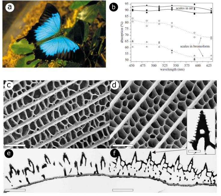
In the scales covering the wings of the male Papilio ulysses butterfly, similar complex photonic structures were found to play a role in intensifying the darkness areas [28], in addition to the porous multilayer structure producing the bright blue colour through interference [46]. They were described to scatter light towards the ridges and the interior of the scale, leading to a longer optical path, resulting in a higher light absorption by the pigments distributed within the scale material. For matt black scales, the absorption reduced from 95% to 55%, upon contact with bromoform serving as index-matching fluid, while for lustrous black scales, it decreased from 90% to 70% (figure 2). Ridges were however demonstrated through simulation to have neglected role in visible-light absorption of the ultra-black scales of Pachliopta aristolochiae [47].
The wings of Troides magellanus butterfly, the Magellan birdwing, present a captivating spectacle of optical properties including light diffraction and controlled fluorescence emission on their hindwings [48, 49, 50, 51]. It inhabits the Philippines and Taiwan’s Orchid Island. Renowned for their impressive size and striking appearance, the forewings showcase a remarkable 98% absorption of visible light as well as reveal two distinctive peaks in the IR spectrum [27]. The presence of chitin imparts the wings with these robust absorption peaks at 3 m and 6 m due to C = O vibrations, strategically positioned within the wavelength range where a black body emits radiation at 40°C, enabling radiative cooling. The architecture of the Magellan birdwing consists of five major elements [27]: a roof-like structure on which are located a series of ridges, holes in the separating structures and pillars, joining the upper membrane to the lower membrane of the wing. A similar structure was also found in the case of the related Troides aeacus [52]. Comparison of numerical simulations between the photonic structure and a non-structured flat slab with equal volume of material showed a 10% increase in electromagnetic radiation absorption and a 17% increase in emissivity at 40°C [27]. This unique combination of optical characteristics suggests that the Magellan birdwing has evolved to manage efficiently both visible and IR light, underscoring the sophisticated adaptation of these butterfly wings for light and thermal radiation purposes.
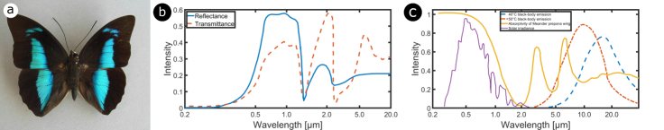
A related thermoregulation was demonstrated in the case of Archaeoprepona meander, the Meander prepona, a tropical butterfly species (figure 3) [23], as well as later on in various butterfly species [53]. These insects employ a sophisticated mechanism to manage its body temperature effectively within a given range such as 36°C-40°C [23] or 20°C-50°C [53], depending on the species. The intricate structure of their wings such as the black wings of Meander prepona serves as a remarkable example of nature’s engineering prowess. The scale structures on the wings, in addition to melanin pigments, play a crucial role in harnessing solar energy efficiently, absorbing approximately 95% of the visible solar spectrum (figure 3c) [23], akin the phenomenon described in the cases of Papilio ulysses and the Magellan birdwing hereabove. In the near-IR range, the absorptance intensity decreases down to less than 2%, ensuring low thermal emissivity, apart from the absorptance peaks at 3 m and 6 m. The 6-m emissivity peak plays a crucial role in thermoregulation [23]. At temperatures below 40°C, the black body peak is located at a longer wavelength (figure 3c). It allows the wing to harvest heat effectively while maintaining low thermal emission. When temperatures exceed 40°C, a significant overlap between the black body spectrum and the 6-m peak appears (figure 3c), leading to higher thermal emission (namely, radiative cooling) and contributing to the butterfly’s fine-tuned response to thermal challenges in its habitat. This thermoregulation mechanism could be employed in applications such as solar energy harvesting as it can help maintaining the devices within an optimal temperature range.
The role of ultra-black colours in butterflies remains the subject of speculation. However, it was hypothesised that they enhance the contrast in the visual signals, as ultra-black areas are always located next to bright areas [44]. Such visual contrast would have implications in terms of aposematism or intraspecific communication.
In addition to structured scales covering the ventral and dorsal sides of lepidopteran wings, many species including butterflies Greta spp., the moth Cacostatia ossa, and the moth Cephonodes hylas display some highly transparent scale-less wings with antireflection properties through photonic structuring of the wing membranes [54, 55, 18, 56, 57, 16, 58, 59, 60]. This structuring curtails reflection of incident light to levels below 2% across the entire visible spectrum, through electromagnetic impedance matching. It consists in a lattice of dome-shaped protuberances, also known as nipples (figure 4). The underlying principle behind this antireflection effect lies in the gradual refractive index matching between the air and the wing membrane, typically composed of chitin. If the protuberances are spaced by less than the incident wavelength, typically less than 200 nm, the non-zero diffraction orders are evanescent. The protuberance structure can be regarded as a slow variation of the effective refractive index along the normal to the wing membrane. Depending on the species, the protuberance lattice can be very well ordered such as the hexagonal compact array in the wings of C. hylas [54, 55, 18] and Hemaris fuciformis [56, 60] hawkmoths or more disordered such as the wings of C. ossa moth (figure 4) [57, 16] and the ones of Greta spp. glasswing butterflies [58, 59]. Interestingly, disorder in the protuberance height, width, and position was found to increase the transparency properties, in the case of G. oto glasswing butterfly [59]. Beyond the order of lepidopterans, antireflection structures manifest in the wings of odonatans, such as Aeshna cyanea dragonfly [61, 60], the American Rubyspot dameselfly Hetaerina americana [58], and Vestalis amabilis damselfly [60], or even in hemipterans like cicadas [62, 63, 64, 16, 65, 60]. In addition, such nipple arrays were observed on the surfaces of compound-eye corneas of several arthropods [66, 67, 68, 56, 21]. They are often referred to as moth-eye structures. A comparative study of 19 species of butterflies led to the classification of the arrays into three categories according to their morphologies: conical, paraboloidal, and Gaussian. The paraboloid profile with protuberances almost touching each other was found to exhibit the lowest reflectance, with the effective refractive index varying quasi-linearly with depth [21]. Highly antireflective wings in insect are often reported to play a likely role in crypsis [63, 64]. Similarly, nipple arrays on the surfaces of compound eyes are assumed to improve camouflage under daylight and improve night vision [66, 67]. In general, some of such lattices of protuberances were reported to combine antireflection with hydrophobic properties [63, 64, 16, 69, 70, 71, 72], bactericidal activity [73, 74, 75, 76, 72], and fluorescence emission [77, 78, 79, 80, 60]. In the case of cicadas, it was shown that the protrusions could be approximated by truncated cones under hemispheres [64, 16]. The cones gave rise to impedance matching and high antireflection whereas the cones favoured hydrophobicity. Fluorescence arises from fluorescent proteins -typically resilin- embedded withing the membrane material [77, 78, 79, 80, 60].
2.2 Elytra of beetles
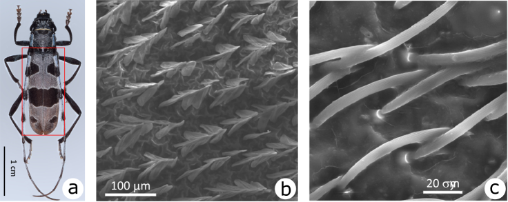
The blue-grey elytra of Rosalia alpina longhorn beetle (family Cerambycidae) exhibit large black spots (figure 5). The micro- and nanostructured setae that cover these elytra contribute, on one side, to the camouflage of this beetle on beech barks and, on the other side, to thermoregulation by allowing a quick heating of the body to the optimal temperature and dissipating excess of heat through IR emission to prevent overheating [81, 82], akin the wings of some butterflies described in the previous section. The setae occurring in the black spots enhance visible light absorption by light trapping, whereas the setae of all the elytra enable the thermoregulation. The former are inclined scales, touching neighbours at the tips and forming tent-like architectures with 1-m period and 100-nm period grating patterns (figure 5a) [81, 82]. The setae occurring on the blue-grey areas consist in hairs [81, 82] (figure 5b). Through optical modelling based on SEM observations, the light trapping role of the scales was demonstrated [81, 82]. Several reflections on opposite inclined patterned scales and high concentration of melanin in these scales account for the high-absorption properties. In addition, the scales and the hairs exhibit absorption (and hence emission) enhancement in the mid-IR range [81, 82].
More recently, Vasiljević and co-workers unveiled a combination of lenslets and micron-sized multilayered spherical black elements located within the elytra of Morimus asper funereus longhorn beetle (family Cerambycidae) [83], which also display black spots on a grey surface. However, in this case, both areas, black and grey, look identical when observed with a thermal camera. The authors concluded from Finite Element Method (FEM) simulations that the combined action of the lenslets and the multilayered spherical elements focuses IR radiation on microchannels containing hemolymph.
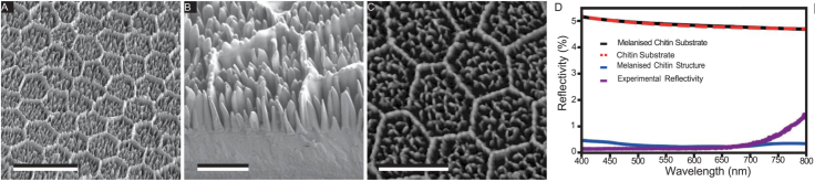
Finally, arrays of ellipsoidal and randomly located micropillars (figure 6A-C) were reported on the elytra of Euprotaetia inexpectata scarab beetle (family Scarabaeidae) [84]. They enhance light absorption by a combination of Mie scattering and optical focusing. This way, incident light reaches absorbing pigment -namely melanin- located within the elytra, giving rise to an absorptance up to 99.5% and a reflectance of 0.1% at 400 nm (figure 6D).
2.3 Bird feathers
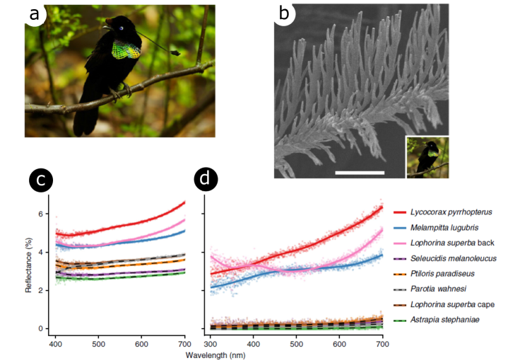
The ultra-black plumage occurring in some species of birds of paradise within the family Paradisaeidae has captivated researchers due to its unparalleled darkness, reaching absorption levels of up to 99.95%. This phenomenon, elucidated by McCoy and colleagues, is a result of structural absorption rather than pigmentation (figure 7) [36]. These feathers appear even darker than typical black feathers due to a significant reduction in specular reflection, as measured through directional reflectance ranging from a mere 0.05% to 0.31%. The secret lies in the microstructure of the feathers, featuring barbules curved up that are tilted vertically by ca. 30° with respect to the normal in the direction of the feathers’ distal tip. This unique arrangement enhances multiple light scattering, creating regularly spaced cavities with dimensions of 5–30 m in width and 200–400 m in depth. Astonishingly, the super-black effect is most pronounced when looked from the distal direction, aligning perfectly with the perspective of a female observing a male. The cavities present a directional reflectance bias, making the feathers even darker when viewed from the distal direction. This natural adaptation showcases the fascinating ways in which birds of paradise have evolved to achieve remarkable visual effects in their plumage.
2.4 Cuticle of Maratus jumping spiders
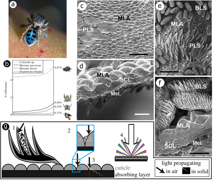
Jumping spiders, specifically the male members of the genus Maratus, commonly referred to as peacock spiders, have evolved a fascinating display strategy to attract their female counterparts (figure 8a). These spiders exhibit a striking combination of brilliant colours arising from pigments or photonic structures [85] and velvety black areas [86] on their bodies. These black regions, described as ultra-black [86], reflect less than 0.5% of light, reaching intensities as low as 0.35% in the case of Maratus karrie, due to microstructures, including densely packed cuticular bumps resembling microlens arrays (figure 8c,d). In addition, M. karrie displays some black scales reassembling brushes (figure 8e,f). Optical modelling revealed a delicate balance between minimising light reflection from the surface and maximising absorption by melanin (figure 8g). Interestingly, McCoy and co-workers proposed that this ultra-black followed a convergent evolution for the success of these spiders and the birds of paradise in the competitive realm of sexual selection [86].
2.5 Skin of West African Gaboon viper
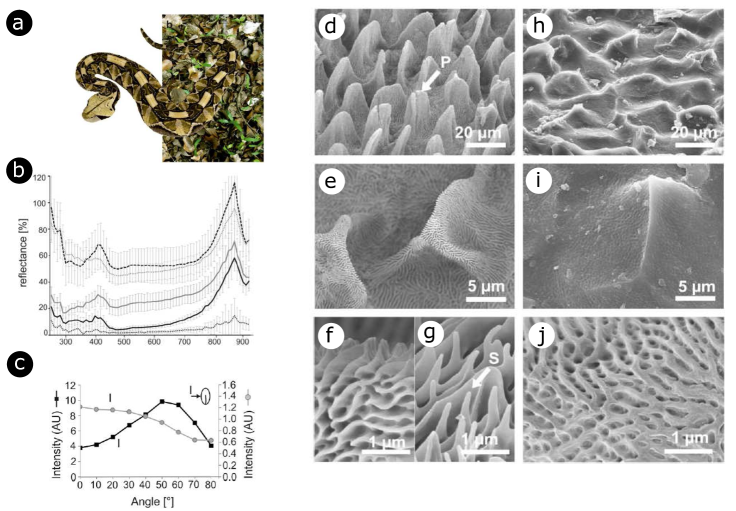
The Gaboon viper Bitis rhinoceros, native to West Africa, exhibits a stunning camouflage in its natural habitat, thanks to its intricate skin pattern [87]. The geometrically arranged velvet black spots, interspersed with pale and light brown regions (figure 9), seamlessly blend into the diverse light and shade patterns of the forest ground under the canopy. Observations revealed that the blackness of the viper’s scales is primarily derived from a hierarchical structure characterised by densely packed, leaf-like microstructures covered with nanoridges. Under microscopic scrutiny, even the areas in between black scales exhibit nanoridge striations (figure 9). Reflectance spectra analysis demonstrates that both black and pale scales have nearly flat profiles across the visible range, with a notable peak around 880 nm (figure 9). Intriguingly, applying an Au-Pd coating to the black scales preserves the black colour and further diminishes reflectance (figure 9). This finding supports the idea that the viper’s original surface works as an effective light-trapping device, utilising multiple reflections of light. The metallic coating further enhances light trapping via light reflections on the metal-coated surfaces. Modelling of diffuse reflection using Lambertian symmetric V-shaped cavities validated the proposed light-trapping mechanism and elucidated the angular dependence of reflectance spectra in pale scales [87]. However, the black scales exhibit a distinct angular characteristic, lacking a specular reflection peak and displaying a gradual decrease in reflectance intensity with increasing emerging angle. This unique angular behaviour imparts a nonglossy visual appearance to the velvet black, attributed to the more isotropic arrangement of scale structure.
2.6 Plant leaves and petals
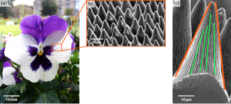
In the realm of plants, surfaces of some petals and leaves reveal a mesmerizing array of structures optimised to enhance light harvesting (figure 10). The interplay of antireflection and light trapping mechanisms unfolds through the subtle architecture of conical-shaped epidermal cells [88, 89, 34, 90]. As sunlight encounters these structures, a gradual increase in the effective refractive index occurs, giving rise to antireflection akin to the cones found on the surfaces of moth eyes and cicada wings. In some plants, additional nanowrinkles were demonstrated to reduce light reflection [34, 90]. In addition to antireflection, light redirection extends the path length within plant integuments, contributing to light trapping. The epidermal cells of flowers were reported to function as lenses and conducting incident light into the integuments comprising pigments [88, 91, 92]. It was also shown that the cone shape of their petals varies, reflecting the plant’s strategic adaptation to either scatter or absorb incident ultraviolet waves, with shorter cones in the former, and taller ones in the latter, respectively [93]. This adaptive variability plays a pivotal role in enhancing light capture for crucial processes like photosynthesis and contributes to the vivid coloration of these botanical wonders, especially in environments with limited light availability.
Venturing into the microscopic world, diatoms, unicellular algae encased in intricate silica frustules (namely, the hard porous structures of diatoms), offer a different yet equally captivating story of solar energy harvesting [94]. The case of Coscinodiscus sp. stands out with its frustule comprising three layers – termed cribellum, cribrum, and the internal plate – each composed of thin slabs housing hexagonal arrays of disk holes. The size and spacing of these holes vary from layer to layer, forming a hierarchical structure that has been finely tuned for optimal light trapping and photosynthesis.
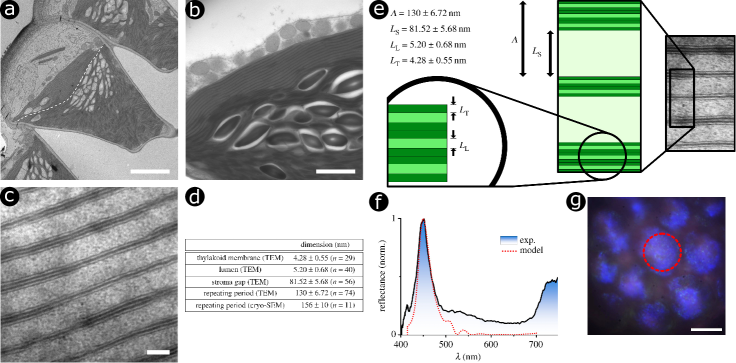
The blue iridescent epidermal chloroplasts occurring in some plant leaves, such as those in shade-dwelling Begonia spp. and Selaginella erythropus, display intricate multilayers that significantly enhance light absorption [35, 95, 96, 97]. Chloroplasts, crucial plant organelles facilitating photosynthesis by absorbing incident light via chlorophyll, play a pivotal role in converting light energy into biochemical energy as adenosine triphosphate (ATP) and nicotinamide adenine dinucleotide phosphate (NADPH). Particularly, the initial light-dependent phase of photosynthesis takes place within the absorbing thylakoid tissues of chloroplasts. Two distinct types of chloroplasts are of interest with respect to photonics and enhanced light absorption: iridoplasts and bizonoplasts [98]. Iridoplasts, exclusive to some plants such as the leaves of Begonia, possess photonic structures consisting in periodic multilayers of thylakoid tissues. Conversely, bizonoplasts have been identified in a single plant species, S. erythropus (figure 11) [99, 100]. They look like a mix of conventional thylakoid tissue present in typical irregular chloroplasts across many plants, and very organised iridoplasts. The unique ordered photonic structures of iridoplasts and bizonoplasts result in enhanced light absorption in the green part of the electromagnetic spectrum due to slow-light effect occurring at the red edge of the photonic bandgap of the multilayered structures. This increased absorption aligns with the incident light environment of these canopy-adapted plants. It leads to an enhanced quantum yield in low-light conditions, bolstering photosynthesis when compared to normal chloroplasts [35, 95, 96, 97].
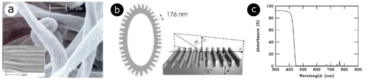
In the ethereal heights of the Alps, the edelweiss flower, Leontopodium nivale, unveils a captivating defense mechanism against intense UV radiation. Located within the wooly cover layer of its bracts (namely, the downy-white “petals” of the edelweiss that actually are specialised leaves), an intersecting pattern of transparent filaments, displaying some faint iridescence can be observed by microscopy (figure 12). These filaments exhibit variations in diameter and morphology of their cross-sections. They are hollow with parallel corrugation running along the main axis of the filaments with a period of ca. 180 nm. The spectral reflectance is rather low from 300 to 400 nm and abruptly increases around 400 nm to form a plateau at ca. 65%. This optical behaviour corresponds to some strong absorption in the near UV range. This intricate filamentary architecture, akin to a two-dimensional corrugated dielectric slab, emerges as a UV-selective waveguide coupling device. Fano resonances within these filaments facilitate the transfer of incident ultraviolet waves. The filaments, characterised by a broadband angular response, act as conduits for UV photon energy, efficiently dissipating it along the hollow guides, as the filament materials absorb UV. This ingenious strategy protects the delicate cellular tissue beneath.
3 Infrared absorbers inspired by natural photonic structures
The underlying mechanisms of various natural structures have been elucidated thus far, as presented in the previous section. If their potential for energy harvesting remains largely untapped, several devices have been suggested through a bioinspiration approach [15, 17, 14]. In addition to their eco-friendly and sustainable materials composition and fabrication, these natural structures present other benefits with respect to current alternative such as their thinness and lightness. They can clearly improve the energy yield of PV cells and solar panels [102, 29, 47], passive radiative cooling [103, 104, 105, 106, 37], photocatalysis [107, 108, 45], or even the efficiency of electromagnetic camouflage, and the capture of stray light in telescopes.
3.1 Bioinspired antireflective coatings: from moth-eye and cicada-wing templates to functional applications
As described in section 2.1, insects employ antireflective features as crucial characteristics in their crypsis strategy. The nipple arrays found on some of their eye and wing surfaces have been replicated to create bioinspired antireflective coatings applicable across various uses, including solar panels, antiglare glasses, screens, light-sensitive detectors, telescopes, thermochromic smart windows and camera lenses [69, 109, 102, 110, 111, 112, 113, 114, 115, 14, 116].
Bottom-up nanofabrication approaches such as self-assembled spherical nanoparticles in non-close-packed hexagonal arrays have been used to mimic moth-eye structures (figure 13) [109]. Simulations indicated that non-close-packed structures result in lower reflectance at wavelengths longer than the nipple interdistance, affirming the suitability of the natural moth-eye design for highly efficient antireflective devices. Spin-coating deposition of colloidal suspensions of silica particles (360 nm in diameter) and their shear alignment allowed to fabricate a template that served as a mould for casting some polydimethylsiloxane (PDMS). This PDMS mould was subsequently pressed onto a ethoxylated trimethylolpropane triacrylate (ETPTA) [109] or a perfluoroacrylate polymer [117] layer laying on glass substrate (figure 13a-d). These monomer films were then polymerised with a pulsed UV light curing system. Such biomimetic structures demonstrated outstanding low reflectance of less than 0.5% across the visible spectrum (figure 13e) [109, 117]. Similarly, such spin-coated monolayer silica colloids were utilised as a mask in a reactive ion etching (RIE) process of silicon wafers with SF6 [110] and of gallium antimonide (GaSb) substrates with Cl2 [102], reducing reflectance to less than 5% in the visible-near-IR range (with respect to ca. 40% for unstructured wafers) [110, 102]. Such biomimetic structures appeared easy to fabricate on solar and TPV cells.
Similarly, top-down synthesis techniques such as nanoimprint lithography (NIL) were employed to develop efficient and cost-effective antireflective coatings inspired from nature. For instance, cicada wings were directly utilised as natural stamps, leveraging the wing chitinous material with commendable thermomechanical characteristics (figure 14a) [69, 111]. This material can indeed be heated up to 200°C without any damage. After heating, a poly(methyl methacrylate) (PMMA) film may be pressed onto the biological template (figure 14b-d) [69]. The negative replica was transferred to a silicon substrate using the array of nanowells in PMMA as a mask for RIE. Upon removal of PMMA, the resulting patterned surface demonstrated antireflective properties, evident from its dark visual appearance [69]. If the structured PMMA film was employed as a mould for gold thermodeposition instead of a mask for RIE, a perfect replica of gold hexagonal nanopilars was produced (figure 14e,f) [69]. Alternatively, a PMMA replica was fabricated through a modified procedure [111]: a first negative replica was obtained from the thermodeposition of gold onto the natural photonic structures of the wings. It was used as a mould to cast PMMA, which was subsequently peeled off [111]. The antireflective capability of the replicated PMMA film was notable. The PMMA film’s reflectance was found to be decreased from approximately 6% to about 2% across the visible-near-IR range due to the unique nipple array [111].
3.2 Advances in bioinspired solar light harvesting: beyond petals, leaves and butterfly wings
The photonic structures found in the petals and leaves occurring in the integuments of certain plants have informed the development of improved bioinspired light-absorbing structures (figure 15a,b) [107, 108, 34, 90, 118, 119]. From enhancing the efficiency of organic solar cells to improving photocatalysis and contributing to artificial photosynthesis systems, these bioinspired designs continue to pave the way for sustainable and innovative energy solutions.
For instance, the microstructures of the epidermal cells of some rose species inspired a polymer thin film replica that was integrated in a solar cell [34]. The biomimetic coating demonstrated a significant reduction in reflectance over the entire spectral range, particularly at grazing incidence, with remarkable 13% and 44% increase in the solar cell’s short-circuit current at normal and grazing incidence, respectively (figure 15c,d). These properties are of high interest for solar cell efficiency with respect to the Sun movement throughout the day. The dome profile of the micropapillae on the petal surface was demonstrated to play the role of microlenses, lengthening the optical path of light rays within the plant integuments. This dual functionality of efficient antireflection and light trapping is crucial for enhancing the performance of thin-film organic solar cells, addressing issues of low optical absorption and spectral drops due to Fabry-Pérot interferences.
While the petal of roses offer a compelling template for biomimetic light harvesting, the sophistication of plant leaves provides an even more complex blueprint. The thin and soft leaves of Vallisneria spp., aquatic grass plants, also known as eelgrass are an very informative study case for bioinspiration [107]. The hierarchical architecture of these leaves includes lens-like epidermal cells, a so-called palisade parenchyma functioning as optical waveguides, and a spongy disordered layer with intertwined veins giving rise to optical multiscattering and extending the optical path length. Utilising a sol-gel method, a silica and titania mimic of these leaves was templated for photocatalitic application [107]. The resulting Ti-Si catalyst exhibited a threefold higher rate constant for the degradation reaction of methylene blue exposed to UV compared to a commercial TiO2 catalyst. The macroporosity and enhanced light scattering properties of Vallisneria leaf structure made it an ideal template for photocatalysis, showcasing the potential for biomimicry in advancing solar-driven environmental applications.
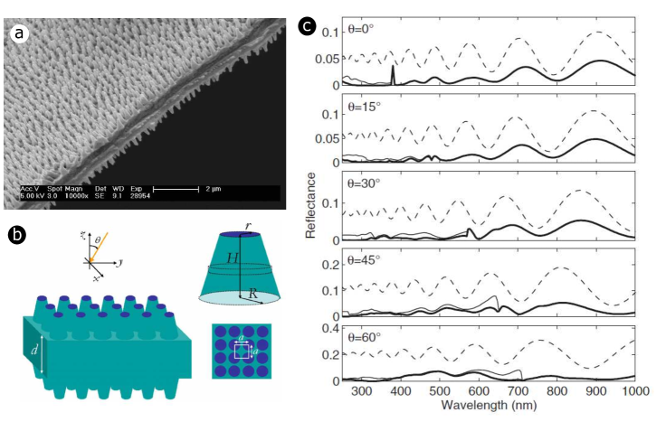
As one could have expected, butterfly wings have naturally inspired the design of visible and IR light absorbers such as an SiO2 negative replica of the black wing scales of Trogonoptera brookiana butterfly that exhibits enhanced light trapping properties (figure 16I) [29]. The solar energy loss of this replica, namely, the integrated solar light intensity reflected by the replica in the 400-900 nm range, was found to be 22.6% of the one of an SiO2 flat surface. Similarly, a hybrid photonic-plasmonic structure was fabricated by bio-templating the black forewings of Troides helena [30]. Silver spherical nanoparticles with various diameters (10 nm, 20 nm, 40 nm, 60 nm, and 80 nm) were deposited on the wings before the chitin structure was carbonised. An enhanced absorption was measured and simulated in the near- and mid-IR ranges. It results from the plasmon resonance of the silver nanoparticles and the coherent coupling among adjacent nanoparticles within the photonic architecture of T. helena. Such a hybrid photonic-plasmonic architecture was designed for photocataltic applications while taking inspiration from P. nireus (figure 16II) [45]. It consisted in gold nanoantennas located onto a bismuth vanadate (BVO) photocatalytic unit with the architecture of P. nireus black wings. This architecture was fabricated through a sol-gel method. Both experiments and simulations demonstrated the enhanced photocatalytic activity arising from the 25%-increased light harvesting within the 700 nm-1200 nm range and the 3.5-fold enhancement of the electric-field intensity of localised surface plasmons (figure 16III) [45]. Whereas these three artificial structures were fabricated by bottom-up methods, a nanostructured absorber film with disordered holes was synthesised as a mimic of the disordered black wing scales of P. aristolochiae butterfly (figure 16IV) [47]. Using phase separation of a two-polymer mixture, a hydrogenated amorphous silicon (a-Si:H) film was patterned for PV applications. The structure exhibited a relative integrated absorption over the range 450 nm-800 nm of 93% and 207%, with a 0°- and a 50°-incidence angle with respect to the normal to the film surface, respectively [47].
4 Conclusions
Photonic structures occurring in the integuments of living organisms such as arthropods, birds, and plants are very sophisticated optical devices that give rise to various striking optical effects, including UV, visible, and IR radiation management and absorption enhancement. They occur in biological tissues encompassing butterfly wings, beetle elytra, bird feathers, spider cuticle, viper skin, as well as plant leaves and petals. These phenomena are often crucial for the survival of animal and plant species. This review article also showcased the promising potential of bioinspiration in the field of energy capture and conversion. Exploiting light trapping, impedance matching or antireflection observed in natural structures is indeed highly interesting in view of developing bioinspired energy-efficient applications such as PV and TPV cells, TEG, artificial photosynthesis, and photocatalysis. With the optimisation of the efficiency of such applications, these advances inspire future research and innovation in the field of bioinspired energy solutions. Ultimately, this research paves the way for a more sustainable and environmentally conscious future by harnessing the beauty of nature’s designs to meet humankind’s energy needs.
Acknowledgements
The author thanks Prof. Olivier Deparis from the University of Namur, Belgium for fruitful discussion and reading the manuscript. The author was supported by a BEWARE Fellowship (Convention n°2110034) of the Walloon Region (COFUND Marie Skłodowska-Curie Actions of the European Union #847587).
Conflicts of interest
The author declares no conflict of interest.
References
- [1] Derya Baran, Raja Shahid Ashraf, David A. Hanifi, Maged Abdelsamie, Nicola Gasparini, Jason A. Röhr, Sarah Holliday, Andrew Wadsworth, Sarah Lockett, Marios Neophytou, Christopher J. M. Emmott, Jenny Nelson, Christoph J. Brabec, Aram Amassian, Alberto Salleo, Thomas Kirchartz, James R. Durrant, and Iain McCulloch. Reducing the efficiency–stability–cost gap of organic photovoltaics with highly efficient and stable small molecule acceptor ternary solar cells. Nature Materials, 16(3):363–369, Mar 2017.
- [2] Christian Breyer, Dmitrii Bogdanov, Ashish Gulagi, Arman Aghahosseini, Larissa S.N.S. Barbosa, Otto Koskinen, Maulidi Barasa, Upeksha Caldera, Svetlana Afanasyeva, Michael Child, Javier Farfan, and Pasi Vainikka. On the role of solar photovoltaics in global energy transition scenarios. Progress in Photovoltaics: Research and Applications, 25(8):727–745, 2017.
- [3] Martin A. Green and Stephen P. Bremner. Energy conversion approaches and materials for high-efficiency photovoltaics. Nature Materials, 16(1):23–34, Jan 2017.
- [4] E. Yablonovitch and G.D. Cody. Intensity enhancement in textured optical sheets for solar cells. IEEE Transactions on Electron Devices, 29(2):300–305, 1982.
- [5] Michael Niggemann, Moritz Riede, Andreas Gombert, and Karl Leo. Light trapping in organic solar cells. physica status solidi (a), 205(12):2862–2874, 2008.
- [6] Aline Herman, Christos Trompoukis, Valérie Depauw, Ounsi El Daif, and Olivier Deparis. Influence of the pattern shape on the efficiency of front-side periodically patterned ultrathin crystalline silicon solar cells. Journal of Applied Physics, 112(11):113107, 12 2012.
- [7] Mark L. Brongersma, Yi Cui, and Shanhui Fan. Light management for photovoltaics using high-index nanostructures. Nature Materials, 13(5):451–460, May 2014.
- [8] Alexandre Mayer, Annick Bay, Lucie Gaouyat, Delphine Nicolay, Timoteo Carletti, and Olivier Deparis. Genetic algorithms used for the optimization of light-emitting diodes and solar thermal collectors. Proceedings of SPIE, 9187, 2014.
- [9] O. Deparis, S. R. Mouchet, and B.-L. Su. Light harvesting in photonic crystals revisited: why do slow photons at the blue edge enhance absorption? Physical Chemistry Chemical Physics, 17:30525–30532, 2015.
- [10] Alexandre Mayer, Jérôme Muller, Aline Herman, and Olivier Deparis. Optimized absorption of solar radiations in nano-structured thin films of crystalline silicon via a genetic algorithm. Proceedings of SPIE, 9546:95461N, 2015.
- [11] Atikur Rahman, Ahsan Ashraf, Huolin Xin, Xiao Tong, Peter Sutter, Matthew D. Eisaman, and Charles T. Black. Sub-50-nm self-assembled nanotextures for enhanced broadband antireflection in silicon solar cells. Nature Communications, 6(1):5963, Jan 2015.
- [12] Thomas Lourdu Madanu, Laroussi Chaabane, Sébastien R. Mouchet, Olivier Deparis, and Bao-Lian Su. Manipulating multi-spectral slow photons in bilayer inverse opal tio2@bivo4 composites for highly enhanced visible light photocatalysis. Journal of Colloid and Interface Science, 647:233–245, 2023.
- [13] Sébastien R. Mouchet and Pete Vukusic. Structural colours in lepidopteran scales. In Advances in Insect Physiology, volume 54, pages 1–53. Elsevier, 2018.
- [14] Sébastien R. Mouchet and Olivier Deparis. Natural Photonics and Bioinspiration. Artech House, 2021.
- [15] L. P. Biró and J.-P. Vigneron. Photonic nanoarchitectures in butterflies and beetles: valuable sources for bioinspiration. Laser & Photonics Reviews, 5(1):27–51, 2011.
- [16] O. Deparis, S. Mouchet, L. Dellieu, J.-F. Colomer, and M. Sarrazin. Nanostructured surfaces: Bioinspiration for transparency, coloration and wettability. Materials Today: Proceedings, 1:122–129, 2014.
- [17] Han Zhou, Jun Xu, Xianghui Liu, Haiwen Zhang, Dantong Wang, Zhihan Chen, Di Zhang, and Tongxiang Fan. Bio-inspired photonic materials: Prototypes and structural effect designs for applications in solar energy manipulation. Advanced Functional Materials, 28(24):1705309, 2018.
- [18] Akihiro Yoshida. Antireflection of the butterfly and moth wings through microstructure. Forma, 17(2):75–89, 2002.
- [19] S.R. Mouchet, J.-P. Vigneron, J.-F. Colomer, C. Vandenbem, and O. Deparis. Additive photonic colors in the Brazilian diamond weevil, Entimus imperialis. Proceedings of SPIE, 8480:848003, 2012.
- [20] Jacques M. Pasteels, Olivier Deparis, Sébastien R. Mouchet, Donald M. Windsor, and Johan Billen. Structural and physical evidence for an endocuticular gold reflector in the tortoise beetle, Charidotella ambita. Arthropod Structure & Development, 45(6):509–518, 2016.
- [21] D.G Stavenga, S Foletti, G Palasantzas, and K Arikawa. Light on the moth-eye corneal nipple array of butterflies. Proceedings of the Royal Society B: Biological Sciences, 273(1587):661–667, 2006.
- [22] L. P. Biró, Zs. Bálint, K. Kertész, Z. Vértesy, G. I. Márk, Z. E. Horváth, J. Balázs, D. Méhn, I. Kiricsi, V. Lousse, and J.-P. Vigneron. Role of photonic-crystal-type structures in the thermal regulation of a lycaenid butterfly sister species pair. Physical Review E, 67:021907, Feb 2003.
- [23] S. Berthier. Thermoregulation and spectral selectivity of the tropical butterfly Prepona meander: a remarkable example of temperature auto-regulation. Applied Physics A, 80(7):1397–1400, Apr 2005.
- [24] Devi Stuart-Fox, Elizabeth Newton, and Susana Clusella-Trullas. Thermal consequences of colour and near-infrared reflectance. Philosophical Transactions of the Royal Society B: Biological Sciences, 372(1724):20160345, 2017.
- [25] Xianghui Liu, Dantong Wang, Zhiwei Yang, Han Zhou, Qibin Zhao, and Tongxiang Fan. Bright silver brilliancy from irregular microstructures in butterfly Curetis acuta moore. Advanced Optical Materials, 7(18):1900687, 2019.
- [26] Dajie Xie, Zhiwei Yang, Xianghui Liu, Shifan Cui, Han Zhou, and Tongxiang Fan. Broadband omnidirectional light reflection and radiative heat dissipation in white beetles Goliathus goliatus. Soft Matter, 15:4294–4300, 2019.
- [27] Aline Herman, Cédric Vandenbem, Olivier Deparis, Priscilla Simonis, and Jean Pol Vigneron. Nanoarchitecture in the black wings of Troides magellanus: a natural case of absorption enhancement in photonic materials. Proceedings of SPIE, 8094:80940H, 2011.
- [28] P. Vukusic, J. R. Sambles, and C. R. Lawrence. Structurally assisted blackness in butterfly scales. Proceedings of the Royal Society of London. Series B: Biological Sciences, 271(suppl_4):S237–S239, 2004.
- [29] Zhiwu Han, Bo Li, Zhengzhi Mu, Meng Yang, Shichao Niu, Junqiu Zhang, and Luquan Ren. An ingenious super light trapping surface templated from butterfly wing scales. Nanoscale Research Letters, 10(1):344, Aug 2015.
- [30] Junlong Tian, Wang Zhang, Xiaotian Fang, Qinglei Liu, Jiajun Gu, Tao Deng, Yuhua Wang, and Di Zhang. Coupling of plasmon and 3d antireflection quasi-photonic crystal structure for enhancement infrared absorption. Journal of Materials Chemistry C, 3:1672–1679, 2015.
- [31] Annick Bay, Peter Cloetens, Heikki Suhonen, and Jean-Pol Vigneron. Improved light extraction in the bioluminescent lantern of a Photuris firefly (lampyridae). Optics Express, 21(1):764–780, Jan 2013.
- [32] Norman Nan Shi, Cheng-Chia Tsai, Fernando Camino, Gary D. Bernard, Nanfang Yu, and Rüdiger Wehner. Keeping cool: Enhanced optical reflection and radiative heat dissipation in saharan silver ants. Science, 349(6245):298–301, 2015.
- [33] Katie Shanks, S. Senthilarasu, Richard H. ffrench-Constant, and Tapas K. Mallick. White butterflies as solar photovoltaic concentrators. Scientific Reports, 5(1):12267, Jul 2015.
- [34] Ruben Hünig, Adrian Mertens, Moritz Stephan, Alexander Schulz, Benjamin Richter, Michael Hetterich, Michael Powalla, Uli Lemmer, Alexander Colsmann, and Guillaume Gomard. Flower power: Exploiting plants’ epidermal structures for enhanced light harvesting in thin-film solar cells. Advanced Optical Materials, 4(10):1487–1493, 2016.
- [35] Matthew Jacobs, Martin Lopez-Garcia, O-Phart Phrathep, Tracy Lawson, Ruth Oulton, and Heather M Whitney. Photonic multilayer structure of Begonia chloroplasts enhances photosynthetic efficiency. Nature Plants, 2(11):16162, oct 2016.
- [36] Dakota E McCoy, Teresa Feo, Todd Alan Harvey, and Richard O Prum. Structural absorption by barbule microstructures of super black bird of paradise feathers. Nature Communications, 9(1):1, jan 2018.
- [37] Jie Xu and Delei Liu. A study on the radiation cooling characteristics of cerambycini latreille. Biomimetics, 9(1), 2024.
- [38] Andrew R. Cossins. Temperature Biology of Animals. Springer Science & Business Media, 2012.
- [39] Stephen G. Bosi, Jacqueline Hayes, Maryanne C. J. Large, and Leon Poladian. Color, iridescence, and thermoregulation in lepidoptera. Applied Optics, 47(29):5235–5241, Oct 2008.
- [40] Andrzej Slominski, Desmond J. Tobin, Shigeki Shibahara, and Jacobo Wortsman. Melanin pigmentation in mammalian skin and its hormonal regulation. Physiological Reviews, 84(4):1155–1228, 2004. PMID: 15383650.
- [41] Alex R. Gunderson, Alicia M. Frame, John P. Swaddle, and Mark H. Forsyth. Resistance of melanized feathers to bacterial degradation: is it really so black and white? Journal of Avian Biology, 39(5):539–545, 2008.
- [42] Helen Nilsson Sköld, Sara Aspengren, Karen L. Cheney, and Margareta Wallin. Chapter four - fish chromatophores—from molecular motors to animal behavior. volume 321 of International Review of Cell and Molecular Biology, pages 171–219. Academic Press, 2016.
- [43] Sébastien R. Mouchet, Fabio Cortesi, Bojana Bokic, Vladimir Lazovic, Pete Vukusic, N. Justin Marshall, and Branko Kolaric. Morphological and optical modification of melanosomes in fish integuments upon oxidation. Optics, 4(4):563–572, 2023.
- [44] Alexander L. Davis, H. Frederik Nijhout, and Sönke Johnsen. Diverse nanostructures underlie thin ultra-black scales in butterflies. Nature Communications, 11(1):1294, Mar 2020.
- [45] Runyu Yan, Min Chen, Han Zhou, Tian Liu, Xingwei Tang, Ke Zhang, Hanxing Zhu, Jinhua Ye, Di Zhang, and Tongxiang Fan. Bio-inspired plasmonic nanoarchitectured hybrid system towards enhanced far red-to-near infrared solar photocatalysis. Scientific Reports, 6(1):20001, Jan 2016.
- [46] Peter Vukusic, Roy Sambles, Christopher Lawrence, and Gavin Wakely. Sculpted-multilayer optical effects in two species of Papilio butterfly. Applied Optics, 40(7):1116–1125, Mar 2001.
- [47] Radwanul H. Siddique, Yidenekachew J. Donie, Guillaume Gomard, Sisir Yalamanchili, Tsvetelina Merdzhanova, Uli Lemmer, and Hendrik Hölscher. Bioinspired phase-separated disordered nanostructures for thin photovoltaic absorbers. Science Advances, 3(10):e1700232, 2017.
- [48] Jean Pol Vigneron, Krisztián Kertész, Zofia Vértesy, Marie Rassart, Virginie Lousse, Zsolt Bálint, and László P. Biró. Correlated diffraction and fluorescence in the backscattering iridescence of the male butterfly Troides magellanus (papilionidae). Physical Review E, 78:021903, Aug 2008.
- [49] R. Todd Lee and Glenn S. Smith. Detailed electromagnetic simulation for the structural color of butterfly wings. Applied Optics, 48(21):4177–4190, Jul 2009.
- [50] Eloise Van Hooijdonk, Serge Berthier, and Jean-Pol Vigneron. Contribution of both the upperside and the underside of the wing on the iridescence in the male butterfly Troïdes magellanus (Papilionidae). Journal of Applied Physics, 112(7):074702, 10 2012.
- [51] Eloise Van Hooijdonk, Carlos Barthou, Jean-Pol Vigneron, and Serge Berthier. Angular dependence of structural fluorescent emission from the scales of the male butterfly Troïdes magellanus (papilionidae). Journal of the Optical Society of America B, 29(5):1104–1111, May 2012.
- [52] Qibin Zhao, Xingmei Guo, Tongxiang Fan, Jian Ding, Di Zhang, and Qixin Guo. Art of blackness in butterfly wings as natural solar collector. Soft Matter, 7:11433–11439, 2011.
- [53] Anirudh Krishna, Xiao Nie, Andrew D. Warren, Jorge E. Llorente-Bousquets, Adriana D. Briscoe, and Jaeho Lee. Infrared optical and thermal properties of microstructures in butterfly wings. Proceedings of the National Academy of Sciences, 117(3):1566–1572, 2020.
- [54] Akihiro Yoshida, Mayumi Motoyama, Akinori Kosaku, and Kiyoshi Miyamoto. Nanoprotuberance Array in the Transparent Wing of a Hawkmoth, Cephonodes hylas. Zoological Science, 13(4):525–526, 1996.
- [55] Akihiro Yoshida, Mayumi Motoyama, Akinori Kosaku, and Kiyoshi Miyamoto. Antireflective Nanoprotuberance Array in the Transparent Wing of a Hawkmoth, Cephonodes hylas. Zoological Science, 14(5):737–741, 1997.
- [56] Pete Vukusic and J. Roy Sambles. Photonic structures in biology. Nature, 424(6950):852–855, Aug 2003.
- [57] Olivier Deparis, Nadia Khuzayim, Andrew Parker, and Jean Pol Vigneron. Assessment of the antireflection property of moth wings by three-dimensional transfer-matrix optical simulations. Physical Review E, 79:041910, Apr 2009.
- [58] Doekele G. Stavenga. Thin film and multilayer optics cause structural colors of many insects and birds. Materials Today: Proceedings, 1:109–121, 2014.
- [59] Radwanul Hasan Siddique, Guillaume Gomard, and Hendrik Hölscher. The role of random nanostructures for the omnidirectional anti-reflection properties of the glasswing butterfly. Nature Communications, 6(1):6909, Apr 2015.
- [60] Sébastien R. Mouchet, Charlotte Verstraete, Bojana Bokic, Dimitrije Mara, Louis Dellieu, Albert G. Orr, Olivier Deparis, Rik Van Deun, Thierry Verbiest, Pete Vukusic, and Branko Kolaric. Revealing natural fluorescence in transparent insect wings by linear and nonlinear optical techniques. Journal of Luminescence, 254:119490, 2023.
- [61] I. R. Hooper, P. Vukusic, and R. J. Wootton. Detailed optical study of the transparent wing membranes of the dragonfly Aeshna cyanea. Optics Express, 14(11):4891–4897, May 2006.
- [62] P R Stoddart, P J Cadusch, T M Boyce, R M Erasmus, and J D Comins. Optical properties of chitin: surface-enhanced raman scattering substrates based on antireflection structures on cicada wings. Nanotechnology, 17(3):680, jan 2006.
- [63] Mingxia Sun, Aiping Liang, Yongmei Zheng, Gregory S Watson, and Jolanta A Watson. A study of the anti-reflection efficiency of natural nano-arrays of varying sizes. Bioinspiration & Biomimetics, 6(2):026003, apr 2011.
- [64] Louis Dellieu, Michaël Sarrazin, Priscilla Simonis, Olivier Deparis, and Jean Pol Vigneron. A two-in-one superhydrophobic and anti-reflective nanodevice in the grey cicada Cicada orni (Hemiptera). Journal of Applied Physics, 116(2):024701, 07 2014.
- [65] Charlotte Verstraete, Sébastien R. Mouchet, Thierry Verbiest, and Branko Kolaric. Linear and nonlinear optical effects in biophotonic structures using classical and nonclassical light. Journal of Biophotonics, 12(1):e201800262, 2019.
- [66] C G Bernhard, W H Miller, and A R Møller. The insect corneal ripple array. a biological broad-band impedance transformer acts as an antireflection coating. Acta Physiologica Scandinavica, 63(Suppl. 243):1–79, 1965.
- [67] C. G. Bernhard, G. Gemne, and J. Sällström. Comparative ultrastructure of corneal surface topography in insects with aspects on phylogenesis and function. Zeitschrift für vergleichende Physiologie, 67(1):1–25, Mar 1970.
- [68] Andrew R. Parker, Zoltan Hegedus, and Richard A. Watts. Solar–absorber antireflector on the eye of an eocene fly (45 ma). Proceedings of the Royal Society of London. Series B: Biological Sciences, 265(1398):811–815, 1998.
- [69] Guoming Zhang, Jin Zhang, Guoyong Xie, Zhongfan Liu, and Huibo Shao. Cicada wings: A stamp from nature for nanoimprint lithography. Small, 2(12):1440–1443, 2006.
- [70] Mingxia Sun, Gregory S. Watson, Yongmei Zheng, Jolanta A. Watson, and Aiping Liang. Wetting properties on nanostructured surfaces of cicada wings. Journal of Experimental Biology, 212(19):3148–3155, 10 2009.
- [71] Katrina M. Wisdom, Jolanta A. Watson, Xiaopeng Qu, Fangjie Liu, Gregory S. Watson, and Chuan-Hua Chen. Self-cleaning of superhydrophobic surfaces by self-propelled jumping condensate. Proceedings of the National Academy of Sciences, 110(20):7992–7997, 2013.
- [72] Jessica Román-Kustas, Jacob B. Hoffman, Julian H. Reed, Andrew E. Gonsalves, Junho Oh, Longnan Li, Sungmin Hong, Kyoo D. Jo, Catherine E. Dana, Nenad Miljkovic, Donald M. Cropek, and Marianne Alleyne. Molecular and topographical organization: Influence on cicada wing wettability and bactericidal properties. Advanced Materials Interfaces, 7(10):2000112, 2020.
- [73] Elena P. Ivanova, Jafar Hasan, Hayden K. Webb, Vi Khanh Truong, Gregory S. Watson, Jolanta A. Watson, Vladimir A. Baulin, Sergey Pogodin, James Y. Wang, Mark J. Tobin, Christian Löbbe, and Russell J. Crawford. Natural bactericidal surfaces: Mechanical rupture of Pseudomonas aeruginosa cells by cicada wings. Small, 8(16):2489–2494, 2012.
- [74] Elena P. Ivanova, Jafar Hasan, Hayden K. Webb, Gediminas Gervinskas, Saulius Juodkazis, Vi Khanh Truong, Alex H.F. Wu, Robert N. Lamb, Vladimir A. Baulin, Gregory S. Watson, Jolanta A. Watson, David E. Mainwaring, and Russell J. Crawford. Bactericidal activity of black silicon. Nature Communications, 4(1):2838, Nov 2013.
- [75] Ting Diu, Nilofar Faruqui, Terje Sjöström, Baptiste Lamarre, Howard F. Jenkinson, Bo Su, and Maxim G. Ryadnov. Cicada-inspired cell-instructive nanopatterned arrays. Scientific Reports, 4(1):7122, Nov 2014.
- [76] S. M. Kelleher, O. Habimana, J. Lawler, B. O’ Reilly, S. Daniels, E. Casey, and A. Cowley. Cicada wing surface topography: An investigation into the bactericidal properties of nanostructural features. ACS Applied Materials & Interfaces, 8(24):14966–14974, 2016. PMID: 26551558.
- [77] Stanislav N. Gorb. Serial elastic elements in the damselfly wing: Mobile vein joints contain resilin. Naturwissenschaften, 86(11):552–555, Nov 1999.
- [78] Esther Appel and Stanislav N Gorb. Resilin-bearing wing vein joints in the dragonfly Epiophlebia superstes. Bioinspiration & Biomimetics, 6(4):046006, oct 2011.
- [79] Esther Appel, Lars Heepe, Chung-Ping Lin, and Stanislav N. Gorb. Ultrastructure of dragonfly wing veins: composite structure of fibrous material supplemented by resilin. Journal of Anatomy, 227(4):561–582, 2015.
- [80] Chin-Jung Chuang, Cheng-Der Liu, Ranjit A. Patil, Chi-Chung Wu, Yao-Chih Chang, Chih-Wen Peng, Ting-Kwuan Chao, Je-Wen Liou, Yung Liou, and Yuan-Ron Ma. Impact of cuticle photoluminescence on the color morphism of a male damselfly Ischnura senegalensis (rambur, 1842). Scientific Reports, 6(1):38051, Dec 2016.
- [81] G. Dikić, D. Pavlović, D. Vasiljević, Lj. Tomić, and D. Pantelić. The thermographic analisys of photonic characteristics of Rosalia alpina surfaces. In Proceedings of 3rd International Conference on Electrical, Electronic and Computing Engineering IcETRAN, pages MOI1.2.1–5, Zlatibor, Serbia, 13-16 of June 2016.
- [82] Danica Pavlović, Darko Vasiljević, Branislav Salatić, Vladimir Lazović, Goran Dikić, Ljubiša Tomić, Srećko Ćurčić, Petar Milovanović, Dajana Todorović, and Dejan V. Pantelić. Photonic structures improve radiative heat exchange of Rosalia alpina (coleoptera: Cerambycidae). Journal of Thermal Biology, 76:126–138, 2018.
- [83] Darko Vasiljević, Danica Pavlović, Vladimir Lazović, Branko Kolarić, Branislav Salatić, Wang Zhang, Di Zhang, and Dejan Pantelić. Thermal radiation management by natural photonic structures: Morimus asper funereus case. Journal of Thermal Biology, 98:102932, 2021.
- [84] Alessandro Parisotto, Viola V. Vogler-Neuling, Ullrich Steiner, Matthias Saba, and Bodo D. Wilts. Structural light absorption in elytral micropillars of Euprotaetia inexpectata beetles. Materials Today Advances, 19:100399, 2023.
- [85] Doekele G. Stavenga, Jürgen C. Otto, and Bodo D. Wilts. Splendid coloration of the peacock spider Maratus splendens. Journal of The Royal Society Interface, 13(121):20160437, 2016.
- [86] Dakota E. McCoy, Victoria E. McCoy, Nikolaj K. Mandsberg, Anna V. Shneidman, Joanna Aizenberg, Richard O. Prum, and David Haig. Structurally assisted super black in colourful peacock spiders. Proceedings of the Royal Society B: Biological Sciences, 286(1902):20190589, 2019.
- [87] Marlene Spinner, Alexander Kovalev, Stanislav N. Gorb, and Guido Westhoff. Snake velvet black: Hierarchical micro- and nanostructure enhances dark colouration in Bitis rhinoceros. Scientific Reports, 3(1):1846, May 2013.
- [88] H. L. Gorton and T. C. Vogelmann. Effects of Epidermal Cell Shape and Pigmentation on Optical Properties of Antirrhinum Petals at Visible and Ultraviolet Wavelengths. Plant Physiology, 112(3):879–888, 11 1996.
- [89] Dimitrios Gkikas, Apostolos Argiropoulos, and Sophia Rhizopoulou. Epidermal focusing of light and modelling of reflectance in floral-petals with conically shaped epidermal cells. Flora - Morphology, Distribution, Functional Ecology of Plants, 212:38–45, 2015.
- [90] Raphael Schmager, Benjamin Fritz, Ruben Hünig, Kaining Ding, Uli Lemmer, Bryce S. Richards, Guillaume Gomard, and Ulrich W. Paetzold. Texture of the viola flower for light harvesting in photovoltaics. ACS Photonics, 4(11):2687–2692, 2017.
- [91] R. A. Bone, D. W. Lee, and J. M. Norman. Epidermal cells functioning as lenses in leaves of tropical rain-forest shade plants. Applied Optics, 24(10):1408–1412, May 1985.
- [92] Bodo D. Wilts, Paula J. Rudall, Edwige Moyroud, Tom Gregory, Yu Ogawa, Silvia Vignolini, Ullrich Steiner, and Beverley J. Glover. Ultrastructure and optics of the prism-like petal epidermal cells of Eschscholzia californica (california poppy). New Phytologist, 219(3):1124–1133, 2018.
- [93] Anna J. Schulte, Matthias Mail, Lisa A. Hahn, and Wilhelm Barthlott. Ultraviolet patterns of flowers revealed in polymer replica – caused by surface architecture. Beilstein Journal of Nanotechnology, 10:459–466, 2019.
- [94] Xiangfan Chen, Chen Wang, Evan Baker, and Cheng Sun. Numerical and experimental investigation of light trapping effect of nanostructured diatom frustules. Scientific Reports, 5(1):11977, Jul 2015.
- [95] Nathan J. Masters, Martin Lopez-Garcia, Ruth Oulton, and Heather M. Whitney. Characterization of chloroplast iridescence in Selaginella erythropus. Journal of The Royal Society Interface, 15(148):20180559, 2018.
- [96] Miguel A. Castillo, William P. Wardley, and Martin Lopez-Garcia. Light-dependent morphological changes can tune light absorption in iridescent plant chloroplasts: A numerical study using biologically realistic data. ACS Photonics, 8(4):1058–1068, 2021.
- [97] William P. Wardley, Johannes W. Goessling, and Martin Lopez-Garcia. Measuring photonics in photosynthesis: Combined micro-fourier image spectroscopy and pulse amplitude modulated chlorophyll fluorimetry at the micrometre-scale. Biomimetics, 7(3), 2022.
- [98] Kevin S. Gould and David W. Lee. Physical and ultrastructural basis of blue leaf iridescence in four malaysian understory plants. American Journal of Botany, 83(1):45–50, 1996.
- [99] Chiou-Rong Sheue, Vassilios Sarafis, Ruth Kiew, Ho-Yih Liu, Alexandre Salino, Ling-Long Kuo-Huang, Yuen-Po Yang, Chi-Chu Tsai, Chun-Hung Lin, Jean W. H. Yong, and Maurice S. B. Ku. Bizonoplast, a unique chloroplast in the epidermal cells of microphylls in the shade plant Selaginella erythropus (selaginellaceae). American Journal of Botany, 94(12):1922–1929, 2007.
- [100] Chiou-Rong Sheue, Jian-Wei Liu, Jia-Fang Ho, Ai-Wen Yao, Yeh-Hua Wu, Sauren Das, Chi-Chu Tsai, Hsiu-An Chu, Maurice S. B. Ku, and Peter Chesson. A variation on chloroplast development: The bizonoplast and photosynthetic efficiency in the deep-shade plant Selaginella erythropus. American Journal of Botany, 102(4):500–511, 2015.
- [101] Jean Pol Vigneron, Marie Rassart, Zofia Vértesy, Krisztián Kertész, Michaël Sarrazin, László P. Biró, Damien Ertz, and Virginie Lousse. Optical structure and function of the white filamentary hair covering the edelweiss bracts. Physical Review E, 71:011906, Jan 2005.
- [102] Wei-Lun Min, Amaury P. Betancourt, Peng Jiang, and Bin Jiang. Bioinspired broadband antireflection coatings on GaSb. Applied Physics Letters, 92(14):141109, 04 2008.
- [103] Azadeh Didari and M. Pinar Mengüç. A biomimicry design for nanoscale radiative cooling applications inspired by Morpho didius butterfly. Scientific Reports, 8(1):16891, Nov 2018.
- [104] Haiwen Zhang, Kally C. S. Ly, Xianghui Liu, Zhihan Chen, Max Yan, Zilong Wu, Xin Wang, Yuebing Zheng, Han Zhou, and Tongxiang Fan. Biologically inspired flexible photonic films for efficient passive radiative cooling. Proceedings of the National Academy of Sciences, 117(26):14657–14666, 2020.
- [105] Zhangbin Yang, Haoxuan Sun, Yulin Xi, Yanli Qi, Zepeng Mao, Ping Wang, and Jun Zhang. Bio-inspired structure using random, three-dimensional pores in the polymeric matrix for daytime radiative cooling. Solar Energy Materials and Solar Cells, 227:111101, 2021.
- [106] Wanlin Wang, Hongyun Xing, Xiaochi Shu, Xinkun Zhao, Xiaoyuan Yan, Binbin Hong, Lei Sun, Wang Zhang, and Guo Ping Wang. Cooling colors below ambient temperature. Optica, 10(8):1059–1066, Aug 2023.
- [107] Jian Liu, Qiang Yang, Wentao Yang, Mingzhu Li, and Yanlin Song. Aquatic plant inspired hierarchical artificial leaves for highly efficient photocatalysis. Journal of Materials Chemistry A, 1:7760–7766, 2013.
- [108] Han Zhou, Jianjun Guo, Peng Li, Tongxiang Fan, Di Zhang, and Jinhua Ye. Leaf-architectured 3d hierarchical artificial photosynthetic system of perovskite titanates towards co2 photoreduction into hydrocarbon fuels. Scientific Reports, 3(1):1667, Apr 2013.
- [109] Nicholas C. Linn, Chih-Hung Sun, Peng Jiang, and Bin Jiang. Self-assembled biomimetic antireflection coatings. Applied Physics Letters, 91(10):101108, 09 2007.
- [110] Chih-Hung Sun, Peng Jiang, and Bin Jiang. Broadband moth-eye antireflection coatings on silicon. Applied Physics Letters, 92(6):061112, 02 2008.
- [111] Guoyong Xie, Guoming Zhang, Feng Lin, Jin Zhang, Zhongfan Liu, and Shichen Mu. The fabrication of subwavelength anti-reflective nanostructures using a bio-template. Nanotechnology, 19(9):095605, feb 2008.
- [112] Z.W. Han, Z. Wang, X.M. Feng, B. Li, Z.Z. Mu, J.Q. Zhang, S.C. Niu, and L.Q. Ren. Antireflective surface inspired from biology: A review. Biosurface and Biotribology, 2(4):137–150, 2016.
- [113] Guanjun Tan, Jiun-Haw Lee, Yi-Hsin Lan, Mao-Kuo Wei, Lung-Han Peng, I-Chun Cheng, and Shin-Tson Wu. Broadband antireflection film with moth-eye-like structure for flexible display applications. Optica, 4(7):678–683, Jul 2017.
- [114] Imran Zada, Wang Zhang, Peng Sun, Muhammad Imtiaz, Waseem Abbas, and Di Zhang. Multifunctional, angle dependent antireflection, and hydrophilic properties of SiO2 inspired by nano-scale structures of cicada wings. Applied Physics Letters, 111(15):153701, 10 2017.
- [115] Sai Liu, Chi Yan Tso, Hau Him Lee, Yi Zhang, Kin Man Yu, and Christopher Y. H. Chao. Bio-inspired tio2 nano-cone antireflection layer for the optical performance improvement of vo2 thermochromic smart windows. Scientific Reports, 10(1):11376, Jul 2020.
- [116] Anastasia Novikova, Aviad Katiyi, and Alina Karabchevsky. Nature-inspired anti-reflective texturization for solar energy applications. Advanced Materials Technologies, 9(2):2301128, 2024.
- [117] Chih-Hung Sun, Adriel Gonzalez, Nicholas C. Linn, Peng Jiang, and Bin Jiang. Templated biomimetic multifunctional coatings. Applied Physics Letters, 92(5):051107, 02 2008.
- [118] Benjamin Fritz, Ruben Hünig, Markus Guttmann, Marc Schneider, K.M. Samaun Reza, Oliver Salomon, Philip Jackson, Michael Powalla, Uli Lemmer, and Guillaume Gomard. Upscaling the fabrication routine of bioreplicated rose petal light harvesting layers for photovoltaic modules. Solar Energy, 201:666–673, 2020.
- [119] Benjamin Fritz, Gábor Horváth, Ruben Hünig, Ádám Pereszlényi, Ádám Egri, Markus Guttmann, Marc Schneider, Uli Lemmer, György Kriska, and Guillaume Gomard. Bioreplicated coatings for photovoltaic solar panels nearly eliminate light pollution that harms polarotactic insects. PLOS ONE, 15(12):1–22, 12 2020.