RadGenome-Chest CT: A Grounded Vision-Language Dataset for Chest CT Analysis.
Abstract
Developing generalist foundation model has recently attracted tremendous attention among researchers in the field of AI for Medicine (AI4Medicine). A pivotal insight in developing these models is their reliance on dataset scaling, which emphasizes the requirements on developing open-source medical image datasets that incorporate diverse supervision signals across various imaging modalities. In this paper, we introduce RadGenome-Chest CT, a comprehensive, large-scale, region-guided 3D chest CT interpretation dataset based on CT-RATE. Specifically, we leverage the latest powerful universal segmentation and large language models, to extend the original datasets (over 25,692 non-contrast 3D chest CT volume and reports from 20,000 patients) from the following aspects: (i) organ-level segmentation masks covering 197 categories, which provide intermediate reasoning visual clues for interpretation; (ii) 665 K multi-granularity grounded reports, where each sentence of the report is linked to the corresponding anatomical region of CT volume in the form of a segmentation mask; (iii) 1.3 M grounded VQA pairs, where questions and answers are all linked with reference segmentation masks, enabling models to associate visual evidence with textual explanations. All grounded reports and VQA pairs in the validation set have gone through manual verification to ensure dataset quality. We believe that RadGenome-Chest CT can significantly advance the development of multimodal medical foundation models, by training to generate texts based on given segmentation regions, which is unattainable with previous relevant datasets. We will release all segmentation masks, grounded reports, and VQA pairs to facilitate further research and development in this field.
Background & Summary
In the recent literature, the evolution of large-scale foundation models [1, 2, 3, 4, 5] has sparked significant interest in the development of generalist medical AI (GMAI) systems [6, 7, 8, 9, 10], particularly within the realm of radiology—a crucial component of medical diagnostics. By training on large-scale visual-language medical datasets, i.e., medical scans paired with global clinical reports, for example, MIMIC-CXR [11] has chest X-ray scans from 227,835 studies, and CT-RATE [12] contains chest CT scans from 20,000 patients. These medical models have demonstrated the preliminary ability for writing clinical reports, aiming to support radiologists throughout their workflow and markedly reducing workloads.
However, existing datasets only provide global reports for the medical scan, which has posed limitations on training models that enables grounded report generation, grounded question answering, i.e., describing regional abnormalities and relevant normal findings, or answer questions corresponding to certain regions. To further push forward the training of more capable generalist models, we propose to extend the existing image-reports datasets with region-wise description, i.e., linking the descriptive labels or findings from diagnostic reports to their corresponding anatomical regions in the images, in the form of segmentation masks for explainability.
In this paper, we introduce RadGenome-Chest CT, a comprehensive, large-scale and fine-grained annotated dataset for 3D chest CT interpretation, built upon the publicly available CT-RATE [12]. Initially, we employ the latest powerful text-prompted universal segmentation model, SAT [13], to segment primary anatomical targets in the image. Subsequently, utilizing large language models and NER models, we break all reports into an anatomically hierarchical structured format, and link the reports’ sentences to visual regions in CT volume. Finally, we further generate visual question-answering pairs closely related to the structured report and segmented image, from both region level and case level. In summary, we have extended the original image-report datasets from the following aspects:
-
i)
Organ-level segmentation masks that covers 197 categories, i.e., all the critical regions existing in clinical CT reports;
-
ii)
665k multi-granularity grounded reports, with each sentence grounded to the corresponding anatomical region.
-
iii)
1.3 M grounded VQA pairs, concerning both critical region-wise findings and comprehensive case-wise impressions. All the questions and answers are linked to segmentation masks for reference.
We believe that RadGenome-Chest CT , with provided region-to-report associations, can significantly advance the development of agent-based multimodal medical foundation models, that enables to generate texts, grounded on the corresponding visual regions, which is unattainable with previous relevant datasets.
Methods
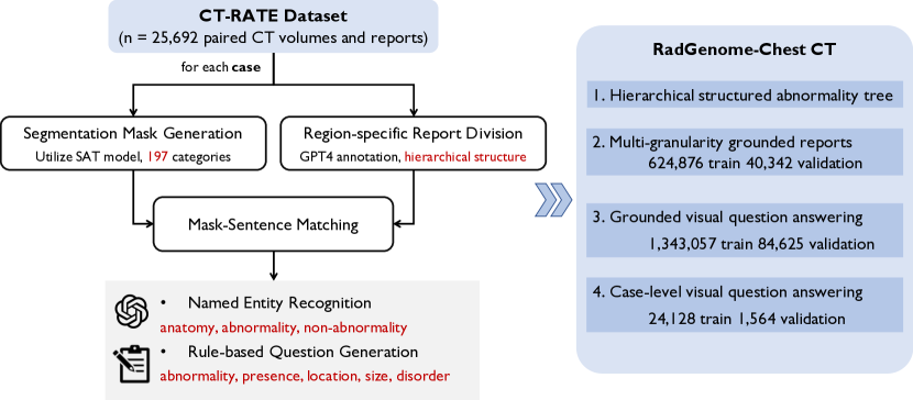
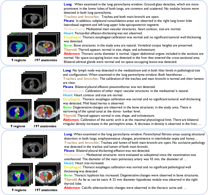
In this section, we start by introducing the source dataset that RadGenome-Chest CT is built on. Next, we provide a detailed description of the collection procedure for obtaining segmentation masks, and region-wise reports, as illustrated in Fig. 1. The outcomes from each step are presented in Fig. 2.
Data Source
We initiate our study with CT-RATE [12] (https://huggingface.co/datasets/ibrahimhamamci/CT-RATE), it is a dataset of 25,692 non-contrast 3D chest CT volumes derived from 21,304 unique patients, each volume is accompanied by a radiology text report and annotated with 18 distinct types of abnormalities. These 25,692 non-contrast 3D chest CT volumes have been reconstructed with various methods to accommodate different window settings, totaling 50,188 images. For consistency in this paper, we have standardized all CT volumes to a uniform voxel spacing of , resulting in only 25,692 paired CT volumes and reports. We follow the official division: 20,000 patients ( 24,128 volumes) were allocated to training and 1,304 (1,564 volumes) for validation.
Constructing RadGenome-Chest CT
The pipeline consists of three major stages, as shown in Fig. 1: (i) segmentation mask generation, where detailed masks for each anatomical region in the chest CT volumes are created; (ii) region-specific report division, that involves the annotation and categorization of radiology text reports by the anatomical regions they refer; (iii) rule-based question generation, which entails extracting entities from the sentence, and formulating visual question answering (VQA) pairs linked to specific segmentation masks.
Segmentation Mask Generation
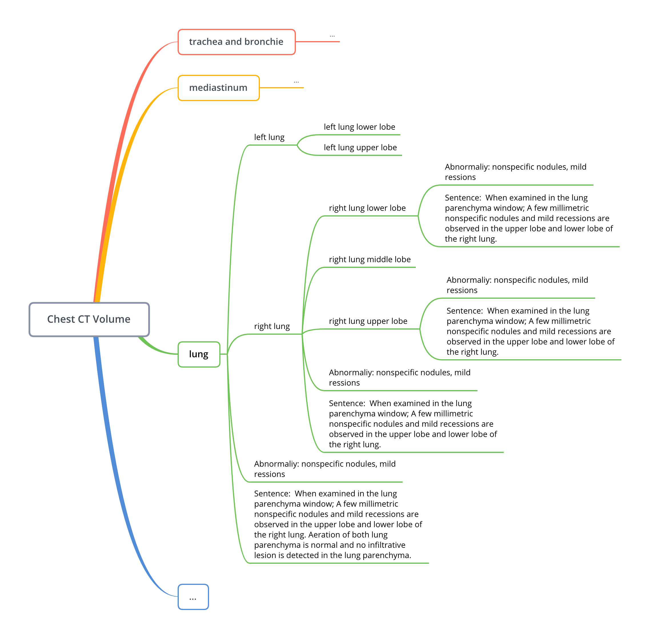
To segment as many anatomical regions as possible, we employ the recent SAT [13] model. It is a knowledge-enhanced segmentation model, that employs natural language as prompts to effectively segment 3D medical volumes. The model has been trained on 72 diverse segmentation datasets, covering 498 classes across various anatomical regions including the brain, head and neck, thorax, spine, abdomen, and limbs. For our research, SAT is adopted to execute detailed segmentation across all volumes of the CT-RATE dataset. Specifically, we focus on segmenting 197 regions pertinent to chest CT scans, enabling precise anatomical analysis. The list of segmented anatomies was organized into a hierarchical tree as shown in Supplementary B. This includes several major regions such as the lungs, trachea and bronchi, mediastinum, heart, pleura, bones, thyroid, breasts, abdomen, and other areas.
Region-wise Reports Generation
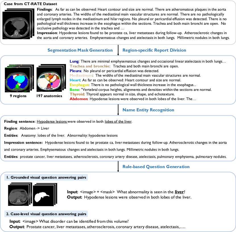
The goal here is to break the entire reports into region-wise descriptions, we observe that the OpenAI GPT-4 [2] model can complete such task in very high accuracy [14, 15]. However, employing GPT-4 on the entire set would be prohibitively expensive, thus, we train a model for such report division. Specifically, we first employ GPT-4 [2] to annotate the anatomical regions of each sentence in the “FINDINGS” section of 2,500 radiology reports, which comprise all reports in the validation set. The prompt used is as follows.
This process results in 15,926 annotated sentences. Subsequently, we divide these sentences into training and validation subsets in an 8:2 ratio and train a GPT-2 model using the annotated sentence along with the two preceding and following sentences from the report as input, if available. The model is designed to output the list of anatomical regions associated with each sentence. For instance, for the target sentence “No pleural effusion was detected on the left.”, the input is “There is minimal pleural effusion on the right. No pleural effusion was detected on the left. Atelectasis is observed in the middle lobe and lower lobe of the right lung. A malignant mass is observed around the lower lobe bronchi of the left lung.”, and the expected output is “left lung”. The model achieves an accuracy of 94.56% on the validation set. Consequently, we employ this model to perform inference on all sentences across the entire dataset of reports. More examples of segmentation results and structured reports can be seen in Supplementary Section A.
Named Entity Recognition
Through the abovementioned process, each “FINDINGS” is divided into multiple sentences, and each is associated with one or multiple segmented regions. To facilitate the generation of detailed question-answer pairs from these sentences, we initially employ an in-house Named-Entity Recognition (NER) model to analyze all sentences. This process involves extracting entities that can be categorized into “anatomy”, “abnormality”, and “non-abnormality”. Here, “anatomy” pertains to the anatomical regions, “abnormality” refers to findings or diseases identified as present, and “non-abnormality” indicates findings or diseases that are reported as absent. Subsequently, all extracted "abnormality" and "non-abnormality" entities undergo quality evaluation using GPT-4, allowing us to filter out and revise any inaccuracies. For instance, in cases where the NER model extracts abnormalities such as “structural distortion and volume loss”, GPT-4 will segment it into “structural distortion” and “volume loss” for more accurate categorization. We filter out abnormalities with a GPT-4 output of “no” and update them to the revised versions provided by GPT-4. The prompt used is as follows.
In addition, for the “IMPRESSION” section, we directly utilize GPT-4 to extract all disorders mentioned, and detailed information regarding the presence of any abnormalities in specific anatomical regions. The prompt used is as follows.
| Level | Question type | Answer type | Example | Train | Validation |
|---|---|---|---|---|---|
| Region | Abnormality | Open | What abnormality is seen in the {region }? | 483,507 | 30,831 |
| Region | Presence | Close | Is there evidence of {abnormality} in the {region }? | 556,050 | 34,785 |
| Region | Location | Close & Open | Where in the image is the {abnormality} seen? | 285,111 | 17,750 |
| Region | Size | Open | What is the size of the {abnormality} in the {region}? | 18,389 | 1,259 |
| Case | Disorder | Open | What disorder can be identified from this volume? | 24,128 | 1,564 |
Rule-based Question Generation
Here, we describe the procedure for generating grounded visual question-answering (VQA) data based on “FINDINGS” extracted from the report section, while case-level visual question-answering data is generated from the “IMPRESSION” section. Tab. 1 presents the 5 question types in the proposed dataset. The detailed rules will be introduced in the following sections.
First, after the region-wise report generation, the sentences in the findings section can be classified as follows:
-
•
Normal Findings: Sentences that report no significant changes from normal health conditions.
-
–
No abnormality entities in the sentence: Sentences that mention specific anatomical regions without noting any abnormalities. For example, “Thoracic aorta diameter is normal.”
-
–
No anatomy entities in the sentence: Sentences that solely note the absence of specific abnormalities without referring to any anatomical regions. For example, “No pleural effusion was detected.”
-
–
With anatomy and abnormality entities in the sentence: Sentences that explicitly state the absence of abnormalities in specific anatomical regions. For example, “Bilateral adrenal glands were normal and no space-occupying lesion was detected.”
-
–
-
•
Abnormal Findings: Sentences that report differences from normal anatomical conditions.
-
–
No anatomy entities in the sentence: Sentences that report an abnormal finding, but do not specify an anatomical region. For example, “Mild hiatal hernia is observed.”
-
–
With anatomy entities in the sentence: Sentences that include both an anatomical reference and describe an abnormality. For example, “There is narrowing of the spinal canal at the dorso-lumbar level.”
-
–
We then construct an anatomical disorder tree for each report, based on the anatomical hierarchical tree introduced in Supplementary Section B. This involves marking any abnormalities on the tree for all nodes within the hierarchy if they are present. As shown in Fig. 3 This comprehensive data enables us to transit to generating questions. Taking inspiration from the previous research [16, 17], we categorize the questions into four types: 1) abnormality, 2) presence, 3) location, 4) size. Tab. 1 shows examples of the different question types. Note that we have designed 50 templates for each question type. The details of all templates are provided in the supplementary materials. For instance, when analyzing a sentence indicating normal findings, such as “Bilateral adrenal glands were normal and no space-occupying lesion was detected.”, questions can include “Is there any evidence of abnormality in adrenal glands?” and “What abnormality is seen in the adrenal glands?”. Conversely, for a sentence in abnormal findings, “There is a narrowing of the spinal canal at the dorso-lumbar level.”, the question can be “Is there any evidence of narrowing in the spinal canal?” “What abnormality is seen at the dorso-lumbar level of the spinal canal?” and “Where in the spinal canal is the narrowing located?”. For impression sentences, since we have already extracted disorders, we can generate case-level questions such as “What disorder can be identified from this volume?” for each case.
In summary, we have successfully generated 1.3M grounded Visual Question Answering (VQA) pairs for training and 85k for validation, along with 24,128 case-level visual question answering pairs for training and 1,564 for validation. Detailed counts for each type of VQA pair in both the training and validation sets are provided in Table 1.
Dataset Analysis
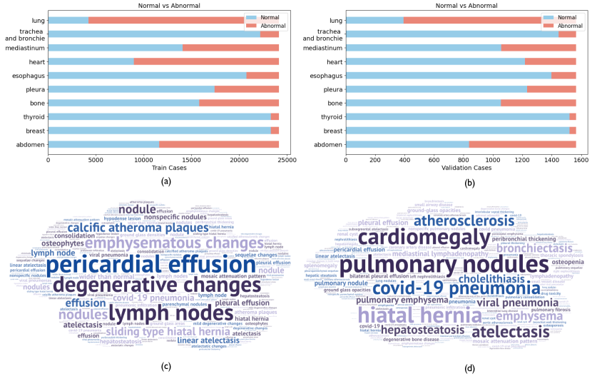
In this section, we analyze the abnormalities of the proposed dataset. The proposed hierarchically structured abnormality tree for each case enables us to systematically extract and analyze the abnormal findings. First, we calculate the normal-to-abnormal case numbers for both the training set and the validation set, as shown in Fig. 4. Second, based on the identified and extracted abnormalities from all anatomical regions, we proceed to visualize these findings using a word cloud. The word cloud of abnormalities is presented in Fig. 4.
Conclusion
In this paper, we develop an automated pipeline for generating grounded datasets and introduce RadGenome-Chest CT, a comprehensive, large-scale, region-guided 3D chest CT interpretation dataset based on CT-RATE. RadGenome-Chest CTinclude 197 organ-level segmentation masks, 665 K multi-granularity grounded reports, and 1.3 M grounded VQA pairs. We anticipate that RadGenome-Chest CTwill significantly advance multimodal medical AI models, enabling them to generate texts based on segmentation regions, thus enhancing interpretability and patient care. We will release all segmentation masks, grounded reports, and VQA pairs to support future research in this field.
References
- [1] Lu, M. Y. et al. Towards a visual-language foundation model for computational pathology. \JournalTitlearXiv preprint arXiv:2307.12914 (2023).
- [2] Achiam, J. et al. Gpt-4 technical report. \JournalTitlearXiv preprint arXiv:2303.08774 (2023).
- [3] Singhal, K. et al. Large language models encode clinical knowledge. \JournalTitleNature 620, 172–180 (2023).
- [4] Team, G. et al. Gemini: a family of highly capable multimodal models. \JournalTitlearXiv preprint arXiv:2312.11805 (2023).
- [5] Alayrac, J.-B. et al. Flamingo: a visual language model for few-shot learning. \JournalTitleAdvances in neural information processing systems 35, 23716–23736 (2022).
- [6] Wu, C., Zhang, X., Zhang, Y., Wang, Y. & Xie, W. Towards generalist foundation model for radiology. \JournalTitlearXiv preprint arXiv:2308.02463 (2023).
- [7] Tu, T. et al. Towards generalist biomedical ai. \JournalTitleNEJM AI 1, AIoa2300138 (2024).
- [8] Li, C. et al. Llava-med: Training a large language-and-vision assistant for biomedicine in one day. \JournalTitleAdvances in Neural Information Processing Systems 36 (2024).
- [9] Huang, Z., Bianchi, F., Yuksekgonul, M., Montine, T. J. & Zou, J. A visual–language foundation model for pathology image analysis using medical twitter. \JournalTitleNature medicine 29, 2307–2316 (2023).
- [10] Moor, M. et al. Med-flamingo: a multimodal medical few-shot learner. In Machine Learning for Health (ML4H), 353–367 (PMLR, 2023).
- [11] Johnson, A. E. et al. Mimic-cxr, a de-identified publicly available database of chest radiographs with free-text reports. \JournalTitleScientific data 6, 317 (2019).
- [12] Hamamci, I. E. et al. A foundation model utilizing chest ct volumes and radiology reports for supervised-level zero-shot detection of abnormalities. \JournalTitlearXiv preprint arXiv:2403.17834 (2024).
- [13] Zhao, Z. et al. One model to rule them all: Towards universal segmentation for medical images with text prompts. \JournalTitlearXiv preprint arXiv:2312.17183 (2023).
- [14] Nori, H., King, N., McKinney, S. M., Carignan, D. & Horvitz, E. Capabilities of gpt-4 on medical challenge problems. \JournalTitlearXiv preprint arXiv:2303.13375 (2023).
- [15] Liu, Q. et al. Exploring the boundaries of gpt-4 in radiology. \JournalTitlearXiv preprint arXiv:2310.14573 (2023).
- [16] Hu, X. et al. Expert knowledge-aware image difference graph representation learning for difference-aware medical visual question answering. In Proceedings of the 29th ACM SIGKDD Conference on Knowledge Discovery and Data Mining, 4156–4165 (2023).
- [17] Lau, J. J., Gayen, S., Ben Abacha, A. & Demner-Fushman, D. A dataset of clinically generated visual questions and answers about radiology images. \JournalTitleScientific data 5, 1–10 (2018).
Appendix A Case Examples
Appendix B Hierarchy of anatomical regions
.1 Chest CT Volume. .2 Lung. .3 Lung. .4 Left Lung. .5 Left Lung Lower Lobe. .5 Left Lung Upper Lobe. .4 Right Lung. .5 Right Lung Lower Lobe. .5 Right Lung Middle Lobe. .5 Right Lung Upper Lobe. .4 Lung Lower Lobe. .5 Left Lung Lower Lobe. .5 Right Lung Lower Lobe. .4 Lung Upper Lobe. .5 Left Lung Upper Lobe. .5 Right Lung Upper Lobe. .2 Trachea and Bronchie. .3 Trachea. .3 Bronchie. .2 Mediastinum. .3 Brachiocephalic Trunk. .3 Brachiocephalic Vein. .4 Left Brachiocephalic Vein. .4 Right Brachiocephalic Vein. .3 Superior Vena Cava. .3 Aorta. .3 Pulmonary Artery. .3 Thymus. .3 Pulmonary Vein. .3 Mediastinal Tissue. .3 Subclavian Artery. .4 Left Subclavian Artery. .4 Right Subclavian Artery. .2 Heart. .3 Heart. .4 Heart Atrium. .5 Left Heart Atrium. .5 Right Heart Atrium. .4 Heart Ventricle. .5 Left Heart Ventricle. .5 Right Heart Ventricle. .4 Heart Ascending Aorta. .4 Heart Tissue. .5 Myocardium. .4 Left Auricle of Heart. .2 Esophagus. .3 Esophagus. .4 Cervical Esophagus. .4 Cricopharyngeal Inlet. .2 Pleura. .2 Bone. .3 Bone. .4 Spinal Cord. .4 Spinal Canal. .4 Vertebrae. .5 Cervical Vertebrae. .6 Cervical Vertebrae 1 (C1). .6 Cervical Vertebrae 2 (C2). .6 Cervical Vertebrae 3 (C3). .6 Cervical Vertebrae 4 (C4). .6 Cervical Vertebrae 5 (C5). .6 Cervical Vertebrae 6 (C6). .6 Cervical Vertebrae 7 (C7). .5 Thoracic Vertebrae. .6 Thoracic Vertebrae 1 (T1). .6 Thoracic Vertebrae 2 (T2). .6 Thoracic Vertebrae 3 (T3). .6 Thoracic Vertebrae 4 (T4). .6 Thoracic Vertebrae 5 (T5). .6 Thoracic Vertebrae 6 (T6). .6 Thoracic Vertebrae 7 (T7). .6 Thoracic Vertebrae 8 (T8). .6 Thoracic Vertebrae 9 (T9). .6 Thoracic Vertebrae 10 (T10). .6 Thoracic Vertebrae 11 (T11). .6 Thoracic Vertebrae 12 (T12). .5 Lumbar Vertebrae. .6 Lumbar Vertebrae 1 (L1). .6 Lumbar Vertebrae 2 (L2). .6 Lumbar Vertebrae 3 (L3). .6 Lumbar Vertebrae 4 (L4). .6 Lumbar Vertebrae 5 (L5). .6 Lumbar Vertebrae 6 (L6). .5 Sacral Vertebrae 1 (S1). .4 Clavicle. .5 Left Clavicle. .5 Right Clavicle. .4 Scapula. .5 Left Scapula. .5 Right Scapula. .4 Humerus. .5 Left Humerus. .5 Right Humerus. .4 Femur. .5 Left Femur. .5 Right Femur. .4 Head of Femur. .5 Left Head of Femur. .5 Right Head of Femur. .4 Rib. .5 Left Rib. .6 Left Rib 1. .6 Left Rib 2. .6 Left Rib 3. .6 Left Rib 4. .6 Left Rib 5. .6 Left Rib 6. .6 Left Rib 7. .6 Left Rib 8. .6 Left Rib 9. .6 Left Rib 10. .6 Left Rib 11. .6 Left Rib 12. .5 Right Rib. .6 Right Rib 1. .6 Right Rib 2. .6 Right Rib 3. .6 Right Rib 4. .6 Right Rib 5. .6 Right Rib 6. .6 Right Rib 7. .6 Right Rib 8. .6 Right Rib 9. .6 Right Rib 10. .6 Right Rib 11. .6 Right Rib 12. .4 Rib 1. .5 Left Rib 1. .5 Right Rib 1. .4 Rib 2. .5 Left Rib 2. .5 Right Rib 2. .4 Rib 3. .5 Left Rib 3. .5 Right Rib 3. .4 Rib 4. .5 Left Rib 4. .5 Right Rib 4. .4 Rib 5. .5 Left Rib 5. .5 Right Rib 5. .4 Rib 6. .5 Left Rib 6. .5 Right Rib 6. .4 Rib 7. .5 Left Rib 7. .5 Right Rib 7. .4 Rib 8. .5 Left Rib 8. .5 Right Rib 8. .4 Rib 9. .5 Left Rib 9. .5 Right Rib 9. .4 Rib 10. .5 Left Rib 10. .5 Right Rib 10. .4 Rib 11. .5 Left Rib 11. .5 Right Rib 11. .4 Rib 12. .5 Left Rib 12. .5 Right Rib 12. .4 Rib Cartilage. .4 Costal Cartilage. .4 Sternum. .5 Manubrium of Sternum. .4 Eustachian Tube Bone. .5 Left Eustachian Tube Bone. .5 Right Eustachian Tube Bone. .2 Thyroid. .3 Thyroid. .4 Thyroid Gland. .5 Left Thyroid. .5 Right Thyroid. .2 Breast. .3 Left Breast. .3 Right Breast. .2 Abdomen. .3 Abdomen. .4 Abdominal Tissue. .4 Adrenal Gland. .5 Left Adrenal Gland. .5 Right Adrenal Gland. .4 Aorta. .4 Colon. .4 Duodenum. .4 Gallbladder. .4 Intestine. .5 Small Bowel. .4 Kidney. .5 Left Kidney. .5 Right Kidney. .4 Liver. .5 Left Lobe of Liver. .6 Left Lateral Inferior Segment of Liver. .6 Left Lateral Superior Segment of Liver. .6 Left Medial Segment of Liver. .5 Right Lobe of Liver. .6 Right Anterior Inferior Segment of Liver. .6 Right Anterior Superior Segment of Liver. .6 Right Posterior Inferior Segment of Liver. .6 Right Posterior Superior Segment of Liver. .4 Liver Vessel. .4 Caudate Lobe. .4 Pancreas. .4 Portal Vein and Splenic Vein. .4 Rectum. .4 Renal Artery. .4 Renal Vein. .4 Spleen. .4 Stomach. .4 Celiac Trunk. .2 Others. .3 Thoracic Cavity. .3 Prostate. .3 Urinary Bladder. .3 Carotid Artery. .4 Common Carotid Artery. .4 Internal Carotid Artery. .4 Left Carotid Artery. .5 Left Common Carotid Artery. .5 Left Internal Carotid Artery. .4 Right Carotid Artery. .5 Right Common Carotid Artery. .5 Right Internal Carotid Artery. .3 Iliac Artery. .3 Iliac Vena. .3 Iliac Vein. .3 Left Iliac Artery. .3 Left Iliac Vena. .3 Right Iliac Artery. .3 Right Iliac Vena. .3 Inferior Vena Cava. .3 Internal Jugular Vein. .3 Larynx. .4 Larynx Glottis. .4 Larynx Supraglottis. .3 Muscle.
Appendix C Question template
Abnormality
-
1.
What are the abnormalities in the {region}?
-
2.
What abnormalities are present in the {region}?
-
3.
What types of abnormality are visible in the {region}?
-
4.
What type of abnormality is visible in the {region}?
-
5.
What kind of abnormalities are observed in the {region}?
-
6.
What kinds of abnormalities can be identified in the {region}?
-
7.
What specific abnormalities are detected in the {region}?
-
8.
What specific types of abnormalities are evident in the {region}?
-
9.
What types of abnormalities are evident upon examination of the {region}?
-
10.
What types of abnormalities can be seen in the {region}?
-
11.
What are the anomalies in the {region}?
-
12.
What anomalies are present in the {region}?
-
13.
What types of anomalies are visible in the {region}?
-
14.
What type of anomalies is visible in the {region}?
-
15.
What kind of anomalies are observed in the {region}?
-
16.
What kinds of anomalies can be identified in the {region}?
-
17.
What specific anomalies are detected in the {region}?
-
18.
What specific types of anomalies are evident in the {region}?
-
19.
What types of anomalies are evident upon examination of the {region}?
-
20.
What types of anomalies can be seen in the {region}?
-
21.
What are the abnormal findings in the {region}?
-
22.
What abnormal findings are present in the {region}?
-
23.
What types of abnormal findings are visible in the {region}?
-
24.
What type of abnormal findings is visible in the {region}?
-
25.
What kind of abnormal findings are observed in the {region}?
-
26.
What kinds of abnormal findings can be identified in the {region}?
-
27.
What specific abnormal findings are detected in the {region}?
-
28.
What specific types of abnormal findings are evident in the {region}?
-
29.
What types of abnormal findings are evident upon examination of the {region}?
-
30.
What types of abnormal findings can be seen in the {region}?
-
31.
What are the irregular findings in the {region}?
-
32.
What irregular findings are present in the {region}?
-
33.
What types of irregular findings are visible in the {region}?
-
34.
What type of irregular findings is visible in the {region}?
-
35.
What kind of irregular findings are observed in the {region}?
-
36.
What kinds of irregular findings can be identified in the {region}?
-
37.
What specific irregular findings are detected in the {region}?
-
38.
What specific types of irregular findings are evident in the {region}?
-
39.
What types of irregular findings are evident upon examination of the {region}?
-
40.
What types of irregular findings can be seen in the {region}?
-
41.
What are the irregularities in the {region}?
-
42.
What irregularities are present in the {region}?
-
43.
What types of irregularities are visible in the {region}?
-
44.
What type of irregularities is visible in the {region}?
-
45.
What kind of irregularities are observed in the {region}?
-
46.
What kinds of irregularities can be identified in the {region}?
-
47.
What specific irregularities are detected in the {region}?
-
48.
What specific types of irregularities are evident in the {region}?
-
49.
What types of irregularities are evident upon examination of the {region}?
-
50.
What types of irregularities can be seen in the {region}?
Presence
-
1.
Can {abnormality} be identified in the {region}?
-
2.
Can {abnormality} be observed in the {region}?
-
3.
Can {abnormality} be detected in the {region}?
-
4.
Can {abnormality} be seen in the {region}?
-
5.
Can {abnormality} be founded in the {region}?
-
6.
Can {abnormality} be recognized in the {region}?
-
7.
Can we detect any signs of {abnormality} in the {region}?
-
8.
Can we observe any signs of {abnormality} in the {region}?
-
9.
Can we recognize any signs of {abnormality} in the {region}?
-
10.
Can we see {abnormality} in the {region}?
-
11.
Can we find {abnormality} in the {region}?
-
12.
Can we detect {abnormality} in the {region}?
-
13.
Can we observe {abnormality} in the {region}?
-
14.
Can we identify {abnormality} in the {region}?
-
15.
Is there any sign of {abnormality} in the {region}?
-
16.
Is there any indication of {abnormality} in the {region}?
-
17.
Is there any evidence of {abnormality} in the {region}?
-
18.
Is there any suggestion of {abnormality} in the {region}?
-
19.
Is there a clear sign of {abnormality} in the {region}?
-
20.
Is there a clear indication of {abnormality} in the {region}?
-
21.
Is there a clear evidence of {abnormality} in the {region}?
-
22.
Is there a clear suggestion of {abnormality} in the {region}?
-
23.
Is {abnormality} visibly present in the {region}?
-
24.
Is {abnormality} clearly visible in the {region}?
-
25.
Is there any visual evidence suggesting {abnormality} in the {region}?
-
26.
Is there any indication of {abnormality} upon examination of the {region}?
-
27.
Is there any indication of {abnormality} in the {region}?
-
28.
Is there visual evidence of {abnormality} in the {region} on this scan?
-
29.
Are there any visible indications of {abnormality} in this {region}?
-
30.
Are there any visible cues indicating {abnormality} in the {region}?
-
31.
Are there any visible indicators of {abnormality} in the {region}?
-
32.
Are there any clear indications of {abnormality} in this {region}?
-
33.
Are there any clear cues indicating {abnormality} in the {region}?
-
34.
Are there any clear indicators of {abnormality} in the {region}?
-
35.
Are there any indications of {abnormality} in this {region}?
-
36.
Are there any cues indicating {abnormality} in the {region}?
-
37.
Are there any indicators of {abnormality} in the {region}?
-
38.
Are there any observable signs of {abnormality} in the {region}?
-
39.
Are there any signs of {abnormality} in the {region}?
-
40.
Are there any features of {abnormality} in the {region}?
-
41.
Does the {region} show the presence of {abnormality}?
-
42.
Does the {region} show the existence of {abnormality}?
-
43.
Does the image suggest the presence of {abnormality} in the {region}?
-
44.
Does the image suggest the existence of {abnormality} in the {region}?
-
45.
Does the {region} exhibit any evidence of {abnormality}?
-
46.
Does the {region} display any features suggestive of {abnormality}?
-
47.
Does the {region} display any characteristics suggestive of {abnormality}?
-
48.
Does the {region} exhibit any characteristics indicative of {abnormality}?
-
49.
Does the {region} exhibit any features indicative of {abnormality}?
-
50.
Does the visual features suggest the presence of {abnormality} in the {region}?
Location
-
1.
Where is the {abnormality} located in the image?
-
2.
Where can the {abnormality} be found within the image?
-
3.
Where in the image is the {abnormality} located?
-
4.
Where in the image is the {abnormality} localized?
-
5.
Where in the image can the {abnormality} be found?
-
6.
Where in the image does the {abnormality} appear?
-
7.
Where in the image does the {abnormality} locate?
-
8.
Where in the image does the {abnormality} locate?
-
9.
Where specifically within the image is the {abnormality} located?
-
10.
Where exactly within the image is the {abnormality} located?
-
11.
Where exactly is the {abnormality} located in the image?
-
12.
Where specifically is the {abnormality} located in the image?
-
13.
Where exactly within the image is the {abnormality} localized?
-
14.
Where specifically within the image is the {abnormality} localized?
-
15.
Where within the image can the {abnormality} be precisely located?
-
16.
Where exactly within the image does the {abnormality} present?
-
17.
Where within the image does the {abnormality} specifically present?
-
18.
Where in the image does the {abnormality} appear?
-
19.
What is the location of the {abnormality} in the image?
-
20.
What is the precise location of the {abnormality} in the image?
-
21.
What is the specific location of the {abnormality} within the image?
-
22.
What is the precise region of the {abnormality} in the image?
-
23.
What is the specific region of the {abnormality} within the image?
-
24.
What particular region within the image does the {abnormality} occupy?
-
25.
What particular location within the image does the {abnormality} occupy?
-
26.
What specific location within the image does the {abnormality} occupy?
-
27.
What specific region within the image does the {abnormality} occupy?
-
28.
What specific area of the image does the {abnormality} occupy?
-
29.
What specific region of the image does the {abnormality} appear?
-
30.
What specific spot within the image contains the {abnormality}?
-
31.
What particular region of the image is affected by the {abnormality}?
-
32.
What specific area within the image is impacted by the {abnormality}?
-
33.
What specific region within the image is impacted by the {abnormality}?
-
34.
What specific location within the image is impacted by the {abnormality}?
-
35.
What particular region within the image is affected by the {abnormality}?
-
36.
What particular area within the image is affected by the {abnormality}?
-
37.
What particular location within the image is affected by the {abnormality}?
-
38.
What specific region within the image does the {abnormality} affect?
-
39.
What specific area within the image does the {abnormality} affect?
-
40.
What specific location within the image does the {abnormality} affect?
-
41.
What specific location within the image does the {abnormality} appear?
-
42.
What specific region within the image does the {abnormality} appear?
-
43.
What specific area within the image does the {abnormality} appear?
-
44.
What particular spot within the image does the {abnormality} present?
-
45.
What particular area within the image does the {abnormality} present?
-
46.
What particular region within the image does the {abnormality} present?
-
47.
What particular location within the image does the {abnormality} present?
-
48.
What specific area within the image does the {abnormality} occur?
-
49.
What specific location within the image does the {abnormality} occur?
-
50.
What specific region within the image does the {abnormality} occur?
Size
-
1.
What is the approximate size of the {abnormality} in the {region}?
-
2.
What is the approximate scale of the {abnormality} in the {region}?
-
3.
What is the approximate size range of the {abnormality} in the {region}?
-
4.
What is the approximate magnitude of the {abnormality} in the {region}?
-
5.
What is the approximate dimension of the {abnormality} in the {region}?
-
6.
What is the approximate measurement of the {abnormality} in the {region}?
-
7.
What is the estimated size of the {abnormality} in the {region}?
-
8.
What is the estimated scale of the {abnormality} in the {region}
-
9.
What is the estimated size range of the {abnormality} in the {region}?
-
10.
What is the estimated magnitude of the {abnormality} in the {region}?
-
11.
What is the estimated dimension of the {abnormality} in the {region}?
-
12.
What is the estimated measurement of the {abnormality} in the {region}?
-
13.
What is the size assessment of the {abnormality} in the {region}?
-
14.
What is the measurement of the {abnormality} in the {region}?
-
15.
What is the scale of the {abnormality} in the {region}?
-
16.
What is the size of the {abnormality} in the {region}?
-
17.
What is the size range of the {abnormality} in the {region}?
-
18.
What is the magnitude of the {abnormality} in the {region}?
-
19.
What is the dimension of the {abnormality} in the {region}?
-
20.
What is the overall size of the {abnormality} in the {region}?
-
21.
What is the overall scale of the {abnormality} in the {region}?
-
22.
What is the overall measurement of the {abnormality} in the {region}?
-
23.
What is the overall size range of the {abnormality} in the {region}?
-
24.
What is the overall magnitude of the {abnormality} in the {region}?
-
25.
What is the overall dimension of the {abnormality} in the {region}?
-
26.
What is the scale of the {abnormality} detected in the {region}?
-
27.
What is the size of the {abnormality} detected in the {region}?
-
28.
What is the size range of the {abnormality} detected in the {region}?
-
29.
What is the measurement of the {abnormality} detected in the {region}?
-
30.
What is the magnitude of the {abnormality} detected in the {region}?
-
31.
What is the dimension of the {abnormality} detected in the {region}?
-
32.
What is the scale of the {abnormality} appeared in the {region}?
-
33.
What is the size of the {abnormality} appeared in the {region}?
-
34.
What is the size range of the {abnormality} appeared in the {region}?
-
35.
What is the magnitude of the {abnormality} appeared in the {region}?
-
36.
What is the dimension of the {abnormality} appeared in the {region}?
-
37.
What is the measurement of the {abnormality} appeared in the {region}?
-
38.
How large is the affected {abnormality} area in the {region}?
-
39.
How large is the observed {abnormality} area in the {region}?
-
40.
How large is the {abnormality} observed in the {region}?
-
41.
How large is the {abnormality} in the {region}?
-
42.
How large does the {abnormality} appear in the {region}?
-
43.
How large does the {abnormality} appear to be in the {region}?
-
44.
How large is the {abnormality} area in the {region}?
-
45.
How large is the affected {abnormality} area in the {region}?
-
46.
How big is the affected {abnormality} area in the {region}?
-
47.
How big is the observed {abnormality} area in the {region}?
-
48.
How big is the {abnormality} observed in the {region}?
-
49.
How big is the {abnormality} in the {region}?