† People involved in the organization of the challenge.
‡ People contributing data from their institutions.
§ People involved in annotation process.
** Corresponding author: 5050email: {mlingura@childrensnational.org}
The Brain Tumor Segmentation in Pediatrics (BraTS-PEDs) Challenge: Focus on Pediatrics (CBTN-CONNECT-DIPGR-ASNR-MICCAI BraTS-PEDs)
Abstract
Pediatric tumors of the central nervous system are the most common cause of cancer-related death in children. The five-year survival rate for high-grade gliomas in children is less than 20%. Due to their rarity, the diagnosis of these entities is often delayed, their treatment is mainly based on historic treatment concepts, and clinical trials require multi-institutional collaborations. Here we present the CBTN-CONNECT-DIPGR-ASNR-MICCAI BraTS-PEDs challenge, focused on pediatric brain tumors with data acquired across multiple international consortia dedicated to pediatric neuro-oncology and clinical trials. The CBTN-CONNECT-DIPGR-ASNR-MICCAI BraTS-PEDs challenge brings together clinicians and AI/imaging scientists to lead to faster development of automated segmentation techniques that could benefit clinical trials, and ultimately the care of children with brain tumors.
Keywords:
BraTS, BraTS-PEDs, challenge, pediatric, brain, tumor, segmentation, volume, deep learning, machine learning, artificial intelligence, AI1 Introduction
Pediatric diffuse midline gliomas (DMGs, including pediatric diffuse intrinsic pontine glioma (DIPGs)) are high grade gliomas with short average overall survival [1, 2]. Many DMGs are located in the pons and often diagnosed between 5 and 10 years of age. Pediatric brain tumors require dedicated tumor segmentation tools that help in their characterization and facilitate their diagnosis, prognosis, and treatment response assessment [3, 4, 5]. There are only a handful of automated tumor segmentation methods explicitly proposed for pediatric brain tumors [3, 6, 7, 8, 9, 10, 11, 12, 13, 14, 15]. However, the majority of these methods have been developed only for the segmentation of the T2 fluid attenuated inversion recovery (FLAIR) abnormal signal [6, 7, 12], also called whole tumor (WT).
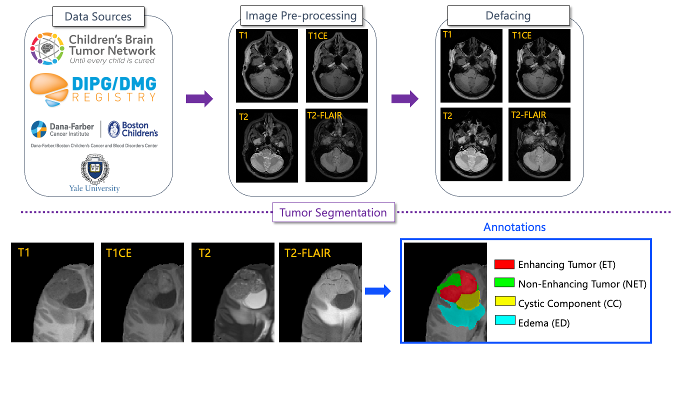
The MICCAI brain tumor segmentation (BraTS) challenges have established a community benchmark dataset and environment for adult glioma over the past 12 years [16, 17, 18, 19]. In 2023 challenge, we launched the first Brain Tumor Segmentation in Pediatrics (BraTS-PEDs) challenge. In 2024, we continue the BraTS-PEDs challenge with modifications in processing pipeline and tumor subregions. We have created a retrospective multi-institutional (multi-consortium) pediatric database, with the data collected through a few consortia, including Children’s Brain Tumor Network (CBTN, https://cbtn.org/ ) [20], DIPG Registry (https://www.dipgregistry.org) and the COllaborative Network for NEuro-oncology Clinical Trials (CONNECT, https://connectconsortium.org/). Additional data from participating pediatric institutions have been included in the BraTS-PEDs cohort. The American Society of Neuroradiology (ASNR, https://www.asnr.org/) collaborated in generating ground truth annotation for the majority of data in this challenge. This manuscript provides an overview of the CBTN-CONNECT-ASNR-MICCAI BraTS-PEDs challenge.
2 Challenge Design
2.1 Data
The BraTS-PEDs dataset includes a retrospective multi-institutional cohort of conventional/structural magnetic resonance imaging (MRI) sequences, including pre- and post-gadolinium T1-weighted (labeled as T1 and T1CE), T2-weighted (T2), and T2-weighted fluid attenuated inversion recovery (T2-FLAIR) images, from 464 pediatric high-grade glioma. These conventional multiparametric MRI (mpMRI) sequences are commonly acquired as part of standard clinical imaging for brain tumors. However, the image acquisition protocols and MRI equipment differ across different institutions, resulting in heterogeneity in image quality in the provided cohort. Inclusion criteria comprised of pediatric subjects with: (1) histologically-approved high-grade glioma, i.e., high-grade astrocytoma and diffuse midline glioma (DMG), including radiologically or histologically-proven diffuse intrinsic pontine glioma (DIPG); (2) availability of all four structural mpMRI sequences on treatment-naive imaging sessions. Exclusion criteria consisted of: (1) images assessed to be of low quality or with artifacts that would not allow for reliable tumor segmentation; and (2) infants younger than one month of age. Data for 464 patients was obtained through CBTN (n = 120), DMG/ DIPG Registry (n = 256), Boston’s Children Hospital (n = 61), and Yale University (n = 27).
The cohort included in the challenge is split into training, validation, and testing datasets. The data shared with the participants comprise mpMRI scans and ground truth labels for the training cohort, as well as mpMRI sequences without any associated ground truth for the validation cohort. Notably, the testing data that will be used for evaluating the performance of the methods submitted by challenge participants will not be shared with the participants, but the containerized submissions of the participants will be evaluated by the synapse.org platform, powered by MedPerf[21].
Participants are prohibited from training their algorithm on any additional public and/or private data (from their own institutions) besides the provided BraTS-PEDs data, or using models pretrained on any other dataset. This restriction is imposed to allow for a fair comparison among the participating methods. However, participants can use additional public and/or private data only for publication of their scientific papers, if they also provide a report of their results on the data from the BraTS-PEDs challenge alone, and discuss potential differences in the obtained results.
2.1.1 Imaging Data Description
For all patients, mpMRI scans were prepared using the following steps:
-
1.
Pre-processing using the ”BraTS Pipeline”, a standardized approach, publicly available through the Cancer Imaging Phenomics Toolkit (CaPTk) 111https://cbica.github.io/CaPTk/ [22, 23, 24] and Federated Tumor Segmentation (FeTS) tool 222https://github.com/FETS-AI/Front-End/. De-identification was performed through removing protected Health Information (PHI) from DICOM headers in DICOM to NIfTI conversion step [25, 26].
-
2.
Defacing using a pediatric-specific automated defacing method 333https://github.com/d3b-center/peds-auto-defacing-public
-
3.
Tumor subregion segmentation using a pediatric autosegmentation method [13] 444https://github.com/d3b-center/peds-brain-seg-pipeline-public/
The result of this step was segmentation of the tumors into four main subregions, recommended by RAPNO working group for evaluation of the treatment response in high-grade gliomas and DIPGs. The annotated tumor subregions comprised of the following regions [3]:
-
1.
Enhancing tumor (ET), described by areas with enhancement (brightness) on T1 post-contrast images as compared to T1 pre-contrast. In case of mild enhancement, checking the signal intensity of normal brain structure can be helpful.
-
2.
Nonenhancing tumor (NET), defined as any other abnormal signal intensity within the tumorous region that cannot be defined as enhancing or cystic. For example, the abnormal signal intensity on T1, T2-FLAIR, and T2 that is not enhancing on T1CE should be considered as nonenhancing portion.
-
3.
Cystic component (CC), typically appearing with hyperintense signal (very bright) on T2 and hypointense signal (dark) on T1CE. The cystic portion should be within the tumor, either centrally or peripherally (as compared to ED which is peritumoral). The brightness of CC is here defined as comparable or close to cerebrospinal fluid (CSF).
-
4.
Peritumoral edema (ED), defined by the abnormal hyperintense signal (very bright) on FLAIR scans. ED is finger-like spreading that preserves underlying brain structure and surrounds the tumor.
The automatically-generated four labels (Figure 1) using the automated segmentation tool were used as preliminary segmentation to be manually revised by volunteer neuroradiology experts of varying rank and experience, in accordance with annotation guidelines. The volunteer neuroradiology expert annotators were provided with four mpMRI sequences (T1, T1CE, T2, FLAIR) along with the fused automated segmentation volume to initiate the manual refinements. The ITK-SNAP [27] software was used for making these refinements. Once the automated segmentations were refined by the annotators, three attending board-certified neuroradiologists, reviewed the segmentations. Depending upon correctness, these segmentations were either approved or returned to the individual annotator for further refinements. This process was followed iteratively until the approvers found the refined tumor subregion segmentations acceptable for public release and the challenge conduction.
2.1.2 Common errors of automated segmentations
In our experience with automated segmentation of pediatric brain tumors, some errors may be noticed:
-
1.
Peri-ventricular edema segmented as ED
-
2.
Remote areas segmented as tumor (far from actual tumor region)
-
3.
Under-segmentation of cysts
-
4.
Non-enhancing tumor segmented as cyst, or vice versa: if there is an enhancing rim around a cyst-looking portion, this is considered NET. If cyst-looking portion is very bright on T2 and dark on T1, then it is cyst.
2.2 Performance Evaluation
As described earlier and exemplified in Figure 2 (bottom panel), for the BraTS-PEDs 2023 challenge, the regions to evaluate the performances are: i) the “enhancing tumor” (ET), ii) the “enhancing tumor/cystic component/necrosis” (NC) and iii) the entire tumorous region, or the so-called “whole tumor” (WT).
The participants are required to send the output of their methods to the evaluation platform for the scoring to occur during the training and the validation phases. At the end of the validation phase the participants are asked to identify the method they would like to evaluate in the final testing/ranking phase. The organizers will then confirm receiving the containerized method and will evaluate it on withheld testing data. The participants will be provided guidelines on the form of the container as we have done in previous years. This will enable confirmation of reproducibility, comparing these algorithms to the previous BraTS instances and comparison with results obtained by algorithms of previous years, thereby maximizing solutions in solving the problem of brain tumor segmentation.
During the training and validation phases, the participants will be able to test the functionality of their submission through three platforms:
-
1.
Cancer Imaging Phenomics Toolkit (CaPTk [5-6], https://github.com/CBICA/CaPTk)
-
2.
Federated Tumor Segmentation (FeTS) Tool [7] (https://fets-ai.github.io/Front-End/)
-
3.
Online evaluation platform (Synapse)
2.3 Participation Timeline
The challenge is composed of three main stages:
-
1.
Training: The four MRI sequences along with the corresponding ground truth labels will be shared with participants to design and train their methods.
-
2.
Validation: The validation data will be released to the participants within three weeks after the training data. The participants will not be provided with the ground truth of the validation data, but will be given the opportunity to submit multiple times to the online evaluation platforms. The participants can generate preliminary results for their trained models in unseen data and report them in their submitted short MICCAI LNCS papers , in addition to their cross-validated results on the training data, to be published in conjunction with the proceedings of the BrainLes workshop. The top-ranked participating teams in the validation phase will be invited to prepare their slides for a short oral presentation of their method during the BraTS challenge at MICCAI 2024.
-
3.
Testing/Ranking: After the participants upload their containerized method in the evaluation platforms, they will be evaluated and ranked on unseen testing data, which will not be made available. The final top-ranked participating teams will be announced at the 2024 MICCAI Annual Meeting.
3 Discussion
In this paper, we outlined the design of BraTS-PEDs challenge, to benchmark methods devised for segmentation of pediatric brain tumors. We are actively working on increasing the number of subjects in this cohort to provide the community with a large dataset of these rare tumors, and to facilitate the future development of tools for computer-aided treatment planning. Future BraTS-PEDs challenges will include data from more institutions and tumor histologies, and will be extended to post-operative or post-treatment scans.
Acknowledgments
Success of any challenge in the medical domain depends upon the quality of well annotated multi-institutional datasets. We are grateful to all the data contributors, annotators, and approvers for their time and efforts. Our profound thanks go to the Children’s Brain Tumor Network (CBTN), the Collaborative Network for Neuro-oncology Clinical Trials (CONNECT), the International DIPG/DMG Registry (DIPGR), the American Society of Neuroradiology (ASNR), and the Medical Image Computing and Computer Assisted Intervention (MICCAI) Society for their invaluable support of this challenge.
Funding
Research reported in this publication was partly supported by the National Institutes of Health (NIH) under award numbers: NCI/ITCR:U01CA242871 and NCI:UH3CA236536, and by grant funding from the Pediatric Brain Tumor Foundation and DIPG/DMG Research Funding Alliance (DDRFA). The content of this publication is solely the responsibility of the authors and does not represent the official views of the NIH.
References
- [1] A. Mackay, A. Burford, D. Carvalho, E. Izquierdo, J. Fazal-Salom, K. R. Taylor, L. Bjerke, M. Clarke, M. Vinci, M. Nandhabalan, et al., “Integrated molecular meta-analysis of 1,000 pediatric high-grade and diffuse intrinsic pontine glioma,” Cancer cell, vol. 32, no. 4, pp. 520–537, 2017.
- [2] M. H. Jansen, S. E. Veldhuijzen van Zanten, E. Sanchez Aliaga, M. W. Heymans, M. Warmuth-Metz, D. Hargrave, E. J. Van Der Hoeven, C. E. Gidding, E. S. de Bont, O. S. Eshghi, et al., “Survival prediction model of children with diffuse intrinsic pontine glioma based on clinical and radiological criteria,” Neuro-oncology, vol. 17, no. 1, pp. 160–166, 2015.
- [3] A. Fathi Kazerooni, S. Arif, R. Madhogarhia, N. Khalili, D. Haldar, S. Bagheri, A. M. Familiar, H. Anderson, S. Haldar, W. Tu, et al., “Automated tumor segmentation and brain tissue extraction from multiparametric mri of pediatric brain tumors: A multi-institutional study,” Neuro-Oncology Advances, p. vdad027, 2023.
- [4] R. Madhogarhia, D. Haldar, S. Bagheri, A. Familiar, H. Anderson, S. Arif, A. Vossough, P. Storm, A. Resnick, C. Davatzikos, et al., “Radiomics and radiogenomics in pediatric neuro-oncology: a review,” Neuro-Oncology Advances, vol. 4, no. 1, p. vdac083, 2022.
- [5] A. Nabavizadeh, M. J. Barkovich, A. Mian, V. Ngo, A. F. Kazerooni, and J. E. Villanueva-Meyer, “Current state of pediatric neuro-oncology imaging, challenges and future directions,” Neoplasia, vol. 37, p. 100886, 2023.
- [6] J. Nalepa, S. Adamski, K. Kotowski, S. Chelstowska, M. Machnikowska-Sokolowska, O. Bozek, A. Wisz, and E. Jurkiewicz, “Segmenting pediatric optic pathway gliomas from mri using deep learning,” Computers in Biology and Medicine, vol. 142, p. 105237, 2022.
- [7] M. Artzi, S. Gershov, L. Ben-Sira, J. Roth, D. Kozyrev, B. Shofty, T. Gazit, T. Halag-Milo, S. Constantini, and D. Ben Bashat, “Automatic segmentation, classification, and follow-up of optic pathway gliomas using deep learning and fuzzy c-means clustering based on mri,” Medical Physics, vol. 47, no. 11, pp. 5693–5701, 2020.
- [8] C. Tor-Diez, A. R. Porras, R. J. Packer, R. A. Avery, and M. G. Linguraru, “Unsupervised mri homogenization: application to pediatric anterior visual pathway segmentation,” in Machine Learning in Medical Imaging: 11th International Workshop, MLMI 2020, Held in Conjunction with MICCAI 2020, Lima, Peru, October 4, 2020, Proceedings 11, pp. 180–188, Springer, 2020.
- [9] A. Mansoor, J. J. Cerrolaza, R. Idrees, E. Biggs, M. A. Alsharid, R. A. Avery, and M. G. Linguraru, “Deep learning guided partitioned shape model for anterior visual pathway segmentation,” IEEE transactions on medical imaging, vol. 35, no. 8, pp. 1856–1865, 2016.
- [10] R. A. Avery, A. Mansoor, R. Idrees, C. Trimboli-Heidler, H. Ishikawa, R. J. Packer, and M. G. Linguraru, “Optic pathway glioma volume predicts retinal axon degeneration in neurofibromatosis type 1,” Neurology, vol. 87, no. 23, pp. 2403–2407, 2016.
- [11] A. Mansoor, I. Li, R. J. Packer, R. A. Avery, and M. G. Linguraru, “Joint deep shape and appearance learning: application to optic pathway glioma segmentation,” in Medical Imaging 2017: Computer-Aided Diagnosis, vol. 10134, pp. 423–429, SPIE, 2017.
- [12] J. Peng, D. D. Kim, J. B. Patel, X. Zeng, J. Huang, K. Chang, X. Xun, C. Zhang, J. Sollee, J. Wu, et al., “Deep learning-based automatic tumor burden assessment of pediatric high-grade gliomas, medulloblastomas, and other leptomeningeal seeding tumors,” Neuro-oncology, vol. 24, no. 2, pp. 289–299, 2022.
- [13] A. Vossough, N. Khalili, A. M. Familiar, D. Gandhi, K. Viswanathan, W. Tu, D. Haldar, S. Bagheri, H. Anderson, S. Haldar, P. B. Storm, A. Resnick, J. B. Ware, A. Nabavizadeh, and A. F. Kazerooni, “Training and comparison of nnu-net and deepmedic methods for autosegmentation of pediatric brain tumors,” 2024.
- [14] X. Liu, E. R. Bonner, Z. Jiang, H. R. Roth, R. Packer, M. Bornhorst, and M. G. Linguraru, “From adult to pediatric: deep learning-based automatic segmentation of rare pediatric brain tumors,” in Medical Imaging 2023: Computer-Aided Diagnosis (K. M. Iftekharuddin and W. Chen, eds.), vol. 12465, p. 1246505, International Society for Optics and Photonics, SPIE, 2023.
- [15] A. Boyd, Z. Ye, S. Prabhu, M. C. Tjong, Y. Zha, A. Zapaishchykova, S. Vajapeyam, H. Hayat, R. Chopra, K. X. Liu, A. Nabavidazeh, A. Resnick, S. Mueller, D. Haas-Kogan, H. J. Aerts, T. Poussaint, and B. H. Kann, “Expert-level pediatric brain tumor segmentation in a limited data scenario with stepwise transfer learning,” medRxiv, 2023.
- [16] B. H. Menze, A. Jakab, S. Bauer, J. Kalpathy-Cramer, K. Farahani, J. Kirby, Y. Burren, N. Porz, J. Slotboom, R. Wiest, et al., “The multimodal brain tumor image segmentation benchmark (brats),” IEEE transactions on medical imaging, vol. 34, no. 10, pp. 1993–2024, 2014.
- [17] S. Bakas, H. Akbari, A. Sotiras, M. Bilello, M. Rozycki, J. S. Kirby, J. B. Freymann, K. Farahani, and C. Davatzikos, “Advancing the cancer genome atlas glioma mri collections with expert segmentation labels and radiomic features,” Scientific data, vol. 4, no. 1, pp. 1–13, 2017.
- [18] S. Bakas, M. Reyes, A. Jakab, S. Bauer, M. Rempfler, A. Crimi, R. T. Shinohara, C. Berger, S. M. Ha, M. Rozycki, et al., “Identifying the best machine learning algorithms for brain tumor segmentation, progression assessment, and overall survival prediction in the brats challenge,” arXiv preprint arXiv:1811.02629, 2018.
- [19] U. Baid, S. Ghodasara, S. Mohan, M. Bilello, E. Calabrese, E. Colak, K. Farahani, J. Kalpathy-Cramer, F. C. Kitamura, S. Pati, et al., “The rsna-asnr-miccai brats 2021 benchmark on brain tumor segmentation and radiogenomic classification,” arXiv preprint arXiv:2107.02314, 2021.
- [20] J. V. Lilly, J. L. Rokita, J. L. Mason, T. Patton, S. Stefankiewiz, D. Higgins, G. Trooskin, C. A. Larouci, K. Arya, E. Appert, et al., “The children’s brain tumor network (cbtn)-accelerating research in pediatric central nervous system tumors through collaboration and open science,” Neoplasia, vol. 35, p. 100846, 2023.
- [21] A. Karargyris, R. Umeton, M. J. Sheller, A. Aristizabal, J. George, S. Bala, D. J. Beutel, V. Bittorf, A. Chaudhari, A. Chowdhury, et al., “Medperf: open benchmarking platform for medical artificial intelligence using federated evaluation,” arXiv preprint arXiv:2110.01406, 2021.
- [22] C. Davatzikos, S. Rathore, S. Bakas, S. Pati, M. Bergman, R. Kalarot, P. Sridharan, A. Gastounioti, N. Jahani, E. Cohen, et al., “Cancer imaging phenomics toolkit: quantitative imaging analytics for precision diagnostics and predictive modeling of clinical outcome,” Journal of medical imaging, vol. 5, no. 1, p. 011018, 2018.
- [23] S. Rathore, S. Bakas, S. Pati, H. Akbari, R. Kalarot, P. Sridharan, M. Rozycki, M. Bergman, B. Tunc, R. Verma, et al., “Brain cancer imaging phenomics toolkit (brain-captk): an interactive platform for quantitative analysis of glioblastoma,” in International MICCAI Brainlesion Workshop, pp. 133–145, Springer, 2017.
- [24] S. Pati, A. Singh, S. Rathore, A. Gastounioti, M. Bergman, P. Ngo, S. M. Ha, D. Bounias, J. Minock, G. Murphy, et al., “The cancer imaging phenomics toolkit (captk): Technical overview,” in International MICCAI Brainlesion Workshop, pp. 380–394, Springer, 2019.
- [25] C. G. Schwarz, W. K. Kremers, T. M. Therneau, R. R. Sharp, J. L. Gunter, P. Vemuri, A. Arani, A. J. Spychalla, K. Kantarci, D. S. Knopman, R. C. Petersen, and C. R. Jack, “Identification of anonymous mri research participants with face-recognition software,” New England Journal of Medicine, vol. 381, no. 17, pp. 1684–1686, 2019. PMID: 31644852.
- [26] C. G. Schwarz, W. K. Kremers, T. M. Therneau, R. R. Sharp, J. L. Gunter, P. Vemuri, A. Arani, A. J. Spychalla, K. Kantarci, D. S. Knopman, R. C. Petersen, and C. R. Jack, “Identification from mri with face-recognition software,” New England Journal of Medicine, vol. 382, no. 5, pp. 489–490, 2020. PMID: 31995706.
- [27] P. A. Yushkevich, J. Piven, H. Cody Hazlett, R. Gimpel Smith, S. Ho, J. C. Gee, and G. Gerig, “User-guided 3D active contour segmentation of anatomical structures: Significantly improved efficiency and reliability,” Neuroimage, vol. 31, no. 3, pp. 1116–1128, 2006.