22email: {wangly2023, taoyt2023, yangqing, liangyan1, zhanghan2}@shanghaitech.edu.cn
Revolutionizing Disease Diagnosis with simultaneous functional PET/MR and Deeply Integrated Brain Metabolic, Hemodynamic, and Perfusion Networks
Abstract
Simultaneous functional PET/MR (sf-PET/MR) presents a cutting-edge multimodal neuroimaging technique. It provides an unprecedented opportunity for concurrently monitoring and integrating multifaceted brain networks built by spatiotemporally covaried metabolic activity, neural activity, and cerebral blood flow (perfusion). Albeit high scientific/clinical values, short in hardware accessibility of PET/MR hinders its applications, let alone modern AI-based PET/MR fusion models. Our objective is to develop a clinically feasible AI-based disease diagnosis model trained on comprehensive sf-PET/MR data with the power of, during inferencing, allowing single modality input (e.g., PET only) as well as enforcing multimodal-based accuracy. To this end, we propose MX-ARM, a multimodal MiXture-of-experts Alignment and Reconstruction Model. It is modality detachable and exchangeable, allocating different multi-layer perceptrons dynamically (”mixture of experts”) through learnable weights to learn respective representations from different modalities. Such design will not sacrifice model performance in uni-modal situation. To fully exploit the inherent complex and nonlinear relation among modalities while producing fine-grained representations for uni-modal inference, we subsequently add a modal alignment module to line up a dominant modality (e.g., PET) with representations of auxiliary modalities (MR). We further adopt multimodal reconstruction to promote the quality of learned features. Experiments on precious multimodal sf-PET/MR data for Mild Cognitive Impairment diagnosis showcase the efficacy of our model toward clinically feasible precision medicine.
Keywords:
Positron Emission Tomography (PET) Magnetic Resonance Imaging (MRI) Alzheimer’s disease (AD) brain connectome early diagnosis modal alignment.1 Introduction
A clinically feasible and accurate artificial intelligence (AI)-based disease diagnosis model based on contemporary neuroimaging techniques is highly desirable for precision medicine[11]. Literature has witnessed significant advances of Magnetic Resonance (MR) Imaging and Positron Emission Tomography (PET)-based individualized diagnosis, not only relieving tedious human labor but also expanding knowledge on disease mechanisms[12]. Recently, integrated PET/MR equipment has provided a unique opportunity for revealing molecular and anatomical changes with a single scan, making PET/MR study a hot clinical research focus[13]. However, brain studies either used anatomical MR with PET[24], or treated the two modalities as separate sources in downstream AI models[8]. The potential of PET/MR has yet to be exploited.
This paper reports a pioneered research with the advent of simultaneous functional PET/MR (namely sf-PET/MR) for comprehensively characterizing brain metabolic, hemodynamic, and perfusion networks in a single scan for early diagnosis of Alzheimer’s disease (AD). It is not only calling for a paradigm shift in neuroimaging study with concurrently modeled brain connectome, but also revolutionizing previous single or multimodal AI modeling methodology by learning deep representatives from the multifaceted brain connectome at micro- and macroscopic levels. On the other hand, although integrating multimodal information with deep learning can take advantage of deep representations of these modalities by focusing on either modality fusion[25, 26], or inter-modality dependency with full modalities[21, 23], limitations in hardware accessibility in real clinical applications and the extra complexity of AI workflows caused by modality absences further hinder sf-PET/MR’s application in real clinical scenarios[9]. The objective of this study is to develop a clinically feasible AI model for multimodal sf-PET/MR integration and uni-model diagnosis in common clinical settings. This model has to be powerful enough to have both multimodal-like accuracy and uni-model flexibility, while capable of modeling concurrently acquired, high-dimensional, non-Euclidean, and complementary brain multifaceted sf-PET/MR connectome.
Instead of using traditional modality fusion strategy that requires full-modality data during inference, we propose a multimodal MiXture-of-experts Alignment and Reconstruction Model (MX-ARM) that adopts a modality-detachable architecture to ease full-modality requirement for inference. The Mixture-of-Experts uses a fingerprint-based router to dynamically allocate modality-specific, learnable weights (”fingerprints”) for a combination of various multi-layer perceptrons (”experts”). This design closes the gap of inherent data bias among different modalities and supports uni-modal inference without sacrificing model performance, since the combination of the experts is modality independent. To fully exploit the inherent dependency among different modalities for regularization in the learning process while ensuring multimodal consistency, a modal alignment module is designed to align single modal (in this work, PET) representations with those from other auxiliary modalities (functional MRI and perfusion MRI). These designs can lead to fine-grained representations for uni-modal disease identification in the testing (clinical implementation) phase. Additionally, we adopt a multimodal reconstruction module to measure and also promote the quality of the learned representations. We test the model on a carefully curated sf-PET/MR dataset for early AD diagnosis.
The contributions of this work include 1) a pioneering design for multimodal brain connectome modeling in a disease population with simultaneously acquired functional PET/MR. To our knowledge, this represents the first medical image analysis study on brain metabolism, hemodynamics and perfusion; 2) A novel AI framework trained on multimodal sf-PET/MR but implemented on single modality data with carefully balanced accuracy and clinical flexibility; 3) A fingerprint-based mixture-of-experts adapter for adaptive multimodal learning and uni-modal inferencing; and 4) Modules for modality Alignment and Reconstruction to improve representation quality and promote diagnostic accuracy.
2 Methods
As shown in Fig.1, MX-ARM consists of four parts: a fingerprint-based Mixture-of-Experts (f-MoE) Adapter, a MultiModal Alignment (MMA) module, a MultiModal Reconstruction (MMR) module, and a Disease Classifier. During the training phase, the input of MX-ARM is the combination of brain connectome derived from sf-PET/MR, representing spatiotemporal covariation of neural activity (i.e., blood-oxygen-level-dependence [BOLD]), metabolic activity (PET), and cerebral blood flow (i.e., perfusion, from Arterial Spin Labeling [ASL] MRI). They take the form of: , where is the batch size and is the dimension of brain connectome (number of brain regions). For inference, the input is only, since the current method integrates three modalities with PET as the main modality. However, without losing generality, it can take more modalities and treat any modality as the main modality.
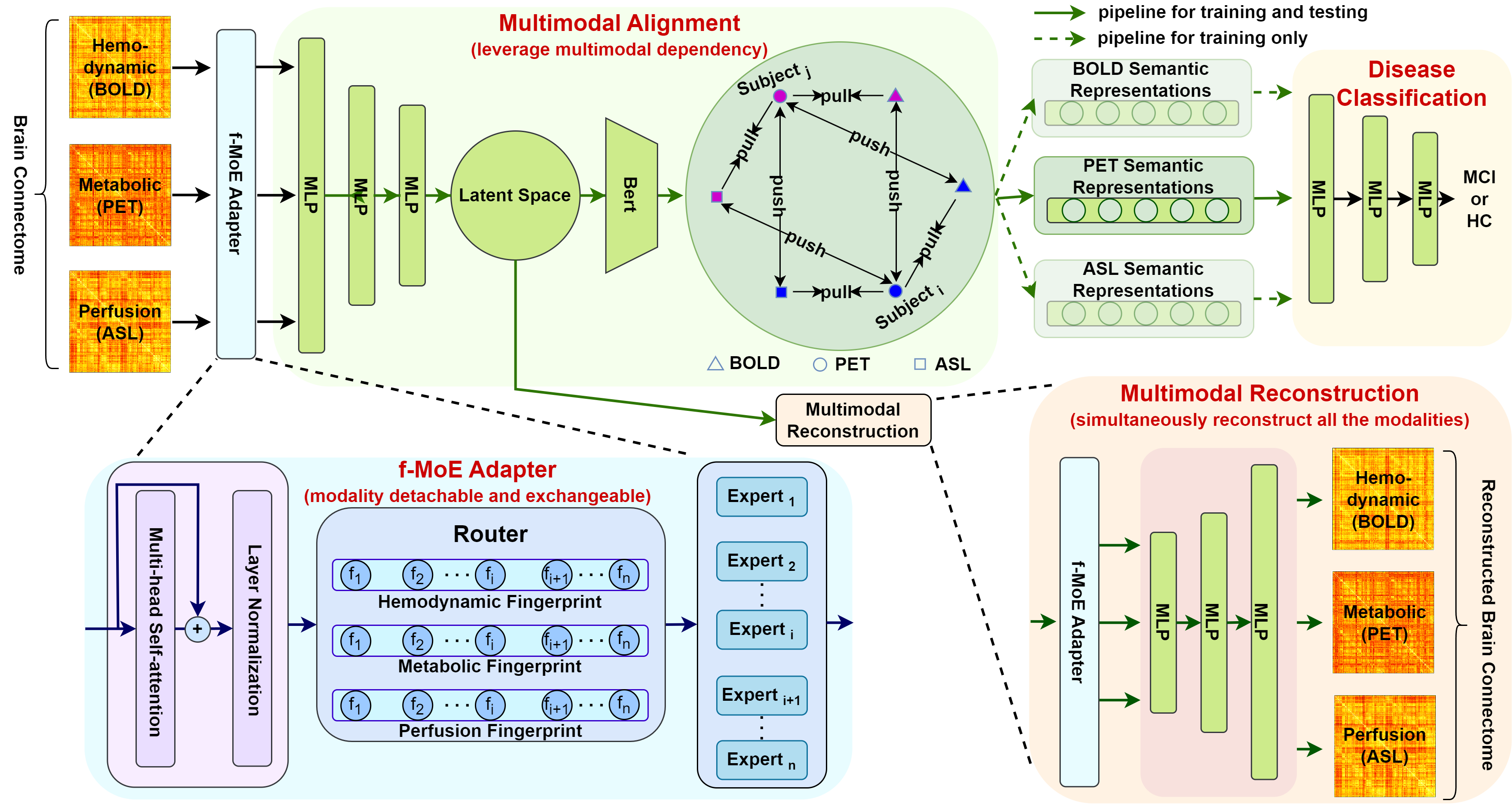
Considering the heterogeneity and scale difference across modalities, we initially employ an f-MoE Adapter for self-adaptive projections across modalities. This nuanced adaptation ensures optimal interpretation of diverse data characteristics inherent to all modalities used for training while creating modality-specific representations. Subsequently, these representations are projected into a latent space using a linear encoder and a Bert encoder with classification () tokens[3], generating semantic embeddings for each modality. These embeddings are then aligned using a triplet alignment loss, with main modality (PET)-derived semantic embeddings specifically utilized for disease classification. Furthermore, we incorporate a multimodal reconstruction task, compelling the reconstruction of all modalities from each modality-specific representations in the latent space, which further regularizes the learning process.
2.1 Fingerprint-based Mixture-of-Experts Adapter
Owing to the heterogeneity in value ranges across brain connectome derived from different modalities, instead of directly fusing all the modalities that requires full-modality data during the inference phase, we use Mixture-of-Experts (MoE) module in a more flexible and modality-detachable manner to dynamically learn deep dependency among modalities. Differed from some recent works that utilize MoE in a modality-specific or sparse routing way[4, 14, 16, 27], we develop a fingerprint-based MoE (f-MoE) algorithm to dynamically allocate experts for all the modalities, which avoids using additional linear layers to calculate routing weights for experts, thus maintaining flexibility. Specifically, in each layer of the f-MoE Adapter, for representations of each modality (where denotes modality), we first leverage self-attention followed by layer normalization[19] to capture intra-dependency among modality-specific features and generate intermediate representations:
| (1) |
where , and are linear transformation matrices to produce Query, Key and Value. , and are layer normalization, dropout and Softmax functions, respectively. To adaptively learn modality-specific representations, we introduce multiple multi-layer-perceptrons (MLP) to act as experts for different modalities: . We also develop a router using modality-specific fingerprints to serve as learnable weights for future combination of the experts. Then, modality-specific representations can be obtained:
| (2) |
where denotes the weighted average of the outputs of experts using as weights. Despite the shared experts among different modalities, the calculation of modality-specific representations is independent for each modality, which allows uni-modal calculation without degenerating model performance during the inference phase. Moreover, due to the simplicity of fingerprint-based router, our algorithm can be naturally extended to applications with more than three modalities without much increase in computation cost when routing.
2.2 Multimodal Alignment
After deriving modality-specific representations, it is essential to fully exploit deep entanglement between the main modality (in this paper, PET) and auxiliary modalities (e.g., BOLD and ASL) during training towards fine-grained representations for classification. Therefore, we invent a multimodal alignment module to align PET representations with those from BOLD and ASL, respectively. In practice, we use a shared linear encoder to encode modality-specific representations into a latent space: . Then, a shared Bert[3] model is adopted to integrate the information of encoded representations and translate it into semantic embeddings (where is the dimension of the latent space) using tokens:
| (3) |
For multimodal alignment, we define the following similarity scores and design a triplet alignment loss inspired by contrastive learning[2, 6, 15]:
| (4) |
| (5) |
As such, multimodal semantic embeddings coming from the same subject will be pulled together (thus promoting modal alignment) and those from different subjects will tend to be pushed apart, which benefits learning of modality-independent, disease-related representations and facilitates downstream tasks.
2.3 Multimodal Reconstruction
Different from traditional reconstruction tasks which reconstruct only one modality at a time[7], to facilitate quality of the learned multimodal representations, we perform simultaneous multimodal reconstruction with an f-MoE Adapter (to avoid ambiguity, we denote the f-MoE Adapter used in this module as f-MoE Decoder Adapter) combined with a shared linear decoder . For each modality, we adaptively reconstruct all the modalities at once using :
| (6) |
| (7) |
| (8) |
Practically, we use less MLPs for reconstruction, which indicates a smaller during reconstruction. For optimizing the reconstruction process, element-wise Mean-Squared Error (MSE) between the reconstructed and the original brain connectome is used:
| (9) |
| (10) |
2.4 Disease Classification
After obtaining fine-grained representations (i.e., semantic embeddings), a linear classifier is adopted for classification: , with Binary CrossEntropy loss (BCE) as the classification loss:
| (11) |
where is the label that indicates whether the subject is diagnosed as patient or not. Collectively, our model is trained by optimizing the joint loss:
| (12) |
3 Experiments
3.1 Datasets and Implementation Details
We test our framework on a precious sf-PET/MR in-house dataset consisting of simultaneously acquired brain functional (metabolic, hemodynamic, and perfusion) images from 48 patients with Mild Cognitive Impairment (MCI, the early stage of AD) and 62 matched healthy controls (HC). Of note, this is the unprecedented data concurrently monitoring AD-related brain function and connectivity changes, collected in a single scan, from a cutting-edge integrated PET/MR scanner. The PET tracer used is 18F-fluorodeoxyglucose (18F-FDG). MCI diagnosis was conducted by experienced neurologist adhering to the established criteria[1]. All subjects provided written informed consent.
Each of the modalities undergoes standard preprocessing detailed elsewhere[5, 17, 22]. The Schaefer’s atlas is used to parcellate the brain into 400 regions of interest, serving as nodes of brain connetome. Brain metabolic and perfusion connectome are constructed by measuring the Kullback-Leibler divergence for each pair of brain regions between region-wise distributions of relative standard uptake value and cerebral blood flow, respectively[20]. Hemodynamic connectome is built using Pearson’s correlation between BOLD signals[28]. Such multimodal connectome building transforms initial raw data into richly detailed brain networks carrying disease-sensitive information.
The dataset is divided into a 7:3 ratio for training and testing. The evaluation metrics for model assessment are Area Under Curve (AUC), accuracy (ACC), F1-score (F1), sensitivity (SEN) and specificity (SPE). During the training phase, multimodal brain connectome is used; For testing, only PET connectome is used. We use a 3-layer f-MoE Adapter with set to 12 and a 1-layer f-MoE Decoder Adapter with its set to 3. is 400 and is 128. The learning rate is set to and the batch size is 36. The model size is 20M and it is trained on an A100 GPU using Adam as the optimizer.
3.2 Results and Discussion
| Method | AUC | ACC | F1 | SEN | SPE |
|---|---|---|---|---|---|
| Linear Regression | 0.600 | 0.597 | 0.562 | 0.631 | 0.562 |
| Support Vector Machine[10] | 0.655 | 0.657 | 0.625 | 0.625 | 0.684 |
| Graph Neural Network[18] | 0.657 | 0.649 | 0.562 | 0.736 | 0.600 |
| Bert[3] | 0.712 | 0.714 | 0.687 | 0.687 | 0.736 |
| MX-ARM (Ours) | 0.827 | 0.828 | 0.812 | 0.842 | 0.812 |
Table 1 shows the comparison results of our method with traditional models. Note that MX-ARM uses multimodal data in training and PET data only in testing; while the competing methods use similar architectures with modality-specific parameters for representation learning and simply concatenate the modality-specific representations for disease classification in training and testing (i.e., use all modalities in testing). Our model not only outperforms other methods but also better suits for the clinical setting with uni-modal inference ability.
Table 2 summarizes ablation studies with the baseline being a transformer model without f-MOE Adapter, MMA and MMR modules . Insights from experiments 1 and 8 elucidate that the specialized modules collectively contribute to substantial improvement across all main metrics.
| Experiment | Method | AUC | ACC | F1 | SEN | SPE |
|---|---|---|---|---|---|---|
| 1 | Base | 0.666 | 0.685 | 0.560 | 0.437 | 0.894 |
| 2 | Base + MMA | 0.572 | 0.600 | 0.363 | 0.250 | 0.894 |
| 3 | Base + MMA + MMR | 0.603 | 0.628 | 0.434 | 0.312 | 0.894 |
| 4 | Base + f-MoE | 0.764 | 0.771 | 0.733 | 0.687 | 0.842 |
| 5 | Base + MMR | 0.659 | 0.657 | 0.647 | 0.687 | 0.631 |
| 6 | Base + f-MoE + MMA | 0.791 | 0.800 | 0.758 | 0.687 | 0.894 |
| 7 | Base + f-MoE + MMR | 0.810 | 0.800 | 0.810 | 0.937 | 0.684 |
| 8 | Base + f-MoE + MMA + MMR | 0.827 | 0.828 | 0.812 | 0.842 | 0.812 |
-
•
Note: Base, a transformer model; MMA, MMR, and f-MoE denote Multimodal Alignment, Multimodal Reconstruction, and f-MoE Adapter, respectively.
The Effect of f-MoE Adapter. Insights from the Experiments 1, 4, 6, 7, and 8 elucidate that 1) The implementation of the f-MoE Adapter significantly enhances model performance (Experiment 1 vs. 4). This underscores the effectiveness of adaptive learning across different modalities; 2) With f-MoE, both MMA and MMR modules make additional contribution (Experiment 4 vs. 6, 7), which implies that fine-grained representations are achieved by both exploiting inherent multimodal dependency and enforcing the reconstruction quality; 3) The concurrent usage of f-MoE along with MMA and MMR achieves more balanced results, indicating more robust and stable learning.
The Effect of MMA and MMR.
From the results in Experiments 1-3, 5-7, specific roles of the MMA and MMR modules are revealed: 1) Without f-MoE, MMA seems not to work properly if used separately from MMR (Experiments 1, 2, 6). This indicates that heterogeneity of multimodal brain connectome indeed creates a bias that cannot be solved by shared linear transformation, thus results in misalignment; 2) Compared with MMA, MMR performs better if used alone (Experiments 1, 5), as the f-MoE decoder Adapter takes effect.
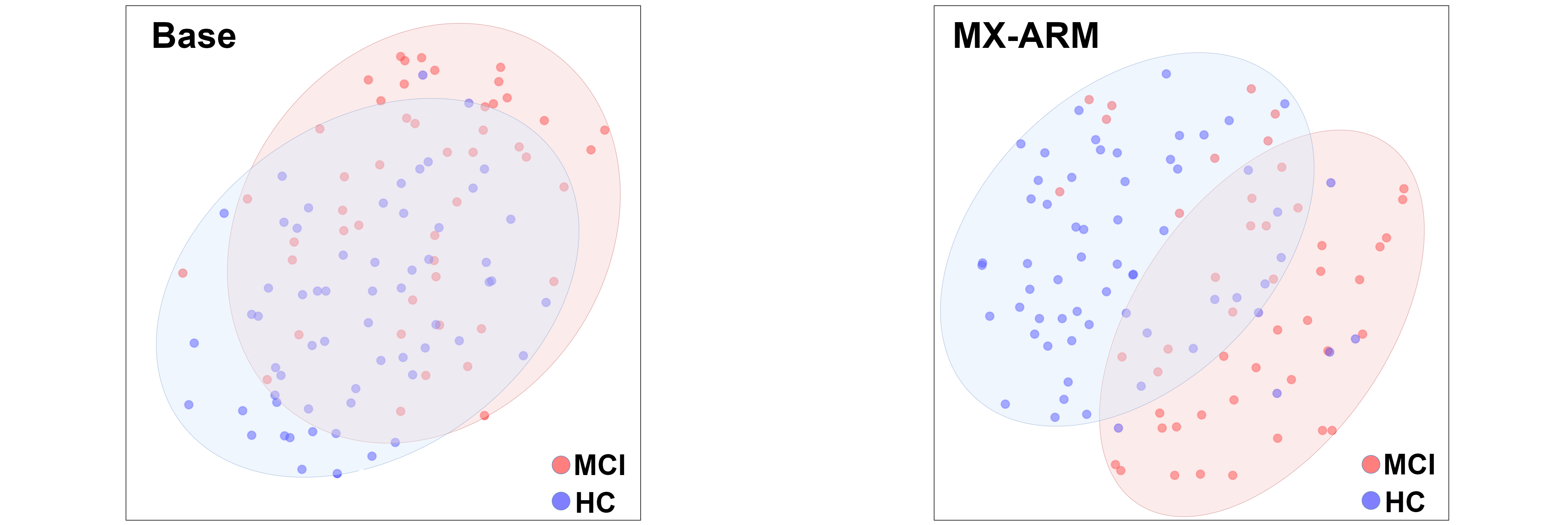
Analysis of the Semantic Embeddings.
It is important that the f-MoE Adapter, MMA and MMR modules should be included in a complete manner. This will promote fine-grained representations for better classification performance. In Fig. 2, we use t-SNE maps to visualize for understanding the learned semantic embeddings. It is clear that the representations learned by our method lead to much separated groups, indicating a better discriminative ability.
4 Conclusion
This study presents an innovative research framework including simultaneous functional PET/MR, multimodal brain connectome construction and learning, clinically feasible multimodal fusion and diagnosis. The superior performance of MX-ARM is demonstrated by a precious, carefully curated sf-PET/MR dataset. The AUC of 0.827 also outperforms current SOTA performance in MCI detection, indicating that concurrent modeling brain metabolic, hemodynamic, and perfusion activity helps with more accurate early AD detection.
References
- [1] Albert, M.S., DeKosky, S.T., Dickson, D., Dubois, B., Feldman, H.H., Fox, N.C., Gamst, A., Holtzman, D.M., Jagust, W.J., Petersen, R.C., et al.: The diagnosis of mild cognitive impairment due to alzheimer’s disease: recommendations from the national institute on aging-alzheimer’s association workgroups on diagnostic guidelines for alzheimer’s disease. Alzheimer’s & dementia 7(3), 270–279 (2011)
- [2] Chen, T., Kornblith, S., Norouzi, M., Hinton, G.: A simple framework for contrastive learning of visual representations. In: International conference on machine learning. pp. 1597–1607. PMLR (2020)
- [3] Devlin, J., Chang, M.W., Lee, K., Toutanova, K.: Bert: Pre-training of deep bidirectional transformers for language understanding. arXiv preprint arXiv:1810.04805 (2018)
- [4] Du, N., Huang, Y., Dai, A.M., Tong, S., Lepikhin, D., Xu, Y., Krikun, M., Zhou, Y., Yu, A.W., Firat, O., et al.: Glam: Efficient scaling of language models with mixture-of-experts. In: International Conference on Machine Learning. pp. 5547–5569. PMLR (2022)
- [5] Esteban, O., Markiewicz, C.J., Blair, R.W., Moodie, C.A., Isik, A.I., Erramuzpe, A., Kent, J.D., Goncalves, M., DuPre, E., Snyder, M., et al.: fmriprep: a robust preprocessing pipeline for functional mri. Nature methods 16(1), 111–116 (2019)
- [6] Girdhar, R., El-Nouby, A., Liu, Z., Singh, M., Alwala, K.V., Joulin, A., Misra, I.: Imagebind: One embedding space to bind them all. In: Proceedings of the IEEE/CVF Conference on Computer Vision and Pattern Recognition. pp. 15180–15190 (2023)
- [7] He, K., Chen, X., Xie, S., Li, Y., Dollár, P., Girshick, R.: Masked autoencoders are scalable vision learners. In: Proceedings of the IEEE/CVF conference on computer vision and pattern recognition. pp. 16000–16009 (2022)
- [8] Huang, Z., Liu, H., Wu, Y., Li, W., Liu, J., Wu, R., Yuan, J., He, Q., Wang, Z., Zhang, K., et al.: Automatic brain structure segmentation for 18f-fluorodeoxyglucose positron emission tomography/magnetic resonance images via deep learning. Quantitative Imaging in Medicine and Surgery 13(7), 4447 (2023)
- [9] Hung, S.C., Liu, M., Yap, P.T., Shen, D., Lin, W., Castillo, M.: Future trends of pet/mr and utility of ai in multi-modal imaging. Hybrid PET/MR Neuroimaging: A Comprehensive Approach pp. 79–86 (2022)
- [10] Khazaee, A., Ebrahimzadeh, A., Babajani-Feremi, A.: Identifying patients with alzheimer’s disease using resting-state fmri and graph theory. Clinical Neurophysiology 126(11), 2132–2141 (2015)
- [11] Lima, A.A., Mridha, M.F., Das, S.C., Kabir, M.M., Islam, M.R., Watanobe, Y.: A comprehensive survey on the detection, classification, and challenges of neurological disorders. Biology 11(3), 469 (2022)
- [12] Liu, G., Yang, X., Zhou, X.: In vivo biomarker imaging: Paving the way for precision medicine (2023)
- [13] Liu, J., Geng, J.: Recent progress on imaging technology and performance testing of pet/mr. Radiation Detection Technology and Methods 7(1), 84–89 (2023)
- [14] Mustafa, B., Riquelme, C., Puigcerver, J., Jenatton, R., Houlsby, N.: Multimodal contrastive learning with limoe: the language-image mixture of experts. Advances in Neural Information Processing Systems 35, 9564–9576 (2022)
- [15] Radford, A., Kim, J.W., Hallacy, C., Ramesh, A., Goh, G., Agarwal, S., Sastry, G., Askell, A., Mishkin, P., Clark, J., et al.: Learning transferable visual models from natural language supervision. In: International conference on machine learning. pp. 8748–8763. PMLR (2021)
- [16] Riquelme, C., Puigcerver, J., Mustafa, B., Neumann, M., Jenatton, R., Susano Pinto, A., Keysers, D., Houlsby, N.: Scaling vision with sparse mixture of experts. Advances in Neural Information Processing Systems 34, 8583–8595 (2021)
- [17] Routier, A., Burgos, N., Díaz, M., Bacci, M., Bottani, S., El-Rifai, O., Fontanella, S., Gori, P., Guillon, J., Guyot, A., et al.: Clinica: An open-source software platform for reproducible clinical neuroscience studies. Frontiers in Neuroinformatics 15, 689675 (2021)
- [18] Song, T.A., Chowdhury, S.R., Yang, F., Jacobs, H., El Fakhri, G., Li, Q., Johnson, K., Dutta, J.: Graph convolutional neural networks for alzheimer’s disease classification. In: 2019 IEEE 16th international symposium on biomedical imaging (ISBI 2019). pp. 414–417. IEEE (2019)
- [19] Vaswani, A., Shazeer, N., Parmar, N., Uszkoreit, J., Jones, L., Gomez, A.N., Kaiser, Ł., Polosukhin, I.: Attention is all you need. Advances in neural information processing systems 30 (2017)
- [20] Wang, M., Jiang, J., Yan, Z., Alberts, I., Ge, J., Zhang, H., Zuo, C., Yu, J., Rominger, A., Shi, K., et al.: Individual brain metabolic connectome indicator based on kullback-leibler divergence similarity estimation predicts progression from mild cognitive impairment to alzheimer’s dementia. European journal of nuclear medicine and molecular imaging 47, 2753–2764 (2020)
- [21] Wang, Y., Tang, S., Ma, R., Zamit, I., Wei, Y., Pan, Y.: Multi-modal intermediate integrative methods in neuropsychiatric disorders: A review. Computational and Structural Biotechnology Journal (2022)
- [22] Wang, Z., Aguirre, G.K., Rao, H., Wang, J., Fernández-Seara, M.A., Childress, A.R., Detre, J.A.: Empirical optimization of asl data analysis using an asl data processing toolbox: Asltbx. Magnetic resonance imaging 26(2), 261–269 (2008)
- [23] Wang, Z., Wu, Z., Agarwal, D., Sun, J.: Medclip: Contrastive learning from unpaired medical images and text. arXiv preprint arXiv:2210.10163 (2022)
- [24] Weber, W.: Clinical pet/mr. Molecular Imaging in Oncology pp. 747–764 (2020)
- [25] Zheng, X., Tang, C., Wan, Z., Hu, C., Zhang, W.: Multi-level confidence learning for trustworthy multimodal classification. In: Proceedings of the AAAI Conference on Artificial Intelligence. vol. 37, pp. 11381–11389 (2023)
- [26] Zhou, H.Y., Chen, X., Zhang, Y., Luo, R., Wang, L., Yu, Y.: Generalized radiograph representation learning via cross-supervision between images and free-text radiology reports. Nature Machine Intelligence 4(1), 32–40 (2022)
- [27] Zhou, Y., Lei, T., Liu, H., Du, N., Huang, Y., Zhao, V., Dai, A.M., Le, Q.V., Laudon, J., et al.: Mixture-of-experts with expert choice routing. Advances in Neural Information Processing Systems 35, 7103–7114 (2022)
- [28] Zhou, Z., Chen, X., Zhang, Y., Hu, D., Qiao, L., Yu, R., Yap, P.T., Pan, G., Zhang, H., Shen, D.: A toolbox for brain network construction and classification (brainnetclass). Human brain mapping 41(10), 2808–2826 (2020)