(eccv) Package eccv Warning: Package ‘hyperref’ is loaded with option ‘pagebackref’, which is *not* recommended for camera-ready version
22institutetext: Robotics Institute, Carnegie Mellon University, USA
33institutetext: Department of Cell and Developmental Biology, University of Michigan, USA
33email: {bxie9, bduan2}@hawk.iit.edu, bjdxtanghao@gmail.com,
dwcai@umich.edu, yyan34@iit.edu
MaskSAM: Towards Auto-prompt SAM with Mask Classification for Medical Image Segmentation
Abstract
Segment Anything Model (SAM), a prompt-driven foundation model for natural image segmentation, has demonstrated impressive zero-shot performance. However, SAM does not work when directly applied to medical image segmentation tasks, since SAM lacks the functionality to predict semantic labels for predicted masks and needs to provide extra prompts, such as points or boxes, to segment target regions. Meanwhile, there is a huge gap between 2D natural images and 3D medical images, so the performance of SAM is imperfect for medical image segmentation tasks. Following the above issues, we propose MaskSAM, a novel mask classification prompt-free SAM adaptation framework for medical image segmentation. We design a prompt generator combined with the image encoder in SAM to generate a set of auxiliary classifier tokens, auxiliary binary masks, and auxiliary bounding boxes. Each pair of auxiliary mask and box prompts, which can solve the requirements of extra prompts, is associated with class label predictions by the sum of the auxiliary classifier token and the learnable global classifier tokens in the mask decoder of SAM to solve the predictions of semantic labels. Meanwhile, we design a 3D depth-convolution adapter for image embeddings and a 3D depth-MLP adapter for prompt embeddings. We inject one of them into each transformer block in the image encoder and mask decoder to enable pre-trained 2D SAM models to extract 3D information and adapt to 3D medical images. Our method achieves state-of-the-art performance on AMOS2022 [27], 90.52% Dice, which improved by 2.7% compared to nnUNet. Our method surpasses nnUNet by 1.7% on ACDC [3] and 1.0% on Synapse [29] datasets.
1 Introduction
Foundation models [13, 18], trained on vast and diverse datasets, have shown impressive capabilities in various tasks [37, 39] and are revolutionizing artificial intelligence. The extraordinary zero-shot and few-shot generalization abilities of foundation models derive a wide range of downstream tasks and achieve numerous and remarkable progress. In contrast to the traditional methods of training task-specific models from scratch, the “pre-training then finetuning” paradigm has proven pivotal, particularly in the realm of computer vision. Segment Anything Model (SAM) [28], pre-trained over 1 billion masks on 11 million natural images, has been recently proposed as a visual foundation model for prompt-driven image segmentation and has gained significant attention. SAM can generate precise object binary masks based on its impressive zero-shot capabilities. As a very important branch of image segmentation, medical image segmentation has been dominated by deep learning medical segmentation methods [41, 1, 2] for the past few years. The existing deep learning models are often tailored, which have a strong inductive bias and limit their capacity, for specific tasks. This raises an intriguing question: Can SAM still have the ability to revolutionize the field of medical image segmentation? Or can SAM still achieve high-performance results in medical image segmentation by properly fine-tuning based on SAM’s strong zero-shot capabilities in natural image segmentation?
To address the aforementioned inquiries, it is imperative to first tackle several intrinsic issues inherent in applying SAM directly to medical image segmentation. The primary concern lies in SAM’s inability to predict semantic labels for the generated binary masks, as it exclusively predicts a singular binary mask per prompt without associating semantic labels. Usually, medical images involve multiple labels and each label carries semantic information. Additionally, the second challenge involves SAM which requires users to input precise prompts to segment target regions. The third issue is that SAM does not always perform well when applied directly to medical image segmentation tasks, even with the appropriate prompts. Therefore, the challenge lies in refining the fine-tuning process for SAM from natural image segmentation to medical image segmentation. This paper aims to investigate the methodology for adapting SAM from its original 2D natural image segmentation to 3D medical image segmentation.
Since it is rigorous to expect all users to have the medical knowledge to provide points in target regions or frame out target regions only given raw images, the pivotal challenge becomes the design of a prompt-free architecture for SAM. Simultaneously, we observe that the image encoder employs the Vision Transformer (ViT) [14] pre-trained with masked auto-encoder [18] as the backbone. Capitalizing on ViT’s representation capabilities, the image encoder extracts essential features of the images with a series of transformer blocks. To address the critical issue of prompt-free usage, we introduce a prompt generator that leverages multiple levels of feature maps extracted from the image encoder. This prompt generator generates a set of auxiliary binary masks and bounding boxes as prompts, obviating the need for manual prompts. This innovative approach effectively resolves the requirements for extra prompts.
To enable the prediction of semantic labels, the prompt generator simultaneously produces a set of auxiliary classifier tokens. However, there are no classifier tokens in the mask decoder to output class predictions. Inspired by MaskFormer [10], we design global learnable classifier tokens, which are summed by auxiliary classifier tokens to each predicted binary mask corresponding to one specific class, in the mask decoder.
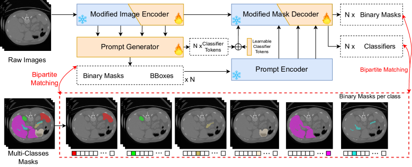
We design a dataset mapping pipeline, shown at the bottom of Fig. 1, which converts a multi-class mask into a set of binary masks with semantic labels per class. The dataset mapping pipeline results in various lengths of binary masks of ground truth. Inspired by DETR [7] and MaskFormer [10], the designed prompt generator would generate a large enough number of prompts so that the number is larger than the maximum of class-level binary masks in datasets. Then bipartite matching is utilized between the set of predictions and ground truth segments to select the most matching predicted masks with ground truth segments to calculate loss functions.
Despite addressing its functionality issues, SAM does not always perform well when applied directly to medical image segmentation tasks, even with appropriate prompts. Many works [12, 22, 50, 34, 42, 43, 20] have demonstrated that SAM is imperfect or even fails when some situations occur, such as weak boundaries, low-contrast, and smaller and irregular shapes, which is consistent with other investigations [25, 26]. Therefore, fine-tuning SAM for medical image segmentation tasks is the main direction. However, fine-tuning the large model of SAM consumes huge computation resources. Many efficient fine-tuning works [32, 45, 30, 15] demonstrate the effectiveness of SAM and the feasibility of efficient fine-tuning by inserting lightweight adapters [21] in medical image segmentation tasks. In this paper, we employ the lightweight adapter for efficient fine-tuning. However, the above works usually abandon the prompt encoder or mask decoder to avoid the requirements of additional prompts provided, which would destroy the consistent system of SAM and abandon the strong prompt encoder and mask decoder, which are trained via large-scale datasets and lots of resources. Therefore, the primary challenge lies in modifying the structure to preserve the inherent capabilities of SAM. Therefore, we keep all structures, freeze all weights, and only insert designed blocks into SAM to adapt. In this way, we retain the zero-shot capabilities of SAM and adapt SAM to medical image segmentation.
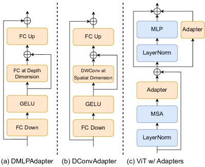
Unlike classic 2D natural images, many medical scans are 3D volumes with an extra depth dimension, such as MRI and CT. To learn extra depth information, we include learnable layers for the extra depth dimension in the lightweight adapters. In SAM, the image encoder and mask decoder contain transformer blocks into which we can insert adapters. The mask decoder contains two types of attention blocks for prompt embeddings and image embeddings, respectively. The original adapter only processes the last dimension, the channel dimension, which cannot learn the information among tokens. However, image embeddings contain spatial information, which is important to let our model understand spatial relationships. we design a 3D depth-convolution adapter (DConvAdapter) that adds a 3D depth-wise convolution layer in the middle of the original adapter with a skip connection for all attention blocks processing image embeddings in the mask decoder. For the rest of the attention blocks for prompt embeddings, we only need to involve a learnable block at depth dimension, since the prompt embeddings do not have any spatial relationship. Therefore, we design a 3D depth-MLP adapter (DMLPAdapter) that adds an invert-bottleneck architecture that consists of two FC layers and an activation layer processing on depth dimension in the middle of the original adapter with a skip connection, which can learn the extra depth information. Both designed adapters are shown in Fig. 3. Since the image encoder only contains the attention blocks for image embeddings, we insert a DConvAdapter for each transformer block in the image encoder. In summary, our contributions to this paper are as follows:
-
•
We propose a novel mask classification prompt-free SAM (MaskSAM) framework for medical image segmentation. To the best of our knowledge, the proposed MaskSAM is the first prompt-free SAM-based image segmentation framework that remains all structure of the original SAM;
-
•
We propose a novel prompt generator that utilizes multiple levels of feature maps from the image encoder to generate auxiliary masks and bounding boxes as prompts to solve the requirements of extra prompts and generate auxiliary classifier tokens summed by learnable global classifier tokens within the mask decoder in SAM to enable the functionality to predict semantic labels for predicted binary masks.
-
•
we design a 3D depth-convolution adapter (DConvAdapter) for image embeddings and a 3D depth-MLP adapter (DMLPAdapter) for prompt embeddings and inject one of them into each transformer block in the image encoder and mask decoder to enable pre-trained 2D SAM models to extract 3D information and adapt to 3D medical image segmentation.
- •
2 Related Work
Deep Learning Methods for Medical Image Segmentation. Deep learning methodologies have dominated medical image segmentation in the past few years. The predominant approaches can be categorized into convolution-based models [41, 51, 33, 11, 24], transformer-based models [49], and hybrid models [9, 6] that integrate convolutional and transformer architectures. The adoption of encoder-decoder networks, particularly U-shaped networks, has dominated the prevailing trend. These methods stand out by explicitly tailoring architectures for medical image segmentation and training from scratch. However, the models exhibit high inductive bias, which also limits their adaptability and overall capacity.
Foundation Models and Parameter-efficient Finetuning. Foundation models [4, 36] are dedicated to the development of large-scale, general-purpose language and vision models. These models derive a wide range of downstream applications, achieving remarkable success following the pre-training and fine-tuning paradigm [19, 23]. The goal of parameter-efficient finetuning [21, 38, 23, 47] is to decrease the number of trainable parameters and reduce the computation cost while achieving or surpassing the performance of full finetuning. Recently, the Segment Anything Model (SAM) [28], pre-trained over 1 billion masks on 11 million natural images, has been proposed as a visual foundation model for image segmentation and has gained a lot of attention. In this paper, we adopt the strategy of parameter-efficient finetuning to adapt SAM from 2D natural image segmentation to 3D medical image segmentation.
SAM-based Medical Image Segmentation. SAM-based medical image segmentation research can be categorized into two primary streams. The first set of studies [12, 22, 50, 34, 42, 43, 20] focuses on assessing SAM’s performance across diverse medical image segmentation tasks and modes through manually provided prompts. Evaluations across different datasets reveal notable variations in SAM’s performance, excelling in specific objects and modalities compared to state-of-the-art methods. However, SAM exhibits imperfections, particularly in scenarios involving weak boundaries, low contrast, and smaller, irregular shapes, rendering its segmentation performance suboptimal for many medical imaging tasks. The second line of research [32, 45, 30, 15] focuses on how to better adapt SAM to medical image segmentation tasks. The requirements of prompts are a difficult problem to deal with. Although some studies opt to circumvent prompt demands by abandoning or redesigning the prompt encoder or mask decoder, this approach risks compromising SAM’s cohesive system and relinquishing the robust capabilities of these components. To address this, we propose a prompt-free SAM to generate auxiliary masks and bounding boxes via the image encoder itself with a designed prompt generator. In this way, our model not only preserves the zero-shot capabilities of SAM, but also obviates the need for additional prompts, offering a comprehensive and efficient framework.
3 The Proposed Method
In this section, we first review SAM. Then, we introduce the whole structure of our proposed MaskSAM. Finally, we describe each component of MaskSAM.
3.1 SAM Preliminaries
SAM is a prompt-driven foundation model for natural image segmentation, which is trained on the large-scale SA-1B dataset of 1B masks and 11M images. The architecture of SAM contains three main components: the image encoder that employs Vision Transformer as the backbone, the prompt encoder that embeds various types of prompts including points, boxes, or texts, and the lightweight mask decoder to generate masks based on the image embedding, prompt embedding, image positional embedding, and output tokens. When utilized to segment a provided 2D image, SAM requires proper prompts, such as points or boxes. Subsequently, SAM generates a singular binary mask for each prompt without any associated semantic labels. However, medical segmentation tasks often involve multiple objects with distinct semantic labels within a given image.
3.2 Overview of the Proposed MaskSAM
In this and the following sections, we introduce the whole pipeline and each component of MaskSAM shown in Fig. 1 and Fig. 4. Our MaskSAM retains all structures of SAM and only inserts designed blocks to adapt the original SAM from 2D natural images to 3D medical images. Therefore, MaskSAM contains the modified image encoder, the designed generator prompt, the original prompt encoder, and the modified mask decoder. Meanwhile, we design a dataset mapping from multi-class labels to binary masks for each class with semantic labels, which ensures that each predicted binary mask is dedicated to a single class.
3.3 Proposed Dataset Mapping
SAM generates a single binary mask without any semantic label for each prompt, while a typical ground truth of medical images comprises multiple classes. Each cropped patch image as input may contain varied classes. The challenge arises from varying the lengths of binary masks within the ground truth. To accommodate this diversity and inspired by DETR [7] and MaskFormer [10], our model generates a sufficiently large number of binary masks, with each mask dedicated to predicting a single class. And then bipartite matching is utilized between the set of predictions and ground truth segments. Therefore, we design a dataset mapping pipeline, shown at the bottom of Fig. 1, which converts a multi-class mask into a set of binary masks with semantic labels per class.
3.4 Proposed Prompt Generator
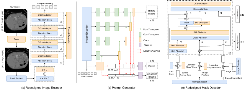
To solve the requirements of the extra proper prompts, we propose a prompt generator, shown in Fig. 4(b), to generate a set of auxiliary binary masks and bounding boxes as prompts instead of manual prompts for the prompt encoder. We adopt box prompts with mask prompts as our prompt way, since the point prompts would bring instabilities that are harmful to medical segmentation tasks. We utilize ViT’s strong representation capabilities to extract multiple levels of feature maps from the image encoder as the input of our proposed prompt generator. From the beginning of the last output of the image encoder, the feature maps are connected with convolution layers, upsampled, and concatenated with the feature maps of the lower level. Following this way, we can obtain feature maps with the same image size as the ground truth. Finally, we utilize a convolutional layer to change the channel size to the fixed number of that is larger than the maximum of object-level binary masks in datasets. Meanwhile, we extract the output of the last convolutional layer at each level, use adaptive average pooling layers to change the spatial dimension to for box queries, concatenate all box queries, and connect with an MLP layer to adjust the channel to . We can obtain number of learnable binary masks and learnable boxes.
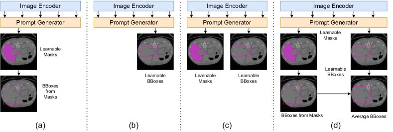
There are many combinations of learnable binary masks and learnable boxes shown in Fig. 2(a)-(d). Fig. 2(a) shows a prompt generator only generates learnable binary masks and uses the binary masks to calculate its bounding boxes. Fig. 2(b) shows a prompt generator only generates boxes as prompts. Fig. 2(c) shows a prompt generator that generates learnable binary masks as mask prompts and learnable boxes as box prompts. Fig. 2(d) shows a prompt generator that generates learnable binary masks as mask prompts and learnable boxes. We average the bounding boxes calculated from binary masks and the learnable boxes as the final box prompts. Through a series of experiments, we found that the best way is Fig. 2(d) since it can involve more information and more robustness.
To solve the inabilities of semantic label predictions, the prompt generator simultaneously produces a set of auxiliary classifier tokens, which are the following in the same way as the generation of auxiliary box prompts, except the use of adaptive average pooling layers to change the spatial dimension to for classifier tokens. The auxiliary classifier tokens will be summed up by our designed learnable global classifier tokens within the mask decoder.
3.5 Proposed Adapters
we adopt the lightweight adapter [21] which is a bottleneck architecture that consists of two fully connected (FC) layers and an activation layer in the middle to modify and inject into each transformer block during fine-tuning. Unlike classic 2D natural images, many medical scans are 3D volumes with extra depth dimensions such as MRI and CT. To learn extra depth information, we involve learnable layers for the extra depth dimension into adapters. In SAM, the image encoder and mask decoder contain transformer blocks that we can insert adapters into. The mask decoder contains two types of attention blocks for prompt embeddings and image embeddings, respectively. The original adapter only processes the last dimension, the channel dimension, which cannot learn the information among the tokens. However, image embeddings contain spatial information, which is important to let our model understand spatial relationships. we design a 3D depth-convolution adapter (DConvAdapter), shown in Fig. 3(a), that adds a 3D depth-wise convolution layer in the middle of the original adapter with a skip connection for all attention blocks processing image embeddings in the mask decoder. For the rest of the attention blocks for prompt embeddings, we only need to involve a learnable block at depth dimension, since the prompt embeddings do not have any spatial relationship. Therefore, we design a 3D depth-MLP adapter (DMLPAdapter), shown in Fig. 3(b), that adds an invert-bottleneck architecture that consists of two FC layers and an activation layer processing in the depth dimension in the middle of the original adapter with a skip connection, which can learn additional depth information. Since the image encoder only contains the attention blocks for the image embeddings, we insert the DConvAdapter for each transformer block into the image encoder. Fig. 3(c) shows the way we insert adapters into vision transformers. We insert an adapter behind the multi-head attention block and parallel MLP block.
3.6 Modified Image Encoder
Fig. 4(a) illustrates the redesigned image encoder. i) SAM works on natural images that have 3 channels for RGB while medical images have varied modalities as channels. There are gaps between the varied modalities of medical images and the RGB channels of natural images. Therefore, we design a sequence of convolutional layers to an invert-bottleneck architecture to learn the adaption from the varied modalities with any size to 3 channels. ii) The image encoder includes one positional embedding. To better understand the extra depth information, we can insert a learnable depth positional embedding with the original positional embedding. iii) Since we use the base ViT backbone, it contains 12 attention blocks. We insert our designed DConvAdapter blocks into each attention block. We extract the feature maps of each three attention blocks and the final output of the image encoder for the prompt generator.
3.7 Modified Mask Encoder
Fig. 4(c) illustrates the redesigned mask encoder. i) we design learnable global classifier tokens, which are summed by auxiliary classifier tokens generated by the prompt generator, concatenated with sparse prompt embedding, and the original mask tokens to equip our model with the functionality to predict semantic labels for each binary mask. ii) The mask decoder also includes a positional embedding. To learn the extra depth information better, we can insert a learnable depth positional embedding with the original image positional embedding. iii) The mask encoder contains two subsequent transformers. Each transformer first applies self-attention to the prompt embedding. We insert a DMLPAdapter behind the self-attention. Then, a cross-attention block is adopted for tokens attending to image embedding. We insert a DMLPAdapter behind the cross-attention. Next, we insert a DMLPAdapter parallel to an MLP block. Finally, a cross-attention block is utilized for image embedding attending to tokens. We insert a DConvAdapter behind the cross-attention. In this way, our model can learn the spatial information with extra depth information for the image embedding and depth information for the prompt embedding.
4 Experiments
4.1 Datasets and Evaluation Metrics
We use three publicly available datasets, AMOS22 Abdominal CT Organ Segmentation [27], Synapse multiorgan segmentation [29] and Automatic Cardiac Diagnosis Challenge (ACDC) [3]. (i) AMOS22 dataset consists of 200 cases of abdominal CT scans with 16 anatomies manually annotated for abdominal multi-organ segmentation, including the spleen, right kidney, left kidney, gallbladder, esophagus, liver, stomach, aorta, inferior vena cava, pancreas, right adrenal gland, left adrenal gland, duodenum, bladder, prostate/uterus. There are 200 testing images, and we evaluate our model on the AMOS22 leaderboard. (ii) Synapse dataset consists of 30 cases of abdominal CT scans. Following the split strategies [9], we use a random split of 18 training cases and 12 cases for validation. We evaluate the model performance via the average Dice score (DSC) on 8 abdominal organs (aorta, gallbladder, spleen, left kidney, right kidney, liver, pancreas, and stomach). (iii) ACDC dataset consists of 100 patients with the cavity of the right ventricle, the myocardium of the left ventricle, and the cavity of the left ventricle to be segmented. The labels involve the right ventricle (RV), myocardium (MYO), and left ventricle (LV). We use a random split of 70 training cases, 10 validation cases, and 20 testing cases. We evaluate the model performance by the average DSC.
| Method | Spleen | R.Kd | L.Kd | GB | Eso. | Liver | Stom. | Aorta | IVC | Panc. | RAG | LAG | Duo. | Blad. | Pros. | Average |
| TransBTS [44] | 0.885 | 0.931 | 0.916 | 0.817 | 0.744 | 0.969 | 0.837 | 0.914 | 0.855 | 0.724 | 0.630 | 0.566 | 0.704 | 0.741 | 0.650 | 0.792 |
| UNETR [17] | 0.926 | 0.936 | 0.918 | 0.785 | 0.702 | 0.969 | 0.788 | 0.893 | 0.828 | 0.732 | 0.717 | 0.554 | 0.658 | 0.683 | 0.722 | 0.762 |
| nnFormer [49] | 0.935 | 0.904 | 0.887 | 0.836 | 0.712 | 0.964 | 0.798 | 0.901 | 0.821 | 0.734 | 0.665 | 0.587 | 0.641 | 0.744 | 0.714 | 0.790 |
| SwinUNETR [16] | 0.959 | 0.960 | 0.949 | 0.894 | 0.827 | 0.979 | 0.899 | 0.944 | 0.899 | 0.828 | 0.791 | 0.745 | 0.817 | 0.875 | 0.841 | 0.880 |
| nn-UNet [24] | 0.965 | 0.959 | 0.951 | 0.889 | 0.820 | 0.980 | 0.890 | 0.948 | 0.901 | 0.821 | 0.785 | 0.739 | 0.806 | 0.869 | 0.839 | 0.878 |
| MaskSAM (Ours) | 0.963 | 0.973 | 0.969 | 0.872 | 0.876 | 0.982 | 0.940 | 0.962 | 0.922 | 0.888 | 0.794 | 0.813 | 0.851 | 0.920 | 0.854 | 0.905 |
4.2 Losses and Matching
Following [10, 7], we build an auxiliary loss, which consists of a combination of binary cross-entropy and dice loss over auxiliary binary mask prediction () and a combination of loss and generalized IoU loss [40] over bounding box predictions (), for our prompt generator. Meanwhile, we build a loss, which consists of the standard classification CE-loss on class predictions and a combination of binary cross-entropy and dice loss on final binary mask predictions (), for the final output of our MaskSAM. To find the lowest cost assignment, we use bipartite matching [10, 7] between the ground truths and the combination of the set auxiliary predictions and the set of final predictions. Then, our model selects the same indexes from auxiliary binary masks and final binary masks by bipartite matching. Finally, we use the indexes to obtain specific predictions to calculate losses with the ground truths.
Specifically, the desired final output contains pairs of binary masks with classes of probability distribution , which contains category labels with an auxiliary ”no object“ label (). Meanwhile, our model produces pairs of auxiliary boxes and masks, . Additionally, the set of ground truth segments is required. Since we set and pad the set of ground truth labels with “no object token” to allow one-to-one matching. To train the model parameters, given a matching , the main loss is expressed as follows,
| (1) |
| Method | Aotra | Gallbladder | Kidnery(L) | Kidnery(R) | Liver | Pancreas | Spleen | Stomach | DSC |
| VNet [33] | 75.34 | 51.87 | 77.10 | 80.75 | 87.84 | 40.04 | 80.56 | 56.98 | 68.81 |
| U-Net [41] | 83.17 | 58.74 | 80.40 | 73.36 | 93.13 | 45.43 | 83.90 | 66.59 | 74.99 |
| AttnUNet [35] | 89.55 | 68.88 | 77.98 | 71.11 | 93.57 | 58.04 | 87.30 | 75.75 | 77.77 |
| TransUNet [9] | 87.23 | 63.16 | 81.87 | 77.02 | 94.08 | 55.86 | 85.08 | 75.62 | 77.48 |
| SwinUNet [6] | 85.47 | 66.53 | 83.28 | 79.61 | 94.29 | 56.58 | 90.66 | 76.6 | 79.13 |
| TransClaw-U-Net [8] | 85.87 | 61.38 | 84.83 | 79.36 | 94.28 | 57.65 | 87.74 | 73.55 | 78.09 |
| LeVit-UNet-384s [46] | 87.33 | 62.23 | 84.61 | 80.25 | 93.11 | 59.07 | 88.86 | 72.76 | 78.53 |
| WAD [31] | 87.73 | 69.93 | 83.95 | 79.78 | 93.95 | 61.02 | 88.86 | 77.16 | 80.30 |
| UNETR [17] | 89.99 | 60.56 | 85.66 | 84.80 | 94.46 | 59.25 | 87.81 | 73.99 | 79.56 |
| nnUNet [24] | 92.39 | 71.71 | 86.07 | 91.46 | 95.84 | 82.92 | 90.31 | 79.01 | 86.21 |
| nnFormer [49] | 92.40 | 70.17 | 86.57 | 86.25 | 96.84 | 83.35 | 90.51 | 86.83 | 86.57 |
| SAMed [48] | 87.77 | 69.11 | 80.45 | 79.95 | 94.80 | 72.17 | 88.72 | 82.06 | 81.88 |
| SAMed_s [48] | 83.62 | 57.11 | 79.63 | 78.92 | 93.98 | 65.66 | 85.81 | 77.49 | 77.78 |
| SAM3D [5] | 89.57 | 49.81 | 86.31 | 85.64 | 95.42 | 69.32 | 84.29 | 76.11 | 79.56 |
| MaskSAM (Ours) | 91.75 | 72.20 | 87.32 | 88.15 | 97.21 | 79.62 | 92.47 | 89.11 | 87.23 |
4.3 Comparison with State-of-the-Art Methods
Results on AMOS 2022 Dataset. We present the quantitative outcomes of our experiments on the AMOS 2022 Dataset Table 1, comparing our proposed MaskSAM against the segmentation methods which are widely used and well-recognized in the community, including convolution-based methods (nnUNet [24]), transformer-based methods (UNETR [17], SwinUNETR [16], and nnFormer [49]). To fair comparison, all methods run 5-fold cross-validation without any ensembles. We observe that our MaskSAM outperforms all existing methods on most organs, achieving a new state-of-the-art performance in DSC. Specifically, it surpasses nnUNet and SwinUNETR by 2.7% and 2.5% in DSC, respectively. In the extremely hard AMOS 2022 dataset, our MaskSAM achieves state-of-the-art performance, which confirms the efficacy of our method.
| Method | RV | Myo | LV | DSC |
| UNETR [17] | 85.29 | 86.52 | 94.02 | 88.61 |
| TransUNet [9] | 88.86 | 84.54 | 95.73 | 89.71 |
| SwinUNet [6] | 88.55 | 85.62 | 95.83 | 90.00 |
| LeViT-UNet-384s [46] | 89.55 | 87.64 | 93.76 | 90.32 |
| nnUNet [24] | 90.24 | 89.24 | 95.36 | 91.61 |
| nnFormer [49] | 90.94 | 89.58 | 95.65 | 92.06 |
| SAM3D [5] | 89.44 | 87.12 | 94.67 | 90.41 |
| MaskSAM (Ours) | 92.30 | 91.37 | 96.49 | 93.39 |
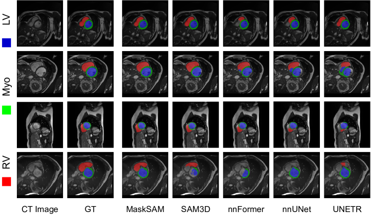
Results on ACDC Dataset. In Table 3, we provide the quantitative experimental results on ACDC dataset. Specifically, we compare the proposed MaskSAM with several leading SAM-based method(i.e. SAM3D [5]), convolution-based methods (i.e., R50-U-Net [41] and nnUNet [24]) and transformer-based methods (i.e., TransUNet [9], SwinUNet [6], and LeViT-UNet-384s [46]). The results show that the proposed MaskSAM outperforms various state-of-the-art approaches, surpassing nnFormer by , , , and in DSC, RV dice, Myo dice, and LV dice. Meanwhile, our method outperforms the SAM-based methods, SAM3D, by 3%, demonstrating the effectiveness of our method. In Fig. 5, we provide qualitative results compared to several state-of-the-art methods. Qualitative results depicted in Fig. 5 further illustrate the model’s accuracy across all labels, achieving near-perfect predictions in the challenging, highly saturated dataset. These outcomes affirm the efficacy of our method, as our proposed modules effectively address the limitations of SAM when adapting to medical image segmentation.
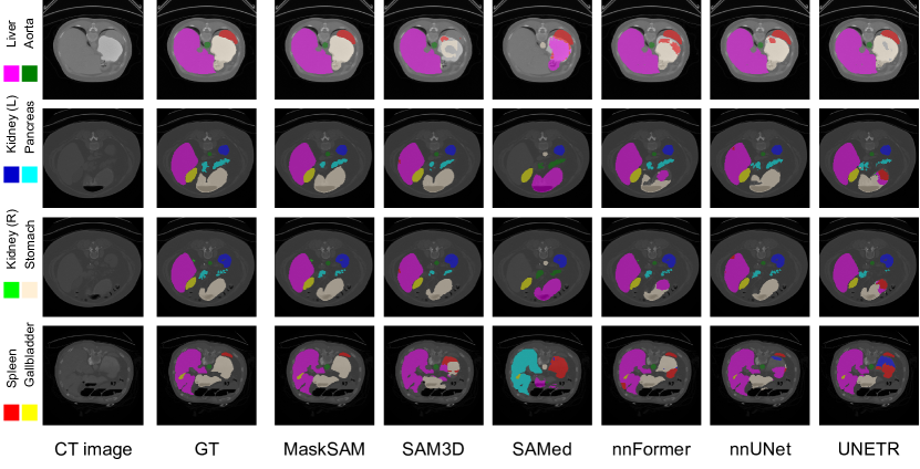
Results on Synapse Dataset. We present the quantitative results of our experiments in the Synapse dataset in Table 2, comparing our proposed MaskSAM against several leading SAM-based method(i.e., SAMed [48] and SAM3D [5]), convolution-based methods (VNet [41] and nnUNet [24]), transformer-based methods (TransUNet [9], SwinUNet [6], and nnFormer [49]). We observe that MaskSAM outperforms all existing methods, achieving a new state-of-the-art performance. Specifically, it surpasses nnFormer by 0.7% in the DSC for the highly saturated dataset. Meanwhile, our method outperforms the SAM-based methods, SAMed and SAM3D, by more than 5%, demonstrating the effectiveness of our method. Notably, our model excels in predicting the large-size ‘Liver,’ ‘Spleen,’ and ‘Stomach’ labels, attributed to our innovative DConvAdapter and DMLPAdapter. These adapters enable the learning of more intricate 3D spatial information and adapt 2D SAM to medical image segmentation. In Fig. 6, we illustrate qualitative results compared to representative methods. These results also demonstrate that our MaskSAM can predict more accurately the ‘Liver’, ‘Spleen’, and ‘Stomach’ labels. In conclusion, the effectiveness of our method is robustly demonstrated by both quantitative and qualitative results.
4.4 Ablation Study
Baseline Models. The proposed MaskSAM has 9 baselines (i.e., B1, B2, B3, B4, B5, B6, B7, B8, B9) as shown in Table 4. All baselines contain the entire SAM structure, a prompt generator, and a learnable class token. (i) B1 adopts the prompt generator that only generates learnable binary masks and uses the binary masks as mask prompts to calculate its bounding boxes as box prompts shown in Fig. 2(a). (ii) B2 adopts the prompt generator that only generates learnable boxes as the prompts shown in Fig. 2(b). (iii) B3 adopts the prompt generator that generates learnable binary masks as mask prompts and learnable boxes as box prompts shown in Fig. 2(c). (iv) B4 adopts the prompt generator that generates learnable binary masks as mask prompts and learnable boxes. We average the bounding boxes calculated from binary masks and the learnable boxes as the final box prompts shown in Fig. 2(d). (v) B5 adds depth positional embedding blocks (DPosEmbed) in the image encoder and mask decoder based on B4. (vi) B6 modifies the vanilla adapter by inserting the invert-bottleneck depth MLPs with a skip connection after the fully-connected layers for upsampling based on B5. (vii) B7 modifies the vanilla adapter by inserting the invert-bottleneck depth MLPs with a skip connection before the fully-connected layers for downsampling based on B5. (viii) B8 replaces the vanilla adapter with our designed DMLPAdapter based on B5. (ix) B9 is our full model, named MaskSAM, illustrated in Fig. 1. B9 adopts the DMLPAdapter for prompt embeddings and the DConvAdapter for image embedding based on B8. The results of the ablation study are shown in Table 4.
Ablation analysis. The results of the ablation study are shown in Table 4. When we use our proposed MaskAvgBBoxPG to first generate auxiliary binary masks and auxiliary boxes, then average the bounding boxes calculated from auxiliary binary masks and learnable auxiliary boxes as the final box prompts, the model achieves the best results and improves by 1.9%, 2.6% and 1.3% compared to B1 with a learnable mask prompt generator, B2 with a learnable box prompt generator, and B3 with a learnable mask and a learnable box prompt generator, respectively. The result confirms the effectiveness of the proposed prompt generator. When inserting depth positional embedding (DPosEmbed) into the image encoder and mask decoder, the performance of B5 improves by more than 0.15% compared to B4, demonstrating the effectiveness of DPosEmbed blocks. The DMLPAdapter (B8) which we insert depth MLPs with a skip connection in the middle of the vanilla adapter achieves the best performance and improves by 0.2% and 0.1% compared to B6 in which we insert depth MLPs with a skip connection after fully-connected layers for upsampling and B7 in which depth MLPs are inserted with a skip connection after fully-connected layers for downsampling, respectively. The result confirms the effectiveness of the proposed DMLPAdapter. B9 is our full model, MaskSAM, utilizing the DMLPAdapter for prompt embeddings and the DConvAdapter that we replace the depth MLP with a 3D depth-wise convolution layer from the DMLPAdapter for image embedding based on B8 as shown in Fig. 1. Compared to B8, our model brings about 0.3% improvements. Therefore, the results demonstrate the effectiveness of our proposed MaskSAM.
| Method | DSC | |
| B1 | SAM + MaskPG + vAdapter | 89.53 |
| B2 | SAM + BBoxPG + vAdapter | 88.78 |
| B3 | SAM + MaskBBoxPG + vAdapter | 90.08 |
| B4 | SAM + MaskAvgBBoxPG + vAdapter | 91.45 |
| B5 | SAM + MaskAvgBBoxPG + vAdapter + DPosEmbed | 91.61 |
| B6 | SAM + MaskAvgBBoxPG + vAdapter w/ D-MLP after FC-Up + DPosEmbed | 92.88 |
| B7 | SAM + MaskAvgBBoxPG + vAdapter w/ D-MLP before FC-Down + DPosEmbed | 92.93 |
| B8 | SAM + MaskAvgBBoxPG + DMLPAdapter + DPosEmbed | 93.10 |
| B9 | Our Full Model (B8 + DConvAdapter) | 93.39 |
5 Conclusion
In this paper, we propose a mask classification prompt-free SAM adaptation framework for medical image segmentation, named MaskSAM, which adapts pre-trained SAM models worked on from 2D natural images to 3D medical images without any prompts provided. By designing a prompt generator combined with the image encoder in SAM to generate a set of auxiliary classifier tokens, auxiliary binary masks, and auxiliary bounding boxes. Each pair of auxiliary mask and box prompts, which can solve the requirements of extra prompts, is associated with class label predictions by the sum of the auxiliary classifier token and the learnable global classifier tokens in the mask decoder of SAM to solve the predictions of semantic labels. By inserting one of our redesigned 3D depth-convolution adapter (DConvAdapter) for image embeddings and 3D depth-MLP adapter (DMLPAdapter) for prompt embeddings into each transformer block in the image encoder and mask decoder, our model enables pre-trained 2D SAM models to extract 3D information and adapt to 3D medical images. Our method achieves state-of-the-art performance on AMOS2022 [27], 90.52% Dice, which improved by 2.7% compared to nnUNet. Meanwhile, our method surpasses nnUNet by 1.7% on ACDC [3] and 1.0% on Synapse [29] datasets.
References
- [1] Akkus, Z., Galimzianova, A., Hoogi, A., Rubin, D.L., Erickson, B.J.: Deep learning for brain mri segmentation: state of the art and future directions. Journal of digital imaging 30(4), 449–459 (2017)
- [2] Avendi, M., Kheradvar, A., Jafarkhani, H.: A combined deep-learning and deformable-model approach to fully automatic segmentation of the left ventricle in cardiac mri. Medical image analysis 30, 108–119 (2016)
- [3] Bernard, O., Lalande, A., Zotti, C., Cervenansky, F., Yang, X., Heng, P.A., Cetin, I., Lekadir, K., Camara, O., Ballester, M.A.G., et al.: Deep learning techniques for automatic mri cardiac multi-structures segmentation and diagnosis: Is the problem solved? IEEE transactions on medical imaging 37(11), 2514–2525 (2018)
- [4] Brown, T., Mann, B., Ryder, N., Subbiah, M., Kaplan, J.D., Dhariwal, P., Neelakantan, A., Shyam, P., Sastry, G., Askell, A., et al.: Language models are few-shot learners. Advances in neural information processing systems 33, 1877–1901 (2020)
- [5] Bui, N.T., Hoang, D.H., Tran, M.T., Le, N.: Sam3d: Segment anything model in volumetric medical images. arXiv preprint arXiv:2309.03493 (2023)
- [6] Cao, H., Wang, Y., Chen, J., Jiang, D., Zhang, X., Tian, Q., Wang, M.: Swin-unet: Unet-like pure transformer for medical image segmentation. arXiv preprint arXiv:2105.05537 (2021)
- [7] Carion, N., Massa, F., Synnaeve, G., Usunier, N., Kirillov, A., Zagoruyko, S.: End-to-end object detection with transformers. In: European conference on computer vision. pp. 213–229. Springer (2020)
- [8] Chang, Y., Menghan, H., Guangtao, Z., Xiao-Ping, Z.: Transclaw u-net: Claw u-net with transformers for medical image segmentation. arXiv preprint arXiv:2107.05188 (2021)
- [9] Chen, J., Lu, Y., Yu, Q., Luo, X., Adeli, E., Wang, Y., Lu, L., Yuille, A.L., Zhou, Y.: Transunet: Transformers make strong encoders for medical image segmentation. arXiv preprint arXiv:2102.04306 (2021)
- [10] Cheng, B., Schwing, A., Kirillov, A.: Per-pixel classification is not all you need for semantic segmentation. Advances in Neural Information Processing Systems 34, 17864–17875 (2021)
- [11] Çiçek, Ö., Abdulkadir, A., Lienkamp, S.S., Brox, T., Ronneberger, O.: 3d u-net: learning dense volumetric segmentation from sparse annotation. In: International conference on medical image computing and computer-assisted intervention. pp. 424–432. Springer (2016)
- [12] Deng, R., Cui, C., Liu, Q., Yao, T., Remedios, L.W., Bao, S., Landman, B.A., Wheless, L.E., Coburn, L.A., Wilson, K.T., et al.: Segment anything model (sam) for digital pathology: Assess zero-shot segmentation on whole slide imaging. arXiv preprint arXiv:2304.04155 (2023)
- [13] Devlin, J., Chang, M.W., Lee, K., Toutanova, K.: Bert: Pre-training of deep bidirectional transformers for language understanding. arXiv preprint arXiv:1810.04805 (2018)
- [14] Dosovitskiy, A., Beyer, L., Kolesnikov, A., Weissenborn, D., Zhai, X., Unterthiner, T., Dehghani, M., Minderer, M., Heigold, G., Gelly, S., et al.: An image is worth 16x16 words: Transformers for image recognition at scale. arXiv preprint arXiv:2010.11929 (2020)
- [15] Gong, S., Zhong, Y., Ma, W., Li, J., Wang, Z., Zhang, J., Heng, P.A., Dou, Q.: 3dsam-adapter: Holistic adaptation of sam from 2d to 3d for promptable medical image segmentation. arXiv preprint arXiv:2306.13465 (2023)
- [16] Hatamizadeh, A., Nath, V., Tang, Y., Yang, D., Roth, H.R., Xu, D.: Swin unetr: Swin transformers for semantic segmentation of brain tumors in mri images. In: International MICCAI Brainlesion Workshop. pp. 272–284. Springer (2021)
- [17] Hatamizadeh, A., Tang, Y., Nath, V., Yang, D., Myronenko, A., Landman, B., Roth, H.R., Xu, D.: Unetr: Transformers for 3d medical image segmentation. In: Proceedings of the IEEE/CVF Winter Conference on Applications of Computer Vision. pp. 574–584 (2022)
- [18] He, K., Chen, X., Xie, S., Li, Y., Dollár, P., Girshick, R.: Masked autoencoders are scalable vision learners. In: Proceedings of the IEEE/CVF conference on computer vision and pattern recognition. pp. 16000–16009 (2022)
- [19] He, K., Girshick, R., Dollár, P.: Rethinking imagenet pre-training. In: Proceedings of the IEEE/CVF International Conference on Computer Vision. pp. 4918–4927 (2019)
- [20] He, S., Bao, R., Li, J., Grant, P.E., Ou, Y.: Accuracy of segment-anything model (sam) in medical image segmentation tasks. arXiv preprint arXiv:2304.09324 (2023)
- [21] Houlsby, N., Giurgiu, A., Jastrzebski, S., Morrone, B., De Laroussilhe, Q., Gesmundo, A., Attariyan, M., Gelly, S.: Parameter-efficient transfer learning for nlp. In: International Conference on Machine Learning. pp. 2790–2799. PMLR (2019)
- [22] Hu, C., Li, X.: When sam meets medical images: An investigation of segment anything model (sam) on multi-phase liver tumor segmentation. arXiv preprint arXiv:2304.08506 (2023)
- [23] Hu, E.J., Shen, Y., Wallis, P., Allen-Zhu, Z., Li, Y., Wang, S., Wang, L., Chen, W.: Lora: Low-rank adaptation of large language models. arXiv preprint arXiv:2106.09685 (2021)
- [24] Isensee, F., Jäger, P.F., Kohl, S.A., Petersen, J., Maier-Hein, K.H.: Automated design of deep learning methods for biomedical image segmentation. arXiv preprint arXiv:1904.08128 (2019)
- [25] Ji, G.P., Fan, D.P., Xu, P., Cheng, M.M., Zhou, B., Van Gool, L.: Sam struggles in concealed scenes–empirical study on" segment anything". arXiv preprint arXiv:2304.06022 (2023)
- [26] Ji, W., Li, J., Bi, Q., Li, W., Cheng, L.: Segment anything is not always perfect: An investigation of sam on different real-world applications. arXiv preprint arXiv:2304.05750 (2023)
- [27] Ji, Y., Bai, H., Ge, C., Yang, J., Zhu, Y., Zhang, R., Li, Z., Zhanng, L., Ma, W., Wan, X., et al.: Amos: A large-scale abdominal multi-organ benchmark for versatile medical image segmentation. Advances in Neural Information Processing Systems 35, 36722–36732 (2022)
- [28] Kirillov, A., Mintun, E., Ravi, N., Mao, H., Rolland, C., Gustafson, L., Xiao, T., Whitehead, S., Berg, A.C., Lo, W.Y., et al.: Segment anything. arXiv preprint arXiv:2304.02643 (2023)
- [29] Landman, B., Xu, Z., Igelsias, J.E., Styner, M., Langerak, T., Klein, A.: Miccai multi-atlas labeling beyond the cranial vault–workshop and challenge. In: Proc. MICCAI: Multi-Atlas Labeling Beyond Cranial Vault-Workshop Challenge (2015)
- [30] Li, C., Khanduri, P., Qiang, Y., Sultan, R.I., Chetty, I., Zhu, D.: Auto-prompting sam for mobile friendly 3d medical image segmentation. arXiv preprint arXiv:2308.14936 (2023)
- [31] Li, Y., Cai, W., Gao, Y., Hu, X.: More than encoder: Introducing transformer decoder to upsample. arXiv preprint arXiv:2106.10637 (2021)
- [32] Ma, J., Wang, B.: Segment anything in medical images. arXiv preprint arXiv:2304.12306 (2023)
- [33] Milletari, F., Navab, N., Ahmadi, S.A.: V-net: Fully convolutional neural networks for volumetric medical image segmentation. In: 2016 fourth international conference on 3D vision (3DV). pp. 565–571. IEEE (2016)
- [34] Mohapatra, S., Gosai, A., Schlaug, G.: Sam vs bet: A comparative study for brain extraction and segmentation of magnetic resonance images using deep learning. arXiv preprint arXiv:2304.04738 2, 4 (2023)
- [35] Oktay, O., Schlemper, J., Folgoc, L.L., Lee, M., Heinrich, M., Misawa, K., Mori, K., McDonagh, S., Hammerla, N.Y., Kainz, B., et al.: Attention u-net: Learning where to look for the pancreas. arXiv preprint arXiv:1804.03999 (2018)
- [36] OpenAI: GPT-4 technical report (2023)
- [37] OpenAI, R.: Gpt-4 technical report. arxiv 2303.08774. View in Article 2 (2023)
- [38] Pan, J., Lin, Z., Zhu, X., Shao, J., Li, H.: St-adapter: Parameter-efficient image-to-video transfer learning. Advances in Neural Information Processing Systems 35, 26462–26477 (2022)
- [39] Radford, A., Kim, J.W., Hallacy, C., Ramesh, A., Goh, G., Agarwal, S., Sastry, G., Askell, A., Mishkin, P., Clark, J., et al.: Learning transferable visual models from natural language supervision. In: International conference on machine learning. pp. 8748–8763. PMLR (2021)
- [40] Rezatofighi, H., Tsoi, N., Gwak, J., Sadeghian, A., Reid, I., Savarese, S.: Generalized intersection over union: A metric and a loss for bounding box regression. In: Proceedings of the IEEE/CVF conference on computer vision and pattern recognition. pp. 658–666 (2019)
- [41] Ronneberger, O., Fischer, P., Brox, T.: U-net: Convolutional networks for biomedical image segmentation. In: International Conference on Medical image computing and computer-assisted intervention. pp. 234–241. Springer (2015)
- [42] Roy, S., Wald, T., Koehler, G., Rokuss, M.R., Disch, N., Holzschuh, J., Zimmerer, D., Maier-Hein, K.H.: Sam. md: Zero-shot medical image segmentation capabilities of the segment anything model. arXiv preprint arXiv:2304.05396 (2023)
- [43] Wang, A., Islam, M., Xu, M., Zhang, Y., Ren, H.: Sam meets robotic surgery: An empirical study in robustness perspective. arXiv preprint arXiv:2304.14674 (2023)
- [44] Wang, W., Chen, C., Ding, M., Yu, H., Zha, S., Li, J.: Transbts: Multimodal brain tumor segmentation using transformer. In: Medical Image Computing and Computer Assisted Intervention–MICCAI 2021: 24th International Conference, Strasbourg, France, September 27–October 1, 2021, Proceedings, Part I 24. pp. 109–119. Springer (2021)
- [45] Wu, J., Fu, R., Fang, H., Liu, Y., Wang, Z., Xu, Y., Jin, Y., Arbel, T.: Medical sam adapter: Adapting segment anything model for medical image segmentation. arXiv preprint arXiv:2304.12620 (2023)
- [46] Xu, G., Wu, X., Zhang, X., He, X.: Levit-unet: Make faster encoders with transformer for medical image segmentation. arXiv preprint arXiv:2107.08623 (2021)
- [47] Yang, T., Zhu, Y., Xie, Y., Zhang, A., Chen, C., Li, M.: Aim: Adapting image models for efficient video action recognition. arXiv preprint arXiv:2302.03024 (2023)
- [48] Zhang, K., Liu, D.: Customized segment anything model for medical image segmentation. arXiv preprint arXiv:2304.13785 (2023)
- [49] Zhou, H.Y., Guo, J., Zhang, Y., Yu, L., Wang, L., Yu, Y.: nnformer: Interleaved transformer for volumetric segmentation. arXiv preprint arXiv:2109.03201 (2021)
- [50] Zhou, T., Zhang, Y., Zhou, Y., Wu, Y., Gong, C.: Can sam segment polyps? arXiv preprint arXiv:2304.07583 (2023)
- [51] Zhou, Z., Siddiquee, M.M.R., Tajbakhsh, N., Liang, J.: Unet++: A nested u-net architecture for medical image segmentation. In: Deep learning in medical image analysis and multimodal learning for clinical decision support, pp. 3–11. Springer (2018)
Appendix 0.A Implementation Details
Our models are based on the codebase of nnUNet for the preprocessing and postprocessing. For the preprocessing, we utilize some data augmentations such as rotation, scaling, Gaussian noise, Gaussian blur, brightness, and contrast adjustment, simulation of low resolution, gamma augmentation, and mirroring. During training, we set the initial learning rate to 0.01 and employ a “poly” decay strategy in Eq. (1).
| (1) |
where means the number of epochs, MAX_EPOCH means the maximum of epochs, set it to 500, 1000, and 1000 for ACDC, AMOS2022, and Synapse dataset respectively, and each epoch includes 250 iterations. We utilize SGD as our optimizer and set the momentum to 0.99. The weighted decay is set to 3e-5. For the postprocessing, we crop the whole image into several patches with half overlapping. For each patch, our model infers 8 times by different three axes (i.e. axial, sagittal, and coronal planes) and then averages all output to get the final predictions. All experiments are conducted using two NVIDIA RTX A6000 GPUs with 40GB memory with a batch size of 2.
| Method | Spleen | R.Kd | L.Kd | GB | Eso. | Liver | Stomach | Aorta | IVC | Veins | Pancreas | AG | Average |
| TransUNet | 95.2 | 92.7 | 92.9 | 66.2 | 75.7 | 96.9 | 88.9 | 92.0 | 83.3 | 79.1 | 77.5 | 63.7 | 83.8 |
| nnUNet | 94.2 | 89.4 | 91.0 | 70.4 | 72.3 | 94.8 | 82.4 | 87.7 | 78.2 | 72.0 | 68.0 | 61.6 | 80.2 |
| 3D UX-Net | 94.6 | 94.2 | 94.3 | 59.3 | 72.2 | 96.4 | 73.4 | 87.2 | 84.9 | 72.2 | 80.9 | 67.1 | 81.4 |
| SwinUNETR | 95.6 | 94.2 | 94.3 | 63.6 | 75.5 | 96.6 | 79.2 | 89.9 | 83.7 | 75.0 | 82.2 | 67.3 | 83.1 |
| nnFormer | 93.5 | 94.9 | 95.0 | 64.1 | 79.5 | 96.8 | 90.1 | 89.7 | 85.9 | 77.8 | 85.6 | 73.9 | 85.6 |
| SAMed_h | 95.3 | 92.1 | 92.9 | 62.1 | 75.3 | 96.4 | 90.2 | 87.6 | 79.8 | 74.2 | 77.9 | 61.0 | 82.1 |
| MaskSAM (Ours) | 97.0 | 95.3 | 95.3 | 68.6 | 78.3 | 97.3 | 92.4 | 91.8 | 87.6 | 80.4 | 86.9 | 72.3 | 86.9 |
Appendix 0.B Theoretical comparison with SAM-based models.
The main contributions of our MaskSAM different from the existing SAM-based models are i) automatic prompt, ii) the classifier to generate semantic labels for each mask, and iii) remain all parameters of the original SAM for zero-shot capabilities. There are several categories of the existing SAM-based models. The first category does not modify SAM, such as MedSAM and Polyp-SAM. These models need manual prompts, such as points or boxes, and cannot classify masks into semantic labels. The second category uses parameter-efficient transfer learning, such as Adapters, into SAM. The popular model, Med-SA, uses the GT to generate prompts during inference, which do not have any practical clinical values. It also includes the non-automatic models of the 3DSAM-Adapter and MA-SAM. These models do not handle the requirements of extra prompts. The third category is that cannot classify masks into semantic labels, such as DeSAM, Med-SA, and MA-SAM. Since SAM only predicts binary masks, these models do not address the lack of classifiers. The fourth category is abandoning the components of SAM, such as Mask Decoder, to handle the inability to classify semantic labels, such as 3DSAM-Adapter. This way inevitably destroys the consistency and zero-shot capabilities of SAM. These models only use the pre-trained ViT encoder, which is not the contribution of SAM.
Appendix 0.C More experiments.
We conducted the experiments on the Beyond the Cranial Vault (BTCV) challenge datase again but different split strategies and the Pancreas Tumor Segmentation task within 2018 MICCAI Medical Segmentation Decathlon Challenge (MSD-Pancreas) dataset. We follow the split in SwinUNETR which contains 24 cases for training and 6 cases for testing for BTCV and the results are shown in Tab. 2. We follow the split in 3DSAM-Adapter that the datasets are randomly split into 70%, 10%, and 20% for training, validation, and testing and the results are shown in Tab. 1. Our model achieves the state-of-the-art performance and outperforms nnUNet by 6.7% and 1.0% on BTCV and MSD-Pancreas dataset. Meanwhile, our model surpasses the SAM-based methods, SAMed_h by 4.9% on BTCV, MA-SAM by 2.4% on MSD-Pancreas and 3DSAM-Adapter by 12.4% on MSD-Pancreas dataset. In conclusion, the effectiveness of our method is robustly demonstrated.
| Methods | DSC | NSD |
| nnUNet | 41.6 | 62.5 |
| 3D UX-Net | 34.8 | 52.6 |
| SwinUNETR | 40.6 | 60.0 |
| nnFormer | 36.5 | 54.0 |
| 3DSAM-Adapter | 30.2 | 45.4 |
| MA-SAM | 40.2 | 59.1 |
| MaskSAM (Ours) | 42.6 | 68.6 |