ROI short=ROI, long=region of interest, \DeclareAcronymIOU short=IOU, long= , \DeclareAcronymcIOU short=cIOU, long=circle intersection over union, \DeclareAcronymDoF short=DoF, long=degrees of freedom, \DeclareAcronymCPL short=CPL, long=Center Point Localization,
Circle Representation for Medical Instance Object Segmentation
Abstract
Recently, circle representation has been introduced for medical imaging, designed specifically to enhance the detection of instance objects that are spherically shaped (e.g., cells, glomeruli, and nuclei). Given its outstanding effectiveness in instance detection, it is compelling to consider the application of circle representation for segmenting instance medical objects. In this study, we introduce CircleSnake, a simple end-to-end segmentation approach that utilizes circle contour deformation for segmenting ball-shaped medical objects at the instance level. The innovation of CircleSnake lies in these three areas: (1) It substitutes the complex bounding box-to-octagon contour transformation with a more consistent and rotation-invariant bounding circle-to-circle contour adaptation. This adaptation specifically targets ball-shaped medical objects. (2) The circle representation employed in CircleSnake significantly reduces the degrees of freedom to two, compared to eight in the octagon representation. This reduction enhances both the robustness of the segmentation performance and the rotational consistency of the method. (3) CircleSnake is the first end-to-end deep instance segmentation pipeline to incorporate circle representation, encompassing consistent circle detection, circle contour proposal, and circular convolution in a unified framework. This integration is achieved through the novel application of circular graph convolution within the context of circle detection and instance segmentation. In practical applications, such as the detection of glomeruli, nuclei, and eosinophils in pathological images, CircleSnake has demonstrated superior performance and greater rotation invariance when compared to benchmarks. The code has been made publicly available: https://github.com/hrlblab/CircleSnake.
Index Terms:
contour-based, CircleSnake, detection, segmentation, image analysis, pathology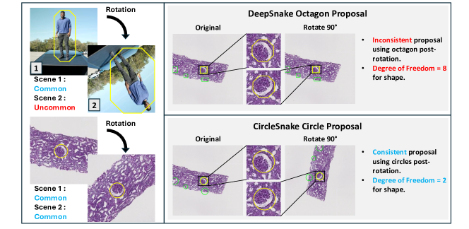
I Introduction
The deep learning model for natural images is frequently applied in the context of medical imaging to identify and highlight regions of interest within medical images [1, 2, 3]. However, medical images often present unique challenges compared to natural images. Unlike natural images which are typically displayed in a fixed orientation, medical images can be viewed at various angles [4, 5], making traditional rectangular bounding boxes less consistent across different angles (Fig. 1). In such cases, circles may offer more flexible and consistent representations of delineating objects or areas of interest. This representation allows for a more consistent and orientation-independent identification of objects, which is crucial in biomedical tissue quantification across different acquisition scenarios. Moreover, the detection method can yield less rotational variances often seen in medical imagery, ensuring that key features are accurately captured regardless of which angle the image is acquired, especially when detecting ball-shaped objects [6].
Recently, the concept of circle representation has been introduced as a representation optimized for medical imaging, particularly for ball-shaped object detection, such as glomeruli, nuclei, inflammatory cells such as eosinophils, and tumors [6, 4, 5, 7, 8, 9]. The exceptional effectiveness of circle representation makes it an attractive candidate for adaptation in the field of instance object segmentation.
In this paper, we propose a simple contour-based end-to-end instance segmentation method that utilizes the circle representation, called CircleSnake, for the robust segmentation of ball-shaped medical objects. The “bounding circle” is introduced for both detection and initial contour representation on the ball-shaped objects. Once the center location of the lesion is obtained, only \acDoF = 2 is required to form the bounding circle, while \acDoF = 8 is required for the bounding octagon. Briefly, the contribution of this study is threefold:
Circle Representation: Our proposal introduces a unified circle representation pipeline for the segmentation of ball-shaped biomedical objects. This pipeline includes three integrated components: (1) circle detection, (2) circle contour proposal, and (3) circular convolution. It achieves superior segmentation performance while requiring a reduced degree of freedom \acDoF for fitting.
Optimized Biomedical Object Segmentation: To the best of our knowledge, CircleSnake, our proposed method, represents the first instance of a contour-based end-to-end segmentation approach that is optimized for ball-shaped biomedical objects.
Rotation Consistency: The proposed circle representation results in less \acDoF of fitting, improved segmentation efficiency, and enhanced rotation consistency. As shown in Fig.1, tissue samples can be viewed at various angles. Consequently, improved rotational consistency could enhance the robustness in detecting identical objects within the same tissue, potentially leading to increased reproducibility in image analysis.
This study expands upon our prior conference paper [6] by introducing fundamental enhancements in this manuscript. (1) More rigorous and comprehensive experiments are conducted via three public and in-house datasets with various ball-shaped objects, including glomeruli, nuclei, and cells; (2) an In-depth assessment and presentation of the methodology is presented, including more benchmarks, comprehensive mathematical derivations, and elaborate experimental design; (3) Detailed introduction is provided with an expanded section on related works and a detailed description on data; (4) Deeper analyses in terms of rotation invariance are conducted. (5) The open-source implementation has been improved to provide both instance object detection and instance segmentation through a single updated codebase: https://github.com/hrlblab/CircleSnake.
II Related Works
II-A Instance Segmentation
II-A1 Mask-Based Methods
Mask-based instance segmentation can be further divided into two-stage [10, 11] and one-stage methods [12, 13].
Two-stage methods usually perform detection in two steps: region proposal and object classification and segmentation mask regression. Mask-RCNN [10] establishes the foundation for two-stage, binary mask-based detection systems. Mask-RCNN [10] consists of a region proposal network (RPN) and a Region-based CNN (R-CNN) approach [14, 15] that is capable of pixel-level object instance segmentation. To achieve pixel-level precision, these binary masks classify pixels as part of the object (foreground) or not (background), simplifying the computational process and allowing for fast and accurate delineation of object boundaries. Following the introduction of Mask-RCNN, numerous algorithms have been developed to enhance its effectiveness, including different architectures [16, 17], different backbone [18], transfer learning strategy [19], different training strategy [20], and feature aggregation mechanisms [21]. While these two-stage detectors can achieve state-of-the-art results, they are often structurally complex and need more time to infer.
One-stage methods eliminate the region proposal step and encapsulate all computations in a single network. With the introduction of YOLACT [22], these types of methods have attracted academic attention for their high computational efficiency. YOLACT employs separate prediction heads that simultaneously identify object classes, bounding boxes, and mask coefficients for each detected object. The final instance masks are dynamically assembled by combining these prototype masks with the specific mask coefficients. After YOLACT, There were many methods to improve its performance by using different feature wrapping module [23], multi-scale feature extraction [24], and adjusting model structure [25]. Now, the performance of one-stage object detectors that use binary masks is nearly equivalent to that of two-stage detectors with anchors, yet they offer quicker inference times.
II-B Medical Object Segmentation
II-B1 Binary Mask-Based Methods
In the existing literature, several works have focused on pixel-based instance segmentation methodologies within regional proposals at the pixel level. Notable among these are studies by [26, 27, 28, 29]. A prominent example of this approach is the Mask R-CNN framework introduced by [10]. This method involves an initial detection of objects, followed by the segmentation of instances within the suggested boxes through the application of a mask predictor. However, despite their effectiveness, these methods are often characterized by structural complexity and a significant demand for processing time, posing challenges for real-time application scenarios.
II-B2 Contour-based Methods
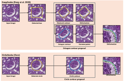
Contour-based methods such as DeepSnake [30] have the potential to be faster and simpler. Huang et al. [31] adapts ideas from U-Net [32] and DeepSnake [30] to right ventricular segmentation. However, this methodology exhibits suboptimal performance when applied to circular objects in pathology images, and the computational simplicity is not adequately achieved (Fig. 2).
III Methods
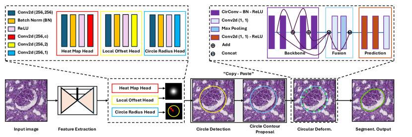
The proposed CircleSnake method, shown in Fig. 3, is an instance segmentation method inspired by the DeepSnake [30] method. CircleSnake aggregates the circle detection [4, 5, 6] and circle contour proposals for initial object segmentation (Fig. 1). This method involves starting with a basic circular contour, which is then refined through a process of circular convolution. This process adjusts the vertices of the contour to align more closely with the actual boundary of the object (Fig. 2). This adjustment is repeated in several iterations to accurately capture the final shape of the object for segmentation purposes. In contrast to the DeepSnake method, which depends on bounding boxes and polygonal contours, the proposed strategy is more straightforward and consistent.
III-A Circle Object Detection
The method for detecting circular objects in this approach is based on the CircleNet framework [6]. The is an input image with height and width . The CPL network produces the center localization of each object within a heatmap where is the number of candidate class and is a downsampling factor. Within the heatmap, is the center of the lesion and is the background. All these terminologies are defined by Zhou et al. [33]. Following convention, [34, 33], the target center point is splat on a heatmap as a 2D Gaussian kernel:
| (1) |
where the and are the center point of the ground truth, and are the downsampled ground truth center point, and is the kernel standard deviation. The training loss is penalty-reduced pixel-wise logistic regression with focal loss [35]:
| (2) |
where the hyper-parameters and are kept the same as [35]. To further refine the prediction location, the -norm offset prediction loss is used.
To ascertain the central point, we propose identifying the top peaks, where each peak’s value is either greater than or equal to its 8-connected neighbors. These selected center points are denoted as . For each object, its center point is represented by an integer coordinate , which is derived from and . Additionally, the offset is calculated from . The formulation of the bounding circle incorporates both the center point and the radius , thereby creating a comprehensive representation of the spatial domain:
| (3) |
where contains the radius prediction for each pixel, optimized by
| (4) |
where is the ground truth radius for each object . Finally, the overall objective is
| (5) |
Following [33], we set and .
III-B Circle Contour Proposal and Deformation
Bounding circle-based detection for each target object is accomplished using CircleNet-based object detection, as described in [5]. Directly derived from the circle representation shown in Fig. 2, the initial circle contour proposal marks a departure from the previously complex deformation process and the use of extreme point-based octagon contour proposals. This innovation simplifies and standardizes the contour proposal approach. Defined by its radius and center point, the circle proposal is established by uniformly sampling initial points from the circle contour, beginning at the top-most point . Correspondingly, the ground truth contour is created by clockwise sampling of vertices along the boundary of the object. Adhering to the guidelines in [30], the value of is fixed at 128, facilitating a consistent and streamlined process.
For a contour with vertices denoted as , we initiate by creating feature vectors for each vertex. The feature corresponding to a vertex comprises a combination of the learning-based features and the vertex’s coordinates, represented as . signifies the feature maps. These input features are then treated as a one-dimensional discrete signal on the contour of the circle. In line with the approach detailed in [30], circular convolution is employed for the learning of features. We define as a periodic signal as:
| (6) |
where is a learnable kernel function, while the operator is the standard convolution. Following [30], the kernel size of the circular convolution is fixed to be nine.
The CircleSnake model incorporates a Graph Convolutional Network (GCN), drawing inspiration from the work presented in [30], for its convolutional implementation. This GCN is organized into three distinct pieces: backbone, fusion, and prediction. The backbone is structured with eight layers, each composed of ”CirConv-Bn-ReLU” sequences. This segment also includes residual skip connections and employs ”CirConv” specifically for circular convolution. The fusion block is designed to integrate information across contour points of varying scales. In this process, features from all the layers in the backbone are amalgamated, subsequently passing through a 11 convolutional layer followed by a max pooling layer. The prediction head, forming the final component of the GCN, is equipped with three 11 convolution layers. Its primary function is to output vertex-wise offsets. As for the loss function, it is specifically formulated to cater to the iterative contour deformation process within the CircleSnake framework:
| (7) |
where is the ground truth boundary point and is the deformed contour point. Following [30], we regress the offsets in 3 iterations.
IV Experimental Design
IV-A Data
IV-A1 Glomeruli Dataset
To construct a dataset for glomeruli analysis, renal biopsy whole slide images (WSIs) were obtained and annotated. Kidney tissues underwent routine processing, embedding in paraffin, and were sectioned to a thickness of 3, followed by staining with hematoxylin and eosin (HE), periodic acid–Schiff (PAS), or Jones. The samples were anonymized, with studies receiving Institutional Review Board (IRB) approval. The dataset comprised 704 glomeruli from 42 biopsies for training, 98 from 7 for validation, and 147 from 7 for testing. Given the relative size of a glomerulus [36], the original high-resolution images (0.25/pixel) were downsampled to 4/pixel. Image patches of 512512 pixels containing at least one glomerulus were randomly sampled, resulting in a comprehensive dataset with 7,040 training, 980 validation, and 1,470 testing images.
IV-A2 Nuclei Dataset
The Nuclei challenge dataset comprises 30 tissue images, each measuring 10001000 pixels, and contains 21,623 manually annotated nuclear boundaries. These images were extracted from various organs sourced from The Cancer Genomic Atlas (TCGA), using H&E stained WSIs at 40 magnification. Additionally, a separate testing dataset with 14 images of the same dimensions was created, including lung and brain tissues exclusively for testing. To create training and validation datasets, we randomly selected 10 patches sized at 512512 pixels from each of the 30 training/validation images, resulting in a total of 300 images. Similarly, 140 images were obtained from the 14 testing images. This dataset comprised 200 training, 100 validation, and 140 testing images. It’s worth noting that the Nuclei 2018 dataset contains more objects (21,623 nuclei) than the glomeruli dataset (802 glomeruli) before any data augmentation. The Nuclei 2018 dataset is publicly accessible for research purposes.
IV-A3 Eosinophils Dataset
All image patches were cut from 50 WSIs from the Pediatric Eosinophils dataset with a 40 objective lens[8]. The Eosinophils dataset contains over 12,000 annotations in 50 annotated WSIs. All human subjects research was approved by the IRB. To enhance the model’s discernment of eosinophils characteristics, we established four distinct classes which include eosinophils (eos), papilla eos, red blood cells (RBC), and RBC clusters. To evaluate the performance of models, the dataset was divided into three subsets: train, val (validation), and test, with a split ratio of 7:1:2.
IV-B Experimental Design
The implementation of CircleSnake’s object instance segmentation and backbone networks followed DeepSnake’s official PyTorch implementations. The Common Objects in Context (COCO) pre-trained model [37] was used to initialize all models. All experiments were conducted on the same workstation with a 24 GB Nvidia RTX A5000. For both the glomeruli and Eos experiments, the hyperparameters were set to maximum epoch = 50, learning rate = , batch size = 16, and optimizer = Adam. For the nuclei experiment, the batch size was set to 4 due to memory constraints.
As baseline methods, Faster-RCNN [38], Mask-RCNN [10], CenterNet [33], DeepSnake [30], were chosen for their superior object detection and object instance segmentation performance. ResNet-50 [39] and deep layer aggregation (DLA) network [40] were used as backbone networks for these different methods. For CircleSnake, we followed the original implementation [5] and used DLA for the backbone networks.
IV-C Evaluation Metrics
In the domain of computer vision, the COCO evaluation metrics have gained prominence as a means to assess the performance of object detection and segmentation algorithms. These metrics were developed as an integral component of the COCO dataset and challenge, offering a standardized framework for quantifying the accuracy and efficacy of such algorithms. In the evaluation process, we employed several key metrics, including Average Precision (AP), (indicating Intersection over Union, or IOU, threshold at 0.5), (IOU threshold at 0.75), (pertaining to objects at a small scale with area less than ), and (associated with objects at a medium scale with area greater than and less than ). In the context of segmentation, the Dice score was also utilized as part of the evaluation criteria. During the evaluation process, all evaluations were conducted using consistent threshold settings.
IV-D Rotation Consistency Score
To rigorously evaluate the resilience and precision of our model across varying angles, we have adopted the Rotation Consistency Score as a key metric [5]. This decision is rooted in the necessity to rotate the original test images by 90 degrees, a choice specifically made to mitigate the effects of intensity interpolation that might arise with arbitrary angles. By employing this structured rotation, we can effectively transform the segmentation masks from the rotated images back to their original orientation. This allows for direct comparison and calculation of the Rotation Consistency Score against the manually segmented results. It is important to note that this rotation process is exclusively implemented during the testing phase and is deliberately not utilized as a data augmentation strategy during the training of all the methods. This approach ensures a focused assessment of the model’s performance in handling rotational variations, providing a clear insight into its robustness and accuracy under controlled conditions.
| Methods | Backbone | |||||
|---|---|---|---|---|---|---|
| CenterNet[34] | DLA-34 | 0.547 | 0.872 | 0.643 | 0.432 | 0.640 |
| CircleNet[6] | DLA-34 | 0.570 | 0.857 | 0.691 | 0.466 | 0.647 |
| Faster-RCNN[38] | ResNet-50 | 0.578 | 0.878 | 0.691 | 0.466 | 0.670 |
| Mask-RCNN (Detection)[10] | ResNet-50 | 0.576 | 0.877 | 0.675 | 0.470 | 0.660 |
| Mask-RCNN (Segmentation)[10] | ResNet-50 | 0.590 | 0.878 | 0.705 | 0.453 | 0.685 |
| DeepSnake (Detection)[30] | DLA-34 | 0.527 | 0.881 | 0.609 | 0.424 | 0.610 |
| DeepSnake (Segmentation)[30] | DLA-34 | 0.548 | 0.877 | 0.669 | 0.411 | 0.642 |
| CircleSnake(Detection) (Ours) | DLA-34 | 0.603 | 0.877 | 0.734 | 0.534 | 0.670 |
| CircleSnake(Segmentation) (Ours) | DLA-34 | 0.623 | 0.894 | 0.762 | 0.488 | 0.719 |
| Methods | Backbone | |||||
|---|---|---|---|---|---|---|
| CenterNet[34] | DLA-34 | 0.404 | 0.818 | 0.342 | 0.440 | 0.290 |
| CircleNet[6] | DLA-34 | 0.461 | 0.843 | 0.462 | 0.473 | 0.332 |
| Faster-RCNN[38] | ResNet-50 | 0.364 | 0.679 | 0.349 | 0.365 | 0.336 |
| Mask-RCNN (Detection)[10] | ResNet-50 | 0.369 | 0.679 | 0.359 | 0.367 | 0.348 |
| Mask-RCNN (Segmentation)[10] | ResNet-50 | 0.366 | 0.678 | 0.369 | 0.365 | 0.446 |
| DeepSnake (Detection)[30] | DLA-34 | 0.401 | 0.827 | 0.315 | 0.408 | 0.345 |
| DeepSnake (Segmentation)[30] | DLA-34 | 0.425 | 0.827 | 0.394 | 0.425 | 0.454 |
| CircleSnake (Detection) (Ours) | DLA-34 | 0.485 | 0.845 | 0.518 | 0.495 | 0.217 |
| CircleSnake (Segmentation) (Ours) | DLA-34 | 0.436 | 0.837 | 0.419 | 0.436 | 0.418 |
| Methods | Backbone | |||||
|---|---|---|---|---|---|---|
| CenterNet[34] | DLA-34 | 0.327 | 0.729 | 0.230 | 0.351 | 0.318 |
| CircleNet[6] | DLA-34 | 0.340 | 0.706 | 0.292 | 0.368 | 0.332 |
| Faster-RCNN[38] | ResNet-50 | 0.331 | 0.696 | 0.237 | 0.332 | 0.343 |
| Mask-RCNN (Detection)[10] | ResNet-50 | 0.335 | 0.700 | 0.246 | 0.336 | 0.354 |
| Mask-RCNN (Segmentation)[10] | ResNet-50 | 0.330 | 0.690 | 0.243 | 0.282 | 0.391 |
| DeepSnake (Detection)[30] | DLA-34 | 0.300 | 0.720 | 0.149 | 0.301 | 0.323 |
| DeepSnake (Segmentation)[30] | DLA-34 | 0.305 | 0.712 | 0.169 | 0.255 | 0.360 |
| CircleSnake(Detection) (Ours) | DLA-34 | 0.344 | 0.727 | 0.298 | 0.340 | 0.358 |
| CIrcleSnake(Segmentation) (Ours) | DLA-34 | 0.332 | 0.723 | 0.263 | 0.282 | 0.403 |
V Results
| Dataset | Methods | Backbone | Dice | Rotation Consistency |
|---|---|---|---|---|
| Glomeruli | Mask-RCNN[10] | ResNet | 0.813 | 0.779 |
| Glomeruli | DeepSnake-DLA[33] | DLA | 0.801 | 0.793 |
| Glomeruli | CircleSnake (Ours) | DLA | 0.828 | 0.796 |
| Nuclei | Mask-RCNN[10] | ResNet | 0.763 | 0.763 |
| Nuclei | DeepSnake-DLA[33] | DLA | 0.765 | 0.767 |
| Nuclei | CircleSnake (Ours) | DLA | 0.800 | 0.799 |
| Eosinophils | Mask-RCNN[10] | ResNet | 0.71 | 0.691 |
| Eosinophils | DeepSnake-DLA[33] | DLA | 0.684 | 0.690 |
| Eosinophils | CircleSnake (Ours) | DLA | 0.743 | 0.752 |
V-A Glomeruli Detection and Segmentation Performance
V-B Nuclei Detection and Segmentation Performance
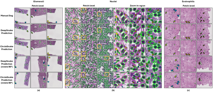
The dataset from the 2018 Multi-Organ Nuclei Segmentation Challenge [41, 42] was also applied in the experiment. As seen in Table II, the proposed CircleSnake-DLA method reach 0.485 detection , 0.845 detection , 0.518 , 0.495 detection , 0.418 segmentation . A qualitative comparison between Mask-RCNN, DeepSnake, and CircleSnake can be seen in Fig. 4 section (b).
V-C Eosinophils Detection and Segmentation Performance
In pursuit of confirming the model’s broad applicability and generalizability, we decided to introduce the Eosinophils dataset[8] into our study. As indicated in Table III, the performance of the CircleSnake achieve 0.344 detection , 0.727 detection , 0.298 , 0.340 detection , 0.403 segmentation . A qualitative comparison between Mask-RCNN, DeepSnake, and CircleSnake can be seen in Fig. 4 part (c).
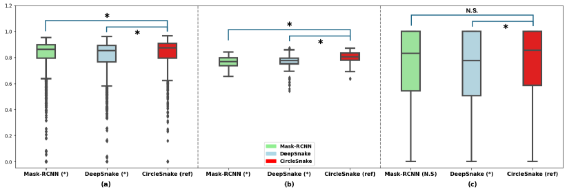
V-D Semantic Segmentation Results and Rotation Consistency
For the semantic segmentation results and rotation consistency comparison. As shown in Table LABEL:table4 and Fig. 5 part (a). Our method, CircleSnake can achieve a Dice score of 0.828 with 0.796 rotation consistency score and the Dice score result has a significant difference from the other two methods in the Glomeruli dataset. For the Nuclei dataset Table LABEL:table4 and Fig. 5 part (b), CircleSnake achieves 0.8 for the Dice score which also has a significant difference from the other two methods and 0.799 in the rotation consistency score. In the Eosinophils dataset Table LABEL:table4 and Fig. 5 part (c), CircleSnake achieved 0.743 for the Dice score which has a significant difference from the DeepSnake method and 0.752 in the rotation consistency score.
VI Discussion
In this study, we propose a contour-based method, CircleSnake, optimized for the segmentation of biomedical ball-shaped objects. Instead of using an octangle as the initial contour, CircleSnake directly uses the detection circle as the initial contour which has a simpler structure and is shown to offer superior detection or segmentation performance and rotation consistency. specifically, the CircleSnake outperforms the other two baseline methods in all evaluation metrics in the Glomeruli dataset in Table I. It also has better performance except in the Nuclei dataset in Table II and also better results in , , and in Table III.
As seen in Table LABEL:table4 the proposed segmentation contour deformed by circle achieved better rotation consistency with higher rotation consistency scores. One explanation for this result is that while length and width metrics are sensitive to rotation, radii are naturally more spatially invariant metrics.
The utilization of a circular contour as the foundational shape for deformation processes may not be ideally suited for objects of varying geometrical forms, such as ellipses or elongated shapes. This assertion is substantiated by the data presented in Table II and Table III, where a discernible discrepancy is observed between the AP of object detection and that of segmentation. The higher AP in detection compared to segmentation might be attributable to the inherent limitations of a circular contour in accommodating the deformation needs of non-circular objects, necessitating a more extensive modification to fit diverse shapes. Furthermore, the current methodology, being predicated on a single-scale feature map, exhibits a marginal reduction in efficacy when dealing with objects of disparate scales, as evidenced by the results in Table Table III. Consequently, it is posited that transitioning from a single-scale to a multi-scale feature map could potentially enhance the overall performance of the model, providing a more nuanced and adaptable approach to object detection and segmentation.
VII Conclusion
In this paper, we propose CircleSnake, a simple contour-based end-to-end deep learning algorithm with simple architecture, that achieves (1) circle detection, (2) circle contour proposal, and (3) circular convolution. The CircleSnake method is optimized for ball-shaped biomedical objects, offering superior glomeruli, nuclei, and eosinophils instance segmentation performance and rotation consistency. The experimental results demonstrate that, in contrast to conventional bounding box and octagonal representations, the reduction of \acDoF has no adverse impact on the model’s accuracy. Furthermore, it effectively sustains detection efficiency across diverse viewing angles.
Acknowledgment
This research was supported by NIH R01DK135597(Huo), DoD HT9425-23-1-0003(HCY), NIH NIDDK DK056942(ABF). This work was also supported by Vanderbilt Seed Success Grant, Vanderbilt Discovery Grant, and VISE Seed Grant. This project was supported by The Leona M. and Harry B. Helmsley Charitable Trust grants G-1903-03793 and G-2103-05128. This research was also supported by NIH grants R01EB033385, R01DK132338, REB017230, R01MH125931, and NSF 2040462. We extend gratitude to NVIDIA for their support by means of the NVIDIA hardware grant.
References
- [1] Y.-C. Lo, C.-F. Juang, I.-F. Chung, S.-N. Guo, M.-L. Huang, M.-C. Wen, C.-J. Lin, and H.-Y. Lin, “Glomerulus detection on light microscopic images of renal pathology with the faster r-cnn,” in International Conference on Neural Information Processing. Springer, 2018, pp. 369–377.
- [2] H. Liang, Z. Cheng, H. Zhong, A. Qu, and L. Chen, “A region-based convolutional network for nuclei detection and segmentation in microscopy images,” Biomedical Signal Processing and Control, vol. 71, p. 103276, 2022. [Online]. Available: https://www.sciencedirect.com/science/article/pii/S1746809421008739
- [3] J. Xiong, Y. Liu, R. Deng, R. N. Tyree, H. Correa, G. Hiremath, Y. Wang, and Y. Huo, “Deep learning-based open source toolkit for eosinophil detection in pediatric eosinophilic esophagitis,” 2023.
- [4] H. Yang, R. Deng, Y. Lu, Z. Zhu, Y. Chen, J. T. Roland, L. Lu, B. A. Landman, A. B. Fogo, and Y. Huo, “Circlenet: Anchor-free glomerulus detection with circle representation,” in International Conference on Medical Image Computing and Computer-Assisted Intervention. Springer, 2020, pp. 35–44.
- [5] E. H. Nguyen, H. Yang, R. Deng, Y. Lu, Z. Zhu, J. T. Roland, L. Lu, B. A. Landman, A. B. Fogo, and Y. Huo, “Circle representation for medical object detection,” IEEE transactions on medical imaging, vol. 41, no. 3, pp. 746–754, 2021.
- [6] E. H. Nguyen, H. Yang, Z. Asad, R. Deng, A. B. Fogo, and Y. Huo, “Circlesnake: Instance segmentation with circle representation,” in International Workshop on Machine Learning in Medical Imaging. Springer, 2022, pp. 298–306.
- [7] X. Luo, T. Song, G. Wang, J. Chen, Y. Chen, K. Li, D. Metaxas, and S. Zhang, “Scpm-net: An anchor-free 3d lung nodule detection network using sphere representation and center points matching,” arXiv preprint arXiv:2104.05215, 2021.
- [8] Y. Liu, R. Deng, J. Xiong, R. N. Tyree, H. Correa, G. Hiremath, Y. Wang, and Y. Huo, “Eosinophils instance object segmentation on whole slide imaging using multi-label circle representation,” 2023.
- [9] H. Zhang, P. Liang, Z. Sun, B. Song, and E. Cheng, “Circleformer: Circular nuclei detection in whole slide images with circle queries and attention,” in International Conference on Medical Image Computing and Computer-Assisted Intervention. Springer, 2023, pp. 493–502.
- [10] K. He, G. Gkioxari, P. Dollár, and R. Girshick, “Mask r-cnn,” 2018.
- [11] Z. Cai and N. Vasconcelos, “Cascade r-cnn: High quality object detection and instance segmentation,” IEEE Transactions on Pattern Analysis and Machine Intelligence, vol. 43, no. 5, pp. 1483–1498, 2021.
- [12] D. Bolya, C. Zhou, F. Xiao, and Y. J. Lee, “Yolact: Real-time instance segmentation,” 2019.
- [13] H. Ying, Z. Huang, S. Liu, T. Shao, and K. Zhou, “Embedmask: Embedding coupling for one-stage instance segmentation,” 2019.
- [14] R. Girshick, J. Donahue, T. Darrell, and J. Malik, “Rich feature hierarchies for accurate object detection and semantic segmentation,” in Proceedings of the IEEE conference on computer vision and pattern recognition, 2014, pp. 580–587.
- [15] R. Girshick, “Fast r-cnn,” in Proceedings of the IEEE international conference on computer vision, 2015, pp. 1440–1448.
- [16] L.-C. Chen, A. Hermans, G. Papandreou, F. Schroff, P. Wang, and H. Adam, “Masklab: Instance segmentation by refining object detection with semantic and direction features,” in 2018 IEEE/CVF Conference on Computer Vision and Pattern Recognition, 2018, pp. 4013–4022.
- [17] K. Chen, J. Pang, J. Wang, Y. Xiong, X. Li, S. Sun, W. Feng, Z. Liu, J. Shi, W. Ouyang, C. C. Loy, and D. Lin, “Hybrid task cascade for instance segmentation,” 2019.
- [18] S. Fang, B. Zhang, and J. Hu, “Improved mask r-cnn multi-target detection and segmentation for autonomous driving in complex scenes,” Sensors, vol. 23, no. 8, 2023. [Online]. Available: https://www.mdpi.com/1424-8220/23/8/3853
- [19] Q. Zhang, X. Chang, and S. B. Bian, “Vehicle-damage-detection segmentation algorithm based on improved mask rcnn,” IEEE Access, vol. 8, pp. 6997–7004, 2020.
- [20] S. Wang, G. Sun, B. Zheng, and Y. Du, “A crop image segmentation and extraction algorithm based on mask rcnn,” Entropy, vol. 23, no. 9, 2021. [Online]. Available: https://www.mdpi.com/1099-4300/23/9/1160
- [21] S. Liu, L. Qi, H. Qin, J. Shi, and J. Jia, “Path aggregation network for instance segmentation,” 2018.
- [22] W. Liu, D. Anguelov, D. Erhan, C. Szegedy, S. Reed, C.-Y. Fu, and A. C. Berg, “Ssd: Single shot multibox detector,” in European conference on computer vision. Springer, 2016, pp. 21–37.
- [23] H. Liu, R. A. R. Soto, F. Xiao, and Y. J. Lee, “Yolactedge: Real-time instance segmentation on the edge,” 2021.
- [24] J. Zeng, H. Ouyang, M. Liu, L. Leng, and X. Fu, “Multi-scale yolact for instance segmentation,” Journal of King Saud University - Computer and Information Sciences, vol. 34, no. 10, Part B, pp. 9419–9427, 2022. [Online]. Available: https://www.sciencedirect.com/science/article/pii/S1319157822003482
- [25] D. Bolya, C. Zhou, F. Xiao, and Y. J. Lee, “Yolact++ better real-time instance segmentation,” IEEE Transactions on Pattern Analysis and Machine Intelligence, vol. 44, no. 2, p. 1108–1121, Feb. 2022. [Online]. Available: http://dx.doi.org/10.1109/TPAMI.2020.3014297
- [26] M. Gadermayr, A.-K. Dombrowski, B. M. Klinkhammer, P. Boor, and D. Merhof, “Cnn cascades for segmenting whole slide images of the kidney,” arXiv preprint arXiv:1708.00251, 2017.
- [27] G. Bueno, M. M. Fernandez-Carrobles, L. Gonzalez-Lopez, and O. Deniz, “Glomerulosclerosis identification in whole slide images using semantic segmentation,” Computer methods and programs in biomedicine, vol. 184, p. 105273, 2020.
- [28] S. Kannan, L. A. Morgan, B. Liang, M. G. Cheung, C. Q. Lin, D. Mun, R. G. Nader, M. E. Belghasem, J. M. Henderson, J. M. Francis et al., “Segmentation of glomeruli within trichrome images using deep learning,” Kidney international reports, vol. 4, no. 7, pp. 955–962, 2019.
- [29] B. Ginley, B. Lutnick, K.-Y. Jen, A. B. Fogo, S. Jain, A. Rosenberg, V. Walavalkar, G. Wilding, J. E. Tomaszewski, R. Yacoub et al., “Computational segmentation and classification of diabetic glomerulosclerosis,” Journal of the American Society of Nephrology, vol. 30, no. 10, pp. 1953–1967, 2019.
- [30] S. Peng, W. Jiang, H. Pi, X. Li, H. Bao, and X. Zhou, “Deep snake for real-time instance segmentation,” in Proceedings of the IEEE/CVF Conference on Computer Vision and Pattern Recognition, 2020, pp. 8533–8542.
- [31] K. Huang, L. Xu, Y. Zhu, and P. Meng, “A u-snake based deep learning network for right ventricle segmentation.” Medical physics, 2022. [Online]. Available: https://api.semanticscholar.org/CorpusID:247520303
- [32] O. Ronneberger, P. Fischer, and T. Brox, “U-net: Convolutional networks for biomedical image segmentation,” 2015.
- [33] X. Zhou, D. Wang, and P. Krähenbühl, “Objects as points,” arXiv preprint arXiv:1904.07850, 2019.
- [34] H. Law and J. Deng, “Cornernet: Detecting objects as paired keypoints,” in Proceedings of the European Conference on Computer Vision (ECCV), 2018, pp. 734–750.
- [35] T.-Y. Lin, P. Goyal, R. Girshick, K. He, and P. Dollár, “Focal loss for dense object detection,” in Proceedings of the IEEE international conference on computer vision, 2017, pp. 2980–2988.
- [36] V. G. Puelles, W. E. Hoy, M. D. Hughson, B. Diouf, R. N. Douglas-Denton, and J. F. Bertram, “Glomerular number and size variability and risk for kidney disease,” Current opinion in nephrology and hypertension, vol. 20, no. 1, pp. 7–15, 2011.
- [37] T.-Y. Lin, M. Maire, S. Belongie, J. Hays, P. Perona, D. Ramanan, P. Dollár, and C. L. Zitnick, “Microsoft coco: Common objects in context,” in European conference on computer vision. Springer, 2014, pp. 740–755.
- [38] S. Ren, K. He, R. Girshick, and J. Sun, “Faster r-cnn: Towards real-time object detection with region proposal networks,” in Advances in neural information processing systems, 2015, pp. 91–99.
- [39] K. He, X. Zhang, S. Ren, and J. Sun, “Deep residual learning for image recognition,” in Proceedings of the IEEE conference on computer vision and pattern recognition, 2016, pp. 770–778.
- [40] F. Yu, D. Wang, E. Shelhamer, and T. Darrell, “Deep layer aggregation,” in Proceedings of the IEEE conference on computer vision and pattern recognition, 2018, pp. 2403–2412.
- [41] N. Kumar, R. Verma, S. Sharma, S. Bhargava, A. Vahadane, and A. Sethi, “A dataset and a technique for generalized nuclear segmentation for computational pathology,” IEEE transactions on medical imaging, vol. 36, no. 7, pp. 1550–1560, 2017.
- [42] N. Kumar, R. Verma, D. Anand, Y. Zhou, O. F. Onder, E. Tsougenis, H. Chen, P.-A. Heng, J. Li, Z. Hu et al., “A multi-organ nucleus segmentation challenge,” IEEE transactions on medical imaging, vol. 39, no. 5, pp. 1380–1391, 2019.