Water-based Quantum Dots Liquid Scintillator for Particle Physics
Abstract
Liquid scintillators are typically composed from organic compounds dissolved in organic solvents. However, usage of such material is often restricted due to fire safety and environmental reasons. Because of this, R&D of water-based liquid scintillators is of extreme relevance; yet, no such scintillators have been made commercially available as yet. Here, we investigate an alternative, water-based quantum dots liquid scintillator. Pre-determined and controllable optical properties of the quantum dots, as well as the existence of large libraries of established protocols for their dispersion in aqueous solutions, make them an attractive option for nuclear and particle physics applications. We characterize the optical properties of water-based quantum dots liquid scintillator and find that most of its optical properties are preserved upon quantum dots’ phase transfer into water, through the addition of an oleic acid hydrophilic layer. Using the developed scintillator, the time and charge responses from atmospheric muons are measured, highlighting the practical viability of water-based quantum dots liquid scintillators for nuclear and particle physics, with special interest in neutrino physics.
1 Introduction
Since their first experimental observation in glass matrices [1], nano-sized crystals of semiconducting materials, aka Quantum Dots (QDs), have been the focus of research in many different areas. Their superior and size-dependent optoelectronic properties make them attractive for many applications, including nanomedicine [2], bioimaging [3], photocatalysis [4], photovoltaics [5], and lighting and display applications [6]. The field of QD-based photonics has advanced rapidly since their first realization, with some examples of translation into commercial products occurring within the last decade [7] which highlight the suitability of QDs for technological applications.
The most attractive properties of QDs for photonics applications are their high extinction cross-sections and photoluminescence quantum yields (PLQY), both of which are typically higher or comparable to molecular equivalents [8]. In addition, they have larger physical cross-sections, which increases the probability of their interaction with charged particles and subsequent generation of photon signals through direct scintillation. Direct scintillation has been observed from QD-doped solid materials under irradiation [9, 10]. Therefore, we expect that direct scintillation through energy exchange with other radiations, for example minimally ionizing particles (MIPs) such as cosmic rays (atmospheric muons), are equally possible.
The last few decades of research into the properties of colloidal QDs established a large library of surface modification protocols [11], including molecular scintillators or dopant-containing molecules, which provide a route to introduce additional components into the system without adverse effects to solvent transparency. Using this approach, QDs can simultaneously act as direct liquid scintillators and as wavelength-shifters for the molecular scintillators or dopant molecules attached to their surfaces, where the energy is transferred resonantly between the two. This presents a new prospect of being able to improve light yields of the solution without modifying the overall concentration of solute in the solution. QDs of appropriate surface functionalisations are also directly dispersible in water [12], allowing them to act as a water-based quantum dots (WbQD) liquid scintillator.
Here we are investigating the possibility of utlizing QDs for particle physics applications [13, 14, 15]. In particular, we are interested in the possibility of WbQDs liquid scintillator in nuclear fission reactor neutrino detection. Water is a safe material to use near the fission reactor core, and such a detector can be a new type of near-field monitor for fission reactors [16, 17, 18, 19, 20, 21, 22]. Typical compositions of the QDs include elements such as cadmium, selenium, and tellurium, all of which have high neutron capture cross-sections. This opens a possibility to tag neutrons, and WbQDs to act as conventional liquid scintillators with neutron detection ability without doping neutron capturing materials, in contrast to the proposed gadolinium-doped water-based liquid scintillator [20].
In preparing the WbQD liquid scintillator, the parameters of interest were (1) the photoluminescence quantum yield (PLQY), (2) the transparency, (3) the emission spectra, (4) concentration and (5) the stability of the scintillating liquid. The PLQY, defined as the ratio of photons emitted per photons absorbed by the fluorophore, has to be high enough to be able to efficiently resolve the incoming particle energy. However, high-PLQY colloidal QDs are typically produced in organic solvents and are therefore not stable in aqueous solutions as-synthesized. It is possible to encapsulate such QDs in an amphiphilic layer via a process called phase transfer, which facilitates their dispersion in water [23]; however, such processes typically result in a diminished PLQY of the QDs and increased scattering by the colloids. Nonetheless, the resulting dispersions are shown to be sufficiently transparent for the emitted photons to reach the photodetectors without being re-absorbed.
Regarding the emission properties of QDs, the vast majority of photodetectors used for radiation detection have maximum quantum efficiencies (QE) in the blue end of the spectrum; therefore, the selection of QDs’ composition and size needs to be such as to yield emission in this spectral range. QDs with Cd-based compositions and small core sizes seem appropriate for proof-of-principle demonstrations. Furthermore, and although not explored in this work, cadmium has a high neutron capture cross-section and Cd-based cores of our QDs open a possibility to employ them as neutron capture materials, allowing tagging of neutrino interactions through the inverse beta-decay processes [24]. Regardless of the application, the concentration of QDs in the scintillating liquid needs to be high enough to facilitate sufficient muon interactions or neutron capture events for experimentation, whilst low enough to maintain the optical transparency of the liquid. As such, there will be an optimum range for QD concentration in WbQD scintillating liquids.
Finally, for application purposes, QDs must remain colloidally and functionally stable for relatively long periods of time. Whilst it is known that phase-transferred QDs might gradually lose their stability over time and that routes to mitigate this do exist [25, 26, 27, 28], little is known about the effect of long-term exposure to radiation on the QDs’ stability and functionality. Although radiation responses of QDs were studied in the past [9, 10], these were for low energy particles and not MIPs such as cosmic rays. As such, measuring MIP responses and therefore demonstrating the scintillating property of WbQDs liquid scintillator - the subject the work presented here - is the first step towards the utilization of WbQDs as particle scintillators.
In Section 2, we describe the selection and preparation of WbQD scintillating liquids used in this work. Section 3 discusses the optical and physical properties of the prepared liquids, as well as their long-term stability. In Section 4, we study the cosmic ray responses from WbQD samples to evaluate their potential as a liquid scintillator for a radiation detector. The results are discussed and critically evaluated in Section 5. Finally, Section 6 summarizes key findings and proposes directions for future studies.
2 Quantum dots sample preparation
2.1 Quantum dots selected
To meet the required characteristics discussed in the previous section, cadmium sulfide (CdS) core zinc sulfide (ZnS) shell type coating quantum dots were selected for the experiments. There were numerous reasons behind this selection. Firstly, the CdS composition of the QD core facilitates shorter wavelengths of QD fluorescence, compared to e.g. the more popular CdSe or CdTe QDs. Secondly, blue-emitting CdS QDs of relatively high PLQY ( 50%) were available for purchase commercially [29], although these QDs could not be dispersed in water directly, requiring further surface modification to transfer them to aqueous environments (as detailed in sec. 2.2). Thirdly, the manufacturer stated the shelf-life of these QDs as being three years, which was sufficient for our purposes due to the short time span of the experiment. And finally, the cadmium isotope 113Cd has a natural abundance of , and a relatively high neutron capture cross-section of barn [30], with a Q-value MeV.
The CdS/ZnS QDs satisfied many of the criteria posed in the previous section, and it was expected that these QDs may allow for particle identification, i.e. to distinguish neutrons from other signals with a suitable concentration. This would be especially useful as a low-energy neutrino detector, such as a fission reactor neutrino detector. This property will be studied in future. Here, our target concentration of cadmium is 0.01% by mass, to match the recent gadolinium doping by the Super-Kamiokande experiment [31]. In our case, this corresponds to roughly 10 mg of CdS/ZnS QDs in 80 mL water.
2.2 Surfactant encapsulation method
Oleic acid (OA) functionalized CdS/ZnS core-shell QDs, supplied in solid form, were purchased from Sigma Aldrich [29]. Since the as-supplied QDs cannot be directly dispersed in water, QDs were first dispersed in toluene and then phase transferred into water using the surfactant encapsulation method. The procedure for the phase transfer closely followed the previously reported methodology[23], involving encapsulation of QDs in a double layer of OA, which is known to produce extremely stable QDs. Briefly, 10 mg of CdS/ZnS QDs powder was dispersed in 1.6 mL toluene and the stock solution was left to completely evaporate under vacuum and at room temperature, in the dark. The QDs solids were then dispersed in 16 mL of hexane, to which 320 L of OA was added. The solvent was then sonicated for 1 min using the Ultrasonic bath XUBA3 [32], after which 200 mL of Milli-Q water (Millipore, 16 M-cm) was added to the mixture, whilst being continuously stirred. Subsequent sonication of the mixture over a period of one hour, with occasional manual shaking of the vial (every 3 minutes), resulted in the formation of an emulsion. This was then kept undisturbed in darkness and at room temperature for 75h, during which time phase separation took place. At this point, QDs should have phase transferred to the water phase (bottom half of the phase separated liquid) and they were removed carefully using a syringe. The dispersion was then centrifuged at 6500 rpm for 15 min, using Eppendorf Centrifuge 5804 [33] to cause precipitation of any large QD aggregates. The remaining supernatant was collected and filtered through a 220 nm pore syringe filter, to remove any remaining smaller QD aggregates. We note that, during the above procedure, a small aliquot of toluene-dispersed QDs was set aside for control measurements.

Three samples were prepared in this manner, one in each of the years of 2021, 2022 and 2023, and labelled accordingly. Figure 1 shows the three samples under natural and UV illumination (recorded in August of 2023). The blue color of solutions under UV illumination (in panel (b)) is indicative of QD presence in the dispersions and therefore of the successful completion of the phase transfer procedure on each occasion. It is clear that in all three cases the concentration of QDs is relatively high; yet, and as is evident from panel (a) of Figure 1, all three samples have a high degree of transparency, as per requirements for successful neutrino experiments. In the following result section, we present a representative set of data from the 2023 sample; the properties of 2021 and 2022 samples will be discussed in detail in section 3.3, where sample aging is considered.
3 Physicochemical properties of CdS/ZnS QDs
In this section we report the results of the photophysical characterization of the 2023 QD dispersion, including measurements of the QDs’ size, surface charge, and their optical properties.
3.1 Optical properties of CdS/ZnS QDs
The extinction and photoluminescence (PL) spectra of QD dispersions were recorded using a 2600i UV-vis spectrometer from Shimadzu [34] and Cary Eclipse Fluorescence Spectrophotometer [35] respectively. In our interpretation of the extinction data, we attributed any strong changes in the extinction of the sample to the absorption by the QDs, since they have relatively small scattering cross-sections [36]. However, below the band gap of the QDs, scattering contributions can and do become dominant.
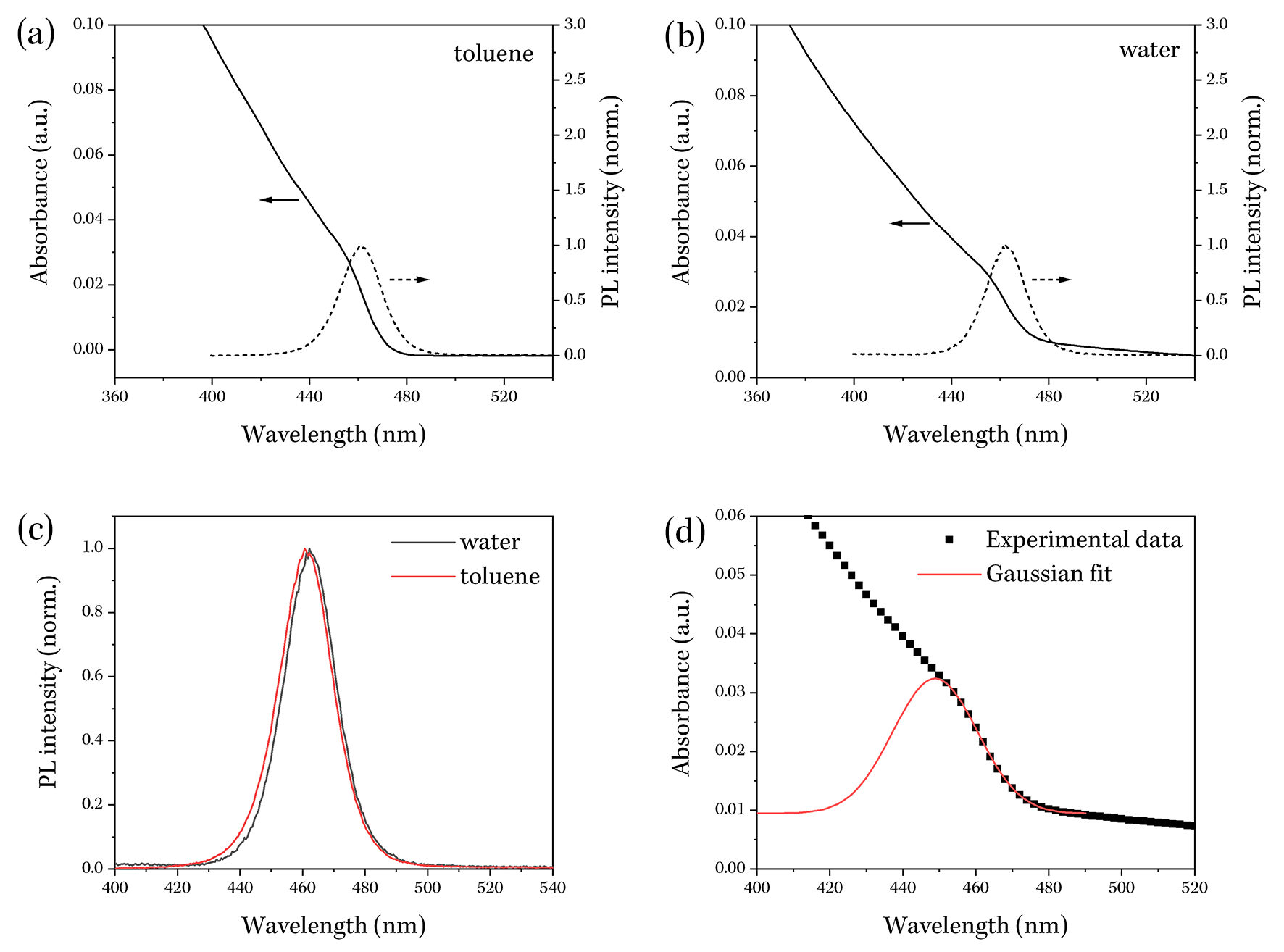
As expected, and as shown in Figs. 2(a) and (b), the absorbance of QDs dispersions in toluene and in water remains negligible at low energies (high wavelengths), but increases rapidly as the energy of the excitation becomes greater than the bandgap of the QDs (at 470 nm for both). No significant changes to the absorption spectra could be observed for the WbQD liquid as compared to the toluene-dispersed QDs, indicating that no significant chemical or physical changes to the QD core occurred as a result of the phase transfer process. However, comparison of the extinction spectra of the two samples at wavelengths above the QDs’ band edge, it is clear that the phase transfer of QDs to an aqueous environment resulted in an increased Rayleigh’s scattering by the QDs - this appears as an elevated but gradually decreasing background at wavelengths larger than 470 nm. This is a typical observation, owing to the size dependency of the Rayleigh scattering process and simply indicates an increased overall size of the QDs upon addition of the surfactant coating that is needed to achieve water-dispersibility [37, 38].
The PL spectra of both WbQD and toluene-based dispersions appeared as Gaussian peaks centred around 460 nm (Fig. 2(a) and (b)). As discussed previously, the observed emission in the blue end of the spectrum is favourable for radiation detection experimentation. The Gaussian shape of the PL spectra is reflective of the size-dispersion of the QDs in the sample, and is very typical and characteristic of colloidal QDs [39, 40]. Of note is also the lack of any significant changes to the emission of the QDs upon phaser transfer, as is evidenced by the normalized PL spectra presented in Fig. 2(c) where only a slight shift in the position of the peak could be observed (). This small shift can be attributed to the slight change in the environmental refractive index of the QDs resulting from the addition of an OA shell during the phase transfer process [41].
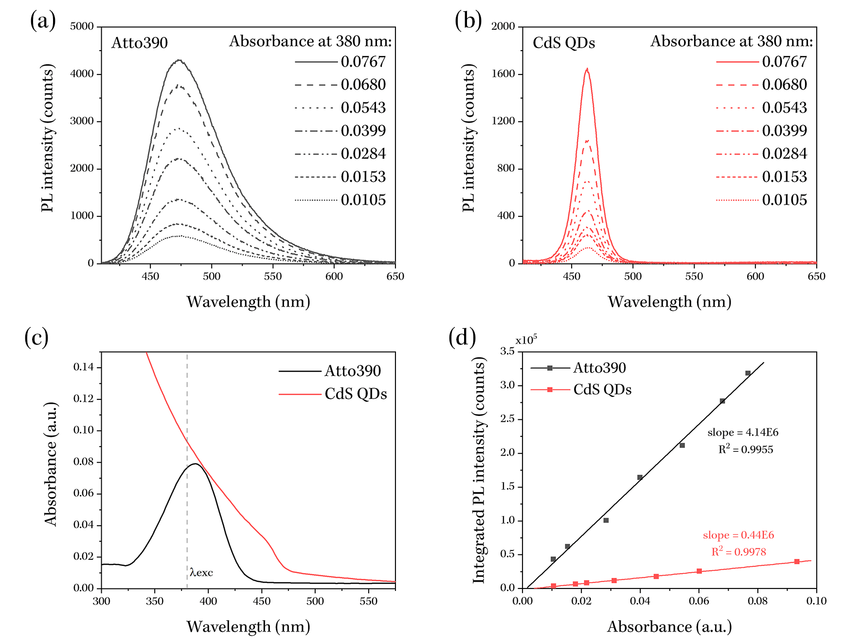
We further determine the PLQY of the WbQD sample, by comparing its emission properties to those of a reference dye with a known PLQY and emission in a wavelength range that coincides with that of the WbQD sample. Atto390 dye was used for the purpose here, which has a PLQY of in aqueous environments. Figure 3 summarizes the measurements performed. PL spectra of both the reference dye and the WbQD, diluted to seven different concentrations, were recorded (Figs. 3(a) and (b) respectively) using 380 nm as the excitation wavelength in all cases. These concentrations corresponded to a range of sample absorbances, which were recorded using extinction measurements (a representative set is shown in Figure 3(c)). The PL spectra were then integrated over the entire spectral range and these integrated values were plotted against corresponding absorbance values at excitation wavelength, as shown in Figure 3(d). The resulting sets of data, one for the reference dye and one for the WbQD sample, were then fitted to linear functions of the form
| (3.1) |
where refers to the integrated PL intensity of the sample at a specific value of sample absorbance, is an offset/background term. The results of the fitting procedure are summarized in Table 1 below.
The ratio of the slopes for the WbQD and reference dye solutions are proportional to the ratio of their PLQYs [42], i.e.
| (3.2) |
Using this relation, and the known value of the PLQY of the reference dye (), the PLQY of our 2023 WbQD liquid was calculated to be
| (3.3) |
Clearly, there was a reduction of the PLQY of the QY upon their phase transfer from organic to aqueous environment, which is a typical observation for such processes [43, 44], often attributed to the introduction of surface defects that act as long-lived trap states for the excited states of the QDs [45]. Nonetheless, the PLQY of the WbQD solution remained sufficiently high for the proposed proof-of-principle demonstrations.
| Sample | Slope, | Intercept, | value |
|---|---|---|---|
| Atto390 dye | 0.9955 | ||
| 2023 WbQDs | 0.9978 |
In addition to information provided above, the absorption spectrum of the QD dispersions was also utilized to estimate the concentration of the prepared dispersions, using a set of empirical equations developed by W. W. Yu et al. for aqueous dispersions of CdS QDs [46]. To do so, the measured absorption spectrum of the WbQD solution was first fitted to a Gaussian function, at the on-set of QD absorption, and as shown in Figure 2(d). Specifically, a subset of the data in the nm spectral range was fitted to a function of the form:
| (3.4) |
where and are the amplitudes of the absorbance background and the Gaussian peak respectively, and and are the central position and the width of the peak, respectively. As part of the fitting analysis, the software also calculated the full-width at half maximum (FWHM ) of the Gaussian peak. The results of the fitting procedure, summarized in Table 2, were then inputted into the empirical equation of reference [46] to calculate the extinction coefficient, of the WbQD dispersion at the position of the first extinction peak, , using the following set of equations:
| (3.5) |
| (3.6) |
where refers to the mean diameter of the QDs in the sample.
Furthermore, and as per protocol described in reference [46], the measured absorbance of the sample at was calibrated to account for the dispersion of QD sizes within the sample according to
| (3.7) |
where is the measured absorbance of the sample at , is the calibrated absorbance and is the half width at half maximum of the first excitonic absorption peak of a typical CdS QD sample ().
| Value | 0.00947 | 0.0229 | 448.8 | 11.6 | 27.3 |
| Error | 0.00006 | 0.0002 | 0.3 | 0.2 | 0.4 |
Following the described above procedure, the our WbQD sample was determined to a calibrated absorbance of , a mean QD diameter of
| (3.8) |
and therefore an an extinction coefficient of
| (3.9) |
at the first extinction peak. Using these two pieces of information, as well as the known path length used in the absorbance measurements (), the concentration of the sample () could then be determined from the Beer-Lambert law,
| (3.10) |
yielding a value of
| (3.11) |
for the WbQD dispersion.
3.2 Physical properties of CdS/ZnS QDs
The hydrodynamic size () of the CdS/ZnS QDs was measured using the dynamic light scattering (DLS) technique, in which intensity fluctuations caused by the scattering of laser radiation by freely diffusing QDs in a dispersion are auto-correlated [47]. DLS measurements were performed on Zetasizer Nano ZS (Malvern), which performs measurements in triplicates and outputs the respective correlation curves, cumulant analysis results (mean particle size and standard deviation) and size distribution curves [48].
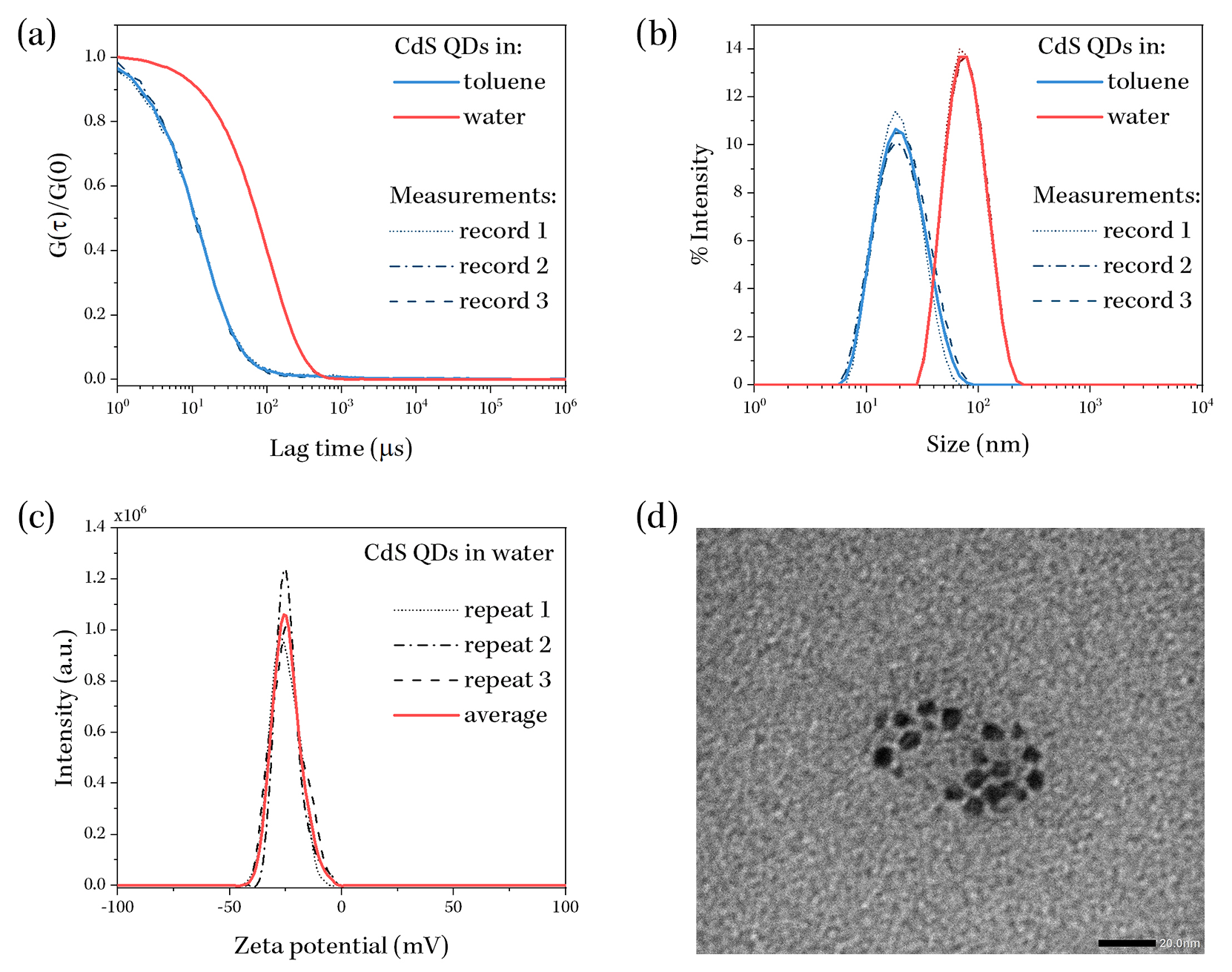
Figure 4(a) shows the normalized correlation curves of individual measurements as well as their average for CdS QDs dispersed in toluene and in water, after phase transfer. From this data, it is clear that correlation in scattered signals persists for longer lag times for WbQDs, which is indicative of their increased hydrodynamic size as compared to QDs dispersed in toluene. This was confirmed by the cumulant analysis, which yielded mean QD sizes of nm and nm pre- and post-phase transfer, with the population standard deviations of nm and nm respectively. The size distribution data, shown in Figure 4(b) further confirms these results - the hydrodynamic size of WbQDs increases strongly compared to their predecessors dispersed in toluene. It is important to note that the observed large increase in the size of the QDs (approx. 3 fold) is due to both their increased physical size, owing to the additional OA layer around each QD, but also due to the emergence of charges on their surfaces which interact with the surrounding polar water molecules. It is known that the electrostatic force between charged particles and the polar water molecules contributes to the friction experienced by the particle when diffusing, resulting in larger effective hydrodynamic sizes [49, 50]. We confirmed the existence of surface charges on the phase-transferred QDs by measuring their zeta potential using DLS. Figure 4(c) shows the results of these measurements for the WbQD sample. Analysis of these results (performed automatically by the Zetasizer software) yielded a mean zeta-potential value of
| (3.12) |
for the sample. The polarity of this potential is consistent with that expected from QD functionalisation with the OA molecules. Furthermore, particles with zeta potentials of more than mV or less than mV tend to repel each other [51]; therefore, the value measured here is sufficient to ensure efficient dispersion of QDs via electrostatic interactions between them and hence to ensure the colloidal stability of WbQDs.
Finally, the physical size of the QDs’ semiconductor core was determined using Transmission Electron Microscopy (TEM). A representative TEM image obtained from the measurements is shown in Fig. 4, which was analysed to obtain the mean QD diameter of nm. This value is comparable to that obtained from Eq. 3.5, and any discrepancy between the two can be accounted for by the limited numbers of QDs that could be imaged by the TEM technique.
3.3 Effects of sample ageing
Given that a typical neutrino experiment requires several years to acquire data, the degradation in scintillating properties of the fluors, caused by ageing, is a concern. For this reason, we performed some preliminary studies of the properties of WbQD samples over the span of 2 years. Three WbQD samples were prepared by the phase-transfer method for these purposes, in each of the years of 2021, 2022 and 2023, and their spectral and physical properties were measured.
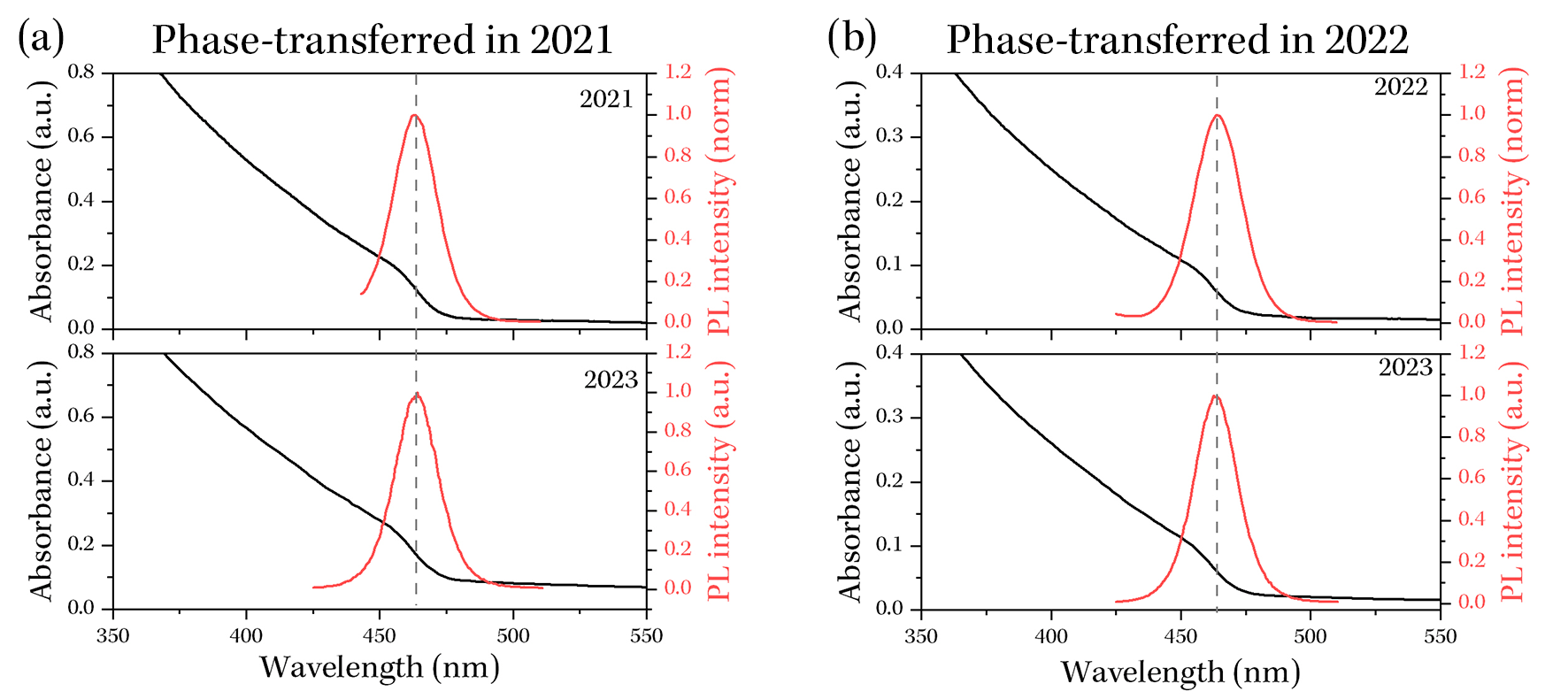
Figure 5 compares the optical properties of the 2021 and 2022 WbQD samples, measured immediately after phase transfer in respective years and then in 2023, corresponding to 2 and 1 years of aging of the samples respectively. As can be seen from this figure, the photoluminescence and absorbance properties of these samples varied very little over time, with no observable changes or shifts in their spectral features. In all cases, the photoluminescence peaked in the nm range and any variations in the position of the peak could easily be explained by the changes in the environmental and experimental conditions under which measurements were performed, such as room temperature and humidity, as well as the calibration of the spectrometer’s detector. That being said, for both samples, a strong change in the PLQY was observed from the moment of their initial preparation and measurements in 2023, with the PLQY of the 2021 sample decreasing from to in the span of two years, whereas the PLQY of the 2022 sample increased from to over a 12 month period.
| Sample | PLQY (%) | (nm) | (mV) | |
| 2021 WbQD | as prepared | not available | ||
| in 2023 | ||||
| 2022 WbQD | as prepared | |||
| in 2023 | ||||
| 2023 WbQD | as prepared |
In view of the lack of any changes to the optical spectra of the samples, it is reasonable to conclude that these result imply that no long-term changes occur to the core of the QDs, since any such changes would cause shifts of spectral features; however, changes to the surface of the QDs - and namely the OA stabilizing shell - do occur, and they lead to changes in the PLQY of the QDs. Evidence to support the latter conclusion can be gained from the measurements of the WbQD samples’ hydrodynamic sizes and zeta potentials () over time. Table 3 shows the variations in these parameters over a period of 2 years. From the data presented in the table, no clear trend can be identified in the changes to the PLQY of the WbQDs samples: although the 2021 sample shows a reduction of PLQY after 2 years, the 2022 sample shows an opposite effect. On the other hand, consistently decreases over time for both samples, with no indication of agglomeration of colloidal QDs. Since the reduction is only a few nm, this is most likely due to the reorganization of OA the surface of WbQDs rather than their detachment/loss. Such surface reorganization would result in a change of potential of the WbQDs, which was observed for the 2022 sample. However, since such data was not available for the as-prepared 2021 sample, the trend remains to be confirmed. In summary, whilst it is not clear what causes the changes in the properties of the WbQD samples from Table 3, three things are confirmed: the hydrodynamic radius of the particles reduces gradually over time, WbQDs remain colloidally and optically stable over time, and no observable aggregation/precipitation of the sample over periods of time as large as two years post-transfer.
4 Quantum Dots water solution cosmic ray test
4.1 Setup
The cosmic ray (atmospheric muon) measurement was performed in 2022 using the 2022 water-based CdS QDs sample. Cosmic rays were tagged by two cosmic ray taggers made with Hamamatsu 6 mm2 silicon photo-multiplier (SiPM) C13367-6050EA [52] and 100 cm cm EJ-200 plastic scintillator [53]. Fig. 6 shows the setup. The QDs sample was placed in front of the North Night Vision Technology (NNVT) negative high-voltage 3-inch photo-multiplier tube (PMT) [54] in a dark box. The signal and high voltage cables from the PMT were fed from the dark box to be connected with electronics outside. The signal cable was connected to the CAEN DT5720 250 MS/s digitizer [55]. The signals from the cosmic ray taggers were processed using two discriminators connected to a coincidence unit, which produced logic signals to trigger the digitizer.
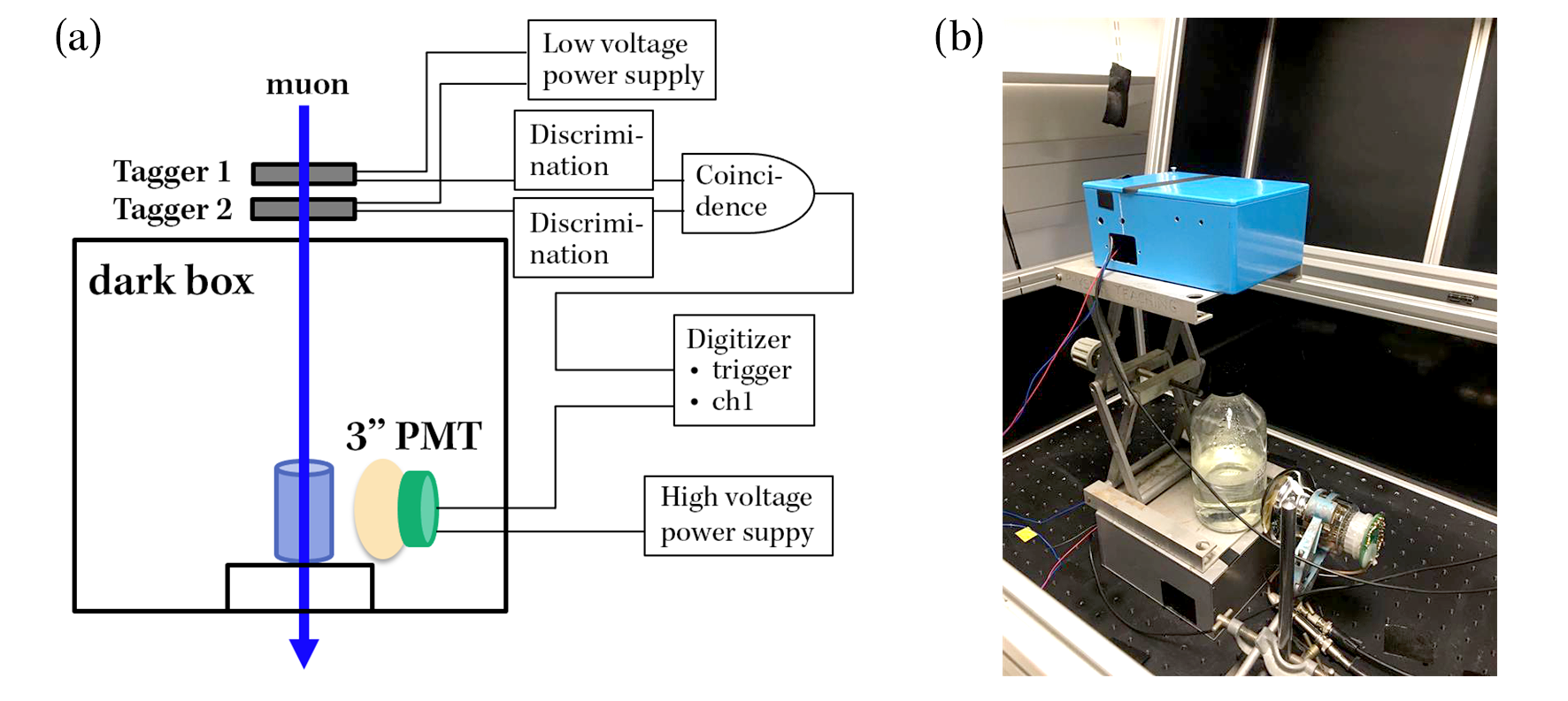
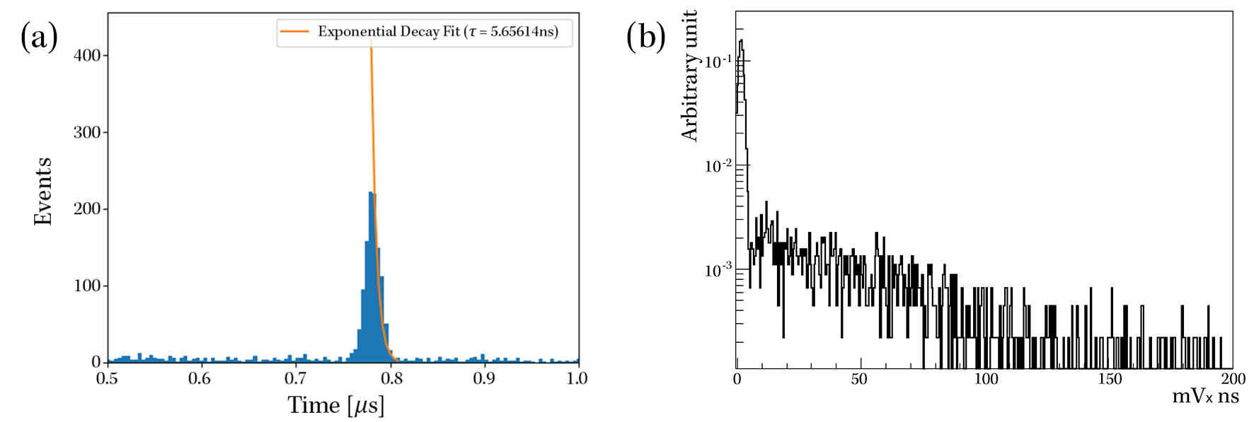
Fig. 7 shows raw data distributions. The left plot shows the timing distributions. Here, "t=0" is defined by the cosmic ray trigger signal. Since the QDs sample has only a few cm depth in a bottle, it takes ns for cosmic rays to penetrate it. Thus the time distribution represents decay time of QDs by excitation due to cosmic rays. The fit gives the decay time of QDs to be 6 ns, which is not atypical for a CdS QD sample [56, 57, 58]. The right plot of Fig. 7 shows the charge distribution in the unit of . The wide charge distribution indicates a wide range in both the angles at which the cosmic rays enter the QD sample, and in their propagation distances once in the water media, resulting in deposition of a wide range of energies across the detected events. To better understand the measured data and their relation to the geometrical factors of the experimental set-up, a simulation was developed to estimate the numbers of photons of different energies that could be produced by the cosmic rays passing through our WbQD scintillating material.
4.2 Simulation
The simulation of this test was created in GEANT4 [59], which is a software toolkit for simulating physics processes relating to particle propagation in matter utilising C++ object-oriented code. The Geant4 toolkit contains applications and core physics header files, which serves as the fundamental framework from which source files and other header files can be created. These source files can be used to construct a vast array of experiments, where the properties and the desired physical processes of the experimental apparatus can be tailored and produced, which in our case concerns a WbQD. In this way, and with the inclusion of the detectors, the behaviour of the particles as they interact with the detector material can be simulated.
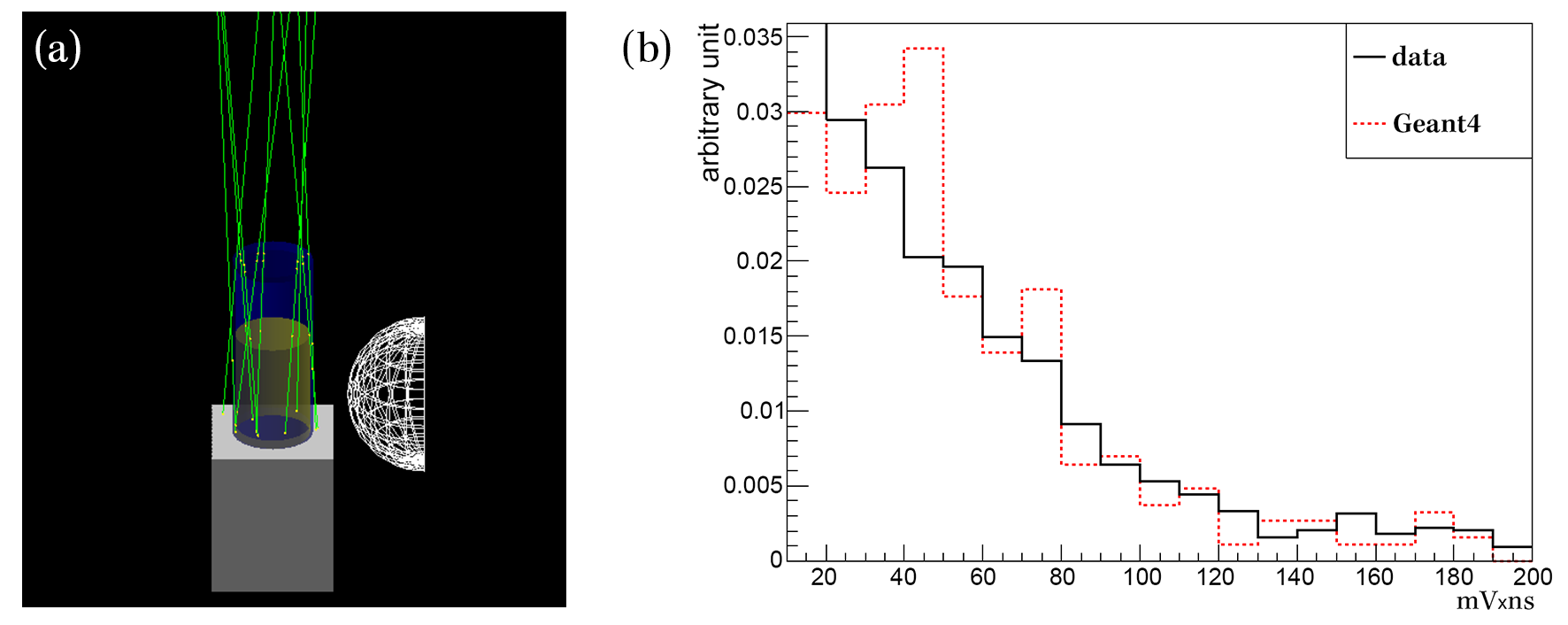
The geometry created is a simplified replica of the physical setup taking into consideration the distance, size, and shape of the objects. Fig. 8 left shows an event display of our simulation in GEANT4. The PMT hemispherical glass surface is set as the sensitive detector. The WbQD material is treated as a liquid scintillator with a similar emission spectrum, located inside of a cylindrical glass. Utilising the necessary GEANT4 libraries such as electromagnetic processes, the physics processes can be constructed. Muons are generated randomly and they penetrate the WbQDs volume and produce scintillation photons that are detected by the PMT. The trajectories of muons are calculated by the solid angle defined by 2 cosmic ray taggers, and for simplicity all muons are 4 GeV in this simulation. In the simulation, we do not simulate PMT responses, instead, all photons which hit the surface of the PMT are registered with constant efficiency and the gain is at V according to our calibration.
Fig. 8 right shows data-MC comparison of their charge distributions above the pedestal. Here, the number of scintillation photons in the simulation is determined by adjusting the two distributions to match. A good agreement between experimental and simulated data is achieved around photons/MeV, although with some deviations in the low charge region. This is likely due to the contamination of background events in the data, and further investigations are required to confirm this. The simulation adopts a simple geometry, and objects in the dark box, except those shown in Fig. 8, left, are ignored. However, we believe they do not affect the time and charge distributions much, as major signals are direct light travelling from the WbQD scintillator to the front of the PMT. Importantly, this is the first demonstration of the water-based quantum dots liquid scintillator with radiation detection; furthermore, the number of photons recorded here is promising for applications in particle and nuclear physics.
5 Discussions
The results of the simulations indicate that several thousands of photons are created by CdSe/ZnS QDs per 1 MeV of energy deposited, which is a surprisingly high number considering the low concentration of the dispersion ( nM). For reference, a typical organic fluor in a liquid scintillator, such as 2,5-diphenyloxazole (PPO), would be present at a concentration of mM and would be expected to produce around 10,000 photons from 1 MeV energy deposit. The larger physical cross-sections of the QDs ( in our case), when compared to organic fluors (), would be expected to somewhat mitigate for the lower concentration, but the differences in sizes are not sufficient to fully account for the observed photon rates. Furthermore, the PLQY of our QDs is also lower to that of a typical organic fluor ( for WbQD sample and for PPO).
It is important to note that the nature of interactions that leads to the excitation of electron-hole pairs in the fluor is the same for both cases; however, the efficiency with which the initial excitation of electron-hole pairs can be achieved, the energy of the created pairs, and/or the processes that proceed this initial excitation are different for inorganic materials such as quantum dots. For example, the much larger number of atoms that constitute nanoparticulate fluors such as QDs, result in much higher electronic state densities being available for the transfer of energy when compared to organic fluors. As a result, excitations with a wide range of energies greater than the bandgap are possible in QDs, with those of energies leading to the generation of deep holes and hot electrons in the valence and conduction bands respectively. Furthermore, relaxation of these excitations, via scattering, Auger and thermalization processes, generates either single or multiple optically-gapped excitons at the band edge. Importantly, excitation of bi-excitons has been shown to be efficient in QDs and it can lead to the emission of two coincidental photons. In contrast, organic fluors do not offer this capability. High scintillation yields from QDs-based scintillators have been reported by other groups [60, 61, 62], suggesting that the optoelectronic properties of QDs offer advantages over organic compounds as scintillating materials. These results open up applications of liquid QD-based scintillators in particle physics [13, 14, 15].
6 Conclusions
In this article, we investigate the potential of water-based quantum dots liquid scintillator for particle physics application. We first performed a phase transfer to CdS/ZnS QDs from hydrophobic phase to hydrophilic phase. Absorbance and emission peaks for CdS QDs are nm and nm for both in toluene and in water. These values fit in the range provided by the manufacturer, and we confirmed optical properties are mostly preserved in water-based quantum dots.
Photoluminescence quantum efficiency of the 2023 WbQD sample is measured to be . The value is lower than that provided by the manufacture, suggesting the loss due to phase transfer. Measured PLQY of other samples varies, and values go up or down over time and further study is needed to understand.
Hydrodynamic size of the toluene and water QD samples are measured to be nm and nm. TEM confirms the QD diameter to be nm, and a consistent diameter is obtained from an empirical equation using the absorption spectrum. Large WbQD size is attributed to the oleic acid layer with polar water molecules. This layer takes over 1 year to reorganize itself and it is stable over 2 years. Furthermore, there is no sign of agglomeration. The phase transfer has a significant impact for both PLQY and QD size, and it is a subject to be optimized in future.
Finally, we performed scintillation measurements from the cosmic rays (atmospheric muons). The time distribution confirms the fast decay ( nm) of QD is preserved in water, and charge distribution shows relatively high scintillation yield. More quantitative studies are needed to confirm these numbers.
In conclusion, water-based quantum dots liquid scintillator is a promising new technology for particle physics. We have a special attention for a water solution of quantum dots to detect inverse beta decay processes from nuclear fission reactor neutrinos, where numerous programs are running all over the world [24]. Although liquid scintillator detectors [63, 64, 65, 66, 67, 68, 69, 70, 71, 72, 73, 74] are popular base designs, other technologies such as solid scintillator [75, 76, 77, 78] are also explored on top of water based detectors [16, 17, 18, 19, 20, 21, 22, 79, 80]. Furthermore, reactor neutrino detection from neutrino-electron elastic scattering [81, 82] and neutrino-nucleus elastic scattering are established [83, 84].
Acknowledgement
We thank Mark Green for provision of characterisation equipment and of his expertise in phase-transfer protocols. We thank Stephen Po, Patrick Stowell, Klaus Suhling, and Lindley Winslow for provision of information and help. This paper is based on research funded by the ‘NuSec’ Nuclear Security Science Network managed by the University of Surrey and funded by the UKRI STFC (ST/S005684/1). Work by JC, SF, DM, AT are supported by the King’s Edge funding via King’s Undergraduate Research Fellowships programme. Work by MT, NB, TK, are supported by the UKRI STFC (ST/W000660/1) and the Royal Society (RGS\R2\180163, IEC\R3\213009). Work by MZ, AR are supported by the UKRI EPSRC (EP/W017075/1) and the Royal Society (URF\R\211023, RF\ERE\231032).
References
- [1] A. I. Ekimov and A. A. Onushchenko, Quantum size effect in the optical-spectra of semiconductor micro-crystals, Soviet Physics Semiconductors-USSR 16 (1982) 775–778.
- [2] A. Rakovich and T. Rakovich, Semiconductor versus graphene quantum dots as fluorescent probes for cancer diagnosis and therapy applications, Journal of Materials Chemistry B 6 (2018) 2690–2712.
- [3] R. Bilan, I. Nabiev and A. Sukhanova, Quantum dot-based nanotools for bioimaging, diagnostics, and drug delivery, ChemBioChem 17 (2016) 2103–2114.
- [4] P. Sun, Z. Xing, Z. Li and W. Zhou, Recent advances in quantum dots photocatalysts, Chemical Engineering Journal 458 (2023) 141399.
- [5] H. Lu, Z. Huang, M. S. Martinez, J. C. Johnson, J. M. Luther and M. C. Beard, Transforming energy using quantum dots, Energy & Environmental Science 13 (2020) 1347–1376.
- [6] D. Tian, H. Ma, G. Huang, M. Gao, F. Cai, Y. Fang et al., A review on quantum dot light-emitting diodes: from materials to applications, Advanced Optical Materials 11 (2023) 2201965.
- [7] “Ultra-slim lcds and quantum-dots enhanced leds enter the market.” https://www.oled-info.com/ultra-slim-lcds-and-quantum-dots-enhanced-leds-enter-market, 2011.
- [8] U. Resch-Genger, M. Grabolle, S. Cavaliere-Jaricot, R. Nitschke and T. Nann, Quantum dots versus organic dyes as fluorescent labels, Nature Methods 5 (2008) 763–775.
- [9] S. E. Létant and T. F. Wang, Semiconductor quantum dot scintillation under ray irradiation, Nano Letters 6 (12, 2006) 2877–2880.
- [10] C. Wang, L. Gou, J. Zaleski and D. Friesel, Zns quantum dot based nanocomposite scintillators for thermal neutron detection, Nuclear Instruments and Methods in Physics Research Section A: Accelerators, Spectrometers, Detectors and Associated Equipment 622 (2010) 186–190.
- [11] R. Bilan, F. Fleury, I. Nabiev and A. Sukhanova, Quantum dot surface chemistry and functionalization for cell targeting and imaging, Bioconjugate Chemistry 26 (04, 2015) 609–624.
- [12] Lesnyak, Vladimir and Gaponik, Nikolai and Eychmüller, Alexander, Colloidal semiconductor nanocrystals: the aqueous approach, Chem. Soc. Rev. 42 (2013) 2905–2929.
- [13] L. Winslow and R. Simpson, Characterizing Quantum-Dot-Doped Liquid Scintillator for Applications to Neutrino Detectors, JINST 7 (2012) P07010, [1202.4733].
- [14] C. Blanco, R. Essig, M. Fernandez-Serra, H. Ramani and O. Slone, Dark matter direct detection with quantum dots, Phys. Rev. D 107 (2023) 095035, [2208.05967].
- [15] M. Doser, E. Auffray, F. M. Brunbauer, I. Frank, H. Hillemanns, G. Orlandini et al., Quantum Systems for Enhanced High Energy Particle Physics Detectors, Front. in Phys. 10 (2022) 887738.
- [16] M. Yeh, S. Hans, W. Beriguete, R. Rosero, L. Hu, R. L. Hahn et al., A new water-based liquid scintillator and potential applications, Nucl. Instrum. Meth. A 660 (2011) 51–56.
- [17] J. R. Alonso et al., Advanced Scintillator Detector Concept (ASDC): A Concept Paper on the Physics Potential of Water-Based Liquid Scintillator, 1409.5864.
- [18] Theia collaboration, M. Askins et al., THEIA: an advanced optical neutrino detector, Eur. Phys. J. C 80 (2020) 416, [1911.03501].
- [19] J. Caravaca, B. J. Land, M. Yeh and G. D. Orebi Gann, Characterization of water-based liquid scintillator for Cherenkov and scintillation separation, Eur. Phys. J. C 80 (2020) 867, [2006.00173].
- [20] L. Kneale, S. T. Wilson, T. Appleyard, J. Armitage, N. Holland and M. Malek, Sensitivity of an antineutrino monitor for remote nuclear reactor discovery, 2210.11224.
- [21] M. J. Ford, N. P. Zaitseva, M. L. Carman, S. A. Dazeley, A. Bernstein, A. Glenn et al., Pulse-shape discrimination in water-based scintillators, Nucl. Instrum. Meth. A 1036 (2022) 166854, [2202.07625].
- [22] J. Hecla, A. Bernstein, S. Dazeley and O. A. Akindele, A long path-length optical property measurement device for highly transparent detector media, JINST 18 (2023) P09004.
- [23] J. Chen, B. Yang, C. Li, K. Zheng, K. Žídek and T. o. Pullerits, Photostability of the oleic acid-encapsulated water-soluble gradient core-shell quantum dots, ACS Omega 2 (2017) 1922–1929, [https://doi.org/10.1021/acsomega.7b00316].
- [24] A. Bernstein, N. Bowden, B. L. Goldblum, P. Huber, I. Jovanovic and J. Mattingly, : Neutrino detectors as tools for nuclear security, Rev. Mod. Phys. 92 (2020) 011003, [1908.07113].
- [25] Raul Calzada, Christopher M. Thompson, Dana E. Westmoreland, Kedy Edme, Emily A. Weiss, Organic-to-aqueous phase transfer of Cadmium Chalcogenide Quantum Dots using a Sulfur-free ligand for enhanced photoluminescence and oxidative stability, Chem. Mater. 28 (2016) 6716–6723.
- [26] Sanjayan C G, Jyothi Mannekote Shivanna, Jessica D. Schiffman, Sakar Mohan, Srinivasa Budagumpi, R. Geetha Balakrishna, Aqueous,non-polymer-based perovskite Quantum Dots for bioimaging: conserving fluorescenceand long-term stability via simple and robust synthesis, ACS Appl. Mater. Interfaces 14 (2022) 38471–38482.
- [27] Jothirmayanantham Pichaandi, Keith A. Abel, Noah J. J. Johnson, and Frank C. J. M. van Veggel, Long-term colloidal stability and photoluminescence retention of Lead-based Quantum Dots in saline buffers and biological media through surface modification, Chem. Mater. 25 (2013) 2035––2044.
- [28] Martin Rudolf, Urs Alexander Peuker, Phase transfer of agglomerated nanoparticles: deagglomeration by adsorbing grafted molecules and colloidal stability in polymer solutions, Chem. Mater. 25 (2012) 990.
- [29] “Cds/zns core-shell type quantum dots.” https://www.sigmaaldrich.com/GB/en/product/aldrich/900282, 2021.
- [30] Y. Liu, Y. Yang, Y. Tai and Z. Zhang, A Capture-gated Fast Neutron Detection Method, Chin. Phys. C 40 (2016) 076201, [1603.03529].
- [31] Super-Kamiokande collaboration, M. Harada et al., Search for Astrophysical Electron Antineutrinos in Super-Kamiokande with 0.01% Gadolinium-loaded Water, Astrophys. J. Lett. 951 (2023) L27, [2305.05135].
- [32] “Ultrasonic water bath grant xuba3.” https://www.wolflabs.co.uk/laboratory-products/water-baths-sonicating/10039541, 2021.
- [33] “Centrifuge 5804 benchtop centrifuge.” https://online-shop.eppendorf.co.uk/UK-en/Centrifugation-44533/Multipurpose-Centrifuges-1007184/Centrifuge-5804-5804R-PF-240993.html, 2021.
- [34] “Uv-2600i, uv-2700i uv-vis-nir spectrophotometer.” https://www.shimadzu.com/an/products/molecular-spectroscopy/uv-vis/uv-vis-nir-spectroscopy/uv-2600i-uv-2700i/index.html, 2021.
- [35] “Cary eclipse fluorescence spectrometer.” https://www.agilent.com/en/product/molecular-spectroscopy/fluorescence-spectroscopy/fluorescence-systems/cary-eclipse-fluorescence-spectrophotometer, 2021.
- [36] J. X. Xu, Y. Yuan, M. Liu, S. Zou, O. Chen and D. Zhang, Quantification of the photon absorption, scattering, and on-resonance emission properties of cdse/cds core/shell quantum dots: Effect of shell geometry and volumes, Analytical chemistry 92 (2020) 5346–5353.
- [37] Lord Rayleigh F.R.S., XXXIV. On the transmission of light through an atmosphere containing small particles in suspension, and on the origin of the blue of the sky, London Edinburgh Dublin Philos. Mag. J. Sci. London 47 (1899) 375–384.
- [38] Craig F. Bohren, Donald R. Huffman, Absorption and Scattering of Light by Small Particles. WILEY-VCH Verlag GmbH & Co. KGaA, 1998, 10.1002/9783527618156.ch5.
- [39] DaeGwi Kim, Tetsuya Kuwabara, Masaaki Nakayama, Photoluminescence properties related to localized states in colloidal PbS quantum dots, J. Lumin. 119–120 (2006) 214–218.
- [40] Victor I. Klimov, Nanocrystal Quantum Dots. CRC Press, 2010.
- [41] K.H. Ibnaouf, Saradh Prasad, M.S. Al Salhi, A. Hamdan, M.B. Zaman, L. El Mir, Influence of the solvent environments on the spectral features of CdSe quantum dots with and without ZnS shell, J. Lumin. (2014) 369–373.
- [42] Glenn A. Crosby, James N. Demas, Measurement of photoluminescence quantum yields. Review, J. Phys. Chem. 75 (1971) 991–1024.
- [43] Yong-Biao Zhao, Mengxia Liu, Oleksandr Voznyy, Bin Sun, Pei-Cheng Li, Haoting Kung, Olivier Ouellette, Min-Jae Choi, Zheng-Hong Lu, F. Pelayo García de Arquer, Edward H. Sargent, Accelerated solution-phase exchanges minimize defects in colloidal quantum dot solids, Nano Energy 63 (2019) 103876.
- [44] Ping Yang, Guangjun Zhou, Phase transfer of hydrophobic QDs for water-soluble and biocompatible nature through silanization, Mater. Res. Bull. 46 (2011) 2367–2372.
- [45] M. J. Danielle L. Woodall, Andrew K. Tobias, Resolving carrier recombination in cds quantum dots: A time-resolved fluorescence study, Chem. Phys. 471 (2016) 2–10.
- [46] W. W. Yu, L. Qu, W. Guo and X. Peng, Experimental determination of the extinction coefficient of cdte, cdse, and cds nanocrystals, Chemistry of Materials 15 (2003) 2854–2860, [https://doi.org/10.1021/cm034081k].
- [47] Jörg Stetefeld, Sean A. McKenna, Trushar R. Patel, Dynamic light scattering: a practical guide and applications in biomedical sciences, Biophys. Rev. 8 (2016) 409–427.
- [48] “Dynamic light scattering: An introduction in 30 minutes.” https://www.malvernpanalytical.com/en/learn/knowledge-center/technical-notes/tn101104dynamiclightscatteringintroduction.
- [49] J. Hubbard and L. Onsager, Dielectric dispersion and dielectric friction in electrolyte solutions. I., The Journal of Chemical Physics 67 (08, 2008) 4850–4857, [https://pubs.aip.org/aip/jcp/article-pdf/67/11/4850/11113849/4850_1_online.pdf].
- [50] P. G. Wolynes, Dynamics of electrolyte solutions, Annual Review of Physical Chemistry 31 (1980) 345–376, [https://doi.org/10.1146/annurev.pc.31.100180.002021].
- [51] F. Hasani Bijarbooneh, Y. Zhao, J. H. Kim, Z. Sun, V. Malgras, S. H. Aboutalebi et al., Aqueous colloidal stability evaluated by zeta potential measurement and resultant tio2 for superior photovoltaic performance, Journal of the American Ceramic Society 96 (2013) 2636–2643, [https://ceramics.onlinelibrary.wiley.com/doi/pdf/10.1111/jace.12371].
- [52] “Mppc module c13367-6050ea.” https://www.hamamatsu.com/jp/en/product/type/C13367-6050EA/index.html, 2021.
- [53] “General purpose ej-200, ej-204, ej-208, ej-212.” https://eljentechnology.com/products/plastic-scintillators/ej-200-ej-204-ej-208-ej-212, 2021.
- [54] “Nnvt photomultiplier tube 2031.” http://yskjnj.com/product/detail-23.html, 2021.
- [55] “Dt5720 2/4 channel 12 bit 250 ms/s digitizer.” https://www.caen.it/products/dt5720, 2021.
- [56] F. Wu, J. Zhang, R. Kho and R. Mehra, Radiative and nonradiative lifetimes of band edge states and deep trap states of cds nanoparticles determined by time-correlated single photon counting, Chemical Physics Letters 330 (2000) 237–242.
- [57] W. Gong, Z. Zheng, J. Zheng, W. Gao, X. Hu and X. Ren, Luminescence study of water-soluble cds nanoparticles produced by femtosecond laserablation at high laser fluence, J. Phys. Chem. C 112 (2008) 9983–9987.
- [58] K. Santhosh, S. Patra, S. Soumya, D. C. Khara and A. Samanta, Fluorescence quenching of cds quantum dots by 4-azetidinyl-7-nitrobenz-2-oxa-1,3-diazole: A mechanisticstudy, ChemPhysChem 12 (2011) 2735–2741.
- [59] S. A. . et al., Geant4—a simulation toolkit, Nuclear Instruments and Methods in Physics Research Section A: Accelerators, Spectrometers, Detectors and Associated Equipment 506 (2003) 250–303.
- [60] S. Oktyabrsky, M. Yakimov, V. Tokranov and P. Murat, Integrated Semiconductor Quantum Dot Scintillation Detector: Ultimate Limit for Speed and Light Yield, IEEE Trans. Nucl. Sci. 63 (2016) 656–663.
- [61] K. M. McCall, K. Sakhatskyi, E. Lehmann, B. Walfort, A. S. Losko, F. Montanarella et al., Fast neutron imaging with semiconductor nanocrystal scintillators, ACS Nano 14 (2020) 14686–14697.
- [62] L. Lu, M. Sun, T. Wu, Q. Lu, B. Chen and B. Huang, All-inorganic perovskite nanocrystals: next-generation scintillation materials for high-resolution x-ray imaging, Nanoscale Adv. 4 (2022) 680–696.
- [63] A. Bernstein, N. S. Bowden, A. Misner and T. Palmer, Monitoring the Thermal Power of Nuclear Reactors with a Prototype Cubic Meter Antineutrino Detector, J. Appl. Phys. 103 (2008) 074905, [0804.4723].
- [64] T. Classen, A. Bernstein, N. S. Bowden, B. Cabrera-Palmer, A. Ho, G. Jonkmans et al., Development of an advanced antineutrino detector for reactor monitoring, Nucl. Instrum. Meth. A 771 (2015) 139–146.
- [65] NUCIFER collaboration, G. Boireau et al., Online Monitoring of the Osiris Reactor with the Nucifer Neutrino Detector, Phys. Rev. D 93 (2016) 112006, [1509.05610].
- [66] KamLAND collaboration, A. Gando et al., Constraints on from A Three-Flavor Oscillation Analysis of Reactor Antineutrinos at KamLAND, Phys. Rev. D 83 (2011) 052002, [1009.4771].
- [67] Double Chooz collaboration, H. de Kerret et al., Double Chooz measurement via total neutron capture detection, Nature Phys. 16 (2020) 558–564, [1901.09445].
- [68] Daya Bay collaboration, F. P. An et al., Precision Measurement of Reactor Antineutrino Oscillation at Kilometer-Scale Baselines by Daya Bay, Phys. Rev. Lett. 130 (2023) 161802, [2211.14988].
- [69] JUNO collaboration, A. Abusleme et al., JUNO physics and detector, Prog. Part. Nucl. Phys. 123 (2022) 103927, [2104.02565].
- [70] RENO collaboration, S. G. Yoon et al., Measurement of reactor antineutrino flux and spectrum at RENO, Phys. Rev. D 104 (2021) L111301, [2010.14989].
- [71] NEOS collaboration, Y. J. Ko et al., Sterile Neutrino Search at the NEOS Experiment, Phys. Rev. Lett. 118 (2017) 121802, [1610.05134].
- [72] PROSPECT, (PROSPECT Collaboration)* collaboration, M. Andriamirado et al., Final Measurement of the U235 Antineutrino Energy Spectrum with the PROSPECT-I Detector at HFIR, Phys. Rev. Lett. 131 (2023) 021802, [2212.10669].
- [73] STEREO collaboration, H. Almazán et al., STEREO neutrino spectrum of 235U fission rejects sterile neutrino hypothesis, Nature 613 (2023) 257–261, [2210.07664].
- [74] NEUTRINO-4 collaboration, A. P. Serebrov et al., First Observation of the Oscillation Effect in the Neutrino-4 Experiment on the Search for the Sterile Neutrino, Pisma Zh. Eksp. Teor. Fiz. 109 (2019) 209–218, [1809.10561].
- [75] S. Oguri, Y. Kuroda, Y. Kato, R. Nakata, Y. Inoue, C. Ito et al., Reactor antineutrino monitoring with a plastic scintillator array as a new safeguards method, Nucl. Instrum. Meth. A 757 (2014) 33–39, [1404.7309].
- [76] NuLat collaboration, C. Lane et al., A new type of Neutrino Detector for Sterile Neutrino Search at Nuclear Reactors and Nuclear Nonproliferation Applications, 1501.06935.
- [77] A. Haghighat, P. Huber, S. Li, J. M. Link, C. Mariani, J. Park et al., Observation of Reactor Antineutrinos with a Rapidly-Deployable Surface-Level Detector, Phys. Rev. Applied 13 (2020) 034028, [1812.02163].
- [78] SoLid collaboration, Y. Abreu et al., SoLid: a short baseline reactor neutrino experiment, JINST 16 (2021) P02025, [2002.05914].
- [79] A. Goldsack, Reactor Neutrinos in a Gadolinium-Loaded Super-Kamiokande. PhD thesis, University of Oxford, Oxford U., 2022.
- [80] SNO+ collaboration, A. Allega et al., Evidence of Antineutrinos from Distant Reactors using Pure Water at SNO+, Phys. Rev. Lett. 130 (2023) 091801, [2210.14154].
- [81] TEXONO collaboration, M. Deniz et al., Measurement of Nu(e)-bar -Electron Scattering Cross-Section with a CsI(Tl) Scintillating Crystal Array at the Kuo-Sheng Nuclear Power Reactor, Phys. Rev. D 81 (2010) 072001, [0911.1597].
- [82] A. G. Beda, E. V. Demidova, A. S. Starostin, V. B. Brudanin, V. G. Egorov, D. V. Medvedev et al., GEMMA experiment: Three years of the search for the neutrino magnetic moment, Phys. Part. Nucl. Lett. 7 (2010) 406–409, [0906.1926].
- [83] J. Liao, H. Liu and D. Marfatia, Implications of the first evidence for coherent elastic scattering of reactor neutrinos, Phys. Rev. D 106 (2022) L031702, [2202.10622].
- [84] COHERENT collaboration, P. An et al., Measurement of Electron-Neutrino Charged-Current Cross Sections on I127 with the COHERENT NaIE Detector, Phys. Rev. Lett. 131 (2023) 221801, [2305.19594].