Training Small Multimodal Models to Bridge Biomedical Competency Gap: A Case Study in Radiology Imaging
Abstract
The scaling laws and extraordinary performance of large foundation models motivate the development and utilization of such large models in biomedicine. However, despite early promising results on some biomedical benchmarks, there are still major challenges that need to be addressed before these models can be used in real-world applications. Frontier models such as GPT-4V still have major competency gaps in multimodal capabilities for biomedical applications. Moreover, pragmatic issues such as access, cost, latency, and compliance make it hard for clinicians to use privately-hosted state-of-the-art large models directly on private patient data. In this paper, we explore training open-source small multimodal models (SMMs) to bridge biomedical competency gaps for unmet clinical needs. To maximize data efficiency, we adopt a modular approach by incorporating state-of-the-art pre-trained models for image and text modalities, and focusing on training a lightweight adapter to ground each modality to the text embedding space, as exemplified by LLaVA-Med. We conduct a comprehensive study of this approach on radiology imaging. For training, we assemble a large dataset with over 1 million image-text pairs. For evaluation, we propose a clinically driven novel approach using GPT-4 and demonstrate its parity with expert evaluation. We also study grounding qualitatively using attention. For best practice, we conduct a systematic ablation study on various choices in data engineering and multimodal training. The resulting LLaVA-Rad (7B) model attains state-of-the-art results on standard radiology tasks such as report generation and cross-modal retrieval, even outperforming much larger models such as GPT-4V and Med-PaLM M (84B). Training on over 1 million image-text pairs took just two days using a standard 8-A100 cluster, which bodes well for further fine-tuning by clinicians using their own data. LLaVA-Rad is fast and can be run on a single V100 GPU in private settings, offering a promising state-of-the-art tool for real-world clinical applications.
Introduction
Foundation models, which are models trained from a massive amount of unlabelled data using self-supervised learning, can be quickly adapted to various downstream tasks using only a small amount of task-specific labeled data [5, 35]. Due to the costly biomedical data annotation [21, 20], foundation models have become a new paradigm in biomedicine by achieving state-of-the-art results on many applications, including medical question answering [46, 35] and medical image classification [4, 3]. Recently, multimodal generative AI has emerged as an exciting frontier in the biomedical domain, expanding the application scope from single-modality to multi-modality (e.g., text and image), such as visual question answering and radiology report generation [28, 46, 43]. While existing models are still largely evaluated on artificial biomedical benchmarks, their promising performance demonstrates their potential in medical decision support.
However, there are still bottlenecks hindering the progress toward using these foundation models in real-world clinical settings. First, sharing patient data with large foundation models hosted on the cloud is subject to privacy and reproducibility concerns [44]. Therefore, clinicians may prefer to run inference and fine-tuning locally. Second, existing state-of-the-art models are often very large and resource-intensive, which make local deployment challenging. Smaller models offer decreased serving costs and latency, which is of particular importance in resource-constrained settings outside of data centers [50], as well as offer a smaller carbon footprint [45]. While such smaller language models111We refer to small language models as a smaller (¡10 billion parameters) yet still performant counterpart to large language models (LMs) have shown success in text-only domains [15, 33, 27], multimodal model performance still lags behind large models [46, 43]. Third, many state-of-the-art models are inaccessible [50], necessitating the development of effective open-source models for biomedicine. Finally, even the best models are still subject to hallucinating false information and there exist limited methods to reliably verify the correctness of model outputs at scale, especially in the specialized field of biomedicine [10].
To bridge this gap between existing medical foundation models and real-world clinical applications, we have developed LLaVA-Rad, a small multimodal model (SMM) that attains state-of-the-art (SOTA) performance in standard radiology imaging tasks. We focus our study on identifying key findings from chest X-ray (CXR) images, the most commonly performed medical imaging examination. Automatically drafting high-quality radiology reports is a challenging, yet clinically meaningful, task which could directly increase radiologist productivity and potentially improve communication and decrease burnout [26]. Existing frontier models such as GPT-4V still have a large competency gap even on such a fundamental medical application. To develop LLaVA-Rad, we adopt a modular approach by incorporating state-of-the-art open-source pretrained models for image and text modalities, and focusing on training a lightweight adapter to ground each modality to the text embedding space, as exemplified by LLaVA-Med [28].
For training, we assemble a large dataset comprising 1,034,660 radiology image-report pairs from 8 diverse sources. Some data sources only contain structured labels of key findings, in which case we use GPT-4 to synthesize the report based on the labels.
For evaluation, we report standard metrics such as n-gram-based BLEU and ROUGE, as well as factuality checks based on CheXpert and RadGraph [42, 24]. Additionally, we propose G-Rad, a novel factuality evaluation method based on GPT-4. Compared to existing automatic metrics, we show that G-Rad is more consistent with error quantification by practicing radiologists, thus demonstrating the potential of using GPT-4 for evaluation in a manner that is both scalable and highly relevant to medical practice.
To establish best practice for biomedical multimodal learning, we conduct a systematic ablation study on various choices in data engineering and multimodal training. The resulting LLaVA-Rad (7B) model attains state-of-the-art results on standard radiology tasks such as report generation and cross-modal retrieval, even outperforming much larger models such as GPT-4V and Med-PaLM M (84B) [46]. E.g., LLaVA-Rad outperforms GPT-4V on G-Rad by more than four-fold.
LLaVA-Rad is fast and can be run on a single V100 GPU in private settings, offering a promising state-of-the-art tool for real-world clinical applications. LLaVA-Rad training is very efficient, taking just two days on over 1 million image-text pairs using a standard 8-A100 cluster. This means that clinicians can further efficiently fine-tune the model as needed using their private data. By examining the model weights, we found that LLaVA-Rad can ground key regions of abnormalities to generated words in the output report, which signals future opportunities to synergize with latest progress in biomedical segmentation and grounded report generation.
In sum, we develop LLaVA-Rad, a lightweight yet high-performing radiology multimodal model for clinical applications. The promising performance of LLaVA-Rad shows that its underlying modular approach can effectively and efficiently bridge the biomedical competency gap in existing frontier models, enabling clinical access with limited computational resources. GPT-4 is used extensively in data processing, synthetic training data generation, and complex evaluation, demonstrating how frontier LLMs can play a pivotal role in training the next generation of biomedical multimodal models.
Results
Overview of LLaVA-Rad
LLaVA-Rad represents an emerging paradigm in exploring SMMs, following the proliferation of small language models (SLMs). Despite its compact size, which is over an order of magnitude smaller than prior state-of-the-art models such as Med-PaLM M, LLaVA-Rad attains state-of-the-art performance on standard radiology tasks. This bodes well for potential clinical applications with limited computational resources.
Our intuition for designing LLaVA-Rad is that a lightweight, specialized SMM can be efficiently developed by decomposing training into unimodal pretraining on individual modalities followed by lightweight cross-modal learning focusing on a small adapter to ground a non-text modality to the text embedding space. For training, we collect one million pairs of fully de-identified CXR images and associated radiology reports from 8 diverse datasets [23, 40, 13, 36, 16, 25, 6]. These de-identified image-text pairs were sourced from approximately 476,565 patients across Oceania and four continents, spanning Asia, Europe, North America, and South America. The diversity of this data facilitates pretraining of robust unimodal models (image encoder in this case) and cross-modal adapters (grounding image to text).
| Dataset | Patients | Images | Label | Text Length | Pre-training | ||
| Types | Min | Max | Avg. | Image-Text Pairs | |||
| Synthetic Findings | |||||||
| CheXpert [23] | |||||||
| BraX [40] | |||||||
| CandidPTX [13] | |||||||
| VinDR [36] | |||||||
| JF Healthcare [16] | |||||||
| Real-World Findings | |||||||
| MIMIC-CXR [25] | – | ||||||
| PadChest [6] | – | ||||||
| Private data | – | 3 | 157 | 47.9 | |||
| Total: | |||||||
LLaVA-Rad can generate radiology report findings given a CXR. Its training is comprised of three stages: a pre-training stage, an alignment stage, and a fine-tuning stage. In the first stage of pre-training, we train a domain-specific vision encoder by using one million pairs of CXR images and associated radiology reports. Since CXR images are often published with a limited number of associated findings or image labels instead of a complete report, we used GPT-4 to derive a set of rule based heuristics that enable us to obtain a synthetic report based on published image labels. Alternatively, reports may be available in other languages, such as the PadChest reports which are available in Spanish, for which we leverage GPT-4 to translate them into English. We also exploit GPT-4 to extract findings from reports when the finding section cannot be reliably extracted using existing rule-based heuristics [25]. These diverse datasets offer us a robust and effective vision encoder for embedding CXR images with the consideration of the associated radiology reports. In the second stage of alignment, we align this pre-trained vision encoder with radiology report findings by training a conditional generative model that generates findings given an input CXR. In this alignment stage we provide only the CXR as the input, without any associated instructions or additional patient information. As noted by other works [29, 28] and also demonstrated by our ablation studies, this strategy can substantially improve the alignment by forcing the decoder model to focus on the images. In the third stage, we fine-tune the model to generate the findings according to both the indication for the exam in addition to the image, more closely reflecting a real-world setting. LLaVA-Rad exploits an efficient technique LoRA [18] for fine-tuning, thus substantially reducing the computational time required for this stage. We further reduce the computational time by only training LLaVA-Rad using MIMIC-CXR data instead of the entire 1 million image-text pairs in the second and third steps since reports in MIMIC-CXR have higher quality. As a result, the three stages of LLaVA-Rad can be finished in 11 hours, 4 hours, and 16 hours respectively using 8 A100 GPUs.
Evaluating LLaVA-Rad using existing report generation benchmarks
We first evaluated LLaVA-Rad on the widely-used radiology report generation benchmark MIMIC-CXR (Figure 2) using metrics assessing lexical similarity and factual accuracy. In particular, lexical similarity metrics, such as BLEU and ROUGE, are used in traditional NLP to assess the model’s ability to produce contextually and stylistically aligned output. On the other hand, factual accuracy metrics, including CheXbert-based and RadGraph-based F1 [42, 24] scores are more clinically relevant because they gauge the extent to which the generated reports accurately reflect imaging findings. A high-quality radiology report necessitates both coherence and factual accuracy, underscoring the importance of evaluating these two types of metrics to ensure the report is both intelligible and clinically precise. Any inaccuracies in the report can potentially lead to adverse patient outcomes, emphasizing the critical need for factual correctness.
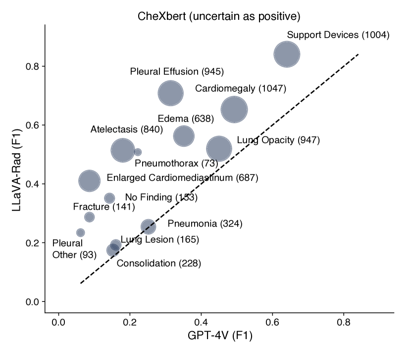
We found that LLaVA-Rad achieved superior performance on both groups of metrics. When compared to other models of equivalent complexity (7B parameters), such as LLaVA-Med [28], CheXagent [8] and MAIRA-1 [22], LLaVA-Rad demonstrates significant advancements in performance across all evaluated metrics. Furthermore, LLaVA-Rad is more efficient than the current overall leading model, Med-PaLM M [46], with an order of magnitude fewer parameters. This efficiency does not come at the cost of effectiveness; LLaVA-Rad outperforms Med-PaLM M in the most important existing lexical similarity and factual correctness metrics for radiology text (ROUGE-L and F1-RadGraph [53]). A more detailed evaluation (Table 4) shows that Med-PaLM M marginally surpasses LLaVA-Rad by F1-5 CheXbert metrics, which assess only a small subset of 5 potential abnormalities, and the performance gap is minimal. We attribute the promising performance of LLaVA-Rad to the continued training on a large-scale radiology dataset. The efficiency and the high degree of factual and lexical precision of LLaVA-Rad demonstrate its clinical implication in real-world applications where large models are computationally too costly.
The key idea of LLaVA-Rad is to conduct pre-training using a large and diverse dataset. To verify the effectiveness of this idea in generating aligned vision and language representations, we examined the learned image encoder in LLaVA-Rad by comparing the performance of using it for retrieval against the image encoders from LLaVA and LLaVA-Med. We observed that LLaVA-Rad attained the best results on both image-to-text and text-to-image retrieval, indicating the high quality of its image encoder by training on one million text-image pairs (Fig. 2C). Moreover, LLaVA-Med performed better than LLaVA, suggesting that better performance can be gained as better performance as increasing domain specialization is performed.
Evaluating LLaVA-Rad using G-Rad, a GPT-based evaluation system
Because existing automated report evaluation methods might be biased to pre-defined conditions and have limited correlation with expert assessments [51], we explore the utility of an LLM-based evaluation system, which has shown success in other domains [31, 47, 14], to more comprehensively evaluate LLaVA-Rad. Specifically, we employ GPT-4 as an evaluator to count how many times the evaluated report presents each of the following six error types, as per [51]: false positive finding, omission of finding, incorrect location/position of finding, incorrect severity of finding, mention of comparison that is not present in the reference report, omission of comparison describing a change from a previous study. For each specific error, we further instruct GPT-4 to distinguish clinically significant and clinically insignificant errors.
We first assessed the rigorousness of G-Rad by examining its consistency with expert scoring. To this end, we exploited the ReXval dataset [52], which contains annotations from 6 board certified radiologists on 200 pairs of ground-truth reports from MIMIC-CXR and AI-generated reports. Each radiologist provides counts of each of the 6 aforementioned error types, also discriminating between clinically significant and insignificant errors. We found that GPT-4-based and GPT-4T-based evaluations Figure 3B were highly correlated with expert scoring by achieving Kendall’s Tau b correlations greater than 0.75 for total errors and greater than 0.70 for clinically significant errors. In contrast, none of the existing preferred metrics (ROUGE-L metric, RadGraph-F1, and RadCliQ) was able to obtain a correlation greater than 0.57. Moreover, we found that a similar evaluation system using GPT-3.5T, a less capable model compared to GPT-4, attains a much lower association with expert scoring due to an overestimation of the number of total and clinically significant errors, demonstrating the difficulty of automatically scoring radiology reports.
We also perform a head-to-head comparison of the calculation of total errors as determined by GPT-4T with manual expert ratings in a leave-one-rater-out fashion. Figure 3A summarizes the results of this comparison, which on average shows a mean absolute difference (MAD) of 0.71 between the left-out rater and the average of the remaining ones, whereas GPT-4T has on average 0.55 MAD. We find that the MAD between GPT-4T G-Rad score and the left-in expert average is smaller compared to the left-out expert in 3/6 cases (P<0.001), and not significantly different (P>0.05) in the remaining 3/6 cases. Altogether, we find that GPT-4T is indistinguishable from expert raters in calculating total number of errors, increasing our confidence in using this proposed automated metric as a new evaluation method that directly aligns with expert opinions.
After assuring the effectiveness of the GPT-based metric, we evaluated the performance of LLaVA-Rad on the held-out MIMIC-CXR test set using G-Rad (Figure 3B. Similar to our observation using existing metrics, LLaVA-Rad outperforms publicly available report generation models, generating fewer clinically significant and total errors compared to GPT-4V and CheXagent. Moreover, by comparing models within the LLaVA family (e.g., LLaVA-Rad, LLaVA-Med, LLaVA), we observed that fewer errors are made in the generated reports as increasing domain specialization is performed. In particular, LLaVA-Rad generates fewer errors than LLaVA-Med, a LLaVA model tailored to medicine, and LLaVA-Med generates fewer errors than the general-domain model LLaVA. This suggests a trade-off between domain-specific performance and broad applicability, supporting our intuition of developing LLaVA-Rad by training a general model using large amounts of domain-specific data. Finally, to determine the clinical utility of LLaVA-Rad, we propose to use the percentage of error-free reports to track the overall performance of report-generation models. A higher percentage of error-free reports increases the utility of a report generation model, given that it directly reflects the number of reports that require little to no radiologist modification following automated generation. Notably, LLaVA-Rad has the highest percentage of error-free reports, with 6.79% reports free of clinically significant errors, and 2.58% free of errors.
Analyzing Components of LLaVA-Rad Using Ablation and Case Studies
Conducting thorough ablation studies for LLMs is often intractable due to the costly training of multiple variants. In contrast, the small size of LLaVA-Rad enables us to efficiently conduct ablation studies that explain the promising performance of LLaVA-Rad, shedding light on the design of larger foundation models. We compared LLaVA-Rad with 8 variants described in Table 5. In Figure 4, we investigate two key technical ideas used in LLaVA-Rad: the effect of pre-training a domain-specific image encoder using 1M diverse CXR image-text pairs and the effect of using GPT-4 to augment and organize the data. Firstly, to understand the effect of pre-training an image encoder, we compare LLaVA-Rad with three increasingly domain-specific variants: an image encoder from OpenAI CLIP, an image encoder from BiomedCLIP, and an image encoder from BiomedCLIP but continually pre-trained using MIMIC-CXR only. We found that the MIMIC-CXR-based image encoder outperforms the other two variants, indicating the effectiveness of training a domain-specific image encoder. BiomedCLIP-CXR-1M substantially outperforms the MIMIC-CXR-based image encoder, necessitating the pre-training using large-scale diverse CXR datasets. Secondly, we studied the effect of using GPT-4 to process and augment the MIMIC-CXR report data. Table 2 summarizes the data that LLaVA-Rad uses for training in the second and the third stage. It is a combination of rule-based and GPT-structured data. We compare LLaVA-Rad with a variant that only uses rule-based data and a variant that only uses GPT-structured data. We found that LLaVA-Rad attains a better performance than both variants, indicating the effectiveness of GPT-4 data augmentation. The variant that only uses GPT-structured data outperforms the one that only uses rule-based data on factual accuracy metrics, reassuring the effectiveness of GPT-4-based structuring in generating clinically precise reports. Lastly, it is expected that rule-based variant outperforms GPT-structured variant on n-gram lexical metrics, because the test data is also from rule-based data. These ablation studies support our intuition that domain-specific data can help us build a small but effective domain-specific model, further revealing key components for designing larger-scale models.
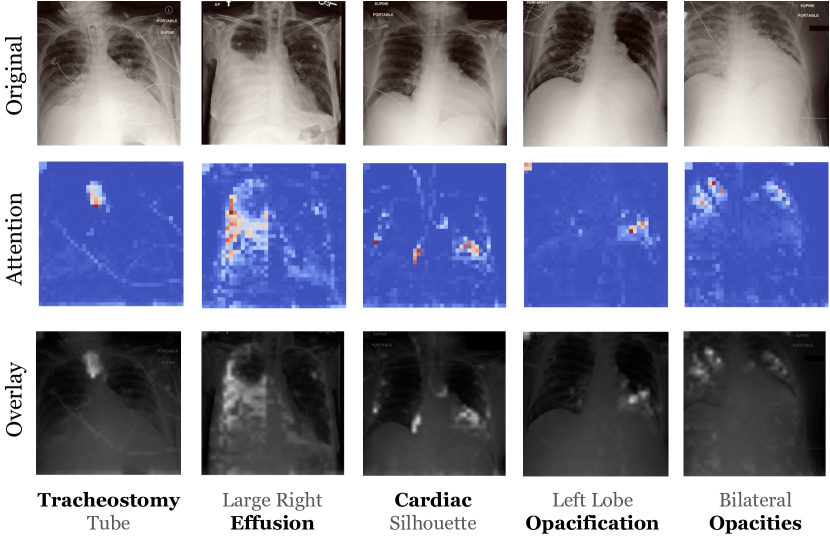
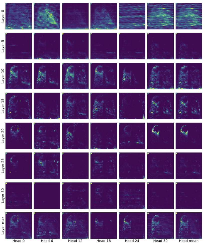
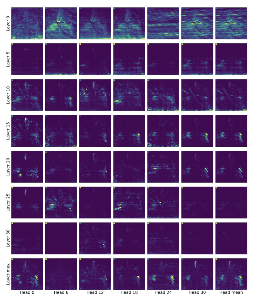
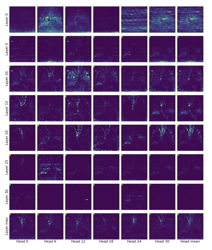
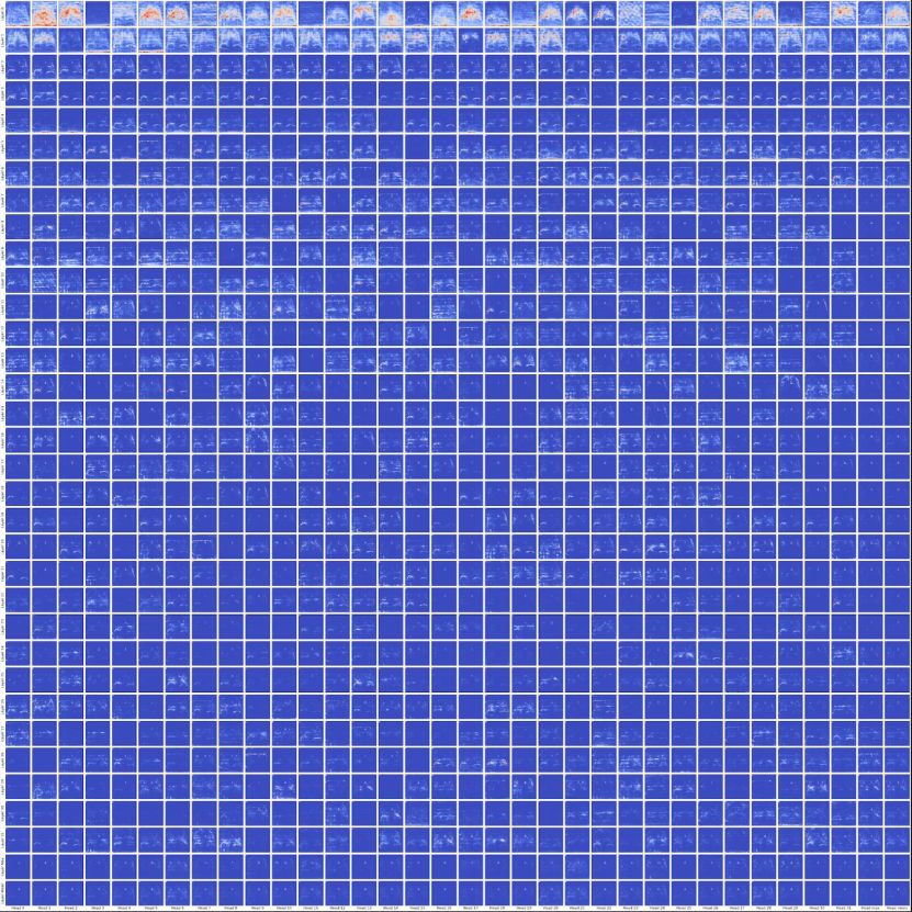
We also qualitatively investigate the correspondence of LLaVA-Rad’s attention on the input image patches for a given generated word in the reports (Figure 5), which highlight the model’s ability to focus on relevant image regions for each generated word in the reports. A detailed examination reveals a significant variability in attention across different layers and attention heads, with different configurations gravitating towards distinct regions of the image (See Appendix). Our evaluation also identifies that the aggregation of attention, particularly through averaging the outputs of all heads within the 20th layer, generally yields the most coherent and relevant focal points across a wide array of scenarios. However, this approach does not uniformly apply, as deviations in alignment were observed in certain instances. Conversely, an alternative strategy of taking the maximum across all layers, coupled with an average across heads, demonstrates a consistently high correlation with pertinent image regions. Our proposed attention visualization indicate a strong alignment between the model’s attention and the specific image regions relevant to the generated words. This alignment underscores the model’s efficacy in synthesizing contextual information from visual cues to ground its linguistic output.
Discussion
To address the significant challenges of developing foundation models for real-world clinical settings, our work introduces LLaVA-Rad, a lightweight radiology SMM that offers open-source accessibility while setting new benchmarks in the domain of radiology report generation. By curating a dataset of 1 million CXR images paired with radiology reports from diverse sources, using GPT-4 for dataset processing, coupled with the innovative training regimen of BiomedCLIP-CXR-1M, we have developed a model that not only surpasses the performance of its larger counterparts, such as GPT-4V and Med-PaLM M, but also demonstrates exceptional proficiency in generating accurate and lexically similar radiology reports on the MIMIC-CXR dataset. Through our attention visualization techniques, LLaVA-Rad offers deep insights into how it prioritizes key regions in chest X-rays, correlating them with specific findings in the generated reports. Furthermore, our utilization of G-Rad for evaluation demonstrates a closer alignment of automated scoring with expert radiologist assessments than traditional metrics, reinforcing LLaVA-Rad’s precision and reliability in clinical report generation.
The landscape of AI-driven radiology report generation has evolved significantly with the advent of transformers and large multimodal models, ushering in a new era of more sophisticated and accurate models [34, 22, 48, 38, 17, 19, 1]. R2Gen stands out as a pioneering effort in leveraging memory-efficient transformers for report generation [7]. A notable leap forward is CheXagent [8], which leverages an instruction fine-tuned foundation model trained across 28 publicly available datasets, demonstrating an enhanced capability for analyzing and summarizing CXR images. Concurrently, Flamingo-CXR fine-tuned the Flamingo vision language model [2] and incorporated regularization and adaptation techniques to tailor their applications to the nuances of radiology report generation [43]. Med-PaLM M pushed the boundaries by creating a versatile 84-billion-parameter biomedical AI system capable of addressing multiple tasks across various medical image modalities [46]. In contrast to these advancements, our method, LLaVA-Rad, distinguishes itself by not only achieving superior performance across several benchmark metrics but also by being comparatively lightweight. This attribute is particularly crucial, as it offers a more accessible and efficient solution for automating radiology report generation, addressing both the need for factual correctness and the practicality of deployment in clinical settings.
While LLaVA-Rad represents a significant advancement in medical foundation models, our research acknowledges several areas for future exploration and improvement. Firstly, the current scope of LLaVA-Rad is limited to CXRs. While CXR is the most common medical image examination, future iterations should evaluate the feasibility of our method on alternative anatomies -such as abdomen or extremities- and modalities -such as computed tomography or ultrasound- to enhance the model’s applicability and utility across diverse diagnostic scenarios. Moreover, although our attention visualization technique sheds light on the model’s decision-making processes, alternative saliency based methods for CXR interpretation algorithms such as Grad-CAM have been shown to have limited correlation with expert assessments and limited robustness to input perturbations [41, 54]. Alternatively, attention-based attribution methods have been found to be more effective at explaining model decisions and to be more useful by radiologists [49]. Despite our method relying on attention-based attribution, there is a pressing need for more exhaustive evaluation of such grounding strategies. These would further improve the model’s explainability and interpretability, making it more transparent and trustworthy for clinical use. Another consideration is the inherently multimodal nature of modern medical practice, which integrates various patient information streams, including historical medical images, medical records, lab tests, and vital signs. Integrating these diverse data sources into medical foundation models like LLaVA-Rad could significantly enrich the model’s understanding and analysis, leading to more nuanced and comprehensive patient assessments.
LLaVA-Rad exemplifies a significant leap toward making advanced diagnostic capabilities accessible with limited computational resources, thereby paving the way for broader clinical application and impact. The pursuit of creating an open-source, lightweight, high-performing model that not only extends to various medical imaging types but also incorporates multimodality and interpretability, embodies the next frontier in medical foundation model development. Such advancements will bridge the gap between current technological capabilities and the real-world demands of medical diagnostics, moving us closer to achieving meaningful improvements in patient outcomes.
Methods
Details of the dataset
CXR-1M We compiled a comprehensive dataset comprising 1 million pairs of CXR images, each accompanied by its corresponding radiology report, for pre-training the image encoder of LLaVA-Rad. This dataset amalgamates data from seven publicly available datasets and one private database as summarized in Table 1. To maintain transparency and reproducibility, we adhere to the original train/val/test splits provided by each contributing public dataset, using only the train split for pre-training the image encoder.
The CheXpert dataset [23] consists of retrospectively collected chest radiographic studies conducted between October 2002 and July 2017, encompassing both inpatient and outpatient centers at Stanford Hospital. BraX [40], obtained from chest radiography studies at Hospital Israelita Albert Einstein (HIAE) before the COVID-19 pandemic in So Paulo, Brazil, was labeled for 14 radiological findings using the CheXpert Label Extraction Algorithm [23], which was adapted to detect findings in Portuguese for this dataset. CandidPTX [13] encompasses data acquired between January 2010 and April 2020 from Dunedin Hospital in New Zealand. This dataset’s chest radiographs were manually annotated by RANZCR radiology trainees and radiologists with respect to pneumothoraces, acute rib fractures, and intercostal chest tubes. VinDR [36] was gathered from HMUH and H108 hospitals in Vietnam between 2018 and 2020, with images labeled for six diagnoses by multiple experienced radiologists from these institutions. JF Healthcare [16] data was collected from approximately 300 township hospitals in China and manually annotated by multiple radiologists to identify foreign objects within the lung field on CXRs. These datasets are comprised of images and associated binary labels that indicate whether common disease entities such as pneumonia, or pneumothorax are present in the image. However, they lack free-text reports. Thus, to enable pre-training of our image encoder using image and text methods, we create synthetic reports grounded on the labels provided. Detailed templates used for this synthetic rule-based generation can be found in Appendix Table 8.
MIMIC-CXR comprises images and their corresponding radiology reports sourced from radiographic studies conducted at the Beth Israel Deaconess Medical Center in Boston, MA, spanning the years 2011 to 2016 [25]. PadChest [6] encompasses CXRs interpreted and reported by 18 radiologists at the Hospital Universitario de San Juan, Alicante (Spain), covering the period from January 2009 to December 2017, alongside their corresponding reports in Spanish. For this data, we harness the capabilities of GPT-4 to translate these reports into English, ensuring linguistic consistency. The private dataset consists of of CXR images along with their corresponding de-identified reports. For all datasets, we utilize AP/PA views only.
MIMIC-CXR free-text reports are utilized for training the text-generation component of LLaVA-Rad. For each report, we extract the Indication, Findings and Impression sections. To do so, we employ rule-based heuristics as supported by the official MIMIC code repository.
Extracting reports in this rule-based manner poses two challenges. First, report structure varies within the dataset, with use of different section headers, merging of findings and impression into the same section, etc., which limits the availability of reports with findings available. Second, reports often contain references to prior examinations, such as "heart size remains unchanged". This poses a challenge for training report generation systems which often hallucinate references to prior examinations that are not available at inference time [39]. To mitigate these challenges, we leverage GPT-4 to extract the reason for exam, findings and impression sections in the free-text reports from MIMIC-CXR. Prompt templates used to instruct GPT-4 for the organization are elaborated in Appendix Table 7. Compared to the standard MIMIC-CXR rule-based extraction method, GPT-4 demonstrates proficiency in enhancing report quality by addressing issues like grammar errors, broken words, and synonymous section headers, while at the same time eliminating redundant phrases and references to previous exams. Table 3 showcases examples of sections structured by GPT and those extracted through rule-based methods. The use of GPT to extract sections augments rule-based data by an additional image-text pairs for the training split and for the validation split, as summarized in Table 2.
| MIMIC-CXR | Training | Validation | Test |
| Patients | |||
| Studies | |||
| DICOMs (AP/PA) | |||
| Rule-based reports | |||
| GPT-structured reports | - | ||
| Total data |
| Original | INDICATION: ___ year old woman with likely ileus after cystectomy // NGT placement confirmation NGT placement confirmation IMPRESSION: No previous images. Nasogastric tube extends to the mid body of the stomach, be for coiling on itself so that the tip lies close to the esophagogastric junction. For more optimal positioning, the to would have to be pulled back almost 10 cm and then hopefully redirected toward the lower stomach. Cardiac silhouette is within normal limits and there is no vascular congestion, pleural effusion, or acute focal pneumonia. |
| Rule-based | INDICATION: ___ year old woman with likely ileus after cystectomy // NGT placement confirmation NGT placement confirmation FINDINGS: nan IMPRESSION: No previous images. Nasogastric tube extends to the mid body of the stomach, be for coiling on itself so that the tip lies close to the esophagogastric junction. For more optimal positioning, the to would have to be pulled back almost 10 cm and then hopefully redirected toward the lower stomach. Cardiac silhouette is within normal limits and there is no vascular congestion, pleural effusion, or acute focal pneumonia. |
| GPT-structured | INDICATION: ___ year old woman with likely ileus after cystectomy // NGT placement confirmation FINDINGS: Nasogastric tube extends to the mid body of the stomach, before coiling on itself so that the tip lies close to the esophagogastric junction. Cardiac silhouette is within normal limits and there is no vascular congestion, pleural effusion, or acute focal pneumonia. IMPRESSION: For more optimal positioning, the tube would have to be pulled back almost 10 cm and then hopefully redirected toward the lower stomach. |
Modeling Approach
Image Encoder Within the LLaVA framework, the image encoder plays a pivotal role in extracting complex image representations, crucial for tasks such as automated medical report generation where standard vision transformer models often do not capture the necessary detail and nuanced representations. To overcome this, we pretrain a domain-specific vision encoder, named BiomedCLIP-CXR-1M, and integrate it into LLaVA to bolster its medical image analysis capabilities. Our method includes several key enhancements: firstly, we increase the image input resolution to 518px, substantially higher than the 224px or 336px resolutions typically used in LLaVA-Med, to capture more detailed image features. Secondly, we compile the CXR-1M dataset, an extensive collection of over 1 million CXR images from various sources, providing a rich foundation for pretraining. Lastly, we employ the BiomedCLIP recipe for training BiomedCLIP-CXR-1M, which involves contrastive vision-language training with PubMedBERT, a text encoder specialized for the medical domain [55]. The initialization of our vision encoder uses a DINOv2 model checkpoint, benefiting from its extensive training on a diverse set of 142 million general-domain images [37].
Small Multimodal Model LLaVA-Rad follows LLaVA [30, 29] and LLaVA-Med [28] which leverage the capabilities of a pre-trained image encoder and a pre-trained LM to create a SMM. We choose BiomedCLIP-CXR-1M as our image encoder and Vicuna-7B-v1.5 [9] as our LM. A multi-layer perceptron (MLP), which is randomly initialized, is introduced to project image features extracted by the image encoder into the word embedding space of the LM. Conditioned on the projected image features (visual tokens) and textual tokens, LLaVA-Rad generates text in an autoregressive manner. We refer the reader to LLaVA [30, 29] for a more in-depth description of the model architecture.
Training Strategy Due to the introduction of our domain-specific image encoder BiomedCLIP-CXR-1M, LLaVA-Rad is not initialized with the pre-trained LLaVA weights. Instead, we initialize LLaVA-Rad with the pre-trained image encoder BiomedCLIP-CXR-1M, the pre-trained LM Vicuna-7B-v1.5, and a randomly initialized MLP. Similar to LLaVA and LLaVA-Med, the SMM training procedure is done in two stages, feature alignment and end-to-end fine-tuning. Given a set of training examples, where each example consists of a CXR and the corresponding indication section and finding section from the processed report, the two-stage training procedure is described as follows:
In the feature alignment stage, we freeze the image encoder and the LM, and only update the MLP projection layer. Given a CXR , we train LLaVA-Rad to generate the corresponding findings section . Note that the indication section is not used in this stage. No text prompt is used, and the image is the only input. Our goal is to align the image features with word embeddings of the LM via the learning of the projection layer. In this stage, we train LLaVA-Rad on the training split of MIMIC-CXR for 1 epoch.
In the end-to-end fine-tuning stage, we train both the MLP projection layer and the LM. However, unlike the majority of existing work that fully fine-tunes the LM, we apply the parameter efficient fine-tuning method LoRA [18], which has recently been shown to achieve comparable performance to full fine-tuning while significantly reducing the training cost [12, 32]. Given a CXR and the corresponding indication section , we train LLaVA-Rad to generate the finding section , using the training split of MIMIC-CXR. Similar to the approaches taken by LLaVA and LLaVA-Med, our training process utilizes cross-entropy loss, applied in an auto-regressive manner, to optimize the generation of reports.
Model Evaluation
Our model evaluation consists of cross-modal retrieval evaluation, where we evaluate the quality of alignment between LLaVA-Rad’s CXR their corresponding reports, attention visualization, which illustrated the level of grounding the model’s text predictions with regions of the input image, and the automated report evaluation which studied factual correctness and lexical similarity metrics and their alignment with radiologist error quantification.
Image-Text Alignment
Cross-modal retrieval evaluation This task consists of matching radiology reports to their corresponding radiology images (text-to-image) and the reverse (image-to-text), thus evaluating the model’s ability to identify corresponding CXRs and reports by calculating similarity scores between images and text. We compared the performance of LLaVA-Rad, which uses the specialized BiomedCLIP-CXR-1M, with more general image encoders used for LLaVA-Med and LLaVA, namely BiomedCLIP and OpenAI CLIP models. We used the official MIMIC-CXR test set for evaluation, quantifying performance using recall at K, a commonly used retrieval evaluation metric that measures the share of relevant items captured within the top K positions.
Attention Visualization To qualitatively examine how well LLaVA-Rad’s image and text aligns, we visualize the model’s attention mechanisms during its generative process. Specifically, we focus on analyzing LLaVA-Rad’s attention to each image token while generating words. This analysis enables us to understand how well each generated word aligns with relevant regions within the image. To achieve this, we conduct an in-depth examination of a fully trained LLaVA-Rad model across all its 32 layers and 32 attention heads. Furthermore, to provide a clearer insight into the model’s attention patterns, we calculate either the mean or maximum values (or both) across all layerws and heads. For visualization purposes, we reformat the attention matrices into a 37x37 grid to mirror the original spatial dimensions of the image tokens.
Quality of Generated Reports
Existing Evaluation Metrics We employ a suite of automatic evaluation metrics to determine the quality of generated reports. We report commonly used lexical similarity metrics (ROUGE-L, BLEU-4) for the sake of comparison with prior methods. However, we focus our model development on factual correctness metrics, employing commonly used metrics such as F1-CheXbert and F1-RadGraph, as well as proposing an automatic GPT scoring-based metric, G-Rad. The F1-CheXbert metric [56] corresponds to the F1 score of extracted disease labels of a generated report compared to the reference, as determined by the CheXbert labeler [42]. In line with prior work, we report F1-CheXbert for all 14 CheXbert classes, in addition to that over 5 classes that represent the most common findings in real-world CXR reports (atelectasis, cardiomegaly, consolidation, edema, and pleural effusion). The F1-RadGraph metric [11] broadens the scope of the factual correctness evaluation by comparing the agreement of anatomy and observation entities extracted from the candidate report with that of the reference. Prior to this work, the F1-RadGraph metric has been considered a reference for evaluation of factual correctness in radiology reports. However, it has limited correlation with manual error scoring as performed by radiologists, which has led to the proposal of composite metrics such as RadCliQ that aim to better reflect human evaluation of factual correctness [51].
G-Rad Given the limitations of existing report evaluation methods and the challenge of accurately evaluating generated reports at scale, we explore the utility of an LLM-based scoring system, which has shown success in other domains [31, 47, 14]. Specifically, we employ GPT as an evaluator that quantifies the presence of the following six error types, as per [51]: false prediction of finding, omission of finding, incorrect location/position of finding, incorrect severity of finding, mention of comparison that is not present in the reference, omission of comparison describing a change from a previous study. We instruct GPT to quantify the number of errors of each of the six error types, keeping a separate count for clinically insignificant and significant errors. In each rating prompt we include a fixed set of five example report evaluation pairs alongside mean error counts for each type to enable the model to leverage in-context examples that quantify errors as requested. We evaluate the validity the proposed G-Rad using the ReXval dataset [52], which is comprised of error annotations from 6 board certified radiologists on 200 pairs of candidate and ground-truth reports, where each radiologist provides counts of each of the 6 aforementioned error types, also discriminating between clinically significant and insignificant errors.
For comparison, we evaluate the performance of three types of GPT models: GPT-3.5 Turbo (GPT-3.5T), GPT-4, and GPT-4-Turbo (GPT-4T). We quantify the alignment between errors quantified by radiologists with that of existing report evaluation methods, in addition to G-Rad, using Kendall’s Tau b coefficient (rank correlation coefficient). Further, we directly compare the performance of G-Rad based on GPT-4T with that of each radiologist in a leave-one-rater-out fashion. For each comparison with a rater, the mean of the remaining left-in radiologist raters was calculated. The paired interobserver difference between the held-out radiologist rater and the mean was compared to the paired interobserver difference between G-Rad and the mean. The mean absolute interobserver difference (MAD) for each left-out radiologist was compared with that of G-Rad, with statistical significance determined using paired t-tests.
Finally, we use the GPT-4T version of G-Rad to quantify the total number of clinically significant and overall errors in each generated report in the official MIMIC-CXR test split. We quantify these totals in reports from publicly accessible models, enabling us to compare LLaVA-Rad with LLaVA-Med, LLaVA, CheXagent, and GPT-4V. Further, we study the proportion of error-free reports in the overall MIMIC test set, reflecting the potential of each model in directly impacting radiology workflows.
References
- [1] Nurbanu Aksoy, Nishant Ravikumar, and Alejandro F Frangi. Radiology report generation using transformers conditioned with non-imaging data. In Medical Imaging 2023: Imaging Informatics for Healthcare, Research, and Applications, volume 12469, pages 146–154. SPIE, 2023.
- [2] Jean-Baptiste Alayrac, Jeff Donahue, Pauline Luc, Antoine Miech, Iain Barr, Yana Hasson, Karel Lenc, Arthur Mensch, Katherine Millican, Malcolm Reynolds, et al. Flamingo: a visual language model for few-shot learning. Advances in Neural Information Processing Systems, 35:23716–23736, 2022.
- [3] Shekoofeh Azizi, Laura Culp, Jan Freyberg, Basil Mustafa, Sebastien Baur, Simon Kornblith, Ting Chen, Patricia MacWilliams, S Sara Mahdavi, Ellery Wulczyn, et al. Robust and efficient medical imaging with self-supervision. arXiv preprint arXiv:2205.09723, 2022.
- [4] Shekoofeh Azizi, Basil Mustafa, Fiona Ryan, Zachary Beaver, Jan Freyberg, Jonathan Deaton, Aaron Loh, Alan Karthikesalingam, Simon Kornblith, Ting Chen, Vivek Natarajan, and Mohammad Norouzi. Big self-supervised models advance medical image classification. arXiv preprint arXiv:2101.05224, 2021.
- [5] Rishi Bommasani, Drew A Hudson, Ehsan Adeli, Russ Altman, Simran Arora, Sydney von Arx, Michael S Bernstein, Jeannette Bohg, Antoine Bosselut, Emma Brunskill, et al. On the opportunities and risks of foundation models. arXiv preprint arXiv:2108.07258, 2021.
- [6] Aurelia Bustos, Antonio Pertusa, Jose-Maria Salinas, and Maria De La Iglesia-Vaya. Padchest: A large chest x-ray image dataset with multi-label annotated reports. Medical image analysis, 66:101797, 2020.
- [7] Zhihong Chen, Yan Song, Tsung-Hui Chang, and Xiang Wan. Generating radiology reports via memory-driven transformer. arXiv preprint arXiv:2010.16056, 2020.
- [8] Zhihong Chen, Maya Varma, Jean-Benoit Delbrouck, Magdalini Paschali, Louis Blankemeier, Dave Van Veen, Jeya Maria Jose Valanarasu, Alaa Youssef, Joseph Paul Cohen, Eduardo Pontes Reis, et al. Chexagent: Towards a foundation model for chest x-ray interpretation. arXiv preprint arXiv:2401.12208, 2024.
- [9] Wei-Lin Chiang, Zhuohan Li, Zi Lin, Ying Sheng, Zhanghao Wu, Hao Zhang, Lianmin Zheng, Siyuan Zhuang, Yonghao Zhuang, Joseph E. Gonzalez, Ion Stoica, and Eric P. Xing. Vicuna: An open-source chatbot impressing gpt-4 with 90%* chatgpt quality, March 2023.
- [10] Jan Clusmann, Fiona R Kolbinger, Hannah Sophie Muti, Zunamys I Carrero, Jan-Niklas Eckardt, Narmin Ghaffari Laleh, Chiara Maria Lavinia Löffler, Sophie-Caroline Schwarzkopf, Michaela Unger, Gregory P Veldhuizen, et al. The future landscape of large language models in medicine. Communications Medicine, 3(1):141, 2023.
- [11] Jean-Benoit Delbrouck, Pierre Chambon, Christian Bluethgen, Emily Tsai, Omar Almusa, and Curtis Langlotz. Improving the factual correctness of radiology report generation with semantic rewards. In Findings of the Association for Computational Linguistics: EMNLP 2022, pages 4348–4360, Abu Dhabi, United Arab Emirates, December 2022. Association for Computational Linguistics.
- [12] Tim Dettmers, Artidoro Pagnoni, Ari Holtzman, and Luke Zettlemoyer. Qlora: Efficient finetuning of quantized llms, 2023.
- [13] Sijing Feng, Damian Azzollini, Ji Soo Kim, Cheng Kai Jin, Eve Kim, Simon Gordon, Jason Yeoh, Min A Han, Andrew Lee, Aakash Patel, Martin Urschler, Amy Fong, Cameron Simmers, Gregory Tarr, Stuart Barnard, and Ben Wilson. CANDID-PTX. Radiology: Artificial Intelligence, 6 2021.
- [14] Fabrizio Gilardi, Meysam Alizadeh, and Maël Kubli. Chatgpt outperforms crowd-workers for text-annotation tasks. arXiv preprint arXiv:2303.15056, 2023.
- [15] Suriya Gunasekar, Yi Zhang, Jyoti Aneja, Caio César Teodoro Mendes, Allie Del Giorno, Sivakanth Gopi, Mojan Javaheripi, Piero Kauffmann, Gustavo de Rosa, Olli Saarikivi, Adil Salim, Shital Shah, Harkirat Singh Behl, Xin Wang, Sébastien Bubeck, Ronen Eldan, Adam Tauman Kalai, Yin Tat Lee, and Yuanzhi Li. Textbooks are all you need, 2023.
- [16] JF Healthcare. Object-cxr - automatic detection of foreign objects on chest x-rays. https://web.archive.org/web/20201127235812/https://jfhealthcare.github.io/object-CXR/.
- [17] Xiaodi Hou, Zhi Liu, Xiaobo Li, Xingwang Li, Shengtian Sang, and Yijia Zhang. Mkcl: Medical knowledge with contrastive learning model for radiology report generation. Journal of Biomedical Informatics, 146:104496, 2023.
- [18] Edward J Hu, yelong shen, Phillip Wallis, Zeyuan Allen-Zhu, Yuanzhi Li, Shean Wang, Lu Wang, and Weizhu Chen. LoRA: Low-rank adaptation of large language models. In International Conference on Learning Representations, 2022.
- [19] Jonathan Huang, Luke Neill, Matthew Wittbrodt, David Melnick, Matthew Klug, Michael Thompson, John Bailitz, Timothy Loftus, Sanjeev Malik, Amit Phull, et al. Generative artificial intelligence for chest radiograph interpretation in the emergency department. JAMA network open, 6(10):e2336100–e2336100, 2023.
- [20] Shih-Cheng Huang, Akshay S Chaudhari, Curtis P Langlotz, Nigam Shah, Serena Yeung, and Matthew P Lungren. Developing medical imaging ai for emerging infectious diseases. nature communications, 13(1):7060, 2022.
- [21] Shih-Cheng Huang, Anuj Pareek, Malte Jensen, Matthew P Lungren, Serena Yeung, and Akshay S Chaudhari. Self-supervised learning for medical image classification: a systematic review and implementation guidelines. NPJ Digital Medicine, 6(1):74, 2023.
- [22] Stephanie L Hyland, Shruthi Bannur, Kenza Bouzid, Daniel C Castro, Mercy Ranjit, Anton Schwaighofer, Fernando Pérez-García, Valentina Salvatelli, Shaury Srivastav, Anja Thieme, et al. Maira-1: A specialised large multimodal model for radiology report generation. arXiv preprint arXiv:2311.13668, 2023.
- [23] Jeremy Irvin, Pranav Rajpurkar, Michael Ko, Yifan Yu, Silviana Ciurea-Ilcus, Chris Chute, Henrik Marklund, Behzad Haghgoo, Robyn Ball, Katie Shpanskaya, et al. Chexpert: A large chest radiograph dataset with uncertainty labels and expert comparison. In Proceedings of the AAAI conference on artificial intelligence, volume 33, pages 590–597, 2019.
- [24] Saahil Jain, Ashwin Agrawal, Adriel Saporta, Steven QH Truong, Du Nguyen Duong, Tan Bui, Pierre Chambon, Yuhao Zhang, Matthew P. Lungren, Andrew Y. Ng, Curtis P. Langlotz, and Pranav Rajpurkar. Radgraph: Extracting clinical entities and relations from radiology reports, 2021.
- [25] Alistair EW Johnson, Tom J Pollard, Seth J Berkowitz, Nathaniel R Greenbaum, Matthew P Lungren, Chih-ying Deng, Roger G Mark, and Steven Horng. Mimic-cxr, a de-identified publicly available database of chest radiographs with free-text reports. Scientific data, 6(1):317, 2019.
- [26] Curtis P Langlotz. The future of ai and informatics in radiology: 10 predictions. Radiology, 309(1):e231114, 2023.
- [27] Eric Lehman, Evan Hernandez, Diwakar Mahajan, Jonas Wulff, Micah J Smith, Zachary Ziegler, Daniel Nadler, Peter Szolovits, Alistair Johnson, and Emily Alsentzer. Do we still need clinical language models? In Bobak J. Mortazavi, Tasmie Sarker, Andrew Beam, and Joyce C. Ho, editors, Proceedings of the Conference on Health, Inference, and Learning, volume 209 of Proceedings of Machine Learning Research, pages 578–597. PMLR, 22 Jun–24 Jun 2023.
- [28] Chunyuan Li, Cliff Wong, Sheng Zhang, Naoto Usuyama, Haotian Liu, Jianwei Yang, Tristan Naumann, Hoifung Poon, and Jianfeng Gao. LLaVA-med: Training a large language-and-vision assistant for biomedicine in one day. In Thirty-seventh Conference on Neural Information Processing Systems Datasets and Benchmarks Track, 2023.
- [29] Haotian Liu, Chunyuan Li, Yuheng Li, and Yong Jae Lee. Improved baselines with visual instruction tuning, 2023.
- [30] Haotian Liu, Chunyuan Li, Qingyang Wu, and Yong Jae Lee. Visual instruction tuning. In Thirty-seventh Conference on Neural Information Processing Systems, 2023.
- [31] Yang Liu, Dan Iter, Yichong Xu, Shuohang Wang, Ruochen Xu, and Chenguang Zhu. Gpteval: Nlg evaluation using gpt-4 with better human alignment. arXiv preprint arXiv:2303.16634, 2023.
- [32] Yadong Lu, Chunyuan Li, Haotian Liu, Jianwei Yang, Jianfeng Gao, and Yelong Shen. An empirical study of scaling instruct-tuned large multimodal models, 2023.
- [33] Arindam Mitra, Luciano Del Corro, Shweti Mahajan, Andres Codas, Clarisse Simoes, Sahaj Agarwal, Xuxi Chen, Anastasia Razdaibiedina, Erik Jones, Kriti Aggarwal, Hamid Palangi, Guoqing Zheng, Corby Rosset, Hamed Khanpour, and Ahmed Awadallah. Orca 2: Teaching small language models how to reason, 2023.
- [34] Mashood Mohammad Mohsan, Muhammad Usman Akram, Ghulam Rasool, Norah Saleh Alghamdi, Muhammad Abdullah Aamer Baqai, and Muhammad Abbas. Vision transformer and language model based radiology report generation. IEEE Access, 11:1814–1824, 2022.
- [35] Michael Moor, Oishi Banerjee, Zahra Shakeri Hossein Abad, Harlan M Krumholz, Jure Leskovec, Eric J Topol, and Pranav Rajpurkar. Foundation models for generalist medical artificial intelligence. Nature, 616(7956):259–265, 2023.
- [36] H Nguyen, HH Pham, NT Nguyen, DB Nguyen, M Dao, V Vu, K Lam, and LT Le. Vinbigdata chest x-ray abnormalities detection. Kaggle Competition https://www. kaggle. com/c/vinbi gdatachest-xray-abnor malit ies-detec tion, 2020.
- [37] Maxime Oquab, Timothée Darcet, Théo Moutakanni, Huy Vo, Marc Szafraniec, Vasil Khalidov, Pierre Fernandez, Daniel Haziza, Francisco Massa, Alaaeldin El-Nouby, Mahmoud Assran, Nicolas Ballas, Wojciech Galuba, Russell Howes, Po-Yao Huang, Shang-Wen Li, Ishan Misra, Michael Rabbat, Vasu Sharma, Gabriel Synnaeve, Hu Xu, Hervé Jegou, Julien Mairal, Patrick Labatut, Armand Joulin, and Piotr Bojanowski. Dinov2: Learning robust visual features without supervision, 2024.
- [38] Renjie Pan, Ruisheng Ran, Wei Hu, Wenfeng Zhang, Qibing Qin, and Shaoguo Cui. S3-net: A self-supervised dual-stream network for radiology report generation. IEEE Journal of Biomedical and Health Informatics, 2023.
- [39] Vignav Ramesh, Nathan A Chi, and Pranav Rajpurkar. Improving radiology report generation systems by removing hallucinated references to non-existent priors. In Machine Learning for Health, pages 456–473. PMLR, 2022.
- [40] Eduardo P Reis, Joselisa PQ de Paiva, Maria CB da Silva, Guilherme AS Ribeiro, Victor F Paiva, Lucas Bulgarelli, Henrique MH Lee, Paulo V Santos, Vanessa M Brito, Lucas TW Amaral, et al. Brax, brazilian labeled chest x-ray dataset. Scientific Data, 9(1):487, 2022.
- [41] Adriel Saporta, Xiaotong Gui, Ashwin Agrawal, Anuj Pareek, Steven QH Truong, Chanh DT Nguyen, Van-Doan Ngo, Jayne Seekins, Francis G Blankenberg, Andrew Y Ng, et al. Benchmarking saliency methods for chest x-ray interpretation. Nature Machine Intelligence, 4(10):867–878, 2022.
- [42] Akshay Smit, Saahil Jain, Pranav Rajpurkar, Anuj Pareek, Andrew Y. Ng, and Matthew P. Lungren. Chexbert: Combining automatic labelers and expert annotations for accurate radiology report labeling using bert, 2020.
- [43] Ryutaro Tanno, David GT Barrett, Andrew Sellergren, Sumedh Ghaisas, Sumanth Dathathri, Abigail See, Johannes Welbl, Karan Singhal, Shekoofeh Azizi, Tao Tu, et al. Consensus, dissensus and synergy between clinicians and specialist foundation models in radiology report generation. arXiv preprint arXiv:2311.18260, 2023.
- [44] Arun James Thirunavukarasu, Darren Shu Jeng Ting, Kabilan Elangovan, Laura Gutierrez, Ting Fang Tan, and Daniel Shu Wei Ting. Large language models in medicine. Nature medicine, 29(8):1930–1940, 2023.
- [45] Daniel Truhn, Gustav Müller-Franzes, and Jakob Nikolas Kather. The ecological footprint of medical ai. European Radiology, pages 1–3, 2023.
- [46] Tao Tu, Shekoofeh Azizi, Danny Driess, Mike Schaekermann, Mohamed Amin, Pi-Chuan Chang, Andrew Carroll, Charles Lau, Ryutaro Tanno, Ira Ktena, Anil Palepu, Basil Mustafa, Aakanksha Chowdhery, Yun Liu, Simon Kornblith, David Fleet, Philip Mansfield, Sushant Prakash, Renee Wong, Sunny Virmani, Christopher Semturs, S. Sara Mahdavi, Bradley Green, Ewa Dominowska, Blaise Aguera y Arcas, Joelle Barral, Dale Webster, Greg S. Corrado, Yossi Matias, Karan Singhal, Pete Florence, Alan Karthikesalingam, and Vivek Natarajan. Towards generalist biomedical ai. NEJM AI, 1(3):AIoa2300138, 2024.
- [47] Jiaan Wang, Yunlong Liang, Fandong Meng, Haoxiang Shi, Zhixu Li, Jinan Xu, Jianfeng Qu, and Jie Zhou. Is chatgpt a good nlg evaluator? a preliminary study. arXiv preprint arXiv:2303.04048, 2023.
- [48] Jun Wang, Abhir Bhalerao, and Yulan He. Cross-modal prototype driven network for radiology report generation. In European Conference on Computer Vision, pages 563–579. Springer, 2022.
- [49] Alessandro Wollek, Robert Graf, Saša Čečatka, Nicola Fink, Theresa Willem, Bastian O. Sabel, and Tobias Lasser. Attention-based saliency maps improve interpretability of pneumothorax classification. Radiology: Artificial Intelligence, 5(2):e220187, 2023.
- [50] Michael Wornow, Yizhe Xu, Rahul Thapa, Birju Patel, Ethan Steinberg, Scott Fleming, Michael A Pfeffer, Jason Fries, and Nigam H Shah. The shaky foundations of large language models and foundation models for electronic health records. npj Digital Medicine, 6(1):135, 2023.
- [51] Feiyang Yu, Mark Endo, Rayan Krishnan, Ian Pan, Andy Tsai, Eduardo Pontes Reis, Eduardo Kaiser Ururahy Nunes Fonseca, Henrique Min Ho Lee, Zahra Shakeri Hossein Abad, Andrew Y Ng, et al. Evaluating progress in automatic chest x-ray radiology report generation. Patterns, 4(9), 2023.
- [52] Feiyang Yu, Mark Endo, Rayan Krishnan, Ian Pan, Andy Tsai, Eduardo Pontes Reis, EKU Fonseca, Henrique Lee, Zahra Shakeri, Andrew Ng, et al. Radiology report expert evaluation (rexval) dataset, 2023.
- [53] Juan M Zambrano Chaves, Nandita Bhaskhar, Maayane Attias, Jean-Benoit Delbrouck, Daniel Rubin, Andreas Loening, Curtis Langlotz, and Akshay Chaudhari. Rales: a benchmark for radiology language evaluations. In A. Oh, T. Neumann, A. Globerson, K. Saenko, M. Hardt, and S. Levine, editors, Advances in Neural Information Processing Systems, volume 36, pages 74429–74454. Curran Associates, Inc., 2023.
- [54] Jiajin Zhang, Hanqing Chao, Giridhar Dasegowda, Ge Wang, Mannudeep K. Kalra, and Pingkun Yan. Revisiting the trustworthiness of saliency methods in radiology ai. Radiology: Artificial Intelligence, 6(1):e220221, 2024. PMID: 38166328.
- [55] Sheng Zhang, Yanbo Xu, Naoto Usuyama, Hanwen Xu, Jaspreet Bagga, Robert Tinn, Sam Preston, Rajesh Rao, Mu Wei, Naveen Valluri, Cliff Wong, Andrea Tupini, Yu Wang, Matt Mazzola, Swadheen Shukla, Lars Liden, Jianfeng Gao, Matthew P. Lungren, Tristan Naumann, Sheng Wang, and Hoifung Poon. Biomedclip: a multimodal biomedical foundation model pretrained from fifteen million scientific image-text pairs, 2024.
- [56] Yuhao Zhang, Derek Merck, Emily Tsai, Christopher D Manning, and Curtis Langlotz. Optimizing the factual correctness of a summary: A study of summarizing radiology reports. In Proceedings of the 58th Annual Meeting of the Association for Computational Linguistics, pages 5108–5120, 2020.
Supplementary Material
| Model | Size | CheXbert | Rad- | BLEU | ROUGE | ||||||||
| ("uncertain" as negative) | ("uncertain" as positive) | Graph | |||||||||||
| Micro-avg | Macro-avg | Micro-avg | Macro-avg | ||||||||||
| predictions, wts = weights | F1-14 | F1-5 | F1-14 | F1-5 | F1-14 | F1-5 | F1-14 | F1-5 | F1 | (1) | (4) | -L | |
| LLaVA-Rad | 7B | 57.1 | 57.3 | 41.4 | 48.5 | 56.7 | 59.7 | 44.4 | 52.3 | 29.6 | 38.6 | 15.5 | 30.8 |
| Med-PaLM M | 84B | 53.56 | 57.9 | 39.8 | 51.6 | - | - | - | - | 26.7 | 32.3 | 11.3 | 27.3 |
| GPT-4V | - | 35.5 | 25.8 | 20.4 | 19.6 | 35.6 | 33.3 | 25.3 | 29.6 | 13.2 | 16.4 | 1.9 | 13.2 |
| MAIRA-1 | 7B | 55.7 | 56.0 | 38.6 | 47.7 | 55.3 | 58.8 | 42.3 | 51.7 | 29.6 | 39.2 | 14.2 | 28.9 |
| LLaVA-Med | 7B | 27.2 | 22.0 | 15.5 | 16.6 | 27.3 | 24.4 | 18.7 | 20.5 | 6.5 | 22.2 | 1.0 | 13.3 |
| LLaVA | 7B | 22.9 | 23.4 | 15.4 | 17.5 | 23.7 | 26.9 | 17.0 | 20.3 | 4.5 | 21.0 | 1.3 | 13.8 |
| CheXagent | 7B | 25.6 | 18.7 | 15.4 | 15.6 | 25.9 | 19.8 | 17.6 | 16.9 | 18.0 | 14.4 | 3.9 | 20.9 |
| Flamingo-CXR | <1B | - | - | - | - | 51.9 | 56.5 | - | - | 20.5 | - | 10.1 | 29.7 |
| CvT2Dist. | <1B | 44.20 | - | 30.70 | - | - | - | - | - | - | 39.3 | 12.7 | 28.6 |
| trans | <1B | - | - | - | - | - | 56.7 | - | - | - | - | 11.4 | - |
| RGRG | <1B | - | - | - | - | - | 54.7 | - | - | - | 37.3 | 12.6 | 26.4 |
| R2Gen | <1B | - | - | - | - | 22.8 | 34.6 | - | - | - | 35.3 | 8.6 | 27.7 |
| TieNet | <1B | - | - | - | - | - | 27.1 | - | - | - | - | 8.1 | - |
| Model Version | CheXbert | Rad- | BLEU | ROUGE | ||||||||
| ("uncertain" as negative) | ("uncertain" as positive) | Graph | ||||||||||
| Micro-avg | Macro-avg | Micro-avg | Macro-avg | |||||||||
| F1-14 | F1-5 | F1-14 | F1-5 | F1-14 | F1-5 | F1-14 | F1-5 | F1 | (1) | (4) | -L | |
| LLaVA-Rad | 57.1 | 57.3 | 41.4 | 48.5 | 56.7 | 59.7 | 44.4 | 52.3 | 29.6 | 38.6 | 15.5 | 30.8 |
| Variant #1 | 55.5 | 55.2 | 38.4 | 46.9 | 55.9 | 58.4 | 42.5 | 51.1 | 29.6 | 38.5 | 15.4 | 30.6 |
| Variant #2 | 55.6 | 56.4 | 38.5 | 48.6 | 55.7 | 59.5 | 41.6 | 52.9 | 28.9 | 37.5 | 15.2 | 30.3 |
| Variant #3 | 55.7 | 56.2 | 39.1 | 47.4 | 55.8 | 59.0 | 43.1 | 51.4 | 29.7 | 37.9 | 15.6 | 31.0 |
| Variant #4 | 56.2 | 56.9 | 38.6 | 48.3 | 56.1 | 60.2 | 43.0 | 54.0 | 27.6 | 29.9 | 10.0 | 26.3 |
| Variant #5 | 50.6 | 53.3 | 30.7 | 42.7 | 49.2 | 54.1 | 33.6 | 45.0 | 25.0 | 16.9 | 5.0 | 16.4 |
| Variant #6 | 51.1 | 52.9 | 31.0 | 43.6 | 51.4 | 56.3 | 35.1 | 48.6 | 26.9 | 35.0 | 13.9 | 29.4 |
| Variant #7 | 49.8 | 52.1 | 30.5 | 42.4 | 50.3 | 55.4 | 34.5 | 48.0 | 26.5 | 34.4 | 13.7 | 29.1 |
| Variant #8 | 49.6 | 52.2 | 31.3 | 42.8 | 50.1 | 55.6 | 35.0 | 47.8 | 26.9 | 34.3 | 13.7 | 29.3 |
| Analysis #1 | 96.7 | 98.3 | 95.1 | 97.8 | 96.9 | 98.4 | 95.5 | 97.8 | 93.2 | 82.2 | 78.8 | 90.9 |
| Stage 1 (image encoder pre-training) Stage 2 (feature alignment) Stage 3 (fine-tuning) | ||||||||||||
| Variant #1: Stage 1 pre-trains the image encoder on MIMIC-CXR only. Stage 2&3 are the same. | ||||||||||||
| Variant #2: No stage-1 pretraining. Stage 2&3 are the same. | ||||||||||||
| Variant #3: Stage 1 is the same. Stage 2&3 only use rule-processed MIMIC-CXR training data. | ||||||||||||
| Variant #4: Stage 1 is the same. Stage 2&3 only use GPT-4 processed MIMIC-CXR training data. | ||||||||||||
| Variant #5: Stage 1&2 are the same. No stage 3. | ||||||||||||
| Variant #6: No stage 1. Stage 2 initializes with LLaVA-v1.5 pre-trained weights. | ||||||||||||
| Variant #7: No stage 1. Stage 2 initializes the image encoder with OpenAI CLIP pre-trained weights. | ||||||||||||
| Variant #8: No stage 1. Stage 2 initializes the image encoder with BiomedCLIP pre-trained weights. | ||||||||||||
| Analysis #1: Evaluate GPT-4 processed test data against rule-processed test data. | ||||||||||||
| Model | Key Training Data | Image Encoder | Key Evaluations |
| LLaVA | Image Captions + GPT4 | CLIP | VQA, LLaVA-Bench |
| LLaVA-Med | PMC Captions + GPT4 | BiomedCLIP | VQA, LLaVA-Bench |
| LLaVA-Rad | Radiology Data + GPT4 | BiomedCLIP-CXR-1M | G-Rad/CheXpert |
You are an expert medical assistant AI capable of modifying clinical documents to
user specifications. You make minimal changes to the original document to satisfy
user requests. You never add information that is not already directly stated in
the original document.

Extract four sections from the input radiology report: ‘Examination’, ‘Indication’,
‘Findings’ and ‘Impression’. Leave an extracted section as null if it does not
exist in the original report. The output should be in JSON format. An Indication
section can refer to the History, Indication or Reason for Study sections in the
original report. Remove any information not directly observable from the current
imaging study. For instance, remove any patient demographic data, past medical
history, or comparison to prior images or studies. The generated ‘Findings’ and
‘Impression’ sections should not reference any changes based on prior images,
studies, or external knowledge about the patient. Rewrite such comparisons as a
status observation based only on the current image or study. Remember to remove
any numbering or bullets.

Examples of inputs and expected outputs:

INPUT:
EXAMINATION: XR CHEST AP PORTABLE
INDICATION: Small right apical pneumothorax after lung biopsy.
FINDINGS: Single portable view of the chest was obtained. Copared with 10:42 AM.
The small right apical pneumothorax has decreased slightly in size, the improvement
best appreciated laterally where it now measures 10 mm compared to 14 mm before. At
the lung apex it now measures 1.6 and compared to 2.1 cm previously. A subtle right
apical pulmonary contusion is grossly stable. Minor chest wall emphysema along the
right exilla has not changed significant delay. There is no metastatic shift. No
pleural effusion is evident.

OUTPUT:
{"EXAMINATION": "XR CHEST AP PORTABLE.",
"INDICATION": "Small right apical pneumothorax after lung biopsy."
"FINDINGS": "Single portable view of the chest was obtained. The small right apical
pneumothorax has decreased slightly in size and measures 10mm. At the lung apex it
measures 1.6cm. A subtle right apical pulmonary contusion is grossly stable. Minor
chest wall emphysema along the right exilla has not changed significantly. There is
no metastatic shift. No pleural effusion is evident."
"IMPRESSION": null}
|
Positive <condition>: 1. The radiograph reveals evidence of <condition>. 2. The radiograph demonstrates areas consistent with <condition>. 3. There are findings suggestive of <condition>. 4. There is presence of <condition>. 5. There is a positive finding of <condition>. 6. The radiographic examination of the chest reveals the presence of <condition>. 7. There is evidence of <condition>. 8. <Condition> is present. Negative <condition>: 1. No evidence of <condition> is observed. 2. <Condition> is not identified. 3. The radiograph does not show any signs of <condition>. 4. There is no indications of <condition> in the radiograph. 5. No <condition> is identified in the examined region. 6. No signs of <condition> is observed. 7. The image does not conclusively indicate <condition>. Uncertain <condition>: 1. There is uncertainty regarding <condition>. 2. The presence of <condition> is uncertain based on the current examination. 3. The image shows uncertainty in <condition>. No findings: 1. The radiographic examination of the chest reveals no significant abnormalities or pathologies. 2. The radiographic examination of the patient does not reveal any significant abnormal findings. No support devices: 1. There is no evidence of any support devices in the chest area. 2. There are no support devices seen in the current study. 3. There are no support devices in place. Pleural others: 1. There are some pleural abnormalities that do not fit into the common categories of pleural diseases. 2. Other pleural abnormalities are also observed. No pleural others: 1. No other pleural abnormalities were detected in the radiograph. 2. There are no findings related to other pleural abnormalities. Uncertain pleural others: 1. There are ambiguous findings related to the pleura. 2. There is no visible pleural abnormality, although the image does not completely exclude all potential pleural conditions. |