[table]capposition=top \newfloatcommandcapbtabboxtable[][\FBwidth]
A Unified Framework for Microscopy Defocus Deblur with Multi-Pyramid Transformer and Contrastive Learning 111Accepted in CVPR 2024. This is not the final camera-ready version.
Abstract
Defocus blur is a persistent problem in microscope imaging that poses harm to pathology interpretation and medical intervention in cell microscopy and microscope surgery. To address this problem, a unified framework including multi-pyramid transformer (MPT) and extended frequency contrastive regularization (EFCR) is proposed to tackle two outstanding challenges in microscopy deblur: longer attention span and feature deficiency. The MPT employs an explicit pyramid structure at each network stage that integrates the cross-scale window attention (CSWA), the intra-scale channel attention (ISCA), and the feature-enhancing feed-forward network (FEFN) to capture long-range cross-scale spatial interaction and global channel context. The EFCR addresses the feature deficiency problem by exploring latent deblur signals from different frequency bands. It also enables deblur knowledge transfer to learn cross-domain information from extra data, improving deblur performance for labeled and unlabeled data. Extensive experiments and downstream task validation show the framework achieves state-of-the-art performance across multiple datasets. Project page: https://github.com/PieceZhang/MPT-CataBlur.
1 Introduction
Microscope offers observers enhanced resolution and magnification [48, 9, 49], which greatly promotes the advancement of cell microscopy [9] and surgical microscopy [48]. Cell microscopy employs various optical techniques to reveal the structure and function of cells [9]. Surgical microscopy assists surgeons in performing delicate operations [48] including neurosurgery [81], ophthalmology [4], dentistry [15], etc. In microscopy, out-of-focus, or defocus, is one of the most common visual impairments caused by inferior optical quality, lens aperture, or object magnification [48, 9], resulting in blurred or distorted imaging. It can be disastrous for the downstream tasks [88], including segmentation [30, 29], detection [61], and classification [6]. While various microscopes with auto-focusing [79, 55, 35], assisted-focusing [67], or multi-focus [82, 39] capabilities have been developed to mitigate the defocus effect on-site, image degradation remains if the objects are distributed non-uniformly and not co-planar [50], or the cavities are too deep to be aligned with the focal plane [48]. Microscopy defocus deblur methods have thus been introduced as an offsite restoration approach.
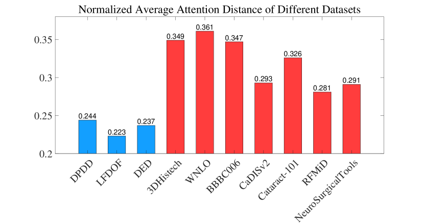
Recent advances in deep learning have led to the development of various deep defocus deblur methods [60, 34, 56, 57], including those designed for microscopy [17, 50, 70, 85, 28, 18, 93, 72, 91, 32, 41]. Microscopy deblurring poses different challenges from real-world deblur tasks, due to the significant discrepancy between the features in the microscope images and natural scene images [88]. This difference can be quantified by calculating the normalized average attention distance for different datasets (attention intensity multiplied by pixel distance then normalize by image size). The evaluation involves real-world datasets (DPDD [1], LFDOF [59], DED [47]), cell microscopy datasets (3DHistech [17], WNLO [17], BBBC006 [44]), and surgical microscopy datasets for cataract surgery (CaDISv2 [19], Cataract-101 [62]), retinal microsurgery (RFMiD [54]), neurosurgery (NeuroSurgicalTools [5]). As shown in Fig. 1, all of the cell microscopy datasets have normalized average attention distance around 0.35, and surgical microscope datasets around 0.3, indicating a much longer attention span than the real-world datasets at under 0.25. This result reveals the substantial discrepancy between the two domains, suggesting that modeling attention in wider areas with larger receptive fields would benefit microscopy tasks. Motivated by this analysis, we introduce a multi-pyramid transformer (MPT) with cross-scale window attention (CSWA), intra-scale channel attention (ISCA) and feature-enhancing feed-forward network (FEFN), to construct multiple pyramids explicitly on each stage of the network, fully exploiting latent cross-scale features in every projection space. CSWA captures the interaction between local and cross-scale - pairs for long-range attention modeling with a quadratically enlarged receptive field while keeping computational efficiency. ISCA builds channel-wise attention on a local scale to provide global channel context, which is then integrated with the spatially correlated feature from CSWA by the proposed FEFN through an asymmetric activation mechanism.
Another problem in microscopy deblur is the insufficient data for training a robust model. Different from natural scene defocus deblur methods that use datasets captured with varying aperture sizes [1] or light field camera [60, 47], the high-quality training data for microscopy deblur can be much harder to obtain[17, 48]. For cell microscopy, insufficient training feature leads to a generalizability problem caused by different staining and imaging methods [17, 88]. The situation is worse for surgical microscopy because the imaging principle of microscope makes it impossible to simultaneously acquire blur-sharp pairs for model training [18, 48]. To alleviate the feature deficiency problem, there exist some training diagrams to learn rich information from extra data, such as extra data pretraining followed by testing data fine-tuning [60]. This paradigm, however, may not be applicable to microscopy deblurring, as there is an inter-domain gap between natural scenes and microscopy images, and intra-domain gaps among different microscopy datasets that are highly task-specific. The extended frequency contrastive regularization (EFCR) is proposed to address the feature deficiency problem by encouraging the model to learn representations from decoupled frequency bands in the wavelet domain [86, 23], and further exploiting latent information leveraging the fact that model trained with synthetic reblurring images can deblur its naturally blurred counterpart [18]. It also enables cross-domain deblur knowledge transfer, facilitating multiple scenarios including extra data training and unlabeled data deblur.
This paper presents a unified deblur framework with MPT and EFCR to address the aforementioned two challenges in microscopy deblur. As the most common surgery worldwide [66, 13, 21], cataract surgery is chosen to illustrate surgical microscopy deblur. Extensive experiments are conducted on various open-source cell and surgical microscopy datasets, along with downstream tasks validation on cell detection and surgery scene semantic segmentation. For surgical microscopy deblur, we present a realistic blur synthesizing method, and collect a new defocus microscope surgery dataset in cataract microscopic surgery, which is the first dataset for surgical microscopy deblur. The method achieves state-of-the-art performance on not only microscope datasets but also real-world datasets, showing the universality of the proposed framework. The deblur result on unlabeled dataset also proves the effectiveness of the proposed EFCR as a knowledge transfer tool. The main contributions are as follows, 1) The multi-pyramid transformer (MPT) is for the first time proposed for microscopy defocus deblur. It models the long-range cross-scale spatial attention in each explicit pyramid using the proposed cross-scale window attention (CSWA) with quadratically enlarged receptive field to adapt to the longer attention span of microscopy datasets that hinders the existing methods. 2) The intra-scale channel attention (ISCA) is presented to model the global channel attention, incorporating channel context in the CSWA spatial information via the proposed feature-enhancing feed-forward network (FEFN), providing additional channel feature to the pyramid. 3) A training strategy with extended frequency contrastive regularization (EFCR) is presented to alleviate feature deficiency by exploiting latent deblur signal beyond the pixel-wise constraint through synthetic reblurring, which is the first implementation of contrastive learning in microscopy deblur. It also enables cross-domain deblur knowledge transfer, facilitating extra data training and enhancing unlabeled image deblur.
2 Related Work
Single image defocus deblurring
For learning-based deblur model, end-to-end method is widely applied [34, 65, 60, 57, 56] for its better performance and robustness [56] than methods based on defocus map estimation [33, 47, 59, 34]. Among them, many deblurring works have been done on cell microscopy to alleviate defocus brought by mechanical axial shift [70] and non-coplanar cells [50], and enhance the imaging quality of human cell [50, 72], pathology [17, 28], parasite [85], etc. Compared with cell microscopy, surgical microscopy deblur has not been well-explored due to the difficult acquisition of blur datasets with ground truth [18], although defocus blur is commonly encountered in microscopic surgery [18, 93].
Multi-scale methods
The image pyramid in existing methods is usually built in two ways, i.e., explicitly stacking multi-scale feature maps in a single pyramid [51, 16, 80], or implicitly applying multi-stage structure [7, 31, 60, 8, 56, 71]. Explicit pyramid methods face single-level feature deficiency since the explicit pyramid is built on downscaling feature in a single latent space [51, 16, 80]. Most of the existing microscope deblur methods [70, 40, 18, 17] adopt implicit multi-stage design to perform aggregation on separated latent space but suffer from inter-level feature discrepancy. The proposed MPT in this work addresses these drawbacks by building multiple explicit pyramids with CSWA, ISCA, and FEFN on each feature level, achieving cross-scale feature aggregation with enlarged receptive field.
Contrastive learning
Contrastive learning has been widely applied in low-level tasks [27, 92, 75, 14, 3, 94, 25]. They construct contrastive pairs on the feature space that take clean and corrupted images as positive and negative pairs, respectively [76, 37]. Some works have leveraged contrastive frequency information [94] and extracted frequency representations by wavelet transformation [86, 3, 75]. Following the fact that sharp and blurry images have similar low-frequency components but differ significantly in the high-frequency part [75], the idea of comparing different frequency bands separately is adopted in [90] by applying loss directly to the frequency bands in contrastive regularization. In this paper, the proposed EFCR adopts basic CR and extended CR to encourage learning of latent deblur signals and transferring of cross-domain deblur information, thus addressing the feature deficiency problem.
3 Method
The proposed framework consists of MPT and EFCR to address the two outstanding problems in microscopy defocus deblur, namely longer attention span and feature deficiency.
3.1 Multi-pyramid Transformer (MPT)
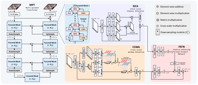
MPT builds multiple explicit pyramids on each feature level, thus avoiding single-level feature deficiency and inter-level feature discrepancy [51, 80, 70, 18]. As shown in Fig. 2, the proposed MPT follows a U-shaped structure [58, 74, 22]. The blur image with the size of first gets input feature projection through a convolution. Then, the feature goes through the network with seven pyramid blocks followed by re-sampling using pixel shuffles [64], and finally projects back to the image with a convolution. The shortcut connection is built between each encoder and decoder stage by element-wise addition.
Pyramid block
The pyramid block receives (), and outputs the map in the same size. There are sub-blocks in pyramid block . Each of them handles a local scale (, ). Multiple stacked sub-blocks build the pyramid in a coarse-to-fine manner, i.e., , . This design achieves progressive multi-scale feature aggregation and ensures the full exploration of each scale. In practice, is set to be an even number. The sub-block in the even level adopts common cyclic window shifting strategy [43] to gain cross-window interaction.
Cross-scale window attention (CSWA)
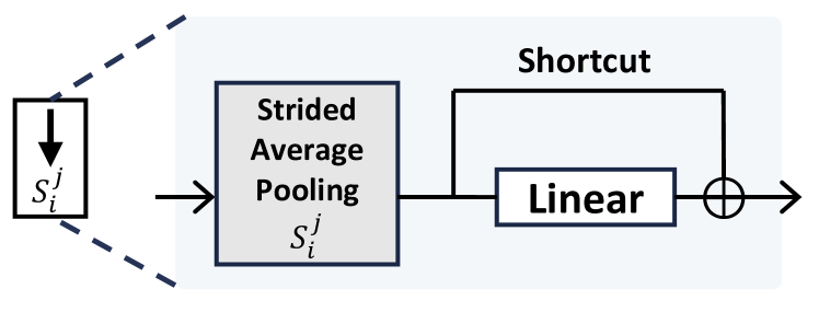
CSWA captures the long-range interaction by modeling the attention between windows from the local-scale map and the down-scale map. The layer normalized [2] feature first passes through a downsampling module to get with the size of . A strided average pooling followed by a linear projection with a shortcut is adopted in the downsampling module. The neighboring padding convolution (NPConv) is proposed to generate projection (, ) with inductive bias[77, 20], where is the patch width. It pads a patch with its neighborhood pixels, providing the isolated edge pixels with neighboring information [69] (all convolutions adopt bias-free grouped convolution by default with group size equal to the feature dimension). To get the cross-scale spatial attention map, the cross-scale multiplication () is introduced as a one-to-many strategy, where each patch in is multiplied with patches in the corresponding location of , as illustrated in Fig. 2. This operation provides an local patch in with interaction with a patch in whose information comes from a region in the original map. Although the downscaled map loses information, it can still be highly instructive, since attention distributions of different scales are highly consistent [36]. It also leverages the pool of sharper patches generated by downscaling which serve as priors for deblurring [95, 53]. It makes receptive field quadratically enlarged by times while keeping the computational complexity unchanged as the vanilla local window attention [43]. A similar strategy is adopted in the multiplication between attention map and . The self-attention in CSWA for a local window in the size of can be defined as:
| (1) |
where , and is the relative positional encoding [43]. In practice, we implement the multi-head self-attention [68] by concatenating the result of parallelly calculated attention. The output is then obtained by a linear projection with a shortcut.
Intra-scale channel attention (ISCA)
ISCA handles a single local scale feature and generates cross-channel interaction with encoded global context [84]. By applying convolutions followed by convolutions, projections are generated from layer normalized , introducing convolutional inductive bias and extracting cross-channel information in both point-wise and spatial-wise manners. The intra-scale channel attention map is then calculated by multiplying with . The self-attention in ISCA can be formulated as:
| (2) |
Similar to CSWA, ISCA implements the multi-head self-attention [68]. The self-attention result finally goes through a linear projection with a shortcut and gets , similar to CSWA.
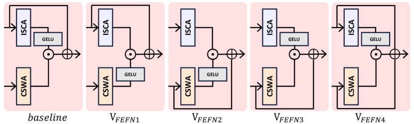
Feature-enhancing feed-forward network (FEFN)
The FEFN aggregates the spatial-wise feature with the channel-wise context . The input features are first projected to and in the size of , where is the expansion ratio. Instead of combining and by simply adding them together [36], FEFN adopts an asymmetrical activation mechanism with GELU [26], where is element-wisely multiplied by GELU activated . The FEFN can be formulated as
| (3) |
where , and refers to linear projection. Compared with the regular FN [12], this asymmetrical operation allows the spatial information from to be guided by the non-linearly activated signal from the channel context in , offering an extra global view in terms of feature channels.
3.2 Extended Frequency Contrastive Regularization (EFCR)
The proposed EFCR contains basic CR and extended CR to explore latent deblur guidance beyond the pixel-wise constraints.
Constructions of contrastive pairs
Given a training pair with ground truth , blur input , and deblurred output , the Haar wavelet transformation [86] decouples the samples into low-low (LL), low-high (LH), high-low (HL), and high-high (HH) bands. For simplicity, here we define as the operator decoupling and concatenating high-frequency bands (LH, HL, HH), and as the operator for low-frequency band (LL). For basic CR, the frequency bands are directly taken as contrastive pairs. The positive and negative basic CR and are given by:
| (4) | |||
| (5) |
Both bands are included in , since both high and low frequencies of are expected to be pulled closer to . Only high frequency is taken for to push the away from as blur degradation mainly happens in the high-frequency parts [8, 42].
The extended CR enforces the model to learn latent information from degraded high-frequency components beyond the pixel-wise constraint. Based on the idea that a model trained with synthetic blurred images can deblur natural blurry images in the dataset [18], the blurred image is generated by applying random Gaussian blur (kernel size in ) on , followed by calculating its deblurred result . The extended CR based on extended training pair () is then formulated as:
| (6) |
The is derived as a relative loss term by normalizing with high-frequency distance between and its blurred counterpart to alleviate the disturbance from the gradient distribution of reblurred image [11].
The overall optimization objective with the proposed EFCR is given by:
| (7) |
where is the supervised pixel loss, is the number of samples, and is the scaling factor.
Knowledge transfer from extra data
Defocus blur mainly causes high-frequency degradation [8, 42], implying that the high-frequency part can provide informative cross-domain deblur guidance. EFCR with extra data (denoted by EFCRex) constructs contrastive pairs on high-frequency components. Given an extra training pair from external dataset and corresponding extended samples , EFCRex with can be formulated as:
| (8) | |||
| (9) | |||
| (10) |
The overall optimization objective follows a similar pattern with Eq. 7, where the supervised training on the testing dataset () proceeds simultaneously with EFCRex () to transfer knowledge without interfering with supervised training.
EFCRex facilitates two important applications. One is to transfer rich deblur signals from a real-world blur dataset to microscopy deblur tasks, in which EFCRex is composed by . Another is to learn latent deblur direction from an unlabeled microscopy dataset thus enhancing the deblur performance, where the model is trained on a labeled dataset with an unlabeled microscopy dataset as the extra data. EFCRex here is reformulated as .
4 Experiments
| Method | BBBC006w1 [44] | BBBC006w2 [44] | 3DHistech [17] | CaDISBlur | ||||||||
| PSNR | SSIM | LPIPS | PSNR | SSIM | LPIPS | PSNR | SSIM | LPIPS | PSNR | SSIM | LPIPS | |
| DRBNet [60] | 32.83 | 0.737 | 0.381 | 26.66 | 0.589 | 0.458 | 32.83 | 0.853 | 0.131 | 42.54 | 0.776 | 0.243 |
| GKMNet [56] | 34.41 | 0.887 | 0.218 | 29.32 | 0.721 | 0.296 | 33.42 | 0.852 | 0.130 | 44.27 | 0.860 | 0.178 |
| MIMO-UNet [7] | 32.73 | 0.725 | 0.412 | 26.90 | 0.601 | 0.457 | 32.40 | 0.837 | 0.169 | 43.36 | 0.823 | 0.197 |
| MSSNet [31] | 34.01 | 0.790 | 0.289 | 28.68 | 0.736 | 0.361 | 33.09 | 0.870 | 0.126 | 44.09 | 0.871 | 0.160 |
| SwinIR [38] | 33.90 | 0.801 | 0.274 | 27.61 | 0.696 | 0.403 | 32.57 | 0.841 | 0.136 | 41.83 | 0.710 | 0.349 |
| PANet [51] | 34.45 | 0.890 | 0.230 | 29.07 | 0.743 | 0.290 | 33.24 | 0.869 | 0.129 | 44.49 | 0.917 | 0.134 |
| GRL [36] | 34.76 | 0.907 | 0.129 | 29.39 | 0.786 | 0.249 | 33.49 | 0.878 | 0.120 | 44.86 | 0.960 | 0.087 |
| Restormer [84] | 34.79 | 0.904 | 0.135 | 29.78 | 0.801 | 0.241 | 33.46 | 0.880 | 0.125 | 44.85 | 0.941 | 0.101 |
| MPT | 34.89 | 0.912 | 0.127 | 29.85 | 0.813 | 0.237 | 33.55 | 0.881 | 0.121 | 44.98 | 0.962 | 0.087 |
| MPT+EFCR | 34.96 | 0.917 | 0.119 | 29.89 | 0.820 | 0.230 | 33.58 | 0.887 | 0.119 | 45.09 | 0.969 | 0.082 |
| MPT+EFCRex | 35.16 | 0.935 | 0.083 | 30.11 | 0.829 | 0.205 | 33.63 | 0.892 | 0.116 | 45.25 | 0.971 | 0.077 |
| Method | PSNR | SSIM | LPIPS |
| KPAC [65] | 25.24 | 0.774 | 0.226 |
| IFAN [34] | 25.37 | 0.789 | 0.217 |
| DRBNet [60] | 25.47 | 0.787 | 0.246 |
| GKMNet [56] | 25.47 | 0.789 | 0.219 |
| Restormer [84] | 25.98 | 0.811 | 0.178 |
| NRKNet [57] | 26.11 | 0.810 | 0.210 |
| GRL [36] | 26.18 | 0.822 | 0.168 |
| MPT | 26.21 | 0.826 | 0.175 |
| MPT+EFCR | 26.23 | 0.829 | 0.172 |
| MPT+EFCRex | 26.27 | 0.831 | 0.161 |
4.1 Datasets and Implementation
Extensive experiments are carried out on various real-world and microscopy datasets, including five labeled datasets: DPDD [1], LFDOF [59], BBBC006 [44], 3DHistech [17], CaDISBlur, and three unlabeled datasets: CUHK [63], WNLO [17], CataBlur. The LFDOF [59] is adopted as an extra training dataset for knowledge transfer using EFCR, since LFDOF has substantial samples with rich information and good cross correlation between defocused and ground truth pairs [60]. For surgical microscopy, two new surgical microscopy deblur datasets are presented, which are CaDISBlur and CataBlur. CaDISBlur is synthesized using images from a high-quality dataset CaDIS [19] by a novel realistic blur simulation method, in which the instruments and anatomies in the surgery scene are blurred respectively leveraging the object segmentation mask in CaDIS to simulate different focal planes. CataBlur is a new surgical microscope defocus blur dataset, including 4614 defocus images collected from 12 cataract surgeries, for evaluation on real surgical defocus blur. More details about the datasets, training settings, and proposed blur synthesizing method are provided in the supplementary material.
The proposed framework employs the same structure in all tests as follows. The MPT adopts a 4-stage design as shown in Fig. 2, with sub-blocks, feature dimensions, and attention heads. The scale set of each pyramid block is set as , , . The expansion ratio in FEFN is set to 2.6, and the scaling factor in EFCR is set to 1e-5. The method is implemented using PyTorch and trained with AdamW optimizer [46] (, , weight decay is 1e-4) for iterations on NVIDIA A800 GPUs. The initial learning rate is set to 1e-4 and gradually decreases to 1e-6 by cosine annealing [45]. The batch size is set to 8 with training patches in the size of 256256 augmented with random scaling, and horizontal and vertical flips. Three implementations are included, which are MPT, MPT with EFCR, and MPT with EFCR using LFDOF as extra data (noted as EFCRex). Then the result is reported in three metrics, including Peak Signal-to-Noise Ratio (PSNR), Structural Similarity (SSIM) [73], and Learned Perceptual Image Patch Similarity (LPIPS) [87].
4.2 Comparison and Analysis
Evaluation on supervised deblur
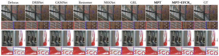
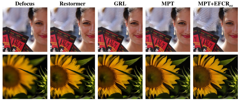
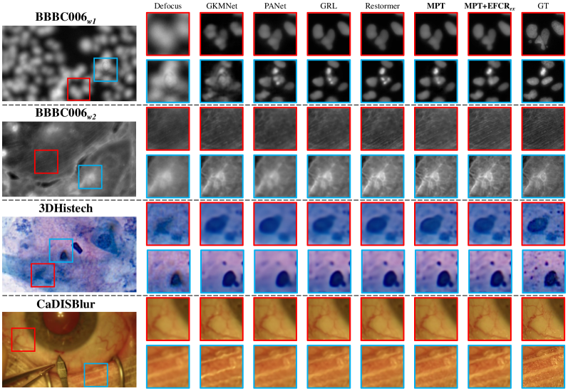
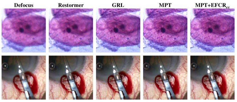
The evaluation on cell microscopy deblur and surgical microscopy deblur is conducted on three microscopy datasets covering a wide range of state-of-the-art defocus deblur methods and image restoration methods. Real-world deblur evaluation is also conducted on DPDD [1] to further prove the generalizability and universality. The result is shown in Tab. 1 and Tab. 2. The proposed framework demonstrates remarkable performance on all microscopy datasets and real-world dataset, showing the superiority of the proposed MPT structure and EFCR training strategy. Compared with Restormer [84], which achieves the second-best performance on BBBC006 [44], MPT (76 FLOPs, 19.80 M) outperforms Restormer (141 FLOPs, 26.12 M) by 0.10 dB and 0.07 dB regarding PSNR while saving 46% FLOPs (for a 256256 input) and 24% parameters. GRL [36] achieves the second-best performance on 3DHistech [17] and CaDISBlur, but it takes 1230 FLOPs for a 256256 input that is 17 larger than ours. Compared to those methods that adopt multi-scale or pyramid design, including MSSNet [31], MIMO-UNet [7], and PANet [51], our method surpasses them significantly in all tests, proving the superiority of our multi-pyramid structure. SwinIR [38] adopts the original local window attention [43], which is hindered by the limited receptive field and fails to build long-range interaction. The visualizations shown in Fig. 3 prove that our method achieves the best restoration of fine details against strong defocus blur, especially for the miniature cell shape and complex cell structure, as well as precise features of surgical anatomies. For real-world deblur on DPDD [1], our method achieves state-of-the-art performance in terms of SSIM and PSNR. Although the LPIPS is slightly lower than the second-best method, it still surpasses the second-best method NRKNet [57] by 17%. It shows that our model is universally applicable to different types of images. Visualization of deblurring on DPDD is provided in supplementary materials.
For MPT trained with EFCR, the performance is improved by learning latent deblur information. By further applying EFCRex to learn cross-domain deblur guidance, the deblur performance is significantly enhanced in all four microscopy datasets, proving that deblurring benefits from cross-domain knowledge, despite the significant feature discrepancy between real-world extra data and the microscope images. Improvements are also observed in SSIM and LPIPS, showing that EFCRex enhances deblurring from the perspective of human visual system, which is of great significance for clinical application. Visualizations in Fig. 3 draw a similar conclusion that the model with EFCRex can restore the fine details more precisely. Real-world deblur can also benefit from EFCR as all three metrics improve with integration of EFCR or EFCRex. Further discussion in Sec. 4.4 shows the superiority of this proposed training diagram against simply pretraining and fine-tuning.
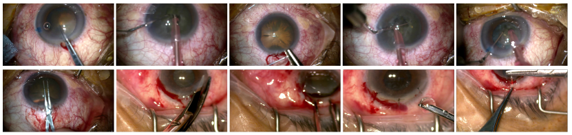
Evaluation on unsupervised deblur
The deblur experiments on unlabeled datasets are conducted to qualitatively evaluate the generalizability of the model, and also to prove the effectiveness of knowledge transfer based on the proposed EFCRex. Two unlabeled microscopy blur datasets are involved, including WNLO [17] and CataBlur. All methods are trained on LFDOF since the rich information in real-world dataset benefits microscopy deblur (see proof in supplementary materials). Unlabeled datasets are adopted as the extra data to learn cross-domain latent information using EFCRex. The qualitative comparison is shown in Fig. 4. Even without EFCRex, our method still shows the best generalizability with fewer artifacts and successfully restores the detail from strong defocus degradation. With the help of EFCRex, the artifacts are further reduced, resulting in clearer deblurred images. Further experiments on the real-world dataset CUHK [63] are conducted (see supplementary materials), which proves the universality of our method.
floatrowsep=qquad,captionskip=10pt {floatrow}
| Configuration | PSNRD | PSNRB | PSNRC |
| (CSWA2) | 26.10 | 34.76 | 44.83 |
| (WA2) | 25.92 | 33.98 | 44.02 |
| (ISCA2) | 26.01 | 34.60 | 44.71 |
| (WA+ISCA) | 26.13 | 34.10 | 44.78 |
| MPT (CSWA+ISCA) | 26.21 | 34.89 | 44.98 |
| Configuration | PSNRD | PSNRB | PSNRC |
| (Cat+GELU) | 26.18 | 34.86 | 44.84 |
| (Add+GELU) | 26.03 | 34.75 | 44.87 |
| (reversed) | 25.98 | 34.72 | 44.50 |
| MPT (FEFN) | 26.21 | 34.89 | 44.98 |
| Configuration | PSNRD | PSNRB | PSNRC |
| MPT+ | +0.01 | +0.04 | +0.05 |
| MPT+EFCR | +0.02 | +0.07 | +0.11 |
| Restormer+EFCR | +0.04 | +0.07 | +0.09 |
| MPT+pretrain | +0.05 | -0.02 | +0.01 |
| MPT+ | +0.05 | +0.21 | +0.18 |
| MPT+EFCRex | +0.06 | +0.27 | +0.27 |
| Restormer+EFCRex | +0.07 | +0.19 | +0.21 |
4.3 Validation on downstream tasks
Cell detection on BBBC006
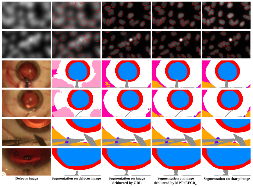
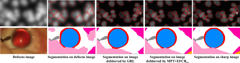
Defocus blur can cause failure in cell detection and segmentation [17, 6] that essential for many biological tasks [52]. The cell segmentation is performed using StarDist [61] on images in BBBC006 before and after deblur. The result is reported in Tab. 6 regarding the average precision (AP) over different intersection-of-union (IoU) thresholds, where higher AP means more cells are successfully detected. Our deblur framework significantly improves the cell detection performance by 19.57% (with extra data) and 18.54% (without extra data) compared with blurry input, surpassing the improvement brought by Restormer (16.05%) and GRL (13.50%). The visualization shown in Fig. 5 also proves that our method achieves better restoration of the cell shape and structure.
Semantic segmentation on CaDISBlur
Semantic segmentation plays an important role in surgical scene understanding [19]. The semantic segmentation of the cataract surgical scene is conducted using OCRNet [83] on CaDISBlur based on CaDIS [19]. Results of images with different focal plane positions (blurry instruments or blurry anatomies, noted as ins or ana) are reported separately in Tab. 7 regarding mean IoU (mIoU) and pixel accuracy (PA). The deblurred images from our method lead to the best performance in most metrics. Visualizations shown in Fig. 5 also prove the superiority of our method.
| Method | Blurry instrument | Blurry anatomies | ||
| mIoUins | PAins | mIoUana | PAana | |
| blur | 0.7577 | 0.8677 | 0.7092 | 0.8194 |
| Restormer [84] | 0.7558 | 0.8803 | 0.8149 | 0.8674 |
| GRL [36] | 0.7606 | 0.8849 | 0.8135 | 0.8689 |
| MPT+EFCR | 0.7610 | 0.8842 | 0.8295 | 0.8830 |
| MPT+EFCRex | 0.7667 | 0.8859 | 0.8361 | 0.8896 |
| sharp | 0.7733 | 0.8886 | 0.8582 | 0.9305 |
4.4 Ablation Studies
For ablation studies, the model variants are evaluated regarding PSNR on DPDD [1], BBBC006w1 [44] and CaDISBlur datasets, which are denoted by PSNRD, PSNRB and PSNRC, respectively. More ablation experiments and analyses are provided in supplementary materials.
Configurations of pyramid block
Ablation studies on CSWA and ISCA are first carried out. As shown in LABEL:tab:ablationcswa, a performance drop is observed when changing CSWA to WA () since WA only models attention within a local window. Although hierarchical network structure in MPT may provide WA with larger receptive field at low-resolution stages, it still causes inferior performance than CSWA since cross-scale interaction are not built. The situation gets worse if the two attention blocks are all changed to WA () since the model totally lost long-range modeling ability in both channel and spatial means. Experiments are then carried out on variants of FEFN, as shown in LABEL:tab:ablationfefn, which proves the superiority of the proposed asymmetrical activation.
Improvements in EFCR
The result is shown in LABEL:tab:ablationefcr. and refer to and . The proposed EFCR and EFCRex yield significant improvements over baseline, not only on our method but also on Restormer [84], proving the effectiveness of the proposed training diagram. Following a similar approach in [60] to pretrain and fine-tune (denoted as MPT+pretrain), the trained model leads to a trivial improvement or even degradation. It further demonstrates the superiority of our EFCRex.
5 Conclusion
This paper presents a unified framework to address the outstanding problems in microscopy defocus deblur. The MPT outperforms existing multi-scale networks by incorporating spatial-channel context from CSWA and ISCA using FEFN. The proposed EFCR enforces the model to explore latent deblur guidance and further learn cross-domain knowledge from the extra data, yielding significant performance gain in both supervised and unsupervised image deblur. In the future, larger-scale datasets, e.g. ImageNet [10], will be adopted for knowledge transfer using EFCR, along with experiments on weakly supervised or unsupervised learning [24] and domain adaptation [89]. Experiments on MPT variants incorporating varied window mechanisms [78] will be carried out.
Acknowledgement
We would like to thank Dr. Danny Siu-Chun Ng and the Department of Ophthalmology and Visual Sciences at the Chinese University of Hong Kong for providing us with the cataract surgery dataset for research.
References
- Abuolaim and Brown [2020] Abdullah Abuolaim and Michael S. Brown. Defocus Deblurring Using Dual-Pixel Data. In Computer Vision – ECCV 2020, pages 111–126, Cham, 2020. Springer International Publishing.
- Ba et al. [2016] Jimmy Lei Ba, Jamie Ryan Kiros, and Geoffrey E Hinton. Layer normalization. arXiv preprint arXiv:1607.06450, 2016.
- Bai and Yuan [2022] Yunpeng Bai and Chun Yuan. Contrastive Learning in Wavelet Domain for Image Dehazing. In 2022 International Joint Conference on Neural Networks (IJCNN), pages 1–7, 2022. ISSN: 2161-4407.
- Bille [2019] Josef F Bille. High resolution imaging in microscopy and ophthalmology: new frontiers in biomedical optics. 2019.
- Bouget et al. [2015] David Bouget, Rodrigo Benenson, Mohamed Omran, Laurent Riffaud, Bernt Schiele, and Pierre Jannin. Detecting Surgical Tools by Modelling Local Appearance and Global Shape. IEEE Transactions on Medical Imaging, 34(12):2603–2617, 2015. Conference Name: IEEE Transactions on Medical Imaging.
- Chen and Ju [2022] Xingyu Chen and Fujiao Ju. Automatic Classification of Pollen Grain Microscope Images Using a Multi-Scale Classifier with SRGAN Deblurring. Applied Sciences, 12(14):7126, 2022. Number: 14 Publisher: Multidisciplinary Digital Publishing Institute.
- Cho et al. [2021] Sung-Jin Cho, Seo-Won Ji, Jun-Pyo Hong, Seung-Won Jung, and Sung-Jea Ko. Rethinking Coarse-To-Fine Approach in Single Image Deblurring. pages 4641–4650, 2021.
- Cui et al. [2023] Yuning Cui, Yi Tao, Wenqi Ren, and Alois Knoll. Dual-Domain Attention for Image Deblurring. Proceedings of the AAAI Conference on Artificial Intelligence, 37(1):479–487, 2023. Number: 1.
- Cuny et al. [2022] Andreas P. Cuny, Fabian P. Schlottmann, Jennifer C. Ewald, Serge Pelet, and Kurt M. Schmoller. Live cell microscopy: From image to insight. Biophysics Reviews, 3(2):021302, 2022.
- Deng et al. [2009] Jia Deng, Wei Dong, Richard Socher, Li-Jia Li, Kai Li, and Li Fei-Fei. Imagenet: A large-scale hierarchical image database. In 2009 IEEE conference on computer vision and pattern recognition, pages 248–255. Ieee, 2009.
- Dong et al. [2019] Han Dong, Aodong Shen, Youyong Kong, Yu Shen, and Huazhong Shu. No-Reference Defocused Image Quality Assessment Based on Human Visual System. In 2019 IEEE International Conference on Signal, Information and Data Processing (ICSIDP), pages 1–6, 2019.
- Dosovitskiy et al. [2021] Alexey Dosovitskiy, Lucas Beyer, Alexander Kolesnikov, Dirk Weissenborn, Xiaohua Zhai, Thomas Unterthiner, Mostafa Dehghani, Matthias Minderer, Georg Heigold, Sylvain Gelly, Jakob Uszkoreit, and Neil Houlsby. An Image is Worth 16x16 Words: Transformers for Image Recognition at Scale, 2021. arXiv:2010.11929 [cs].
- Fang et al. [2022] Rui Fang, Yang-Fan Yu, En-Jie Li, Ning-Xin Lv, Zhao-Chuan Liu, Hong-Gang Zhou, and Xu-Dong Song. Global, regional, national burden and gender disparity of cataract: findings from the global burden of disease study 2019. BMC Public Health, 22(1):2068, 2022.
- Feng et al. [2023] Xin Feng, Yifeng Xu, Guangming Lu, and Wenjie Pei. Hierarchical Contrastive Learning for Pattern-Generalizable Image Corruption Detection. pages 12076–12085, 2023.
- García Calderón et al. [2007] Manuel García Calderón, Daniel Torres Lagares, Carmen Calles Vázquez, Jesús Usón Gargallo, and José Luis Gutiérrez Pérez. The application of microscopic surgery in dentistry. Medicina Oral, Patología Oral y Cirugía Bucal (Internet), 12(4):311–316, 2007.
- Gendy et al. [2022] Garas Gendy, Nabil Sabor, Jingchao Hou, and Guanghui He. Balanced Spatial Feature Distillation and Pyramid Attention Network for Lightweight Image Super-resolution. Neurocomputing, 509:157–166, 2022.
- Geng et al. [2022] Xiebo Geng, Xiuli Liu, Shenghua Cheng, and Shaoqun Zeng. Cervical cytopathology image refocusing via multi-scale attention features and domain normalization. Medical Image Analysis, 81:102566, 2022.
- Ghamsarian et al. [2020] Negin Ghamsarian, Mario Taschwer, and Klaus Schoeffmann. Deblurring Cataract Surgery Videos Using a Multi-Scale Deconvolutional Neural Network. In 2020 IEEE 17th International Symposium on Biomedical Imaging (ISBI), pages 872–876, 2020. out of focus blurry ISSN: 1945-8452.
- Grammatikopoulou et al. [2022] Maria Grammatikopoulou, Evangello Flouty, Abdolrahim Kadkhodamohammadi, Gwenol’e Quellec, Andre Chow, Jean Nehme, Imanol Luengo, and Danail Stoyanov. CaDIS: Cataract Dataset for Image Segmentation, 2022. arXiv:1906.11586 [cs].
- Guo et al. [2022] Jianyuan Guo, Kai Han, Han Wu, Yehui Tang, Xinghao Chen, Yunhe Wang, and Chang Xu. CMT: Convolutional Neural Networks Meet Vision Transformers. pages 12175–12185, 2022.
- Han et al. [2023] Xiaotong Han, Jiaqing Zhang, Zhenzhen Liu, Xuhua Tan, Guangming Jin, Mingguang He, Lixia Luo, and Yizhi Liu. Real-world visual outcomes of cataract surgery based on population-based studies: a systematic review. British Journal of Ophthalmology, 107(8):1056–1065, 2023.
- He et al. [2023a] Chunming He, Chengyu Fang, Yulun Zhang, Kai Li, Longxiang Tang, Chenyu You, Fengyang Xiao, Zhenhua Guo, and Xiu Li. Reti-diff: Illumination degradation image restoration with retinex-based latent diffusion model. 2023a.
- He et al. [2023b] Chunming He, Kai Li, Yachao Zhang, Longxiang Tang, Yulun Zhang, Zhenhua Guo, and Xiu Li. Camouflaged object detection with feature decomposition and edge reconstruction. In Proceedings of the IEEE/CVF Conference on Computer Vision and Pattern Recognition, pages 22046–22055, 2023b.
- He et al. [2024a] Chunming He, Kai Li, Yachao Zhang, Guoxia Xu, Longxiang Tang, Yulun Zhang, Zhenhua Guo, and Xiu Li. Weakly-supervised concealed object segmentation with sam-based pseudo labeling and multi-scale feature grouping. Advances in Neural Information Processing Systems, 36, 2024a.
- He et al. [2024b] Chunming He, Kai Li, Yachao Zhang, Yulun Zhang, Zhenhua Guo, Xiu Li, Martin Danelljan, and Fisher Yu. Strategic preys make acute predators: Enhancing camouflaged object detectors by generating camouflaged objects. 2024b.
- Hendrycks and Gimpel [2016] Dan Hendrycks and Kevin Gimpel. Gaussian error linear units (gelus). arXiv preprint arXiv:1606.08415, 2016.
- Jan and Tang [2022] Feng-Kai Jan and Chih-Wei Tang. Contrastive Learning Aided Single Image Deblurring. In 2022 IEEE International Conference on Consumer Electronics-Asia (ICCE-Asia), pages 1–3, 2022.
- Jiang et al. [2020] Cheng Jiang, Jun Liao, Pei Dong, Zhaoxuan Ma, De Cai, Guoan Zheng, Yueping Liu, Hong Bu, and Jianhua Yao. Blind deblurring for microscopic pathology images using deep learning networks, 2020. arXiv:2011.11879 [cs, eess].
- Kalavakonda et al. [2019] Niveditha Kalavakonda, Blake Hannaford, Zeeshan Qazi, and Laligam Sekhar. Autonomous neurosurgical instrument segmentation using end-to-end learning. In Proceedings of the IEEE/CVF Conference on Computer Vision and Pattern Recognition Workshops, pages 0–0, 2019.
- Keaton et al. [2023] Matthew R Keaton, Ram J Zaveri, and Gianfranco Doretto. Celltranspose: Few-shot domain adaptation for cellular instance segmentation. In Proceedings of the IEEE/CVF Winter Conference on Applications of Computer Vision, pages 455–466, 2023.
- Kim et al. [2023] Kiyeon Kim, Seungyong Lee, and Sunghyun Cho. MSSNet: Multi-Scale-Stage Network for Single Image Deblurring. In Computer Vision – ECCV 2022 Workshops, pages 524–539, Cham, 2023. Springer Nature Switzerland.
- Kornilova et al. [2021] Anatasiia Kornilova, Mikhail Salnikov, Olga Novitskaya, Maria Begicheva, Egor Sevriugov, Kirill Shcherbakov, Valeriya Pronina, and Dmitry V. Dylov. Deep Learning Framework For Mobile Microscopy. In 2021 IEEE 18th International Symposium on Biomedical Imaging (ISBI), pages 324–328, 2021. ISSN: 1945-8452.
- Lee et al. [2019] Junyong Lee, Sungkil Lee, Sunghyun Cho, and Seungyong Lee. Deep Defocus Map Estimation Using Domain Adaptation. pages 12222–12230, 2019. DMENet+SYNDOF.
- Lee et al. [2021] Junyong Lee, Hyeongseok Son, Jaesung Rim, Sunghyun Cho, and Seungyong Lee. Iterative Filter Adaptive Network for Single Image Defocus Deblurring. In 2021 IEEE/CVF Conference on Computer Vision and Pattern Recognition (CVPR), pages 2034–2042, Nashville, TN, USA, 2021. IEEE.
- Li et al. [2021] Chen Li, Adele Moatti, Xuying Zhang, H Troy Ghashghaei, and Alon Greenbaum. Deep learning-based autofocus method enhances image quality in light-sheet fluorescence microscopy. Biomedical Optics Express, 12(8):5214–5226, 2021.
- Li et al. [2023] Yawei Li, Yuchen Fan, Xiaoyu Xiang, Denis Demandolx, Rakesh Ranjan, Radu Timofte, and Luc Van Gool. Efficient and Explicit Modelling of Image Hierarchies for Image Restoration. pages 18278–18289, 2023.
- Liang et al. [2022] Dong Liang, Ling Li, Mingqiang Wei, Shuo Yang, Liyan Zhang, Wenhan Yang, Yun Du, and Huiyu Zhou. Semantically contrastive learning for low-light image enhancement. In Proceedings of the AAAI Conference on Artificial Intelligence, pages 1555–1563, 2022.
- Liang et al. [2021] Jingyun Liang, Jiezhang Cao, Guolei Sun, Kai Zhang, Luc Van Gool, and Radu Timofte. SwinIR: Image Restoration Using Swin Transformer, 2021. arXiv:2108.10257 [cs, eess].
- Lin et al. [2019] Wei Lin, Dongping Wang, Yunlong Meng, and Shih-Chi Chen. Multi-focus microscope with hilo algorithm for fast 3-d fluorescent imaging. PloS one, 14(9):e0222729, 2019.
- Liu et al. [2022] Gaosheng Liu, Huanjing Yue, and Jingyu Yang. A coarse-to-fine convolutional neural network for light field angular super-resolution. In CAAI International Conference on Artificial Intelligence, pages 268–279. Springer, 2022.
- Liu et al. [2020a] Jiahao Liu, Xiaoshuai Huang, Liangyi Chen, and Shan Tan. Deep learning–enhanced fluorescence microscopy via degeneration decoupling. Optics Express, 28(10):14859–14873, 2020a. Publisher: Optica Publishing Group.
- Liu et al. [2020b] Keng-Hao Liu, Chia-Hung Yeh, Juh-Wei Chung, and Chuan-Yu Chang. A Motion Deblur Method Based on Multi-Scale High Frequency Residual Image Learning. IEEE Access, 8:66025–66036, 2020b. Conference Name: IEEE Access.
- Liu et al. [2021] Ze Liu, Yutong Lin, Yue Cao, Han Hu, Yixuan Wei, Zheng Zhang, Stephen Lin, and Baining Guo. Swin Transformer: Hierarchical Vision Transformer using Shifted Windows, 2021. arXiv:2103.14030 [cs].
- Ljosa et al. [2012] Vebjorn Ljosa, Katherine L. Sokolnicki, and Anne E. Carpenter. Annotated high-throughput microscopy image sets for validation. Nature Methods, 9(7):637–637, 2012. Number: 7 Publisher: Nature Publishing Group.
- Loshchilov and Hutter [2016] Ilya Loshchilov and Frank Hutter. Sgdr: Stochastic gradient descent with warm restarts. arXiv preprint arXiv:1608.03983, 2016.
- Loshchilov and Hutter [2017] Ilya Loshchilov and Frank Hutter. Decoupled weight decay regularization. arXiv preprint arXiv:1711.05101, 2017.
- Ma et al. [2022] Haoyu Ma, Shaojun Liu, Qingmin Liao, Juncheng Zhang, and Jing-Hao Xue. Defocus Image Deblurring Network With Defocus Map Estimation as Auxiliary Task. IEEE Transactions on Image Processing, 31:216–226, 2022. Conference Name: IEEE Transactions on Image Processing.
- Ma and Fei [2021] Ling Ma and Baowei Fei. Comprehensive review of surgical microscopes: technology development and medical applications. Journal of Biomedical Optics, 26(1):010901, 2021. Publisher: SPIE.
- Masters [2008] Barry R Masters. History of the optical microscope in cell biology and medicine. eLS, 2008.
- Mazilu et al. [2023] Ioana Mazilu, Shunxin Wang, Sven Dummer, Raymond Veldhuis, Christoph Brune, and Nicola Strisciuglio. Defocus Blur Synthesis and Deblurring via Interpolation and Extrapolation in Latent Space. In Computer Analysis of Images and Patterns, pages 201–211, Cham, 2023. Springer Nature Switzerland.
- Mei et al. [2023] Yiqun Mei, Yuchen Fan, Yulun Zhang, Jiahui Yu, Yuqian Zhou, Ding Liu, Yun Fu, Thomas S. Huang, and Humphrey Shi. Pyramid Attention Network for Image Restoration. International Journal of Computer Vision, 2023.
- Meijering [2012] Erik Meijering. Cell segmentation: 50 years down the road [life sciences]. IEEE signal processing magazine, 29(5):140–145, 2012.
- Michaeli and Irani [2014] Tomer Michaeli and Michal Irani. Blind Deblurring Using Internal Patch Recurrence. In Computer Vision – ECCV 2014, pages 783–798, Cham, 2014. Springer International Publishing.
- Pachade et al. [2021] Samiksha Pachade, Prasanna Porwal, Dhanshree Thulkar, Manesh Kokare, Girish Deshmukh, Vivek Sahasrabuddhe, Luca Giancardo, Gwenolé Quellec, and Fabrice Mériaudeau. Retinal Fundus Multi-Disease Image Dataset (RFMiD): A Dataset for Multi-Disease Detection Research. Data, 6(2):14, 2021. Number: 2 Publisher: Multidisciplinary Digital Publishing Institute.
- Pinkard et al. [2019] Henry Pinkard, Zachary Phillips, Arman Babakhani, Daniel A Fletcher, and Laura Waller. Deep learning for single-shot autofocus microscopy. Optica, 6(6):794–797, 2019.
- Quan et al. [2021] Yuhui Quan, Zicong Wu, and Hui Ji. Gaussian Kernel Mixture Network for Single Image Defocus Deblurring. In Advances in Neural Information Processing Systems, pages 20812–20824. Curran Associates, Inc., 2021.
- Quan et al. [2023] Yuhui Quan, Zicong Wu, and Hui Ji. Neumann Network With Recursive Kernels for Single Image Defocus Deblurring. pages 5754–5763, 2023.
- Ronneberger et al. [2015] Olaf Ronneberger, Philipp Fischer, and Thomas Brox. U-Net: Convolutional Networks for Biomedical Image Segmentation. In Medical Image Computing and Computer-Assisted Intervention – MICCAI 2015, pages 234–241, Cham, 2015. Springer International Publishing.
- Ruan et al. [2021] Lingyan Ruan, Bin Chen, Jizhou Li, and Miu-Ling Lam. AIFNet: All-in-Focus Image Restoration Network Using a Light Field-Based Dataset. IEEE Transactions on Computational Imaging, 7:675–688, 2021. Conference Name: IEEE Transactions on Computational Imaging.
- Ruan et al. [2022] Lingyan Ruan, Bin Chen, Jizhou Li, and Miuling Lam. Learning to Deblur Using Light Field Generated and Real Defocus Images. pages 16304–16313, 2022.
- Schmidt et al. [2018] Uwe Schmidt, Martin Weigert, Coleman Broaddus, and Gene Myers. Cell Detection with Star-Convex Polygons. In Medical Image Computing and Computer Assisted Intervention – MICCAI 2018, pages 265–273, Cham, 2018. Springer International Publishing.
- Schoeffmann et al. [2018] Klaus Schoeffmann, Mario Taschwer, Stephanie Sarny, Bernd Münzer, Manfred Jürgen Primus, and Doris Putzgruber. Cataract-101: video dataset of 101 cataract surgeries. In Proceedings of the 9th ACM Multimedia Systems Conference, pages 421–425, New York, NY, USA, 2018. Association for Computing Machinery.
- Shi et al. [2014] Jianping Shi, Li Xu, and Jiaya Jia. Discriminative Blur Detection Features. pages 2965–2972, 2014.
- Shi et al. [2016] Wenzhe Shi, Jose Caballero, Ferenc Huszar, Johannes Totz, Andrew P. Aitken, Rob Bishop, Daniel Rueckert, and Zehan Wang. Real-Time Single Image and Video Super-Resolution Using an Efficient Sub-Pixel Convolutional Neural Network. pages 1874–1883, 2016.
- Son et al. [2021] Hyeongseok Son, Junyong Lee, Sunghyun Cho, and Seungyong Lee. Single Image Defocus Deblurring Using Kernel-Sharing Parallel Atrous Convolutions. pages 2642–2650, 2021.
- Trikha et al. [2013] Sameer Trikha, Andrew Michael John Turnbull, RJ Morris, David F Anderson, and Parwez Hossain. The journey to femtosecond laser-assisted cataract surgery: new beginnings or a false dawn? Eye, 27(4):461–473, 2013.
- Trukhova et al. [2022] Anna Trukhova, Marina Pavlova, Olga Sinitsyna, and Igor Yaminsky. Microlens-assisted microscopy for biology and medicine. Journal of Biophotonics, 15(9):e202200078, 2022.
- Vaswani et al. [2017] Ashish Vaswani, Noam Shazeer, Niki Parmar, Jakob Uszkoreit, Llion Jones, Aidan N Gomez, Łukasz Kaiser, and Illia Polosukhin. Attention is All you Need. In Advances in Neural Information Processing Systems. Curran Associates, Inc., 2017.
- Vaswani et al. [2021] Ashish Vaswani, Prajit Ramachandran, Aravind Srinivas, Niki Parmar, Blake Hechtman, and Jonathon Shlens. Scaling Local Self-Attention for Parameter Efficient Visual Backbones. pages 12894–12904, 2021.
- Wang and Han [2023] Jiahe Wang and Boran Han. Defocus Deblur Microscopy via Head-to-Tail Cross-scale Fusion, 2023. arXiv:2201.02876 [cs, eess].
- Wang et al. [2021] Wenhai Wang, Enze Xie, Xiang Li, Deng-Ping Fan, Kaitao Song, Ding Liang, Tong Lu, Ping Luo, and Ling Shao. Pyramid Vision Transformer: A Versatile Backbone for Dense Prediction Without Convolutions. pages 568–578, 2021.
- Wang et al. [2023] Yanqi Wang, Zheng Xu, Yifan Yang, Xiaodong Wang, Jiaheng He, Tongqun Ren, and Junshan Liu. Deblurring microscopic image by integrated convolutional neural network. Precision Engineering, 82:44–51, 2023.
- Wang et al. [2004] Zhou Wang, Alan C Bovik, Hamid R Sheikh, and Eero P Simoncelli. Image quality assessment: from error visibility to structural similarity. IEEE transactions on image processing, 13(4):600–612, 2004.
- Wang et al. [2022] Zhendong Wang, Xiaodong Cun, Jianmin Bao, Wengang Zhou, Jianzhuang Liu, and Houqiang Li. Uformer: A General U-Shaped Transformer for Image Restoration. In 2022 IEEE/CVF Conference on Computer Vision and Pattern Recognition (CVPR), pages 17662–17672, New Orleans, LA, USA, 2022. IEEE.
- Wu et al. [2023] Gang Wu, Junjun Jiang, and Xianming Liu. A Practical Contrastive Learning Framework for Single-Image Super-Resolution. IEEE Transactions on Neural Networks and Learning Systems, pages 1–12, 2023. Conference Name: IEEE Transactions on Neural Networks and Learning Systems.
- Wu et al. [2021a] Haiyan Wu, Yanyun Qu, Shaohui Lin, Jian Zhou, Ruizhi Qiao, Zhizhong Zhang, Yuan Xie, and Lizhuang Ma. Contrastive learning for compact single image dehazing. In Proceedings of the IEEE/CVF Conference on Computer Vision and Pattern Recognition, pages 10551–10560, 2021a.
- Wu et al. [2021b] Haiping Wu, Bin Xiao, Noel Codella, Mengchen Liu, Xiyang Dai, Lu Yuan, and Lei Zhang. CvT: Introducing Convolutions to Vision Transformers. pages 22–31, 2021b.
- Xia et al. [2022] Zhuofan Xia, Xuran Pan, Shiji Song, Li Erran Li, and Gao Huang. Vision Transformer With Deformable Attention. pages 4794–4803, 2022.
- Xu et al. [2017] Jing Xu, Xiaolin Tian, Xin Meng, Yan Kong, Shumei Gao, Haoyang Cui, Fei Liu, Liang Xue, Cheng Liu, and Shouyu Wang. Wavefront-sensing-based autofocusing in microscopy. Journal of Biomedical Optics, 22(8):086012–086012, 2017.
- Xu et al. [2021] Ruikang Xu, Zeyu Xiao, Jie Huang, Yueyi Zhang, and Zhiwei Xiong. EDPN: Enhanced Deep Pyramid Network for Blurry Image Restoration. pages 414–423, 2021.
- Yaşargil [2013] Mahmut Gazi Yaşargil. Microsurgery: applied to neurosurgery. Elsevier, 2013.
- Yoo et al. [2018] Seunghwan Yoo, Pablo Ruiz, Xiang Huang, Kuan He, Nicola J Ferrier, Mark Hereld, Alan Selewa, Matthew Daddysman, Norbert Scherer, Oliver Cossairt, et al. 3d image reconstruction from multi-focus microscope: axial super-resolution and multiple-frame processing. In 2018 IEEE International Conference on Acoustics, Speech and Signal Processing (ICASSP), pages 1453–1457. IEEE, 2018.
- Yuan et al. [2020] Yuhui Yuan, Xilin Chen, and Jingdong Wang. Object-Contextual Representations for Semantic Segmentation. In Computer Vision – ECCV 2020, pages 173–190, Cham, 2020. Springer International Publishing.
- Zamir et al. [2022] Syed Waqas Zamir, Aditya Arora, Salman Khan, Munawar Hayat, Fahad Shahbaz Khan, and Ming-Hsuan Yang. Restormer: Efficient Transformer for High-Resolution Image Restoration. pages 5728–5739, 2022.
- Zhang et al. [2022a] Chi Zhang, Hao Jiang, Weihuang Liu, Junyi Li, Shiming Tang, Mario Juhas, and Yang Zhang. Correction of out-of-focus microscopic images by deep learning. Computational and Structural Biotechnology Journal, 20:1957–1966, 2022a.
- Zhang and Zhang [2019] Dengsheng Zhang and Dengsheng Zhang. Wavelet transform. Fundamentals of image data mining: Analysis, Features, Classification and Retrieval, pages 35–44, 2019.
- Zhang et al. [2018] Richard Zhang, Phillip Isola, Alexei A Efros, Eli Shechtman, and Oliver Wang. The unreasonable effectiveness of deep features as a perceptual metric. In Proceedings of the IEEE conference on computer vision and pattern recognition, pages 586–595, 2018.
- Zhang et al. [2022b] Yunlong Zhang, Yuxuan Sun, Honglin Li, Sunyi Zheng, Chenglu Zhu, and Lin Yang. Benchmarking the Robustness of Deep Neural Networks to Common Corruptions in Digital Pathology. In Medical Image Computing and Computer Assisted Intervention – MICCAI 2022, pages 242–252, Cham, 2022b. Springer Nature Switzerland.
- Zhang et al. [2022c] Yuelin Zhang, Sihao Xiang, Zehuan Wang, Xiaoyan Peng, Yutong Tian, Shukai Duan, and Jia Yan. Tdacnn: Target-domain-free domain adaptation convolutional neural network for drift compensation in gas sensors. Sensors and Actuators B: Chemical, 361:131739, 2022c.
- Zhang et al. [2023a] Yanni Zhang, Qiang Li, Miao Qi, Di Liu, Jun Kong, and Jianzhong Wang. Multi-scale frequency separation network for image deblurring. IEEE Transactions on Circuits and Systems for Video Technology, 2023a.
- Zhang et al. [2023b] Yulun Zhang, Donglai Wei, Richard Schalek, Yuelong Wu, Stephen Turney, Jeff Lichtman, Hanspeter Pfister, and Yun Fu. High-Throughput Microscopy Image Deblurring with Graph Reasoning Attention Network. In 2023 IEEE 20th International Symposium on Biomedical Imaging (ISBI), pages 1–5, 2023b. ISSN: 1945-8452.
- Zhao et al. [2023] Bingxin Zhao, Weihong Li, and Weiguo Gong. Real-aware motion deblurring using multi-attention CycleGAN with contrastive guidance. Digital Signal Processing, 135:103953, 2023.
- Zhao et al. [2020] Huangxuan Zhao, Ziwen Ke, Ningbo Chen, Songjian Wang, Ke Li, Lidai Wang, Xiaojing Gong, Wei Zheng, Liang Song, Zhicheng Liu, Dong Liang, and Chengbo Liu. A new deep learning method for image deblurring in optical microscopic systems. Journal of Biophotonics, 13(3):e201960147, 2020. _eprint: https://onlinelibrary.wiley.com/doi/pdf/10.1002/jbio.201960147.
- Zhao et al. [2022] Suiyi Zhao, Zhao Zhang, Richang Hong, Mingliang Xu, Yi Yang, and Meng Wang. FCL-GAN: A Lightweight and Real-Time Baseline for Unsupervised Blind Image Deblurring. In Proceedings of the 30th ACM International Conference on Multimedia, pages 6220–6229, New York, NY, USA, 2022. Association for Computing Machinery.
- Zontak et al. [2013] Maria Zontak, Inbar Mosseri, and Michal Irani. Separating signal from noise using patch recurrence across scales. In proceedings of the IEEE conference on computer vision and pattern recognition, pages 1195–1202, 2013.
Supplementary Material
6 Dataset Description
This section provides supplementary information about the datasets involved in the evaluation. The details are shown in Tab. 8.
For real-world scenarios, three datasets are included. For DPDD [1], the sharp-blur training pairs are collected sequentially by a DSLR camera with different aperture sizes. A labeled defocus deblur dataset LFDOF [59] collected by a light field camera is adopted in this paper as extra data for knowledge transfer since the training pairs captured by the light field camera have strict pixel-wise consistency [60]. LFDOF contains many more images than DPDD, which also benefits the microscopy deblur by transferring rich cross-domain information. For unsupervised deblur, an unlabeled blur dataset CUHK [63] is adopted, which is collected from the internet.
Three datasets are involved in cell microscopy deblur. BBBC006 comes from the Broad Bioimage Benchmark Collection [44], which contains images in two sub-sets stained by Hoechst and phalloidin captured by fluorescence microscope. It contains images with different focal planes (denoted as different z-stacks). Following dataset description [44], images collected on the optimal focal length (z-stack = 16) are set as the ground truth, and images above the optimal focal plane are used for training. To avoid redundancy, images with z-stack = [2, 6, 10] are set as blurry input for training. Since the images in BBBC006 only contain a single grayscale channel, for EFCRex in BBBC006, the images in LFDOF are converted into grayscale with one channel. 3DHistech and WNLO [17] are two cell imaging datasets for cytopathology scanned by digital scanners. The labeled dataset 3DHistech is scanned using different focal planes, where the focal plane with the most cells in focus is set as the ground truth. WNLO is an unlabeled dataset with defocus images only.
Regarding the surgical microscopy deblur, two new datasets are presented, which are the labeled synthesized dataset CaDISBlur and the unlabeled cataract surgery defocus blur dataset CataBlur. CaDISBlur is synthesized based on CaDIS [19], which is a dataset for surgical scene semantic segmentation. Leveraging segmentation masks, the instruments and anatomies are blurred separately to simulate different focal planes. The original images in CaDIS are of high quality thus they can be treated as the sharp ground truth. CataBlur is an unlabeled real defocus blur dataset acquired during cataract surgeries, from which the severity of defocus blur in microscope surgery can be observed.
| Scenario | Dataset | #Image | Resolution | Label |
| Real world | DPDD [1] | 500 | 16801120 | Labeled |
| LFDOF [59] | 12,826 | 1008688 | Labeled | |
| CUHK [63] | 704 | 610470 | Unlabeled | |
| Cell microscopy | BBBC006 [44] | 6144 | 696520 | Labeled |
| 3DHistech [17] | 94,973 | 256256 | Labeled | |
| WNLO [17] | 108,065 | 256256 | Unlabeled | |
| Surgical microscopy | CaDISBlur | 9340 | 960540 | Labeled |
| CataBlur | 4614 | 1280720 | Unlabeled |
7 Supplementary Experiments
Feature deficiency
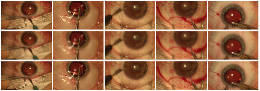
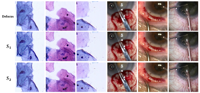
As addressed in Sec. 1, the feature deficiency in microscopy datasets can pose harm to generalizability. To further demonstrate the drawbacks brought by feature deficiency, experiments are conducted on unlabeled microscopy datasets WNLO [17] and CataBlur regarding two settings. For the first setting , the MPT is trained on intra-domain microscopy images and tested on unlabeled microscopy datasets, i.e., trained on 3DHistech [17] then tested on WNLO [17], and trained on CaDISBlur then tested on CataBlur. For , the MPT is trained on cross-domain real-world images (LFDOF [59]) and tested on unlabeled microscopy datasets.
The deblur results are shown in Fig. 6, from which it can be observed that the model trained with cross-domain real-world dataset () leads to fewer artifacts and more fine details in its deblurred results than the model trained with intra-domain microscopy dataset (). This phenomenon proves the existence of feature deficiency in microscopy dataset, i.e., the model trained with microscopy dataset suffers from poor generalizability caused by the insufficient features contained in microscopy dataset. From visualization of , the necessity of learning cross-domain rich deblur guidance is also proved.
Ablation studies on CSWA and FEFN
| Configuration | PSNRD | PSNRB | PSNRC |
| (w/o shortcut) | -0.14 | -0.02 | -0.15 |
| (w/o linear) | -0.05 | -0.04 | -0.07 |
| (max pooling) | -0.37 | -0.49 | -0.63 |
| (convolution) | -0.03 | -0.09 | -0.06 |
| (interpolate) | -0.01 | +0.02 | -0.04 |
| -0.09 | -0.07 | -0.12 | |
| -0.06 | -0.13 | -0.09 | |
| -0.23 | -0.17 | -0.48 | |
| -0.09 | -0.12 | -0.15 | |
| -0.60 | -0.74 | -0.91 | |
| -0.24 | -0.31 | -0.20 |
Additional ablation studies regarding the structure of the proposed CSWA and FEFN are conducted. The results are shown in Tab. 9. For the downsampling module adopted in CSWA (Fig. 7), a series of variants are evaluated, including variant without shortcut connection (), variant without linear projection and shortcut connection (), and variants changing the downsampling operation from strided average pooling to strided maximum pooling (), strided convolution (), and bicubic interpolation (). The results in Tab. 9 show that the current baseline outperforms the variants in most of the situations. Although , which adopts bicubic interpolation, achieves almost the same performance as the baseline or even trivial improvement, it leads to complex computation that hinders the parallel training and inference. Ablation studies on NPConv and shifting window mechanism in CSWA are also carried out, which are denoted by and , respectively. The results prove the superiority of our design.
Going further from the experiments in Sec. 4.4, more ablation studies regarding the asymmetric activation mechanism and shortcut connection in FEFN are conducted, including four variants as shown in Fig. 8. Based on the result reported in Tab. 9, we can conclude that the baseline structure achieves the best performance among all the variants.
Ablation studies on pyramid scales
Following the description in Sec. 4.1 to keep the sub-block number, feature dimensions, and attention heads unchanged, ablation studies on pyramid scales () are carried out regarding variants with similar FLOPs and parameters:
1) V1: . In this variant, CSWA actually downgrades to the original WA (the variant with WA+ISCA shown in LABEL:tab:ablationcswa), and the image pyramid is not constructed.
2) V2: , , . This variant explores the pyramid structure with smaller scales than the baseline.
3) V3: , , . This variant adopts larger-scale pyramids than the baseline.
As shown by the results in Tab. 10, all the variants lead to performance drop. For V1 with no pyramid structure, significant performance degradation is observed on BBBC006, which is the dataset with one of the longest attention spans. A similar phenomenon is observed in SwinIR [38] that does not feature a multi-scale pyramid. It achieves inferior performance on BBBC006 as shown in Tab. 1. These together prove the effectiveness of our multi-pyramid design, especially for the microscopy deblur tasks. The performance degradation in V2 and V3 shows the superiority of the pyramid scales in the baseline model.
| Pyramid | PSNRD | PSNRB | PSNRC |
| V1 | -0.08 | -0.79 | -0.20 |
| V2 | -0.05 | -0.02 | -0.07 |
| V3 | -0.01 | -0.04 | -0.03 |
Additional visualization
Demonstrations of supervised real-world deblur are shown in Fig. 10 for labeled datasets DPDD [1]. Illustrations of unsupervised deblur on microscopy dataset and real-world dataset are provided in Fig. 11(a) and Fig. 11(b), respectively. The visualization proves that our method achieves the best performance on microscopy datasets and real-world datasets regarding various patterns in both supervised and unsupervised scenarios. More visualizations of the results of cell detection on BBBC006 [44] and surgical scene semantic segmentation on CaDISBlur are provided in Fig. 12, from which it can be concluded that the downstream tasks results on deblurred images from our method achieves more satisfactory outcomes.