Harnessing Intra-group Variations Via a Population-Level Context for Pathology Detection
Abstract
Realizing sufficient separability between the distributions of healthy and pathological samples is a critical obstacle for pathology detection convolutional models. Moreover, these models exhibit a bias for contrast-based images, with diminished performance on texture-based medical images. This study introduces the notion of a population-level context for pathology detection and employs a graph theoretic approach to model and incorporate it into the latent code of an autoencoder via a refinement module we term PopuSense. PopuSense seeks to capture additional intra-group variations inherent in biomedical data that a local or global context of the convolutional model might miss or smooth out. Experiments on contrast-based and texture-based images, with minimal adaptation, encounter the existing preference for intensity-based input. Nevertheless, PopuSense demonstrates improved separability in contrast-based images, presenting an additional avenue for refining representations learned by a model.
Keywords Pathology Detection Anomaly Detection Latent Code Refinement Population-level Context Hypergraph
1 Introduction
The dichotomy between contrast-based and texture-based images is inadvertently apparent in anomaly or pathology detection models employing auto-encoders, variational encoders and generative adversarial models, which we collectively refer to as pathology detection convolutional models (PDCs). Evaluation of these models on medical images highlights a notable preference for intensity-driven variations, as evidenced by the relatively superior performance of state-of-the-art (SOTA) models on contrast-based images 1, 2, 3.
Moreover, empirical evidence shows that PDCs may still reconstruct anomalous regions, hindering the separability of healthy and pathological distributions, and resulting in high false positive rates 1, 2, 4, 3. One of the cited challenges lies in striking a balance between learning a compact latent code and one that is also sufficiently informative to prevent the reconstruction of out-of-distribution data points. Relatedly, medical images exhibit internal variations within subjects and lesion types that are typically smoothed out in favor of population-level factors. As a result, a compact and informative code for a PDC would not only go beyond intensity-driven optimization but also incorporate the internal diversity within a healthy population.
This study models a population-level context for pathology detection, employing a hypergraph in the latent space of a PDC to harness underutilized associations and augment the latent code of the PDC. We term the associated module PopuSense and apply it to both intensity-based and texture-based datasets to explore its utility under the different input types. Specifically, this research
-
•
Introduces a latent code refinement module (PopuSense) that embodies a population-level context in the latent space of a pathology detection convolution autoencoder and models it using higher-order relational associations.
-
•
Conducts experiments on the proposed method applying it to a brain tumor dataset and a retinal fundus dataset, which are intensity-based and texture-based inputs respectively.
-
•
Under vanilla conditions, empirical results demonstrate improvement for intensity-based input and a challenge extending similar performance to textural input, a recurring theme in existing research.
2 Background
The core structure of a pathology detection convolutional model (PDC) is a standard auto-encoder convolution model. Variational autoencoders (VAEs) and generative adversarial networks (GANs) extend the autoecoder (AE) and are capable of image synthesis. These models have been utilized in pathology or anomaly detection to estimate the latent representation of normal or healthy distributions, and then determine anomalous or out-of-distribution observations based on the magnitude of the reconstruction error 5, 1.
To realize a latent code that is both concise and sufficiently informative, some methods employ input-centric tactics to make the model resilient to spurious perturbations in the input 6, 7, 8, 1. Other methods incorporate auxiliary branches into the model, providing control, conditioning or guidance to facilitate learning a representation that aligns with some desired prior such as structural correspondence 9, 10, 8, 1. Furthermore, some techniques entail learning a dictionary (also referred to as a codebook or memory bank) of latent codes making the PDC model adaptable to variations or nuances in the data 11, 12, 10.
PDCs, by employing convolutional neural networks (CNNs), inherently entail a local spatial context by virtue of the convolution kernel and operation, and have been extended to capture global spatial contexts via techniques such as dilated kernels, skip connections, and attention modules 13. By learning a set of possible latent codes, the dictionary-learning technique seems to factor in an additional context; a population- or sample-level context. We envisage this context as a potential avenue for accommodating intra-group heterogeneities in medical data.
Nevertheless, learning a dictionary of latent codes increases model parameters as a function of the codebook size, complicating training and data needs 12. Additionally, determining an optimal codebook size and promoting its diversity may call for considerable hyper-parameter tuning and empirical analysis 12. Instead, we consider a graph theoretic approach to capture and incorporate the relational structure of hidden or underutilized associations in the population context.
Graph learning concepts offer adaptable frameworks for addressing graph data as well as modeling relationships in non-graphical domains 14, 15, 16, 17. Moreover, they facilitate a mathematical framework for representing joint probability distributions when combined with probability theory 18. We leverage a hypergraph to incorporate relational assumptions in a non-graphical domain for the purposes of enhancing representations learned by a pathology detection model, fostering the separability of healthy and pathological reconstruction errors.
3 Method
Motivated by the notion of a population-level context and its potential utility, we integrate it into a reasonably capable PDC. This section details the associated model design and evaluation approach.
3.1 Model
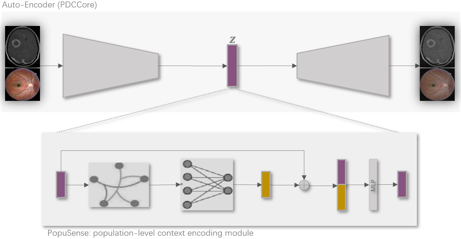
The proposed method operates within the latent space of a pathology detection convolutional model (PDC). We refer to the core architecture of the PDC as the PDCCore and to the module that refines the latent code as the PopuSense module. Figure Figure 1 illustrates the model components.
PDCCore: This part of the model entails a convolutional auto-encoder (AE) with a tunable bottleneck. A ResNet101 backbone is adopted for the encoder function of the AE and augmented with multi-scale features to enhance the detection of small lesions. A bottleneck module is appended to further constrain the size of the latent code.
In contrast, the decoder function is underspecified relative to its partner encoder by having fewer layers and not incorporating skip connections from all the layers of the encoder. Specifically, the additional bottleneck component of the encoder does not have a matching module in the decoder, and instead, the decoder simply upsamples through bilinear interpolation. In addition, the decoder contains no skip connections from the early layers of the encoder, which capture lower-level features.
Consequently, the AE has a powerful encoder for our medical images and the asymmetry regularizes against learning an identity function. Moreover, for additional regularization, a denoising layer is introduced just before the encoder and initially set to a dropout unit for simplicity.
PopuSense: This part of the model employs a graph-theoretic approach to represent and encode a population-level context for normal samples. Firstly, a population-level similarity context is modeled using a hypergraph. The observations in a batch are depicted as nodes while similarity scores between the latent codes from the PDCCore correspond to edges. Similarity scores are computed as the L2 norm between latent codes and edges are only activated for the top k scores. The result is a binary incidence matrix and an associated weight matrix that is equivalent to the identity matrix as our hypergraph is unweighted. The dimensions and correspond to the number of observations in a batch and the size of a neighborhood.
Thereafter, we apply a graph convolutional neural network to the latent codes of the base model, denoted , and the estimated hypergraph . Finally, the initial latent codes are augmented with the output of the graph convolution using a single fully connected layer and projected to a lower dimension of the same size as the bottleneck of the base model PDCCore, producing the enhanced latent code. Equation 1 depicts this process, where the component is derived from the hypergraph Laplacian and serves as a transition matrix for propagating messages during the graph convolution operation. In addition, is the embedding of the encoded population-level context, denotes the hypergraph convolution operation, is the projection matrix from layer to , and and are nonlinearity functions.
| (1) | ||||
Loss function: The loss function, as outlined in equation Equation 2, combines the mean squared error (MSE) and the structural similarity index metric (SSIM). The SSIM offers a more comprehensive assessment of image quality since it considers luminance, contrast and structure. We empirically find that supplementing it with the MSE loss yields reconstructions with contrast and luminance that closely resemble the original input. The contribution of the two metrics can be adjusted using hyperparameters and . In the equation, is the original input and is the reconstructed output. There is no additional regularization for the latent code as we first explore the capacity of PopuSense unconfounded.
| (2) |
We primarily implement using PyTorch 19 and leverage existing packages for the foundational elements in well-established tasks. The PyTorch Segmentation Models package 20 provides access to the ResNet101 backone through its model zoo. To compute SSIM for both grayscale and color images, we utilize MS-SSIM 21. Furthermore, Deep Hypergraphs (DHG) 22 serves as a PyTorch-compatible graph learning module that extends existing graph neural networks to hypergraphs.
3.2 Evaluation
The difference between an original input and its reconstruction , denoted as the residual , represents the erroneous pixels, pinpointing pathological regions. Examining the distribution of these residuals provides insights into the model’s ability to distinguish between healthy and pathological samples. Distribution plots characterize the residuals, and the goal is minimal overlap between the healthy and pathological distributions. Additionally, the Kruskal-Wallis test assesses the statistical difference between the distributions.
Furthermore, thresholding the residuals can categorize samples as healthy or pathological, facilitating the exploration of false positive rates via the accompanying ROC (receiver operating characteristic) curves. Thresholds are based on the statistics of the training sample residuals.
4 Experiments
4.1 Input setup
Leveraging publicly available sources, we assemble two datasets corresponding to contrast and textural input types. The contrast-based dataset is a public brain tumor dataset that is a collection of three other datasets and has diversity in intensity levels 23. We select axial plane images and drop low-quality images that have been overly preprocessed, resulting in 481 images that are marked healthy. For the texture-based dataset, we adopt a retinal fundus public dataset comprising of a diverse collection of other datasets and with a unified definition of what is normal or healthy 24. We access a total of 3515 healthy images.
Images undergo noise reduction preprocessing using median blur and CLAHE, and get resized and center cropped to yield a final resolution of 160x160. In addition, the green color channel is selected for the fundus dataset and all images are standardized to the range [0,1]. Furthermore, sample sizes are bolstered via six offline augmentations that entail combinations of blurring, vertical and horizontal flipping, and random masking using a random number and size of square-shaped masks that are filled with the median value of the input image.
The training phase exclusively utilizes healthy samples, while a combination of healthy and pathological images is used for evaluation. The healthy images are randomly split into 80% training set and a 20% test set.
4.2 Comparison groups
As described in the method section, we build a reasonably capable autoencoder (PDCCore) and then explore the contribution of PopuSense, which incorporates a population-based context. Three models are configured as per below and trained with a batch size of 32.
-
•
PDCCore: This represents a conventional pathology detection autoencoder and is the baseline case
-
•
Narrow PopuSense: Here we implement PopuSense using 35% of the batch-size as the number of closest neighbors to consider when representing the population-level context.
-
•
Wide PopuSense: In this case PopuSense uses 70% of the batch-size as the number of closest neighbors to consider when representing the population-level context.
4.3 Results and discussion
Figure 2 presents distribution plots of the residuals or error scores for the two types of input and across the three model configurations. Ideally, we expect the distributions for new normal observations and training normal samples to align, while their overlap with the distribution of pathological samples is minimized. This expectation is evident for the contrast-based dataset but not for the texture-based images. Additionally, the Wide PopuSense configuration has the least overlap between healthy and pathological distributions for contrast-based input, while Narrow PopuSense has a distribution overlap pattern close to the baseline case (PDCCore).
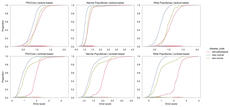
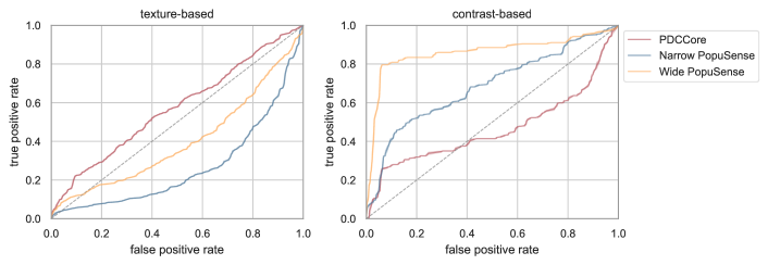
Figure 3 illustrates the ROC curves associated with classifying observations as healthy or pathological based on the thresholded error scores. The ideal model is characterized by points near the top-left corner, representing a good balance of high specificity (true positive rates) and reduced false positive rates. Similar to the results from the distribution plots, the contrast-based dataset has relatively better performance under the Wide PopuSense configuration. Performance under texture-based input appears no better than chance.
Relatedly, the Kruskal-Wallis tests were statistically significant for all pairs of distributions, and a qualitative review of the reconstructed images revealed ongoing reconstruction of anomalous regions by the pathology detection convolution encoder.
Applying Wide PopuSense to contrast-based input attains the best separability between healthy and pathological distributions. Moreover, a wide-reaching population-level context yields better performance than a small one. This demonstrates the utility of the proposed method in enhancing the representations learned by a pathology detection convolutional autoencoder. Furthermore, performance under texture-based input suggests that the vanilla version of PopuSense, a version without extra guidance or optimization support, seems inadequate for textural input. Overall, PopuSense addresses the separability problem for contrast-based images with minimal adaptation, and requires further investigation for textural input
5 Conclusion
This study presents a population-level context for pathology detection and takes on a graph theoretic approach to model and encode it into the latent representation of the underlying convolutional autoencoder. The aim is to enhance the code’s capacity to capture additional intra-group variations inherent in biomedical data, which a local or global context of the convolutional model might miss or smooth out. Experimental results reveal improved separability between healthy and pathological observations in contrast-based medical images and struggle with extending that performance to textural input, which is a recurring challenge in existing research. All in all, such an approach avails an alternate avenue for enhancing the representations learned by a pathology detection convolutional model.
Acknowledgements
We thank the support from the National Natural Science Foundation of China 31970752; 32350410397; Science, Technology, Innovation Commission of Shenzhen Municipality JSGG20200225150707332, JCYJ20220530143014032, WDZC20200820173710001, WDZC20200821150704001; Shenzhen Medical Academy of Research and Translation, D2301002; Shenzhen Bay Laboratory Open Funding, SZBL2020090501004; Department of Chemical Engineering-iBHE special cooperation joint fund project, DCE-iBHE-2022-3; Tsinghua Shenzhen International Graduate School Cross-disciplinary Research and Innovation Fund Research Plan, JC2022009; and Bureau of Planning, Land and Resources of Shenzhen Municipality (2022) 207.
References
- Tschuchnig and Gadermayr [2022] Maximilian E. Tschuchnig and Michael Gadermayr. Anomaly Detection in Medical Imaging - A Mini Review. In Peter Haber, Thomas J. Lampoltshammer, Helmut Leopold, and Manfred Mayr, editors, Data Science – Analytics and Applications, pages 33–38, Wiesbaden, 2022. Springer Fachmedien. ISBN 9783658362959. doi:10.1007/978-3-658-36295-9_5.
- Bercea et al. [2023a] Cosmin I. Bercea, Benedikt Wiestler, Daniel Rueckert, and Julia A. Schnabel. Generalizing Unsupervised Anomaly Detection: Towards Unbiased Pathology Screening. April 2023a. URL https://openreview.net/forum?id=8ojx-Ld3yjR.
- Lagogiannis et al. [2024] Ioannis Lagogiannis, Felix Meissen, Georgios Kaissis, and Daniel Rueckert. Unsupervised Pathology Detection: A Deep Dive Into the State of the Art. IEEE Transactions on Medical Imaging, 43(1):241–252, January 2024. ISSN 0278-0062, 1558-254X. doi:10.1109/TMI.2023.3298093. URL http://arxiv.org/abs/2303.00609. arXiv:2303.00609 [cs].
- Bercea et al. [2023b] Cosmin I. Bercea, Daniel Rueckert, and Julia A. Schnabel. What Do AEs Learn? Challenging Common Assumptions in Unsupervised Anomaly Detection. In Hayit Greenspan, Anant Madabhushi, Parvin Mousavi, Septimiu Salcudean, James Duncan, Tanveer Syeda-Mahmood, and Russell Taylor, editors, Medical Image Computing and Computer Assisted Intervention – MICCAI 2023, Lecture Notes in Computer Science, pages 304–314, Cham, 2023b. Springer Nature Switzerland. ISBN 9783031439049. doi:10.1007/978-3-031-43904-9_30.
- Bulusu et al. [2020] Saikiran Bulusu, Bhavya Kailkhura, Bo Li, Pramod K. Varshney, and Dawn Song. Anomalous Example Detection in Deep Learning: A Survey. IEEE Access, 8:132330–132347, 2020. ISSN 2169-3536. doi:10.1109/ACCESS.2020.3010274. URL https://ieeexplore.ieee.org/document/9144212.
- Tan et al. [2023] Zhiyong Tan, Yuhai Yu, Jiana Meng, Shuang Liu, and Wei Li. Self-supervised learning with self-distillation on COVID-19 medical image classification. Computer Methods and Programs in Biomedicine, 243:107876, October 2023. ISSN 1872-7565. doi:10.1016/j.cmpb.2023.107876.
- Zhou et al. [2023] Lei Zhou, Huidong Liu, Joseph Bae, Junjun He, Dimitris Samaras, and Prateek Prasanna. Self Pre-training with Masked Autoencoders for Medical Image Classification and Segmentation, April 2023. URL http://arxiv.org/abs/2203.05573. arXiv:2203.05573 [cs, eess].
- Fragemann et al. [2022] Jana Fragemann, Lynton Ardizzone, Jan Egger, and Jens Kleesiek. Review of Disentanglement Approaches for Medical Applications – Towards Solving the Gordian Knot of Generative Models in Healthcare, March 2022. URL http://arxiv.org/abs/2203.11132. arXiv:2203.11132 [cs].
- Zhou et al. [2022a] Kang Zhou, Jing Li, Yuting Xiao, Jianlong Yang, Jun Cheng, Wen Liu, Weixin Luo, Jiang Liu, and Shenghua Gao. Memorizing Structure-Texture Correspondence for Image Anomaly Detection. IEEE Transactions on Neural Networks and Learning Systems, 33(6):2335–2349, June 2022a. ISSN 2162-2388. doi:10.1109/TNNLS.2021.3101403. URL https://ieeexplore.ieee.org/abstract/document/9513473.
- Zhou et al. [2022b] Kang Zhou, Jing Li, Weixin Luo, Zhengxin Li, Jianlong Yang, Huazhu Fu, Jun Cheng, Jiang Liu, and Shenghua Gao. Proxy-Bridged Image Reconstruction Network for Anomaly Detection in Medical Images. IEEE Transactions on Medical Imaging, 41(3):582–594, March 2022b. ISSN 0278-0062, 1558-254X. doi:10.1109/TMI.2021.3118223. URL https://ieeexplore.ieee.org/document/9570167/.
- Gong et al. [2019] Dong Gong, Lingqiao Liu, Vuong Le, Budhaditya Saha, Moussa Reda Mansour, Svetha Venkatesh, and Anton Van Den Hengel. Memorizing Normality to Detect Anomaly: Memory-Augmented Deep Autoencoder for Unsupervised Anomaly Detection. In 2019 IEEE/CVF International Conference on Computer Vision (ICCV), pages 1705–1714, October 2019. doi:10.1109/ICCV.2019.00179. URL https://ieeexplore.ieee.org/document/9010977. ISSN: 2380-7504.
- ILIE-ABLACHIM and DUMITRESCU [2022] Denis C. ILIE-ABLACHIM and Bogdan DUMITRESCU. Anomaly Detection with Selective Dictionary Learning. In 2022 8th International Conference on Control, Decision and Information Technologies (CoDIT), volume 1, pages 255–260, May 2022. doi:10.1109/CoDIT55151.2022.9803930. URL https://ieeexplore.ieee.org/document/9803930. ISSN: 2576-3555.
- Alzubaidi et al. [2021] Laith Alzubaidi, Jinglan Zhang, Amjad J. Humaidi, Ayad Al-Dujaili, Ye Duan, Omran Al-Shamma, J. Santamaría, Mohammed A. Fadhel, Muthana Al-Amidie, and Laith Farhan. Review of deep learning: concepts, CNN architectures, challenges, applications, future directions. Journal of Big Data, 8(1):53, March 2021. ISSN 2196-1115. doi:10.1186/s40537-021-00444-8. URL https://doi.org/10.1186/s40537-021-00444-8.
- [14] Feng Xia, Ke Sun, Shuo Yu, Abdul Aziz, Liangtian Wan, Shirui Pan, and Huan Liu. Graph learning: A survey. 2(2):109–127. ISSN 2691-4581. doi:10.1109/TAI.2021.3076021. URL http://arxiv.org/abs/2105.00696.
- Ma et al. [2021] Xiaoxiao Ma, Jia Wu, Shan Xue, Jian Yang, Chuan Zhou, Quan Z. Sheng, Hui Xiong, and Leman Akoglu. A Comprehensive Survey on Graph Anomaly Detection with Deep Learning. IEEE Transactions on Knowledge and Data Engineering, pages 1–1, 2021. ISSN 1041-4347, 1558-2191, 2326-3865. doi:10.1109/TKDE.2021.3118815. URL https://ieeexplore.ieee.org/document/9565320/.
- [16] Qionghai Dai and Yue Gao. Hypergraph Computation. Artificial Intelligence: Foundations, Theory, and Algorithms. Springer Nature Singapore. ISBN 978-981-9901-84-5 978-981-9901-85-2. doi:10.1007/978-981-99-0185-2. URL https://link.springer.com/10.1007/978-981-99-0185-2.
- Ahmedt-Aristizabal et al. [2022] David Ahmedt-Aristizabal, Mohammad Ali Armin, Simon Denman, Clinton Fookes, and Lars Petersson. A survey on graph-based deep learning for computational histopathology. Computerized Medical Imaging and Graphics, 95:102027, January 2022. ISSN 0895-6111. doi:10.1016/j.compmedimag.2021.102027. URL https://www.sciencedirect.com/science/article/pii/S0895611121001762.
- Dietrichstein et al. [2022] Marc Dietrichstein, David Major, Martin Trapp, Maria Wimmer, Dimitrios Lenis, Philip Winter, Astrid Berg, Theresa Neubauer, and Katja Bühler. Anomaly Detection Using Generative Models and Sum-Product Networks in Mammography Scans. In Anirban Mukhopadhyay, Ilkay Oksuz, Sandy Engelhardt, Dajiang Zhu, and Yixuan Yuan, editors, Deep Generative Models, Lecture Notes in Computer Science, pages 77–86, Cham, 2022. Springer Nature Switzerland. ISBN 9783031185762. doi:10.1007/978-3-031-18576-2_8.
- Paszke et al. [2017] Adam Paszke, Sam Gross, Soumith Chintala, Gregory Chanan, Edward Yang, Zachary DeVito, Zeming Lin, Alban Desmaison, Luca Antiga, and Adam Lerer. Automatic differentiation in pytorch. 2017.
- Iakubovskii [2019] Pavel Iakubovskii. Segmentation models pytorch. https://github.com/qubvel/segmentation_models.pytorch, 2019.
- [21] lartpang. A better pytorch-based implementation for the mean structural similarity. Differentiable simpler SSIM and MS-SSIM. URL https://github.com/lartpang/mssim.pytorch. (Jun 21, 2022).
- Gao et al. [2022] Yue Gao, Yifan Feng, Shuyi Ji, and Rongrong Ji. Hgnn+: General hypergraph neural networks. IEEE Transactions on Pattern Analysis and Machine Intelligence, 2022.
- [23] Brain Tumor MRI Dataset. URL https://www.kaggle.com/datasets/masoudnickparvar/brain-tumor-mri-dataset.
- Githinji et al. [2024] P. Bilha Githinji, Keming Zhao, Jiantao Wang, and Peiwu Qin. IRFundusSet: An Integrated Retinal Fundus Dataset with a Harmonized Healthy Label, February 2024. URL http://arxiv.org/abs/2402.11488. arXiv:2402.11488 [cs].