Spectroscopic investigations on trivalent ruthenium ions in ruthenium perovskite oxide thin films
Abstract
The electron configurations under the crystal field, spin-orbit coupling, and Coulomb interaction give rise to a plethora of profound ground states. Ruthenium perovskite oxides exhibit a number of unconventional properties yet the Ru4+ state () is usually stable in these materials. In this regard, Ru3+ ions in perovskite materials are expected to be a mesmerising playground of electron configurations. Here, we report measurements of x-ray photoemission spectroscopy on recently synthesized perovskite ruthenium oxide thin films, LaRuO3 and NdRuO3, whose valence state of the ruthenium ions is trivalent. We discuss correlation and spin-orbit effects from the valence-band spectra, in particular an additional peak structure around 3-5 eV, reminiscent of the so-called 3 eV peak observed in Sr2RuO4. Moreover, we find that the core-level spectra of these materials are quantitatively different from those in other ruthenates which possess Ru4+ ions, e.g., SrRuO3. We therefore argue that the core level spectra of LaRuO3 and NdRuO3 are peculiar to the Ru3+ states.
The interplay between the spin-orbit interaction and Coulomb interaction has been studied as one of central topics in modern condensed matter physics Witczak-Krempa et al. (2014); Georges, Medici, and Mravlje (2013). Notably, the physical properties of transition-metal compounds strongly depend on the number of electrons in the orbitals de’ Medici, Mravlje, and Georges (2011). In particular, the electron configuration under the crystal field of the honeycomb lattice structure, which gives rise to the filled quartet and half-filled doublet manifolds due to the spin-orbit entanglement between the orbitals and electron spin , has been attracted in part because the ground state of these materials have theoretically been proposed as a Kitaev spin liquid state Jackeli and Khaliullin (2009) and experimental evidence to support this proposal have recently been accumulated in several candidate materials, e.g., -RuCl3 Plumb et al. (2014) and -Li2IrO3 Takayama et al. (2015).
Alongside the research avenue on the spin liquid candidate materials, the system in the perovskite structure is another mesmerizing playground with the spin-orbit interaction. For instance, Sr2IrO4 is understood to be a Mott insulator owing to the Mott-Hubbard splitting of the doublet energy level although one would naively expect a metallic state because of the spatially extended 5 orbitals resulting in the large bandwidth without considering the spin-orbit interaction Kim et al. (2008). In the 4 element series, the electronic configuration is chemically stable in rhodium compounds, e.g., SrRhO3 Yamaura and Takayama-Muromachi (2001), Sr2RhO4 Perry et al. (2006), and Sr3Rh2O7 Yamaura et al. (2002), yet these materials are paramagnetic metals. A number of ruthenium perovskite compounds including a ferromagnetic metal SrRuO3, an unconventional superconductor Sr2RuO4 Maeno et al. (1994), a Mott insulator Ca2RuO4 Nakatsuji, Ikeda, and Maeno (1997) exhibit profound properties and therefore their electronic structures have extensively been studied. Nevertheless, the valence number of ruthenium ions in these materials is typically four and trivalent ruthenium ions are usually unstable in a solid. As a consequence, the 4 analogues in perovskite ruthenium oxides have rarely been studied. Recently, Zhang et al. reported the fabrication of epitaxially stabilized perovskite ruthenium oxide thin films: LaRuO3 and NdRuO3 Zhang et al. (2023). Remarkably, Ru -edge x-ray absorption spectroscopy (XAS) revealed that the valence state of ruthenium ions in these compounds is trivalent (4) despite chemical instabilities. These high-quality thin films also exhibited the substantially better conductivity in electrical transport measurements than powder samples previously reported.
In contrast to other 4 elements such as molybdenum whose different valence states yield distinct core-level photoemission spectra Wadati et al. (2014), little was known other than Ru4+ ions in the case of ruthenates. Ru 3 core-level spectra yield a wealth of information of electron correlation effects in terms of well-screened and poorly-screend states Takizawa et al. (2005); Kim et al. (2004). To this end, we have performed soft x-ray photoemission spectroscopy (SXPES) and hard x-ray photoemission spectroscopy (HAXPES) measurements on LaRuO3 and NdRuO3 thin films. The good conductivity of these new ruthenate thin films enables us to scrutinize the trivalent state of ruthenium ions in the perovskite oxides by means of photoemission experiments without ambiguities of charging effects. We find that all the ruthenium and oxygen core-level spectra in both compounds are similar each other but clearly distinct from those of a SrRuO3 thin film (4) Fujioka et al. (1997). We thus attribute these observations in LaRuO3 and NdRuO3 to the trivalent state of ruthenium ions. The core-level spectra of cations (La and Nd) also support this argument. Moreover, we find an additional peak structure near the Fermi level, reminiscent of the well known 3 eV peak observed in Sr2RuO4 whose origin has been debated for decades Yokoya et al. (1996); Pchelkina et al. (2007, 2008); Singh (2008); Ryee et al. (2016); Tran et al. (2004).
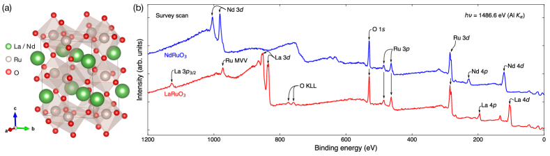
Epitaxial ruthenium oxide thin films (LaRuO3 and NdRuO3) were fabricated on SrTiO3 (001) substrates by means of the pulsed laser deposition technique. Details for the sample growth and subsequent annealing processes can be seen in the recent report by Zhang et al. Zhang et al. (2023). The crystal structure of both compounds was confirmed to be a perovskite structure (Fig. 1 (a)) by x-ray diffraction. The in-plane sample dimensions are 5 5 mm2. The film thickness of LaRuO3 and NdRuO3 was controlled to be approximately 25 nm and 14 nm, respectively.
SXPES measurements on the LaRuO3 and NdRuO3 films were carried out using a spectrometer with a monochromatic Al source ( = 1486.6 eV) implemented in PHI 5000 VersaProbe system (ULVAC-PHI Inc.). The combined instrumental energy resolution was set to be 450 meV. The angle of the photoelectron trajectory is normal to the sample surface. HAXPES measurements on the LaRuO3 and NdRuO3 films were carried out at BL24XU of SPring-8. The incidence angle of the horizontally linearly polarized hard x-ray ( = 7994 eV) was set at 2∘. The photoemission spectra were collected with the Scienta R-4000 electron energy analyzer. The combined energy resolution is around 270 meV. All SXPES and HAXPES spectra were collected at room temperature. The binding energy of the spectra was calibrated using the Fermi edge of silver in electrically contact with samples.
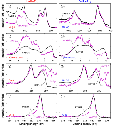
Figure 1 (b) displays survey scans of the LaRuO3 and NdRuO3 thin films. The core-level peaks expected from the nominal compositions are clearly recognizable. Before discussing the valence band and core-level spectra in detail, we first make a comparison between the SXPES and HAXPES data in Fig. 2, as also done for SrRhO3 thin films Zhang et al. (2020). The peak positions in all the SXPES and HAXPES spectra are nearly identical despite the significantly different electron inelastic mean free path of electrons excited by the soft and hard x-rays that we employed Hüfner (2003). The spectral weight in the valence band spectra is yet different between SXPES and HAXPES (Fig. 2 (c,d)). The atomic photoemission cross section ratio of Ru 4 O 2 is 40 : 1 at 1486.6 eV and 70 : 1 at 8047.8 eV Yeh and Lindau (1985), therefore the different spectral weight in these two measurements is generally anticipated. However, this atomic picture is quantitatively inconsistent with our experiments if one merely associates the intensity near the Fermi energy and at 5-7 eV with the Ru 4 and O orbitals, respectively. This disagreement with the atomic picture indicates a hybridization between the Ru 4 and O 2 orbitals as in the case of other ruthenium compounds such as SrRuO3 and Sr2RuO4 Ryee et al. (2016). Moreover, the non-negligible intensity is discernible at 3-5 eV in both spectra. A similar feature is not clearly observed in SrRuO3 but in Sr2RuO4 and this aspect will be further discussed later. In Fig. 2 (e, f), the HAXPES intensity below 280 eV is pronounced and could be attributed to Sr 3 spectra of the SrTiO3 substarates due to the longer inelastic mean free path. We ascribe a high energy shoulder in the O 1 core level appearing in the SXPES spectra to surface contamination since this feature is substantially suppressed in bulk-sensitive HAXPES (Fig. 2 (g,h)). We therefore attribute the main peak at 529.7 eV to the oxygen ions in the ruthenate thin films in the following discussion.
In light of these observations that our SXPES essentially measures bulk properties of the films, let us hereafter discuss the SXPES spectra of LaRuO3 and NdRuO3 in conjunction with an archetypical ruthenium oxide thin film SrRuO3 whose rutenium valence number is four (Fig. 3).
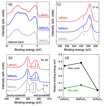
We first directly compare valence-band spectra (Fig. 3 (a)). The valence band spectra show the non-zero Fermi edge, indicative of the metallic states of these samples and consistent with transport measurements. The pseudospin splitting into the =1/2 doublets and =3/2 quartets was however not experimentally recognizable most likely because the energy resolution is not good enough to resolve these low energy features in the present work. In fact, the transition energy between these two spin states in another ruthenium compound -RuCl3 investigated by resonant inelastic x-ray scattering (RIXS) was found to be approximately 250 meV Suzuki et al. (2021) although more complicated band structures are anticipated in LaRuO3 and NdRuO3 in part because these materials are much more metallic than -RuCl3.
The binding energy of the Ru 3 core levels of LaRuO3 and NdRuO3 are downwards shifted in comparison with SrRuO3 (Fig. 3 (b)). By substituting a cation of SrRuO3, such spectral shift was not observed Takizawa et al. (2005). We therefore interpret that these shifts are not caused by the cation radius of La and Nd larger than Sr. We attribute this spectral shift to the difference between tetravalent and trivalent ruthenium valence states as also observed in tetravalent and sexivalent molybdenum compounds Wadati et al. (2014). A similar energy shift was reported for Ca2RuO4 thin films upon fluorination and interpreted as the transition from a tetravalent to trivalent valence state of ruthenium ions Fukuma et al. (2022). Besides the energy shifts, the spectral shape of Ru 3 provides spectroscopic insights regarding well-screened and poorly-screened features Kim et al. (2004). In LaRuO3 and NdRuO3, the poorly-screened feature is more pronounced than the well-screened feature compared to SrRuO3, in which both features are comparable. Since it is known that the stronger electron correlation is the more (less) pronounced well-screened (poorly-screened) feature is, the electron correlation in LaRuO3 and NdRuO3 is expected to be weaker than SrRuO3. This disparity could yield insight into the fact that SrRuO3 exhibits the ferromagnetic ordering despite the absence of magnetic ordering in LaRuO3 and NdRuO3.
Compared to SrRuO3 Guedes et al. (2012), another salient feature in the valence band spectra in both LaRuO3 and NdRuO3 is an additional peak structure around 3-5 eV (Fig. 3 (a)), which is reminiscent of the well known 3 eV peak of Sr2RuO4 whose origin has been discussed for decades Yokoya et al. (1996); Pchelkina et al. (2007, 2008); Singh (2008); Ryee et al. (2016); Tran et al. (2004). This feature has been associated with the lower Hubbard band of Ru 4 states from SXPES and resonant SXPES measurements on the one hand Pchelkina et al. (2007, 2008), this is mainly ascribed to the oxygen states from the local density approximation calculations on the other hand Singh (2008). Based on the discussion of the Ru 3 core level spectra Kim et al. (2004), Sr2RuO4 is more correlated than SrRuO3. In the present work, we pointed out the correlation of LaRuO3 and NdRuO3 is even weaker than SrRuO3 (Fig. 3 (b)). The 3 eV peak observed in LaRuO3 and NdRuO3 could thus yield another implications on the relationship between the correlation and 3 eV peak in Sr2RuO4.
The clear chemical shifts were observed in the O 1 level in comparison with SrRuO3 (Fig. 3 (c)). In particular, the binding energy of the O 1 peak in LaRuO3 and NdRuO3 ( 529 eV) shifts upwards by 350 meV while the peak width is almost identical (Fig. 3 (d)). This shift and unchanged peak shape are qualitatively akin to hole-doped cuprates Ino et al. (1997). From the chemical point of view Wu et al. (2015), another O 1 peak corresponding to the O1- ions, which could experimentally be observed as a broadened peak, should be present if the average valence of oxygen ions are different among these compounds. Nevertheless, we did not observe such effect, suggesting that the oxygen valence number in LaRuO3 and NdRuO3 is comparable with that of SrRuO3 Wu et al. (2015). We thus associate the shift of the O 1 peak with the different ruthenium valence states (i.e., trivalent and tetravalent) rather than the speculative change of the oxygen valence states. The binding energy of the shoulder peak is nearly identical among these three samples (Fig. 3 (d)). This sample-independent peak position also supports our argument that the shoulder peak is ascribed to surface contamination containing oxygen absorbed on the sample surface, also verified by our HAXPES data (Fig. 2 (g,h)).
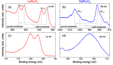
Spectral shapes of 3/4 core levels in lanthanoid elements are also sensitive to their electronic states. From the chemical point of view, the valence number of La and Nd ions tends to be +3 in solids, but it is not trivial in the present case because ruthenium ions in oxides tend to be tetravalent. Figures 4 (a,b) display the 3 core level spectra of La and Nd ions. In Fig. 4 (a), the peak splitting is notable besides the spin-orbit splitting between and peaks due to the Coulomb attraction between core holes and valence electrons peculiar to the photoemission process, also known as the final state effect. As discussed in Ref. Fuggle et al. (1980), the well-screened and poorly-screened features are very clearly observed in both compounds. Even if the valence number is the same, the spectral shape could differ depending on the subtle balance between the charge transfer energy and core-hole potential. Nevertheless, the intensity ratio of these two features in LaRuO3 and NdRuO3 is comparable with that in La2O3 and Nd2O3, respectively. Our data thus imply that the trivalent La and Nd ions are surrounded by cations and anions in a similar fashion to La2O3 and Nd2O3 Schneider et al. (1985). The 4 core level spectra of La and Nd (Fig. 4 (c,d)) can also be understood in the same manner Kotani and Ogasawara (1992). Overall, these SXPES spectral shapes are akin to those of trivalent La and Nd ions. Considering that the oxygen valence state is similar to the one in SrRuO3 (Fig. 2 (d)), we conclude that the spectral shifts of Ru and O core levels in LaRuO3 and NdRuO3 are peculiar to the trivalent state of ruthenium ions, which were previously revealed by the Ru -edge XAS Zhang et al. (2023).
In conclusion, we have investigated the core-level and valence band spectra of the LaRuO3 and NdRuO3 thin films by means of SXPES and HAXPES experiments. We found that the core level spectra are markedly different from those in Ru4+ ions in SrRuO3. We therefore claim that these spectra in LaRuO3 and NdRuO3 are peculiar to the trivalent ruthenium valence states. This argument is supported by the core-level spectra of the cations (La and Nd) as well. We also found the 3 eV peak near the Fermi level, reminiscent of that observed in Sr2RuO4. This peak feature could yield insights into the origin of the 3 eV peak in Sr2RuO4 in a fresh fashion. Although the pseudospin splitting near the Fermi level was not detected in the present study, it is highly desirable to investigate low-energy electronic states with high energy-resolution spectroscopy such as RIXS, which could potentially probe the excitation from the doublet to the quartet in a similar fashion to other 4 systems, e.g., -RuCl3 Lebert et al. (2020); Suzuki et al. (2021) in part because LaRuO3 and NdRuO3 films are rare materials accommodating 4 electrons in the square lattice.
We thank Arata Tanaka for informative discussions. The present work is supported by JSPS Grant-in-Aid for Scientific Research on Innovative Areas “Quantum Liquid Crystals” No. 19H05824, JSPS Grants-in-Aid for Scientific Research (S) No. JP22H04958, JSPS Grant-in-Aid for Early-Career Scientists No. JP20K15168, The Mitsubishi Foundation, The Izumi Science and Technology Foundation, and The Tokuyama Science Foundation. The HAXPES measurements were performed using the beamline BL24XU at SPring-8 (Proposal Nos. 2022A3231 and 2022B3231). We thank Kento Takenaka and Koji Takahara for their technical supports during the HAXPES measurements.
The data that support the findings of this study are available from the corresponding author upon reasonable request.
References
References
- Witczak-Krempa et al. (2014) W. Witczak-Krempa, G. Chen, Y. B. Kim, and L. Balents, “Correlated quantum phenomena in the strong spin-orbit regime,” Annu. Rev. Condens. Matter Phys. 5, 57–82 (2014).
- Georges, Medici, and Mravlje (2013) A. Georges, L. d. Medici, and J. Mravlje, “Strong Correlations from Hund’s Coupling,” Annu. Rev. Condens. Matter Phys. 4, 137–178 (2013).
- de’ Medici, Mravlje, and Georges (2011) L. de’ Medici, J. Mravlje, and A. Georges, “Janus-faced influence of hund’s rule coupling in strongly correlated materials,” Phys. Rev. Lett. 107, 256401 (2011).
- Jackeli and Khaliullin (2009) G. Jackeli and G. Khaliullin, “Mott insulators in the strong spin-orbit coupling limit: From heisenberg to a quantum compass and kitaev models,” Phys. Rev. Lett. 102, 017205 (2009).
- Plumb et al. (2014) K. W. Plumb, J. P. Clancy, L. J. Sandilands, V. V. Shankar, Y. F. Hu, K. S. Burch, H.-Y. Kee, and Y.-J. Kim, “-: A spin-orbit assisted Mott insulator on a honeycomb lattice,” Phys. Rev. B 90, 041112 (2014).
- Takayama et al. (2015) T. Takayama, A. Kato, R. Dinnebier, J. Nuss, H. Kono, L. S. I. Veiga, G. Fabbris, D. Haskel, and H. Takagi, “Hyperhoneycomb Iridate as a Platform for Kitaev Magnetism,” Phys. Rev. Lett. 114, 077202 (2015).
- Kim et al. (2008) B. J. Kim, H. Jin, S. J. Moon, J.-Y. Kim, B.-G. Park, C. S. Leem, J. Yu, T. W. Noh, C. Kim, S.-J. Oh, J.-H. Park, V. Durairaj, G. Cao, and E. Rotenberg, “Novel Mott State Induced by Relativistic Spin-Orbit Coupling in ,” Phys. Rev. Lett. 101, 076402 (2008).
- Yamaura and Takayama-Muromachi (2001) K. Yamaura and E. Takayama-Muromachi, “Enhanced paramagnetism of the itinerant electrons in the rhodium oxide perovskite ,” Phys. Rev. B 64, 224424 (2001).
- Perry et al. (2006) R. S. Perry, F. Baumberger, L. Balicas, N. Kikugawa, N. J. C. Ingle, A. Rost, J. F. Mercure, Y. Maeno, Z. X. Shen, and A. P. Mackenzie, “Sr2RhO4: a new, clean correlated electron metal,” New J. Phys. 8, 175–175 (2006).
- Yamaura et al. (2002) K. Yamaura, Q. Huang, D. P. Young, Y. Noguchi, and E. Takayama-Muromachi, “Crystal structure and electronic and magnetic properties of the bilayered rhodium oxide ,” Phys. Rev. B 66, 134431 (2002).
- Maeno et al. (1994) Y. Maeno, H. Hashimoto, K. Yoshida, S. Nishizaki, T. Fujita, J. G. Bednorz, and F. Lichtenberg, “Superconductivity in a layered perovskite without copper,” Nature 372, 532–534 (1994).
- Nakatsuji, Ikeda, and Maeno (1997) S. Nakatsuji, S.-i. Ikeda, and Y. Maeno, “Ca2RuO4: New Mott Insulators of Layered Ruthenate,” J. Phys. Soc. Jpn. 66, 1868–1871 (1997).
- Zhang et al. (2023) L. F. Zhang, T. C. Fujita, Y. Masutake, M. Kawamura, T. Arima, H. Kumigashira, M. Tokunaga, and M. Kawasaki, “Unconventional anomalous Hall effect in epitaxially stabilized orthorhombic Ru3+ perovskite thin films,” (2023), arXiv:2305.09201 .
- Momma and Izumi (2011) K. Momma and F. Izumi, “VESTA3 for three-dimensional visualization of crystal, volumetric and morphology data,” J. Appl. Crystallogr. 44, 1272–1276 (2011).
- Wadati et al. (2014) H. Wadati, J. Mravlje, K. Yoshimatsu, H. Kumigashira, M. Oshima, T. Sugiyama, E. Ikenaga, A. Fujimori, A. Georges, A. Radetinac, K. S. Takahashi, M. Kawasaki, and Y. Tokura, “Photoemission and DMFT study of electronic correlations in : Effects of Hund’s rule coupling and possible plasmonic sideband,” Phys. Rev. B 90, 205131 (2014).
- Takizawa et al. (2005) M. Takizawa, D. Toyota, H. Wadati, A. Chikamatsu, H. Kumigashira, A. Fujimori, M. Oshima, Z. Fang, M. Lippmaa, M. Kawasaki, and H. Koinuma, “Manifestation of correlation effects in the photoemission spectra of ,” Phys. Rev. B 72, 060404 (2005).
- Kim et al. (2004) H.-D. Kim, H.-J. Noh, K. H. Kim, and S.-J. Oh, “Core-level x-ray photoemission satellites in ruthenates: A new mechanism revealing the mott transition,” Phys. Rev. Lett. 93, 126404 (2004).
- Fujioka et al. (1997) K. Fujioka, J. Okamoto, T. Mizokawa, A. Fujimori, I. Hase, M. Abbate, H. J. Lin, C. T. Chen, Y. Takeda, and M. Takano, “Electronic structure of ,” Phys. Rev. B 56, 6380–6383 (1997).
- Yokoya et al. (1996) T. Yokoya, A. Chainani, T. Takahashi, H. Katayama-Yoshida, M. Kasai, Y. Tokura, N. Shanthi, and D. D. Sarma, “Evidence for correlation effects in from resonant and x-ray photoemission spectroscopy,” Phys. Rev. B 53, 8151–8154 (1996).
- Pchelkina et al. (2007) Z. V. Pchelkina, I. A. Nekrasov, T. Pruschke, A. Sekiyama, S. Suga, V. I. Anisimov, and D. Vollhardt, “Evidence for strong electronic correlations in the spectra of ,” Phys. Rev. B 75, 035122 (2007).
- Pchelkina et al. (2008) Z. V. Pchelkina, I. A. Nekrasov, T. Pruschke, S. Suga, V. I. Anisimov, and D. Vollhardt, “Reply to “Comment on ‘Evidence for strong electronic correlations in the spectra of ’ ",” Phys. Rev. B 77, 046102 (2008).
- Singh (2008) D. J. Singh, “Comment on “Evidence for strong electronic correlations in the spectra of ”,” Phys. Rev. B 77, 046101 (2008).
- Ryee et al. (2016) S. Ryee, S. W. Jang, H. Kino, T. Kotani, and M. J. Han, “Quasiparticle self-consistent GW calculation of and ,” Phys. Rev. B 93, 075125 (2016).
- Tran et al. (2004) T. T. Tran, T. Mizokawa, S. Nakatsuji, H. Fukazawa, and Y. Maeno, “Correlation effects in and : Valence-band photoemission spectra and self-energy calculations,” Phys. Rev. B 70, 153106 (2004).
- Zhang et al. (2020) Y. Zhang, M. Kim, J. Mravlje, C. Sohn, Y. Choi, J. Strempfer, Y. Hotta, A. Yasui, J. Nichols, H. N. Lee, and H. Wadati, “Photoemission and dynamical mean field theory study of electronic correlations in a metal thin film,” Phys. Rev. B 101, 085134 (2020).
- Hüfner (2003) S. Hüfner, Photoelectron spectroscopy: principles and applications (Springer, Berlin, 2003).
- Yeh and Lindau (1985) J. J. Yeh and I. Lindau, “Atomic subshell photoionization cross sections and asymmetry parameters: 1 103,” At. Data Nucl. Data Tables 32, 1–155 (1985).
- Suzuki et al. (2021) H. Suzuki, H. Liu, J. Bertinshaw, K. Ueda, H. Kim, S. Laha, D. Weber, Z. Yang, L. Wang, H. Takahashi, K. Fürsich, M. Minola, B. V. Lotsch, B. J. Kim, H. Yavaş, M. Daghofer, J. Chaloupka, G. Khaliullin, H. Gretarsson, and B. Keimer, “Proximate ferromagnetic state in the Kitaev model material -RuCl3,” Nat. Commun. 12, 4512 (2021).
- Fukuma et al. (2022) S. Fukuma, A. Chikamatsu, T. Katayama, T. Maruyama, K. Yanagisawa, K. Kimoto, M. Kitamura, K. Horiba, H. Kumigashira, Y. Hirose, and T. Hasegawa, “Crystal structure and electronic property modification of thin films via fluorine doping,” Phys. Rev. Mater. 6, 035002 (2022).
- Guedes et al. (2012) E. B. Guedes, M. Abbate, K. Ishigami, A. Fujimori, K. Yoshimatsu, H. Kumigashira, M. Oshima, F. C. Vicentin, P. T. Fonseca, and R. J. O. Mossanek, “Core level and valence band spectroscopy of SrRuO3: Electron correlation and covalence effects,” Phys. Rev. B 86, 235127 (2012).
- Ino et al. (1997) A. Ino, T. Mizokawa, A. Fujimori, K. Tamasaku, H. Eisaki, S. Uchida, T. Kimura, T. Sasagawa, and K. Kishio, “Chemical Potential Shift in Overdoped and Underdoped ,” Phys. Rev. Lett. 79, 2101–2104 (1997).
- Wu et al. (2015) L. Q. Wu, Y. C. Li, S. Q. Li, Z. Z. Li, G. D. Tang, W. H. Qi, L. C. Xue, X. S. Ge, and L. L. Ding, “Method for estimating ionicities of oxides using O1s photoelectron spectra,” AIP Adv. 5, 097210 (2015).
- Fuggle et al. (1980) J. C. Fuggle, M. Campagna, Z. Zolnierek, R. Lässer, and A. Platau, “Observation of a relationship between core-level line shapes in photoelectron spectroscopy and the localization of screening orbitals,” Phys. Rev. Lett. 45, 1597–1600 (1980).
- Schneider et al. (1985) W.-D. Schneider, B. Delley, E. Wuilloud, J.-M. Imer, and Y. Baer, “Electron-spectroscopic manifestations of the 4f states in light rare-earth solids,” Phys. Rev. B 32, 6819–6831 (1985).
- Kotani and Ogasawara (1992) A. Kotani and H. Ogasawara, “Theory of core-level spectroscopy of rare-earth oxides,” J. Electron. Spectrosc. Relat. Phenom. 60, 257–299 (1992).
- Lebert et al. (2020) B. W. Lebert, S. Kim, V. Bisogni, I. Jarrige, A. M. Barbour, and Y.-J. Kim, “Resonant inelastic x-ray scattering study of -RuCl3: a progress report,” J. Phys.: Condens. Matter 32, 144001 (2020).