Controlled creation of point defects in 3D colloidal crystals
Abstract
Crystal defects crucially influence the properties of crystalline materials and have been extensively studied. Even for the simplest type of defect—the point defect—however, basic properties such as their diffusive behavior, and their interactions, remain elusive on the atomic scale. Here we demonstrate in-situ control over the creation of isolated point defects in a 3D colloidal crystal allowing insight on a single particle level. Our system consists of thermoresponsive microgel particles embedded in a crystal of non-responsive colloids. Heating this mixed particle system triggers the shrinking of the embedded microgels, which then vacate their lattice positions creating vacancy-interstitial pairs. We use temperature-controlled confocal laser scanning microscopy to verify and visualize the formation of the point defects. In addition, by re-swelling the microgels we quantify the local lattice distortion around an interstitial defect. Our experimental model system provides a unique opportunity to shed new light on the interplay between point defects, on the mechanisms of their diffusion, on their interactions, and on collective dynamics.
I Introduction
In any crystal, structural imperfections (defects) are thermodynamically bound to occur. Defects crucially influence the mechanical, structural and optical properties of materials and for this reason have remained extensively studied [1, 2]. While defects may have desirable effects — they may enhance the conductive properties of semiconductors [3], or may act as active sites in catalysis [4] — their presence can also be undesirable, causing metal fatigue or the softening of nanocrystalline metals [5]. Of particular interest are point defects (vacancies and interstitials). While these distort the lattice only locally, they play a crucial role in bulk properties such as diffusion creep and material degradation [2]. A dramatic example of this occurs in radiation shielding materials, where high energy particles generate vacancy-interstitial pairs (Frenkel pairs) that are known to greatly compromise material strength and integrity [6, 7, 8]. However, despite remarkable progress in high-resolution electron microscopy studies of defect dynamics [9, 10, 11, 12], the atomic details of the local lattice distortion, diffusion and collective organization of point defects remain elusive.
Here, we present an experimental platform offering a window into the basic properties and dynamics of point defects in crystalline materials using colloids. Colloids have played a central role as convenient experimental model systems to study many physical phenomena, such as phase transitions and the glass transition [13, 14, 15, 16, 17, 18, 19]. Colloidal particles are small enough to experience Brownian motion, causing them to exhibit equilibrium phase behavior similar to atomic and molecular systems. Importantly, though, they are large enough to be readily observed, with single-particle resolution, using optical microscopy. For example, experiments on defects in 3D colloidal crystals have already provided unique insight into dislocations (line defects); their nucleation [20], dynamics [21], interactions [22] and width [23]. Experiments on point defects, however, have so far been limited to 2D systems [24], for instance employing laser optical tweezers to artificially introduce vacancies and interstitials revealing the equilibrium configurations [25], diffusion [26, 27], defect string formation [28] and kinetics [29, 30]. So far, point defects in 3D colloidal crystals remain underexamined because experimental point defect concentrations are low [31] and optical tweezers cannot be used to remove or add particles in 3D crystals without distorting the full crystal [32].
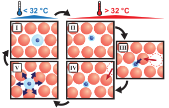
In this Article, we overcome the challenge of the controlled generation of point defects in 3D in an experimental responsive colloidal system that breaks ground for point defect studies in 3D. We demonstrate that our system can induce both vacancies and interstitials, and we present the experimental determination of local lattice distortions around an interstitial in 3D. Our system consists of thermoresponsive poly(N-isopropyl acrylamide) (PNIPAM) microgels embedded in a crystal of non-responsive latex colloids and is schematically depicted in Fig. 1. The size of PNIPAM microgels is reversibly tunable with temperature. At low temperatures the microgels are swollen with water, while above the Volume Phase Transition Temperature (VPTT, around 32 °C) the microgels collapse [33, 34] resulting in a change in their volume fraction that has been extensively studied in bulk [35, 36, 17]. In our system, by employing a low concentration of microgels, they will take up a lattice position in a non-responsive crystal at low temperatures. Heating the system across the VPTT leads to a significant decrease of the microgel size and allows them to migrate to an interstitial lattice site, which effectively results in the formation of a vacancy-interstitial pair. Subsequently, by lowering the temperature, re-swelling of a microgel that is located on an interstitial lattice site results in a significant local strain that we can visualize on a single-particle level. We measured the resulting lattice distortion around these interstitials and found an anisotropic strain with a decay that is comparable to results from theory [2, 37] and simulations [38, 39].
II Results and discussion
Colloidal model system. To realize the system presented in Fig. 1, two types of colloidal particles were synthesized. The microgels consist of a fluorescent, non-responsive core of poly(2,2,2-trifluoro ethyl methacrylate) (PTFEMA) and a crosslinked PNIPAM microgel shell [40]. Fluorescent latex particles consisting of PTFEMA and coated with a thin layer of poly(oligo-ethylene glycol methacrylate) were used as non-responsive colloids (see Methods and Supplementary Information for details about the synthesis). Fig. 2 shows the hydrodynamic diameter of the particles versus temperature as determined with dynamic light scattering (DLS). The VPTT of the microgels is determined as 32.3 °C (see Supplementary Fig. 1). Below the VPTT (20.0 °C), both particles have a similar size , ensuring that the microgels can replace a PTFEMA particle in the crystal. Above the VPTT, the microgels decrease significantly in size to = (37.0 °C) making them small enough to occupy an octahedral site of in a close-packed crystal of colloids.
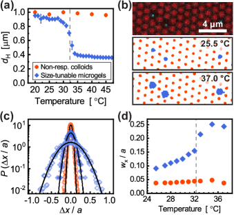
The mixed system consists of a small fraction of microgels (0.5 - 1 %) in a crystal of non-responsive colloids with volume fraction (see Methods for details). The colloids adopt a random-hexagonal close packed (rhcp) structure upon sedimentation, which is essentially a mixture of face-centered cubic (fcc) and hexagonal close packed (hcp) crystal structures. Fig. 2 (upper panel) shows a 2D confocal laser scanning microscopy (CLSM) image of the mixed crystal, where the hexagonal rhcp planes are parallel to the flat substrate. Here, the microgels clearly take up a single lattice site, although only the fluorescent microgel cores are visible, since the shells are not fluorescently labeled. To change the microgel size in the mixed crystal, temperature-controlled CLSM experiments were performed in 2D and 3D using a VAHEAT (Interherence) temperature controller [41]. Particle positions were obtained using Trackpy [42] based on the Crocker-Grier centroid-finding algorithm [43].
To confirm that temperature changes only influence the microgel size and not affect the colloidal crystal structure or dynamics, we studied the mixed system behavior between 25.5 °C and 37.0 °C. Fig. 2 shows 2D trajectories of both particles at 25.5 °C (middle panel) and 37.0 °C (lower panel). The trajectories clearly show that the microgels, due to their smaller size, are more mobile on their lattice site above the VPTT. In addition, we did not observe any change in the overall crystal lattice positions.
To quantify the particle dynamics, we calculate the self-part of the Van Hove correlation function from the particle trajectories, which gives the probability distribution of particle displacements along the coordinate within a lag time . Fig. 2 shows the probability distributions for for both particles (the width of the distributions does not increase for larger ; see Supplementary Fig. 2). Only for the microgels the Van Hove function shows a significant broadening with increasing temperature. The width of the distributions at temperatures around the VPTT was obtained by fitting
| (1) |
Fig. 2 shows for both the PTFEMA colloids and PNIPAM microgels at each investigated temperature. As expected, no substantial change in was observed for the non-responsive colloids. The sharp increase in for the PNIPAM microgels is a result of their collapse around the VPTT. The size change of the microgels clearly still happens in a similar temperature regime in the dense colloidal crystal of PTFEMA particles as under dilute conditions (see DLS measurements in Fig. 2). These results confirm that by controlling the temperature we can finely tune the size of the embedded microgels without affecting the rest of the crystal.
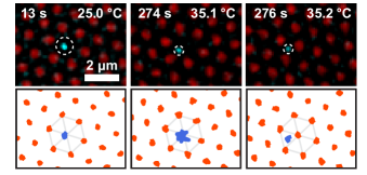
Formation of vacancy-interstitial pairs. To create point defects, we increased the temperature of mixed colloidal crystal above the VPTT slowly. Over time, the collapsed microgels are able to spontaneously move from a regular lattice site to an interstitial position, forming the expected vacancy-interstitial pair. Fig. 3 shows CLSM images (upper panels) and 2D particle trajectories (lower panels) of this observed vacancy-interstitial pair formation. At the start at low temperature, the microgel is clearly embedded in the non-responsive crystal with reduced mobility. After heating the crystal to above the VPTT, the microgel starts to rattle in its cage. Since the microgel collapse leads to a reduced local pressure (similar to the case of a true vacancy [44]) at a certain moment a colloid from an adjacent layer ’hops’ and takes up the original lattice site, thereby trapping the microgel in an interstitial site. We found that the formation of a vacancy-interstitial pair can happen in two ways. The first mechanism is that two spontaneous movements of the microgel and non-responsive particle occur at the same time, as shown in Fig. 3. The second mechanism is the diffusion of the collapsed microgel to an octahedral interstitial site further away by passing between two non-responsive colloids, leaving behind a vacancy in the originally occupied lattice site, which we observed less frequently. For both processes there is an associated energy barrier that depends on the density of the crystal and the size of the collapsed microgel relative to the other colloids. In addition, we observed that after the creation of the vacancy-interstitial pair, the pair can split up by diffusion, resulting in isolated vacancies and interstitials which allows us to study these defects separately as well.
The vacancy-interstitial pairs induced in this colloidal system are analogous to Frenkel pairs found in atomic systems. In metals, this type of defect does generally not occur under normal conditions due to the high formation energy of interstitials, but is predominantly generated during particle irradiation. In ionic solids Frenkel defects can be formed spontaneously because there is usually a significant size difference between the cation and anion. For the same reason, in the system presented here, only collapsed microgels are small enough to move to an interstitial site without significantly distorting the lattice.
Inducing strain with interstitial microgels. In our colloidal system we have the additional control that, once a collapsed microgel is on an interstitial site, lowering the temperature across the VPTT will result in re-swelling of the microgel. This local swelling provides the opportunity to apply a stress around the interstitial site and to visualize how this distorts the local crystal structure, a process that lies at the core of interstial interactions and agglomeration. To investigate the local strain due to re-swelling of interstitial microgels, we slowly cooled a mixed system with interstitial microgels across the VPTT, while simultaneously imaging the system in 3D with CLSM. Fig. 4 shows an overlay of CLSM images of an interstitial microgel and the hexagonal planes directly above and below the microgel (in a pure fcc crystal these are the (111) planes).
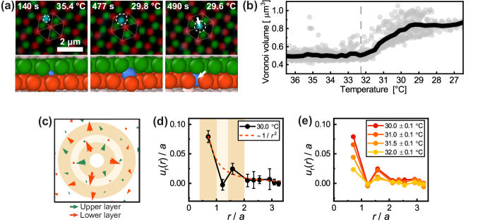
For clarity, a 3D reconstruction [45] using the tracked particle positions is given below the CLSM images. The images show that cooling across the VPTT results indeed in the swelling of the microgel and a distortion of the local lattice. We note that this distortion is relatively small and therefore difficult to observe by merely looking at the CLSM images. At a certain point, a stress relaxation event occurs that results in a jump of the (nearly-) swollen microgel to a regular lattice site, pushing away the colloid that originally occupies this site. These stress relaxations happen so fast that we have not been able to fully visualize the event in 3D with the CLSM with high temporal resolution (our fastest frame rate was 0.23 frames per second for a 256x256x101 image stack). The pushed particles appear to move toward the disordered layer of colloids at the substrate, which is comparable to how in atomic systems disordered interfaces such as grain boundaries act as defect sinks.
To determine the exact temperature range in which the buildup of lattice strain takes place, we first analyzed the Voronoi cell volume of the interstitial microgel as function of temperature [46]. Fig. 4 shows the Voronoi cell volume and the average value during the cooling process for 14 interstitial microgels. We used cooling rates of -1.0 and -3.2 °C/min for the experiments, but we do not observe a significant difference in the results (see Supplementary Fig. 3(a)). The curve shows that by decreasing temperature, the microgel swells and causes an increase in the Voronoi cell volume, indicating the interstitial microgel indeed pushes its direct nearest neighbors. The Voronoi cell volume of the microgels remains constant above the VPTT (32.3 °C - dashed vertical line) and only increases once the temperature is below the VPTT. Hence, only from this temperature onward the microgels start to significantly strain the crystal. For °C, a constant Voronoi cell volume is present as the microgels typically have moved from an interstitial site to a regular crystal lattice site, with some exceptions that remained at the interstitial site (see Supplementary Fig. 3(b)).
To quantify the strain caused by the swelling of interstitial microgels, we measured the local distortion that occurs in the surrounding crystal around a single interstitial microgel between 32 to 30 °C. Fig. 4 shows the typical particle displacements measured around a single microgel by comparing the average non-responsive particle positions in the range 37–35 °C (i.e. fully collapsed microgel) to their positions at °C (nearly-swollen microgel on interstitial site). Similar to Fig. 4, the hexagonal planes above (green arrows) and below (red arrows) the interstitial microgel are shown. The arrows show that the strain is not isotropic: the displacements are strongest in the direction of the nearest neighbors of the interstitial microgel as these are pushed to accommodate the swelling microgel. Assuming a pure fcc structure this displacement occurs along the cubic axes (or directions). In the second shell of colloids around the interstitial (i.e. its next-nearest neighbors) the displacements are found to be negligible, since these colloids are not pushed by the nearest-neighbors. In contrast, the particles in the third shell are pushed by the nearest-neighbors and show a clear displacement. This non-uniform nature of the strain is also observed in the average radial component of the particle displacements around a swelling interstitial microgel as plotted in Fig. 4 (see Supplementary Information for more elaboration). Such anisotropic strain due to the crystal lattice has also been found with simulations [38, 39] and theory [37]. We further note that the amount of distortion around an interstitial microgel can be tuned via the temperature as shown in Fig. 4. The results in Fig. 4 indicate that the measured displacements around microgels at °C decay within a few lattice sites. The decay in the maxima of the radial displacement appears consistent with , which can be rationalized using linear elasticity theory and, although an oversimplification, approximating the crystal as an isotropic continuum [2]. Interestingly, simulations of hard-sphere systems [38] suggest an exponential decay of the displacements near a self-interstitial that ranges over more lattice sites than measured here. This discrepancy is likely due to the fact that in our results obtained at 30.0 °C, the microgels are not fully swollen and are therefore slightly smaller than the colloids in the crystal (see DLS results in Fig. 2). Moreover, it is difficult to predict the actual size of a swelling microgel on an interstitial site, as microgels have a soft and deformable character, and are able to osmotically deswell in dense systems [47]. It is clear, however, that the interstitials can induce a local strain which ranges over several lattice sites into the non-responsive crystal.
In summary, we present here a colloidal model system in which the creation of point defects in 3D crystals can be controlled in-situ and visualized on a single-particle level. In this system, PNIPAM microgels are embedded into a non-responsive colloidal crystal where these are able to take up interstitial positions when heated above 32 °C (VPTT), resulting in the formation of vacancy-interstitial pairs that are similar to Frenkel pairs found in atomic systems. Due to the colloidal nature and unique temperature control the system allows us to visualize the exact moment of point defect creation and their diffusion. In addition, re-swelling of the microgels by lowering the temperature across the VPTT allows us to measure the local distortion induced by interstitials, which is otherwise problematic due to their rarity. We find that the local strain displays an anisotropy that is in agreement with results from theory and simulations. The system presented in this Article opens the possibility to gain new insight into point defect phenomena, such as defect interactions, as it offers in-situ control over the formation of both vacancies and interstitials, while their concentration can be controlled via the mixing ratio. This insight is valuable for understanding defect dynamics in atomic crystals, such as radiation shielding materials, while it could also help to adjust and improve the rational design and controlled defect engineering of complex crystals.
III Methods
Materials and characterization
Colloidal particles: The synthesis procedure used for the thermoresponsive composite microgels was based on the procedure reported by Appel et al. in Ref. [40]. The composite microgels consist of a 192 2 nm poly(2,2,2-trifluoroethyl methacrylate) (PTFEMA) core containing Pyrromethene 546 (BODIPY) dye, and a non-fluorescent PNIPAM microgel shell with 1.0 wt% N,N’-methylenebisacrylamide as crosslinker. The non-responsive colloids consist of PTFEMA and contain a Rhodamine B methacrylate [48] dye. The PTFEMA colloids are coated with thin shell of poly(oligo-ethylene glycol methacrylate) to prevent aggregation with the microgels. A comprehensive description of the synthesis procedures of both particles is given in the Supplementary Information.
Dynamic light scattering: Dilute particle dispersions (0.01-0.1 wt%) were prepared using MilliQ water (18.2 Mcm). Particle sizes were determined with an Anton Paar Litesizer 500, using a scattering angle of 90° and laser wavelength of 658 nm, and using disposable polystyrene cuvettes. The mean hydrodynamic diameter and polydispersity index was obtained from the scattering data using the method of cumulants [49].
Temperature-controlled microscopy experiments
Mixed dispersion and sample preparation: Several mixed particle dispersions were prepared for the experiments. Typically, 4 mg of concentrated microgel dispersion (3.2 wt%) was added to 0.4 g concentrated non-responsive PTFEMA colloid dispersion (40 wt%) in an 1.5 mL Eppendorf tube. Sample cells were prepared using Grace Bio-Labs SecureSeal Hybridization Chambers (SKU: 621503) combined with VAHEAT smart substrates (standard range, 18x18x0.17 mm) with a 5x5 mm heated area. The mixed dispersions were vortexed thoroughly before filling the sample cells and sealing the cells using SecureSeal stickers. Images of a sealed sample cell are depicted in Supplementary Fig. 4.
Equipment and analysis details: Confocal laser scanning microscopy (CLSM) experiments were performed using a Nikon A1R HD25 microscope equipped with a 100x oil immersion objective (Nikon CFI Plan Apo VC, NA = 1.4). The non-responsive PTFEMA colloids containing Rhodamine B were visualized using a 561 nm laser and GaAsP PMT detector, and the composite microgels containing Pyrromethene 546 (BODIPY) were visualized with a 488 nm laser and GaAsP PMT detector. Precise control over the sample temperature was achieved by using a VAHEAT (Interherence) temperature control system. A resonant scanner and piezo objective nanopositioning system (Mad City Labs F200W) were used for fast 3D acquisition. For the 3D acquisition, we used a typical image size of 256x256 pixels in xy and 101 pixels in z (voxel size: 0.084x0.084x0.100 ), resulting in a typical temporal resolution of 0.23 frames per second. Particle tracking was performed using Trackpy (v0.5.0) [42]. In order to account for the distortion of the axial distances due to the mismatch of the refractive index of the immersion oil and the sample, a scaling factor of 0.85 was used [50]. The nearest neighbor distance was determined using the first peak of the pair correlation function, which was found to be in the range of to in our experiments. Considering a particle size of (see Supplementary Table I), we find an average volume fraction of .
IV Acknowledgments
We thank C. Storm and L. Filion for fruitful discussions. We acknowledge T.F. Sparidans for performing DLS measurements. J.M.M. acknowledges financial support from the Netherlands Organization for Scientific Research (NWO) (016.Veni.192.119).
References
- Hull and Bacon [2011] D. Hull and D. Bacon, Introduction to Dislocations, 5th ed. (Elsevier, 2011).
- Phillips [2001] R. Phillips, Crystals, Defects and Microstructures (Cambridge University Press, 2001).
- Koenraad and Flatté [2011] P. M. Koenraad and M. E. Flatté, Single dopants in semiconductors, Nature Materials 10, 91 (2011).
- Xie et al. [2020] C. Xie, D. Yan, H. Li, S. Du, W. Chen, Y. Wang, Y. Zou, R. Chen, and S. Wang, Defect Chemistry in Heterogeneous Catalysis: Recognition, Understanding, and Utilization, ACS Catalysis 10, 11082 (2020).
- Schiøtz et al. [1998] J. Schiøtz, F. D. Di Tolla, and K. W. Jacobsen, Softening of nanocrystalline metals at very small grain sizes, Nature 391, 561 (1998).
- Knaster et al. [2016] J. Knaster, A. Moeslang, and T. Muroga, Materials research for fusion, Nature Physics 12, 424 (2016).
- Bai et al. [2010] X.-M. Bai, A. F. Voter, R. G. Hoagland, M. Nastasi, and B. P. Uberuaga, Efficient Annealing of Radiation Damage Near Grain Boundaries via Interstitial Emission, Science 327, 1631 (2010).
- Lu et al. [2016] C. Lu, L. Niu, N. Chen, K. Jin, T. Yang, P. Xiu, Y. Zhang, F. Gao, H. Bei, S. Shi, M.-R. He, I. M. Robertson, W. J. Weber, and L. Wang, Enhancing radiation tolerance by controlling defect mobility and migration pathways in multicomponent single-phase alloys, Nature Communications 7, 13564 (2016).
- Arakawa et al. [2007] K. Arakawa, K. Ono, M. Isshiki, K. Mimura, M. Uchikoshi, and H. Mori, Observation of the One-Dimensional Diffusion of Nanometer-Sized Dislocation Loops, Science 318, 956 (2007).
- Matsukawa and Zinkle [2007] Y. Matsukawa and S. J. Zinkle, One-Dimensional Fast Migration of Vacancy Clusters in Metals, Science 318, 959 (2007).
- Moll et al. [2013] S. Moll, T. Jourdan, and H. Lefaix-Jeuland, Direct Observation of Interstitial Dislocation Loop Coarsening in -Iron, Physical Review Letters 111, 015503 (2013).
- Chu et al. [2022] S. Chu, P. Liu, Y. Zhang, X. Wang, S. Song, T. Zhu, Z. Zhang, X. Han, B. Sun, and M. Chen, In situ atomic-scale observation of dislocation climb and grain boundary evolution in nanostructured metal, Nature Communications 13, 4151 (2022).
- Gasser et al. [2001] U. Gasser, E. R. Weeks, A. Schofield, P. N. Pusey, and D. A. Weitz, Real-space imaging of nucleation and growth in colloidal crystallization, Science 292, 258 (2001).
- Zhang and Liu [2014] T. H. Zhang and X. Y. Liu, Experimental modelling of single-particle dynamic processes in crystallization by controlled colloidal assembly, Chem. Soc. Rev. 43, 2324 (2014).
- Alsayed et al. [2005] A. M. Alsayed, M. F. Islam, J. Zhang, P. J. Collings, and A. G. Yodh, Premelting at Defects Within Bulk Colloidal Crystals, Science 309, 1207 (2005).
- Peng et al. [2010] Y. Peng, Z. Wang, A. M. Alsayed, A. G. Yodh, and Y. Han, Melting of Colloidal Crystal Films, Physical Review Letters 104, 205703 (2010).
- Wang et al. [2016] F. Wang, D. Zhou, and Y. Han, Melting of Colloidal Crystals, Advanced Functional Materials 26, 8903 (2016).
- Yunker et al. [2010] P. Yunker, Z. Zhang, and A. G. Yodh, Observation of the Disorder-Induced Crystal-to-Glass Transition, Physical Review Letters 104, 015701 (2010).
- Weeks [2017] E. R. Weeks, Introduction to the colloidal glass transition, ACS Macro Letters 6, 27 (2017).
- Schall et al. [2006] P. Schall, I. Cohen, D. A. Weitz, and F. Spaepen, Visualizing dislocation nucleation by indenting colloidal crystals, Nature 440, 319 (2006).
- Schall et al. [2004] P. Schall, I. Cohen, D. A. Weitz, and F. Spaepen, Visualization of Dislocation Dynamics in Colloidal Crystals, Science 305, 1944 (2004).
- Svetlizky et al. [2023] I. Svetlizky, S. Kim, D. A. Weitz, and F. Spaepen, Dislocation interactions during plastic relaxation of epitaxial colloidal crystals, Nature Communications 14, 5760 (2023).
- Hilhorst and Petukhov [2011] J. Hilhorst and A. V. Petukhov, Variable Dislocation Widths in Colloidal Crystals of Soft Thermosensitive Spheres, Physical Review Letters 107, 095501 (2011).
- Cao et al. [2020] X. Cao, E. Panizon, A. Vanossi, N. Manini, E. Tosatti, and C. Bechinger, Pile-up transmission and reflection of topological defects at grain boundaries in colloidal crystals, Nature Communications 11, 3079 (2020).
- Pertsinidis and Ling [2001a] A. Pertsinidis and X. S. Ling, Equilibrium Configurations and Energetics of Point Defects in Two-Dimensional Colloidal Crystals, Physical Review Letters 87, 098303 (2001a).
- Pertsinidis and Ling [2001b] A. Pertsinidis and X. S. Ling, Diffusion of point defects in two-dimensional colloidal crystals, Nature 413, 147 (2001b).
- Kim et al. [2020] S.-C. Kim, L. Yu, A. Pertsinidis, and X. S. Ling, Dynamical processes of interstitial diffusion in a two-dimensional colloidal crystal, Proceedings of the National Academy of Sciences 117, 13220 (2020).
- Lechner et al. [2013] W. Lechner, D. Polster, G. Maret, P. Keim, and C. Dellago, Self-organized defect strings in two-dimensional crystals, Physical Review E 88, 060402 (2013).
- Lechner et al. [2015] W. Lechner, D. Polster, G. Maret, C. Dellago, and P. Keim, Entropy and kinetics of point defects in two-dimensional dipolar crystals, Physical Review E 91, 032304 (2015).
- Van Der Meer et al. [2014] B. Van Der Meer, W. Qi, R. G. Fokkink, J. Van Der Gucht, M. Dijkstra, and J. Sprakel, Highly cooperative stress relaxation in two-dimensional soft colloidal crystals, Proceedings of the National Academy of Sciences 111, 15356 (2014).
- Pronk and Frenkel [2001] S. Pronk and D. Frenkel, Point Defects in Hard-Sphere Crystals, The Journal of Physical Chemistry B 105, 6722 (2001).
- Liu et al. [2016] Y. Liu, K. V. Edmond, A. Curran, C. Bryant, B. Peng, D. G. A. L. Aarts, S. Sacanna, and R. P. A. Dullens, Core–Shell Particles for Simultaneous 3D Imaging and Optical Tweezing in Dense Colloidal Materials, Advanced Materials 28, 8001 (2016).
- Pelton and Chibante [1986] R. H. Pelton and P. Chibante, Preparation of aqueous latices with N-isopropylacrylamide, Colloids and Surfaces 20, 247 (1986).
- Bae et al. [1990] Y. H. Bae, T. Okano, and S. W. Kim, Temperature dependence of swelling of crosslinked poly(N,N‐alkyl substituted acrylamides) in water, Journal of Polymer Science Part B: Polymer Physics 28, 923 (1990).
- Karg et al. [2019] M. Karg, A. Pich, T. Hellweg, T. Hoare, L. A. Lyon, J. J. Crassous, D. Suzuki, R. A. Gumerov, S. Schneider, I. I. Potemkin, and W. Richtering, Nanogels and Microgels: From Model Colloids to Applications, Recent Developments, and Future Trends, Langmuir 35, 6231 (2019).
- Yunker et al. [2014] P. J. Yunker, K. Chen, M. D. Gratale, M. A. Lohr, T. Still, and A. G. Yodh, Physics in ordered and disordered colloidal matter composed of poly(N-isopropylacrylamide) microgel particles, Reports on Progress in Physics 77, 056601 (2014).
- Hall [1957] G. L. Hall, Distortion around point imperfections in simple crystals, Journal of Physics and Chemistry of Solids 3, 210 (1957).
- van der Meer et al. [2017] B. van der Meer, M. Dijkstra, and L. Filion, Diffusion and interactions of point defects in hard-sphere crystals, The Journal of Chemical Physics 146, 244905 (2017).
- Alkemade et al. [2021] R. M. Alkemade, M. de Jager, B. van der Meer, F. Smallenburg, and L. Filion, Point defects in crystals of charged colloids, The Journal of Chemical Physics 154, 164905 (2021).
- Appel et al. [2015] J. Appel, N. De Lange, H. M. Van Der Kooij, T. Van De Laar, J. B. Ten Hove, T. E. Kodger, and J. Sprakel, Temperature Controlled Sequential Gelation in Composite Microgel Suspensions, Particle and Particle Systems Characterization 32, 764 (2015).
- Icha et al. [2022] J. Icha, D. Böning, and P. Türschmann, Precise and Dynamic Temperature Control in High-Resolution Microscopy with VAHEAT, Microscopy Today 30, 34 (2022).
- Allan et al. [2021] D. B. Allan, T. Caswell, N. C. Keim, C. M. van der Wel, and R. W. Verweij, soft-matter/trackpy: Trackpy v0.5.0 (v0.5.0), Zenodo https://doi.org/10.5281/zenodo.4682814 (2021).
- Crocker and Grier [1996] J. C. Crocker and D. G. Grier, Methods of digital video microscopy for colloidal studies, Journal of Colloid and Interface Science 179, 298 (1996).
- Lin et al. [2016] N. Y. Lin, M. Bierbaum, P. Schall, J. P. Sethna, and I. Cohen, Measuring nonlinear stresses generated by defects in 3D colloidal crystals, Nature Materials 15, 1172 (2016).
- Stukowski [2010] A. Stukowski, Visualization and analysis of atomistic simulation data with OVITO–the Open Visualization Tool, Modelling and Simulation in Materials Science and Engineering 18, 015012 (2010).
- Ramasubramani et al. [2020] V. Ramasubramani, B. D. Dice, E. S. Harper, M. P. Spellings, J. A. Anderson, and S. C. Glotzer, freud: A software suite for high throughput analysis of particle simulation data, Computer Physics Communications 254, 107275 (2020).
- Scotti et al. [2016] A. Scotti, U. Gasser, E. S. Herman, M. Pelaez-Fernandez, J. Han, A. Menzel, L. A. Lyon, and A. Fernández-Nieves, The role of ions in the self-healing behavior of soft particle suspensions, Proceedings of the National Academy of Sciences of the United States of America 113, 5576 (2016).
- Kodger et al. [2015] T. E. Kodger, R. E. Guerra, and J. Sprakel, Precise colloids with tunable interactions for confocal microscopy, Scientific Reports 5, 14635 (2015).
- Frisken [2001] B. J. Frisken, Revisiting the method of cumulants for the analysis of dynamic light-scattering data, Applied Optics 40, 4087 (2001).
- Besseling et al. [2015] T. H. Besseling, J. Jose, and A. V. Blaaderen, Methods to calibrate and scale axial distances in confocal microscopy as a function of refractive index, Journal of Microscopy 257, 142 (2015).